
Advancements and Opportunities in Characterizing Patient-Specific Wall Shear Stress Imposed by Coronary Artery Stenting
 ,
, 
Abstract
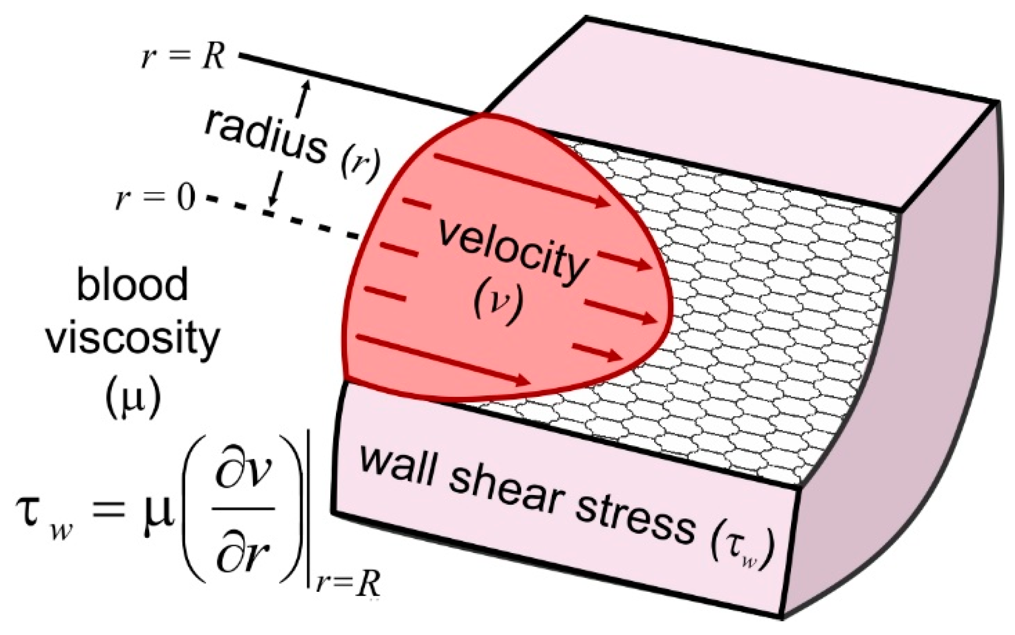
:1. Background
2. Requirements and Best Practices
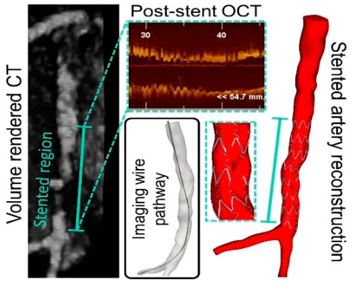
3. Intravascular Reconstruction for Computational Simulations
4. WSS Findings to Date and Related Indices of Interest
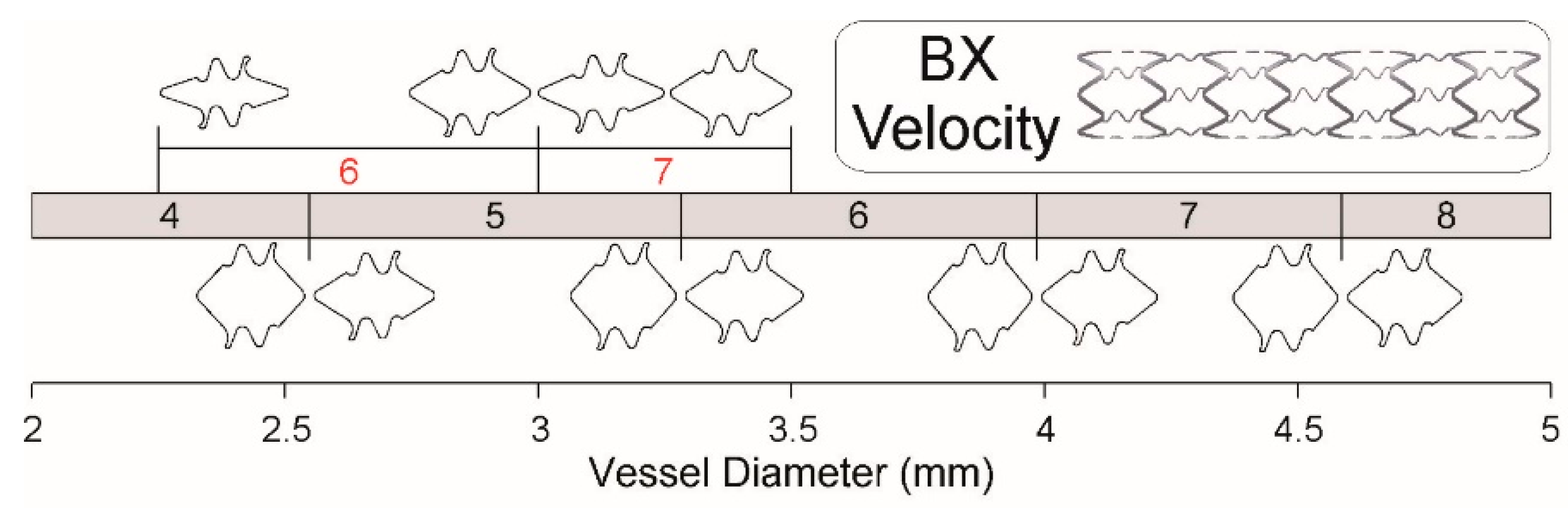
5. Optimizing the Stenting Procedure
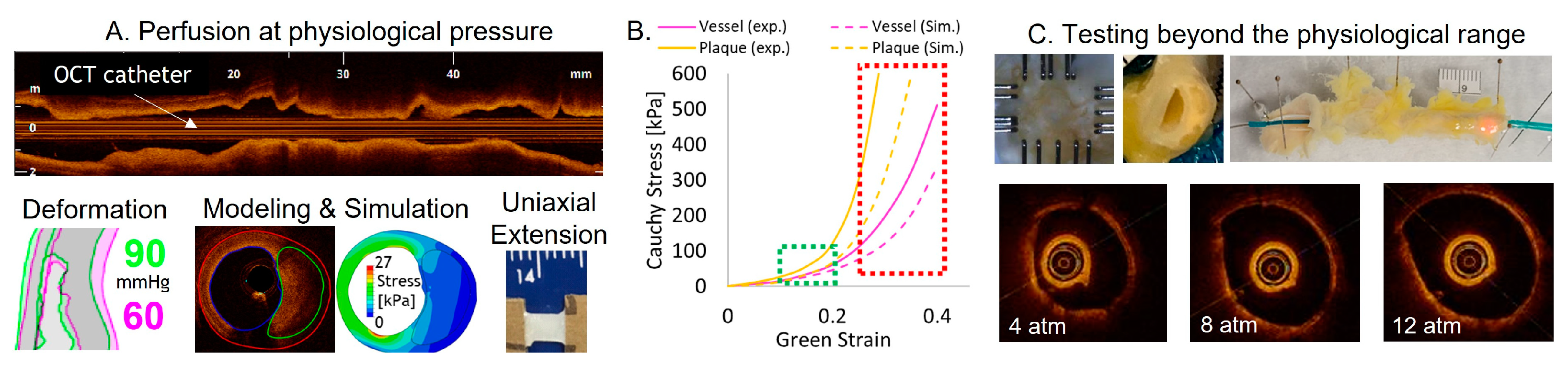
6. Limited Data from Atherosclerotic Arteries
7. Application of Machine Learning (ML) and Artificial Intelligence (AI)
8. Clinical Applications Using Patient-Specific Stenting
9. Future Directions
Author Contributions
Funding
Institutional Review Board Statement
Informed Consent Statement
Data Availability Statement
Conflicts of Interest
References
- Tsao, C.W.; Aday, A.W.; Almarzooq, Z.I.; Alonso, A.; Beaton, A.Z.; Bittencourt, M.S.; Boehme, A.K.; Buxton, A.E.; Carson, A.P.; Commodore-Mensah, Y.; et al. Heart Disease and Stroke Statistics-2022 Update: A Report From the American Heart Association. Circulation 2022, 145, e153–e639. [Google Scholar] [CrossRef]
- Navarese, E.P.; Austin, D.; Gurbel, P.A.; Andreotti, F.; Tantry, U.; James, S.; Buffon, A.; Kozinski, M.; Obonska, K.; Bliden, K.; et al. Drug-coated balloons in treatment of in-stent restenosis: A meta-analysis of randomised controlled trials. Clin. Res. Cardiol. 2013, 102, 279–287. [Google Scholar] [CrossRef] [Green Version]
- Lee, S.J.; Kim, B.K.; Kim, J.S.; Ko, Y.G.; Choi, D.; Jang, Y.; Hong, M.K. Evaluation of neointimal morphology of lesions with or without in-stent restenosis: An optical coherence tomography study. Clin. Cardiol. 2011, 34, 633–639. [Google Scholar] [CrossRef]
- Hoffmann, R.; Mintz, G.S.; Dussaillant, G.R.; Popma, J.J.; Pichard, A.D.; Satler, L.F.; Kent, K.M.; Griffin, J.; Leon, M.B. Patterns and mechanisms of in-stent restenosis. A serial intravascular ultrasound study. Circulation 1996, 94, 1247–1254. [Google Scholar] [CrossRef] [PubMed]
- Krone, R.J.; Rao, S.V.; Dai, D.; Anderson, H.V.; Peterson, E.D.; Brown, M.A.; Brindis, R.G.; Klein, L.W.; Shaw, R.E.; Weintraub, W.S. Acceptance, panic, and partial recovery the pattern of usage of drug-eluting stents after introduction in the, U.S. (a report from the American College of Cardiology/National Cardiovascular Data Registry). JACC Cardiovasc. Interv. 2010, 3, 902–910. [Google Scholar] [CrossRef] [PubMed] [Green Version]
- Holmes, D.R., Jr.; Kereiakes, D.J.; Garg, S.; Serruys, P.W.; Dehmer, G.J.; Ellis, S.G.; Williams, D.O.; Kimura, T.; Moliterno, D.J. Stent thrombosis. J. Am. Coll. Cardiol. 2010, 56, 1357–1365. [Google Scholar] [CrossRef]
- Finn, A.V.; Nakazawa, G.; Joner, M.; Kolodgie, F.D.; Mont, E.K.; Gold, H.K.; Virmani, R. Vascular responses to drug eluting stents: Importance of delayed healing. Arterioscler. Thromb. Vasc. Biol. 2007, 27, 1500–1510. [Google Scholar] [CrossRef]
- Joner, M.; Finn, A.V.; Farb, A.; Mont, E.K.; Kolodgie, F.D.; Ladich, E.; Kutys, R.; Skorija, K.; Gold, H.K.; Virmani, R. Pathology of drug-eluting stents in humans: Delayed healing and late thrombotic risk. J. Am. Coll. Cardiol. 2006, 48, 193–202. [Google Scholar] [CrossRef] [Green Version]
- Kotani, J.; Awata, M.; Nanto, S.; Uematsu, M.; Oshima, F.; Minamiguchi, H.; Mintz, G.S.; Nagata, S. Incomplete neointimal coverage of sirolimus-eluting stents: Angioscopic findings. J. Am. Coll. Cardiol. 2006, 47, 2108–2111. [Google Scholar] [CrossRef] [PubMed] [Green Version]
- Kimura, T.; Morimoto, T.; Kozuma, K.; Honda, Y.; Kume, T.; Aizawa, T.; Mitsudo, K.; Miyazaki, S.; Yamaguchi, T.; Hiyoshi, E.; et al. Comparisons of baseline demographics, clinical presentation, and long-term outcome among patients with early, late, and very late stent thrombosis of sirolimus-eluting stents: Observations from the Registry of Stent Thrombosis for Review and Reevaluation (RESTART). Circulation 2010, 122, 52–61. [Google Scholar]
- Ong, A.T.; Hoye, A.; Aoki, J.; van Mieghem, C.A.; Rodriguez Granillo, G.A.; Sonnenschein, K.; Regar, E.; McFadden, E.P.; Sianos, G.; van der Giessen, W.J.; et al. Thirty-day incidence and six-month clinical outcome of thrombotic stent occlusion after bare-metal, sirolimus, or paclitaxel stent implantation. J. Am. Coll. Cardiol. 2005, 45, 947–953. [Google Scholar] [CrossRef] [PubMed]
- Van Werkum, J.W.; Heestermans, A.A.; de Korte, F.I.; Kelder, J.C.; Suttorp, M.J.; Rensing, B.J.; Zwart, B.; Brueren, B.R.; Koolen, J.J.; Dambrink, J.H.; et al. Long-term clinical outcome after a first angiographically confirmed coronary stent thrombosis: An analysis of 431 cases. Circulation 2009, 119, 828–834. [Google Scholar] [CrossRef] [Green Version]
- Yeo, K.K.; Mahmud, E.; Armstrong, E.J.; Bennett, W.E.; Shunk, K.A.; MacGregor, J.S.; Li, Z.; Low, R.I.; Rogers, J.H. Contemporary clinical characteristics, treatment, and outcomes of angiographically confirmed coronary stent thrombosis: Results from a multicenter California registry. Catheter. Cardiovasc. Interventions Off. J. Soc. Cardiac Angiogr. Interv. 2012, 79, 550–556. [Google Scholar] [CrossRef] [PubMed]
- Chechi, T.; Vecchio, S.; Vittori, G.; Giuliani, G.; Lilli, A.; Spaziani, G.; Consoli, L.; Baldereschi, G.; Biondi-Zoccai, G.G.; Sheiban, I.; et al. ST-segment elevation myocardial infarction due to early and late stent thrombosis a new group of high-risk patients. J. Am. Coll. Cardiol. 2008, 51, 2396–2402. [Google Scholar] [CrossRef] [PubMed] [Green Version]
- De Cock, D.; Bennett, J.; Ughi, G.J.; Dubois, C.; Sinnaeve, P.; Dhooge, J.; Desmet, W.; Belmans, A.; Adriaenssens, T. Healing course of acute vessel wall injury after drug-eluting stent implantation assessed by optical coherence tomography. Eur. Heart J. Cardiovasc. Imaging 2014, 15, 800–809. [Google Scholar] [CrossRef]
- Garg S and Serruys, P.W. Coronary stents: Current status. J. Am. College Cardiol. 2010, 56, S1–S42. [Google Scholar] [CrossRef] [Green Version]
- Bonaa, K.H.; Mannsverk, J.; Wiseth, R.; Aaberge, L.; Myreng, Y.; Nygard, O.; Nilsen, D.W.; Klow, N.E.; Uchto, M.; Trovik, T.; et al. Drug-Eluting or Bare-Metal Stents for Coronary Artery Disease. N. Engl. J. Med. 2016, 375, 1242–1252. [Google Scholar] [CrossRef]
- Byrne, R.A.; Joner, M.; Kastrati, A. Stent thrombosis and restenosis: What have we learned and where are we going? The Andreas Grüntzig Lecture ESC 2014. Eur. Heart J. 2015, 36, 3320–3331. [Google Scholar] [CrossRef] [Green Version]
- Ragosta, M.; Dee, S.; Sarembock, I.J.; Lipson, L.C.; Gimple, L.W.; Powers, E.R. Prevalence of unfavorable angiographic characteristics for percutaneous intervention in patients with unprotected left main coronary artery disease. Catheter. Cardiovasc. Interv. 2006, 68, 357–362. [Google Scholar] [CrossRef]
- Seyahi, N.; Altiparmak, M.R.; Tascilar, K.; Pekpak, M.; Serdengecti, K.; Erek, E. Ultrasonographic maturation of native arteriovenous fistulae: A follow-up study. Ren. Fail. 2007, 29, 481–486. [Google Scholar] [CrossRef]
- Shlofmitz, E.; Iantorno, M.; Waksman, R. Restenosis of Drug-Eluting Stents: A New Classification System Based on Disease Mechanism to Guide Treatment and State-of-the-Art Review. Circ. Cardiovasc. Interv. 2019, 12, e007023, reprinted in Circ. Cardiovasc. Interv. 2019, 12, e000044. [Google Scholar] [CrossRef] [PubMed]
- Nakagawa, M.; Otake, H.; Shinke, T.; Takaya, T.; Kozuki, A.; Hariki, H.; Inoue, T.; Osue, T.; Taniguchi, Y.; Iwasaki, M.; et al. Analysis by Optical Coherence Tomography of Long-term Arterial Healing After Implantation of Different Types of Stents. Can. J. Cardiol. 2014, 30, 904–911. [Google Scholar] [CrossRef] [PubMed] [Green Version]
- Gijsen, F.J.; Oortman, R.M.; Wentzel, J.J.; Schuurbiers, J.C.; Tanabe, K.; Degertekin, M.; Ligthart, J.M.; Thury, A.; de Feyter, P.J.; Serruys, P.W.; et al. Usefulness of shear stress pattern in predicting neointima distribution in sirolimus-eluting stents in coronary arteries. Am. J. Cardiol. 2003, 92, 1325–1328. [Google Scholar] [CrossRef]
- Celi, S.; Vaghetti, M.; Palmieri, C.; Berti, S. Superficial coronary calcium analysis by OCT: Looking forward an imaging algorithm for an automatic 3D quantification. Int. J. Cardiol. 2013, 168, 2958–2960. [Google Scholar] [CrossRef] [PubMed]
- Mortier, P.; De Beule, M.; Dubini, G.; Hikichi, Y.; Murasato, Y.; Ormiston, J.A. Coronary bifurcation stenting: Insights from in vitro and virtual bench testing. EuroIntervention 2010, 6, 53–60. [Google Scholar] [CrossRef] [PubMed]
- Moussa, I.; Di Mario, C.; Reimers, B.; Akiyama, T.; Tobis, J.; Colombo, A. Subacute stent thrombosis in the era of intravascular ultrasound-guided coronary stenting without anticoagulation: Frequency, predictors and clinical outcome. J. Am. Coll. Cardiol. 1997, 29, 6–12. [Google Scholar] [CrossRef] [Green Version]
- Kuntz, R.E.; Safian, R.D.; Carrozza, J.P.; Fishman, R.F.; Mansour, M.; Baim, D.S. The importance of acute luminal diameter in determining restenosis after coronary atherectomy or stenting. Circulation 1992, 86, 1827–1835. [Google Scholar] [CrossRef] [Green Version]
- Ng, J.; Bourantas, C.V.; Torii, R.; Ang, H.Y.; Tenekecioglu, E.; Serruys, P.W.; Foin, N. Local Hemodynamic Forces After Stenting: Implications on Restenosis and Thrombosis. Arterioscler. Thromb. Vasc. Biol. 2017, 37, 2231–2242. [Google Scholar] [CrossRef] [Green Version]
- De Santis, G.; Conti, M.; Trachet, B.; De Schryver, T.; De Beule, M.; Degroote, J.; Vierendeels, J.; Auricchio, F.; Segers, P.; Verdonck, P.; et al. Haemodynamic impact of stent-vessel (mal)apposition following carotid artery stenting: Mind the gaps! Comput. Methods Biomech. Biomed. Engin. 2013, 16, 648–659. [Google Scholar] [CrossRef]
- Rikhtegar, F.; Pacheco, F.; Wyss, C.; Stok, K.S.; Ge, H.; Choo, R.J.; Ferrari, A.; Poulikakos, D.; Muller, R.; Kurtcuoglu, V. Compound ex vivo and in silico method for hemodynamic analysis of stented arteries. PLoS ONE 2013, 8, e58147. [Google Scholar] [CrossRef] [Green Version]
- Garasic, J.M.; Edelman, E.R.; Squire, J.C.; Seifert, P.; Williams, M.S.; Rogers, C. Stent and artery geometry determine intimal thickening independent of arterial injury. Circulation 2000, 101, 812–818. [Google Scholar] [CrossRef] [PubMed] [Green Version]
- LaDisa, J.F., Jr.; Olson, L.E.; Molthen, R.C.; Hettrick, D.A.; Pratt, P.F.; Hardel, M.D.; Kersten, J.R.; Warltier, D.C.; Pagel, P.S. Alterations in wall shear stress predict sites of neointimal hyperplasia after stent implantation in rabbit iliac arteries. Am. J. Physiol. Heart Circ. Physiol. 2005, 288, H2465–H2475. [Google Scholar] [CrossRef] [Green Version]
- Liu, S.Q.; Goldman, J. Role of blood shear stress in the regulation of vascular smooth muscle cell migration. IEEE Trans. Biomed. Eng. 2001, 48, 474–483. [Google Scholar] [CrossRef]
- Liu, S.Q.; Tang, D.; Tieche, C.; Alkema, P.K. Pattern formation of vascular smooth muscle cells subjected to nonuniform fluid shear stress: Mediation by the gradient of cell density. Am. J. Physiol. 2003, 285, H1072–H1080. [Google Scholar]
- Liu, S.Q.; Zhong, L.; Goldman, J. Control of the shape of a thrombus-neointima-like structure by blood shear stress. J. Biomech. Eng. 2002, 124, 30–36. [Google Scholar] [CrossRef] [PubMed]
- Hamuro, M.; Palmaz, J.C.; Sprague, E.A.; Fuss, C.; Luo, J. Influence of stent edge angle on endothelialization in an in vitro model. J. Vascular Interv. Radiol. 2001, 12, 607–611. [Google Scholar] [CrossRef]
- Papafaklis, M.I.; Bourantas, C.V.; Theodorakis, P.E.; Katsouras, C.S.; Naka, K.K.; Fotiadis, D.I.; Michalis, L.K. The effect of shear stress on neointimal response following sirolimus- and paclitaxel-eluting stent implantation compared with bare-metal stents in humans. JACC Cardiovasc. Interv. 2010, 3, 1181–1189. [Google Scholar] [CrossRef]
- Suzuki, N.; Nanda, H.; Angiolillo, D.J.; Bezerra, H.; Sabate, M.; Jimenez-Quevedo, P.; Alfonso, F.; Macaya, C.; Bass, T.A.; Ilegbusi, O.J.; et al. Assessment of potential relationship between wall shear stress and arterial wall response after bare metal stent and sirolimus-eluting stent implantation in patients with diabetes mellitus. Int. J. Cardiovasc. Imaging 2008, 24, 357–364. [Google Scholar] [CrossRef]
- Lee, R.T.; Grodzinsky, A.J.; Frank, E.H.; Kamm, R.D.; Schoen, F.J. Structure-dependent dynamic mechanical behavior of fibrous caps from human atherosclerotic plaques. Circulation 1991, 83, 1764–1770. [Google Scholar] [CrossRef] [Green Version]
- Loree, H.M.; Grodzinsky, A.J.; Park, S.Y.; Gibson, L.J.; Lee, R.T. Static circumferential tangential modulus of human atherosclerotic tissue. J. Biomech. 1994, 27, 195–204. [Google Scholar] [CrossRef]
- Kragel, A.H.; Reddy, S.G.; Wittes, J.T.; Roberts, W.C. Morphometric analysis of the composition of atherosclerotic plaques in the four major epicardial coronary arteries in acute myocardial infarction and in sudden coronary death. Circulation 1989, 80, 1747–1756. [Google Scholar] [CrossRef] [PubMed] [Green Version]
- Akyildiz, A.C.; Speelman, L.; Gijsen, F.J. Mechanical properties of human atherosclerotic intima tissue. J. Biomech. 2014, 47, 773–783. [Google Scholar] [CrossRef] [PubMed]
- Chen H and Kassab, G.S. Microstructure-based biomechanics of coronary arteries in health and disease. J. Biomech. 2016, 49, 2548–2559. [Google Scholar] [CrossRef] [Green Version]
- McKittrick, C.M.; Kennedy, S.; Oldroyd, K.G.; McGinty, S.; McCormick, C. Modelling the Impact of Atherosclerosis on Drug Release and Distribution from Coronary Stents. Ann. Biomed. Eng. 2016, 44, 477–487. [Google Scholar] [CrossRef] [PubMed] [Green Version]
- De Nevers, N. Fluid Mechanics for Chemical Engineers, 3rd ed.; McGraw Hill: New York, NY, USA, 2004. [Google Scholar]
- Chiastra, C.; Wu, W.; Dickerhoff, B.; Aleiou, A.; Dubini, G.; Otake, H.; Migliavacca, F.; LaDisa, J.F., Jr. Computational replication of the patient-specific stenting procedure for coronary artery bifurcations: From OCT and CT imaging to structural and hemodynamics analyses. J. Biomech. 2016, 49, 2102–2111. [Google Scholar] [CrossRef] [PubMed] [Green Version]
- Williams, A.R.; Koo, B.K.; Gundert, T.J.; Fitzgerald, P.J.; LaDisa, J.F., Jr. Local hemodynamic changes caused by main branch stent implantation and subsequent virtual side branch balloon angioplasty in a representative coronary bifurcation. J. Appl. Physiol. 2010, 109, 532–540. [Google Scholar] [CrossRef] [Green Version]
- Muller, J.; Sahni, O.; Li, X.; Jansen, K.E.; Shephard, M.S.; Taylor, C.A. Anisotropic adaptive finite element method for modelling blood flow. Comput. Methods Biomech. Biomed. Eng. 2005, 8, 295–305. [Google Scholar] [CrossRef]
- Sahni, O.; Muller, J.; Jansen, K.E.; Shephard, M.S.; Taylor, C.A. Efficient anisotropic adaptive discretization of the cardiovascular system. Comput. Methods Appl. Mech. Eng. 2006, 195, 5634–5655. [Google Scholar] [CrossRef]
- Ellwein, L.; Samyn, M.M.; Danduran, M.; Schindler-Ivens, S.; Liebham, S.; LaDisa, J.F., Jr. Toward translating near-infrared spectroscopy oxygen saturation data for the non-invasive prediction of spatial and temporal hemodynamics during exercise. Biomech. Modeling Mechanobiol. 2017, 16, 75–96. [Google Scholar] [CrossRef] [Green Version]
- Kwon, S.; Feinstein, J.A.; Dholakia, R.J.; LaDisa, J.F., Jr. Quantification of local hemodynamic alterations caused by virtual implantation of three commercially available stents for the treatment of aortic coarctation. Pediatric Cardiol. 2014, 35, 732–740. [Google Scholar] [CrossRef] [Green Version]
- Samyn, M.M.; LaDisa, J.F., Jr. Novel Applications of Cardiovascular Magnetic Resonance Imaging-Based Computational Fluid Dynamics Modeling in Pediatric Cardiovascular and Congenital Heart Disease. Assess. Cell. Organ Funct. Dysfunc. Using Direct Derived MRI Methodol. 2016, 27–56. [Google Scholar]
- Benard, N.; Perrault, R.; Fau-Coisne, D.; Coisne, D. Computational approach to estimating the effects of blood properties on changes in intra-stent flow. Ann. Biomed. Eng. 2006, 34, 1259–1271. [Google Scholar] [CrossRef] [PubMed]
- Arzani, A. Accounting for residence-time in blood rheology models: Do we really need non-Newtonian blood flow modelling in large arteries? J. R. Soc. Interface 2018, 15, 20180486. [Google Scholar] [CrossRef]
- Chiastra, C.; Morlacchi, S.; Gallo, D.; Morbiducci, U.; Cardenes, R.; Larrabide, I.; Migliavacca, F. Computational fluid dynamic simulations of image-based stented coronary bifurcation models. J. R. Soc. Interface 2013, 10, 20130193. [Google Scholar] [CrossRef] [PubMed] [Green Version]
- Thondapu, V.; Tenekecioglu, E.; Poon, E.K.W.; Collet, C.; Torii, R.; Bourantas, C.V.; Chin, C.; Sotomi, Y.; Jonker, H.; Dijkstra, J.; et al. Endothelial shear stress 5 years after implantation of a coronary bioresorbable scaffold. Eur. Heart J. 2018, 39, 1602–1609. [Google Scholar] [CrossRef]
- Lee, S.W.; Steinman, D.A. On the relative importance of rheology for image-based CFD models of the carotid bifurcation. J. Biomech. Eng. 2007, 129, 273–278. [Google Scholar] [CrossRef]
- Li, Y.; Li, Z.; Holck, E.N.; Xu, B.; Karanasos, A.; Fei, Z.; Chang, Y.; Chu, M.; Dijkstra, J.; Christiansen, E.H.; et al. Local flow patterns after implantation of bioresorbable vascular scaffold in coronary bifurcations- novel findings by computational fluid dynamics. Circ. J. 2018, 82, 1575–1583. [Google Scholar] [CrossRef] [Green Version]
- Nichols, W.W.; O’Rourke, M.F. McDonald’s Blood Flow in Arteries: Theoretical, Experimental and Clinical Principles, 5th ed.; Hodder Arnold Group: London, UK, 2005. [Google Scholar]
- Kim, H.J.; Figueroa, C.A.; Hughes, T.J.R.; Jansen, K.E.; Taylor, C.A. Augmented Lagrangian method for constraining the shape of velocity profiles at outlet boundaries for three-dimensional Finite Element simulations of blood flow. Comput. Methods Appl. Mech. Eng. 2009, 198, 3551–3566. [Google Scholar] [CrossRef]
- Esmaily Moghadam, M.; Bazilevs, Y.; Hsia, T.-Y.; Vignon-Clementel, I.E.; Marsden, A.L.; Modeling of Congenital Hearts Alliance. A comparison of outlet boundary treatments for prevention of backflow divergence with relevance to blood flow simulations. Comput. Mech. 2011, 48, 277–291. [Google Scholar] [CrossRef]
- Bovendeerd, P.H.; Borsje, P.; Arts, T.; van De Vosse, F.N. Dependence of intramyocardial pressure and coronary flow on ventricular loading and contractility: A model study. Ann. Biomed. Eng. 2006, 34, 1833–1845. [Google Scholar] [CrossRef] [PubMed] [Green Version]
- Sankaran, S.; Esmaily Moghadam, M.; Kahn, A.M.; Tseng, E.E.; Guccione, J.M.; Marsden, A.L. Patient-specific multiscale modeling of blood flow for coronary artery bypass graft surgery. Ann. Biomed. Eng. 2012, 40, 2228–2242. [Google Scholar] [CrossRef] [PubMed] [Green Version]
- Chiastra, C.; Dubini, G.; Migliavacca, F. Hemodynamic perturbations due to the presence of stents. In Biomechanics of Living Organs, Biomechanics of Coronary Atherosclerotic Plaque; Jacques Ohayon, G.F., Pettigrew, R.I., Eds.; Academic Press: Cambridge, MA, USA, 2021; pp. 251–271. [Google Scholar]
- Figueroa, C.A.; Vignon-Clementel, I.E.; Jansen, K.E.; Hughes, T.J.R.; Taylor, C.A. A coupled momentum method for modeling blood flow in three-dimensional deformable arteries. Comput. Methods Appl. Mech. Eng. 2006, 195, 5685–5706. [Google Scholar] [CrossRef]
- Gundert, T.J.; Shadden, S.C.; Williams, A.R.; Koo, B.K.; Feinstein, J.A.; LaDisa, J.F., Jr. A rapid and computationally inexpensive method to virtually implant current and next-generation stents into subject-specific computational fluid dynamics models. Ann. Biomed. Eng. 2011, 39, 1423–1437. [Google Scholar] [CrossRef] [PubMed]
- LaDisa, J.F., Jr.; Hettrick, D.A.; Olson, L.E.; Guler, I.; Gross, E.R.; Kress, T.T.; Kersten, J.R.; Warltier, D.C.; Pagel, P.S. Stent implantation alters coronary artery hemodynamics and wall shear stress during maximal vasodilation. J. Appl. Physiol. 2002, 93, 1939–1946. [Google Scholar] [CrossRef] [Green Version]
- Bukač, M.; Čanić, S.; Tambača, J.; Wang, Y. Fluid–structure interaction between pulsatile blood flow and a curved stented coronary artery on a beating heart: A four stent computational study. Comput. Methods Appl. Mech. Eng. 2019, 350, 679–700. [Google Scholar] [CrossRef]
- Wu, W.; Samant, S.; de Zwart, G.; Zhao, S.; Khan, B.; Ahmad, M.; Bologna, M.; Watanabe, Y.; Murasato, Y.; Burzotta, F.; et al. 3D reconstruction of coronary artery bifurcations from coronary angiography and optical coherence tomography: Feasibility, validation, and reproducibility. Sci. Rep. 2020, 10, 18049. [Google Scholar] [CrossRef] [PubMed]
- Slager, C.J.; Wentzel, J.J.; Schuurbiers, J.C.; Oomen, J.A.; Kloet, J.; Krams, R.; von Birgelen, C.; van der Giessen, W.J.; Serruys, P.W.; de Feyter, P.J. True 3-dimensional reconstruction of coronary arteries in patients by fusion of angiography and IVUS (ANGUS) and its quantitative validation. Circulation 2000, 102, 511–516. [Google Scholar] [CrossRef] [Green Version]
- Athanasiou, L.; Nezami, F.R.; Galon, M.Z.; Lopes, A.C.; Lemos, P.A.; de la Torre Hernandez, J.M.; Ben-Assa, E.; Edelman, E.R. Optimized Computer-Aided Segmentation and Three-Dimensional Reconstruction Using Intracoronary Optical Coherence Tomography. IEEE J. Biomed. Health Inform. 2018, 22, 1168–1176. [Google Scholar] [CrossRef]
- Timmins, L.H.; Suo, J.; Eshtehardi, P.; Molony, D.S.; McDaniel, M.C.; Oshinski, J.N.; Giddens, D.P.; Samady, H. Comparison of angiographic and IVUS derived coronary geometric reconstructions for evaluation of the association of hemodynamics with coronary artery disease progression. Int. J. Cardiovasc. Imaging 2016, 32, 1327–1336. [Google Scholar] [CrossRef]
- Ellwein, L.M.; Otake, H.; Gundert, T.J.; Koo, B.K.; Shinke, T.; Honda, Y.; Shite, J.; LaDisa, J.F., Jr. Optical coherence tomography for patient-specific 3D artery reconstruction and evaluation of wall shear stress in a left circumflex coronary artery. Cardiovasc. Eng. Tech. 2011, 2, 212–217. [Google Scholar] [CrossRef]
- Chiastra, C.; Migliori, S.; Burzotta, F.; Dubini, G.; Migliavacca, F. Patient-Specific Modeling of Stented Coronary Arteries Reconstructed from Optical Coherence Tomography: Towards a Widespread Clinical Use of Fluid Dynamics Analyses. J. Cardiovasc. Transl. Res. 2018, 11, 156–172. [Google Scholar] [CrossRef] [PubMed] [Green Version]
- Zhao, S.; Wu, W.; Samant, S.; Khan, B.; Kassab, G.S.; Watanabe, Y.; Murasato, Y.; Sharzehee, M.; Makadia, J.; Zolty, D.; et al. Patient-specific computational simulation of coronary artery bifurcation stenting. Sci. Rep. 2021, 11, 1–17. [Google Scholar] [CrossRef]
- Migliori, S.; Rampat, R.; Bologna, M.; Montin, E.; Burzotta, F.; Hildick-Smith, D.; Dubini, G.; Mainardi, L.; Migliavacca, F.; Cockburn, J.; et al. A Patient-Specific Study Investigating the Relation between Coronary Hemodynamics and Neo-Intimal Thickening after Bifurcation Stenting with a Polymeric Bioresorbable Scaffold. Appl. Sci. 2018, 8, 1510. [Google Scholar] [CrossRef]
- Gogas, B.D.; Yang, B.; Piccinelli, M.; Giddens, D.P.; King, S.B., 3rd; Kereiakes, D.J.; Ellis, S.G.; Stone, G.W.; Veneziani, A.; Samady, H. Novel 3-Dimensional Vessel and Scaffold Reconstruction Methodology for the Assessment of Strut-Level Wall Shear Stress After Deployment of Bioresorbable Vascular Scaffolds From the ABSORB III Imaging Substudy. JACC Cardiovasc. Interv. 2016, 9, 501–503. [Google Scholar] [CrossRef] [PubMed]
- Gogas, B.D.; King, S.B., 3rd; Timmins, L.H.; Passerini, T.; Piccinelli, M.; Veneziani, A.; Kim, S.; Molony, D.S.; Giddens, D.P.; Serruys, P.W.; et al. Biomechanical assessment of fully bioresorbable devices. JACC Cardiovasc. Interv. 2013, 6, 760–761. [Google Scholar] [CrossRef] [PubMed] [Green Version]
- Tu, S.; Holm, N.R.; Koning, G.; Huang, Z.; Reiber, J.H. Fusion of 3D QCA and IVUS/OCT. Int. J. Cardiovasc. Imaging. 2011, 27, 197–207. [Google Scholar] [CrossRef] [Green Version]
- Li, Y.; Gutierrez-Chico, J.L.; Holm, N.R.; Yang, W.; Hebsgaard, L.; Christiansen, E.H.; Maeng, M.; Lassen, J.F.; Yan, F.; Reiber, J.H.; et al. Impact of Side Branch Modeling on Computation of Endothelial Shear Stress in Coronary Artery Disease: Coronary Tree Reconstruction by Fusion of 3D Angiography and, O.C.T. J. Am. Coll. Cardiol. 2015, 66, 125–135. [Google Scholar] [CrossRef] [PubMed] [Green Version]
- Papafaklis, M.I.; Bourantas, C.V.; Yonetsu, T.; Vergallo, R.; Kotsia, A.; Nakatani, S.; Lakkas, L.S.; Athanasiou, L.S.; Naka, K.K.; Fotiadis, D.I.; et al. Anatomically correct three-dimensional coronary artery reconstruction using frequency domain optical coherence tomographic and angiographic data: Head-to-head comparison with intravascular ultrasound for endothelial shear stress assessment in humans. EuroIntervention 2015, 11, 407–415. [Google Scholar] [CrossRef]
- Bourantas, C.V.; Papafaklis, M.I.; Lakkas, L.; Sakellarios, A.; Onuma, Y.; Zhang, Y.J.; Muramatsu, T.; Diletti, R.; Bizopoulos, P.; Kalatzis, F.; et al. Fusion of optical coherence tomographic and angiographic data for more accurate evaluation of the endothelial shear stress patterns and neointimal distribution after bioresorbable scaffold implantation: Comparison with intravascular ultrasound-derived reconstructions. Int. J. Cardiovasc. Imaging 2014, 30, 485–494. [Google Scholar]
- Geith, M.A.; Swidergal, K.; Hochholdinger, B.; Schratzenstaller, T.G.; Wagner, M.; Holzapfel, G.A. On the importance of modeling balloon folding, pleating, and stent crimping: An FE study comparing experimental inflation tests. Int. J. Numer. Method Biomed. Eng. 2019, 35, e3249. [Google Scholar] [CrossRef] [Green Version]
- Kleinstreuer, C.; Hyun, S.; Buchanan, J.R.; Longest, P.W.; Archie, J.P.; Truskey, G.A. Hemodynamic parameters and early intimal thickening in branching blood vessels. Crit. Rev. Biomed. Eng. 2001, 29, 1–64. [Google Scholar] [CrossRef] [PubMed]
- Ku, D.N.; Giddens, D.P.; Zarins, C.K.; Glagov, S. Pulsatile flow and atherosclerosis in the human carotid bifurcation. Positive correlation between plaque location and low oscillating shear stress. Arteriosclerosis 1985, 5, 293–302. [Google Scholar] [CrossRef] [Green Version]
- Malek, A.M.; Alper, S.L.; Izumo, S. Hemodynamic shear stress and its role in atherosclerosis. JAMA 1999, 282, 2035–2042. [Google Scholar] [CrossRef]
- Moore, J.E.; Xu, C.; Glagov, S.; Zarins, C.K.; Ku, D.N. Fluid wall shear stress measurements in a model of the human abdominal aorta: Oscillatory behavior and relationship to atherosclerosis. Atherosclerosis 1994, 110, 225–240. [Google Scholar] [CrossRef]
- Ojha, M. Spatial and temporal variations of wall shear stress within an end-to-side arterial anastomosis model. J. Biomech. 1993, 26, 1377–1388. [Google Scholar] [CrossRef]
- LaDisa, J.F.; Olson, L.E.; Guler, I.; Hettrick, D.A.; Kersten, J.R.; Warltier, D.C.; Pagel, P.S. Circumferential vascular deformation after stent implantation alters wall shear stress evaluated with time-dependent 3D computational fluid dynamics models. J. Appl. Physiol. 2005, 98, 947–957. [Google Scholar] [CrossRef] [PubMed] [Green Version]
- White, C.R.; Haidekker, M.; Bao, X.; Frangos, J.A. Temporal gradients in shear, but not spatial gradients, stimulate endothelial cell proliferation. Circulation 2001, 103, 2508–2513. [Google Scholar] [CrossRef] [PubMed] [Green Version]
- Arzani, A.; Gambaruto, A.M.; Chen, G.; Shadden, S.C. Wall shear stress exposure time: A Lagrangian measure of near-wall stagnation and concentration in cardiovascular flows. Biomech. Modeling Mechanobiol. 2017, 16, 787–803. [Google Scholar] [CrossRef] [PubMed] [Green Version]
- Morbiducci, U.; Ponzini, R.; Grigioni, M.F.; Redaelli, A. Helical flow as fluid dynamic signature for atherogenesis risk in aortocoronary bypass. A numeric study. J. Biomech. 2007, 40, 519–534. [Google Scholar] [CrossRef]
- Himburg, H.A.; Grzybowski, D.M.; Hazel, A.L.; LaMack, J.A.; Li, X.; Friedman, M.H. Spatial comparison between wall shear stress measures and porcine arterial endothelial permeability. Am. J. Physiol. Heart Circ. Physiol. 2004, 286, H1916–H1922. [Google Scholar] [CrossRef] [PubMed] [Green Version]
- Hoi, Y.; Zhou, Y.; Zhang, X.; Henkelman, R.M.; Steinman, D.A. Correlation between local hemodynamics and lesion distribution in a novel aortic regurgitation murine model of atherosclerosis. Ann. Biomed. Eng. 2011, 39, 1414–1422. [Google Scholar] [CrossRef] [PubMed]
- Bashar, A.H.; Suzuki, K.; Kazui, T.; Okada, M.Y.; Suzuki, T.; Washiyama, N.; Terada, H.; Yamashita, K. Changes in cerebrospinal fluid and blood lactate concentrations after stent-graft implantation at critical aortic segment: A preliminary study. Interact. Cardiovasc. Thorac. Surg. 2008, 7, 262–266. [Google Scholar] [CrossRef] [PubMed]
- He, Y.; Duraiswamy, N.; Frank, A.O.; Moore, J.E., Jr. Blood flow in stented arteries: A parametric comparison of strut design patterns in three dimensions. J. Biomech. Eng. 2005, 127, 637–647. [Google Scholar] [CrossRef] [PubMed]
- Kawaguchi, R.; Sabate, M.; Angiolillo, D.J.; Jimenez-Quevedo, P.; Suzuki, N.; Corros, C.; Futamatsu, H.; Alfonso, F.; Hernandez-Antolin, R.; Macaya, C.; et al. Angiographic and 3D intravascular ultrasound assessment of overlapping bare metal stent and three different formulations of drug-eluting stents in patients with diabetes mellitus. Int. J. Cardiovasc. Imaging 2008, 24, 125–132. [Google Scholar] [CrossRef]
- Murphy, J.B.; Boyle, F.J. A full-range, multi-variable, CFD-based methodology to identify abnormal near-wall hemodynamics in a stented coronary artery. Biorheology 2010, 47, 117–132. [Google Scholar] [CrossRef] [Green Version]
- LaDisa, J.F., Jr.; Olson, L.E.; Guler, I.; Hettrick, D.A.; Audi, S.H.; Kersten, J.R.; Warltier, D.C.; Pagel, P.S. Stent design properties and deployment ratio influence indexes of wall shear stress: A three-dimensional computational fluid dynamics investigation within a normal artery. J. Appl. Physiol. 2004, 97, 424–430. [Google Scholar] [CrossRef] [Green Version]
- LaDisa, J.F., Jr.; Olson, L.E.; Hettrick, D.A.; Warltier, D.C.; Kersten, J.R.; Pagel, P.S. Axial stent strut angle influences wall shear stress after stent implantation: Analysis using 3D computational fluid dynamics models of stent foreshortening. Biomed. Eng. Online 2005, 4, 59. [Google Scholar] [CrossRef] [Green Version]
- Briguori, C.; Sarais, C.; Pagnotta, P.; Liistro, F.; Montorfano, M.; Chieffo, A.; Sgura, F.; Corvaja, N.; Albiero, R.; Stankovic, G.; et al. In-stent restenosis in small coronary arteries: Impact of strut thickness. J. Am. Coll. Cardiol. 2002, 40, 403–409. [Google Scholar] [CrossRef] [Green Version]
- Kastrati, A.; Mehilli, J.; Dirschinger, J.; Dotzer, F.; Schuhlen, H.; Neumann, F.J.; Fleckenstein, M.; Pfafferott, C.; Seyfarth, M.; Schomig, A. Intracoronary stenting and angiographic results: Strut thickness effect on restenosis outcome (ISAR-STEREO) trial. Circulation 2001, 103, 2816–2821. [Google Scholar] [CrossRef] [Green Version]
- Beier, S.; Ormiston, J.; Webster, M.; Cater, J.; Norris, S.; Medrano-Gracia, P.; Young, A.; Cowan, B. Hemodynamics in Idealized Stented Coronary Arteries: Important Stent Design Considerations. Ann. Biomed. Eng. 2016, 44, 315–329. [Google Scholar] [CrossRef] [Green Version]
- Gundert, T.J.; Dholakia, R.J.; McMahon, D.; LaDisa, J.F. Computational fluid dynamics evaluation of equivalency in hemodynamic alterations between Driver, Integrity, and similar stents implanted into an idealized coronary artery. J. Med. Devices 2013, 7, 011004. [Google Scholar] [CrossRef]
- LaDisa, J.F., Jr.; Guler, I.; Olson, L.E.; Hettrick, D.A.; Kersten, J.R.; Warltier, D.C.; Pagel, P.S. Three-dimensional computational fluid dynamics modeling of alterations in coronary wall shear stress produced by stent implantation. Ann. Biomed. Eng. 2003, 31, 972–980. [Google Scholar] [CrossRef] [PubMed]
- Chiastra, C.; Mazzi, V.; Lodi Rizzini, M.; Calo, K.; Corti, A.; Acquasanta, A.; De Nisco, G.; Belliggiano, D.; Cerrato, E.; Gallo, D.; et al. Coronary Artery Stenting Affects Wall Shear Stress Topological Skeleton. J. Biomech. Eng. 2022, 144, 061002. [Google Scholar] [CrossRef] [PubMed]
- Ng, J.C.K.; Lian, S.S.; Zhong, L.; Collet, C.; Foin, N.; Ang, H.Y. Stent malapposition generates stent thrombosis: Insights from a thrombosis model. Int. J. Cardiol. 2022, 353, 43–45. [Google Scholar] [CrossRef] [PubMed]
- Gasior, P.; Lu, S.; Ng, C.K.J.; Toong, W.Y.D.; Wong, E.H.P.; Foin, N.; Kedhi, E.; Wojakowski, W.; Ang, H.Y. Comparison of overexpansion capabilities and thrombogenicity at the side branch ostia after implantation of four different drug eluting stents. Sci. Rep. 2020, 10, 20791. [Google Scholar] [CrossRef]
- Katritsis, D.; Kaiktsis, L.; Chaniotis, A.; Pantos, J.; Efstathopoulos, E.P.; Marmarelis, V. Wall shear stress: Theoretical considerations and methods of measurement. Prog. Cardiovasc. Dis. 2007, 49, 307–329. [Google Scholar] [CrossRef]
- Pizarro, C.; De Leval, M.R. Surgical variations and flow dynamics in cavopulmonary connections: A historical review. Semin. Thorac. Cardiovasc. Surg. Pediatr. Card Surg. Annu. 1998, 1, 53–60. [Google Scholar] [CrossRef]
- Gundert, T.J.; Marsden, A.L.; Yang, W.; LaDisa, J.F., Jr. Optimization of cardiovascular stent design using computational fluid dynamics. J. Biomech. Eng. 2012, 134, 011002. [Google Scholar] [CrossRef]
- Gundert, T.J.; Marsden, A.L.; Yang, W.; Marks, D.S.; LaDisa, J.F., Jr. Identification of hemodynamically optimal coronary stent designs based on vessel caliber. IEEE Trans. Biomed. Eng. 2012, 59, 1992–2002. [Google Scholar] [CrossRef] [Green Version]
- LaDisa, J.F.; Bowers, M.; Harmann, L.; Prost, R.; Doppalapudi, A.V.; Mohyuddin, T.; Zaidat, O.; Migrino, R.Q. Time-efficient patient-specific quantification of regional carotid artery fluid dynamics and spatial correlation with plaque burden. Med. Phys. 2010, 37, 784–792. [Google Scholar] [CrossRef] [Green Version]
- Gharleghi, R.; Wright, H.; Luvio, V.; Jepson, N.; Luo, Z.; Senthurnathan, A.; Babaei, B.; Prusty, B.G.; Ray, T.; Beier, S. A multi-objective optimization of stent geometries. J. Biomech. 2021, 125, 110575. [Google Scholar] [CrossRef] [PubMed]
- Xue, H.; Saha, S.C.; Beier, S.; Jepson, N.; Luo, Z. Topological Optimization of Auxetic Coronary Stents Considering Hemodynamics. Front. Bioeng. Biotechnol. 2021, 9, 728914. [Google Scholar] [CrossRef] [PubMed]
- Ozaki, Y.; Okumura, M.; Ismail, T.F.; Naruse, H.; Hattori, K.; Kan, S.; Ishikawa, M.; Kawai, T.; Takagi, Y.; Ishii, J.; et al. The fate of incomplete stent apposition with drug-eluting stents: An optical coherence tomography-based natural history study. Eur. Heart J. 2010, 31, 1470–1476. [Google Scholar] [CrossRef] [PubMed]
- Ragkousis, G.E.; Curzen, N.; Bressloff, N.W. Multi-objective optimisation of stent dilation strategy in a patient-specific coronary artery via computational and surrogate modelling. J. Biomech. 2016, 49, 205–215. [Google Scholar] [CrossRef] [PubMed]
- Ragkousis, G.E.; Curzen, N.; Bressloff, N.W. Simulation of longitudinal stent deformation in a patient-specific coronary artery. Med. Eng. Phys. 2014, 36, 467–476. [Google Scholar] [CrossRef] [PubMed] [Green Version]
- Wong, H.C.; Cho, K.N.; Tang, W.C. Bending of a stented atherosclerotic artery. In COMSOL Conference; Comsol: Boston, MA, USA, 2009. [Google Scholar]
- Welch, T.R.; Eberhart, R.C.; Banerjee, S.; Chuong, C.J. Mechanical Interaction of an Expanding Coiled Stent with a Plaque-Containing Arterial Wall: A Finite Element Analysis. Cardiovasc. Eng. Technol. 2016, 7, 58–68. [Google Scholar] [CrossRef]
- Chiastra, C.; Migliavacca, F.; Martinez, M.A.; Malve, M. On the necessity of modelling fluid-structure interaction for stented coronary arteries. J. Mech. Behav. Biomed. Mater. 2014, 34, 217–230. [Google Scholar] [CrossRef]
- Schroeder, S.; Kuettner, A.; Leitritz, M.; Janzen, J.; Kopp, A.F.; Herdeg, C.; Heuschmid, M.; Burgstahler, C.; Baumbach, A.; Wehrmann, M.; et al. Reliability of differentiating human coronary plaque morphology using contrast-enhanced multislice spiral computed tomography: A comparison with histology. J. Comput. Assist. Tomogr. 2004, 28, 449–454. [Google Scholar] [CrossRef]
- Brodoefel, H.; Reimann, A.; Heuschmid, M.; Tsiflikas, I.; Kopp, A.F.; Schroeder, S.; Claussen, C.D.; Clouse, M.E.; Burgstahler, C. Characterization of coronary atherosclerosis by dual-source computed tomography and HU-based color mapping: A pilot study. Eur. Radiol. 2008, 18, 2466–2474. [Google Scholar] [CrossRef] [Green Version]
- Holzapfel, G.A.; Sommer, G.; Gasser, C.T.; Regitnig, P. Determination of layer-specific mechanical properties of human coronary arteries with nonatherosclerotic intimal thickening and related constitutive modeling. Am. J. Physiol. Heart Circ. Physiol. 2005, 289, H2048–H2058. [Google Scholar] [CrossRef] [Green Version]
- Pericevic, I.; Lally, C.; Toner, D.; Kelly, D.J. The influence of plaque composition on underlying arterial wall stress during stent expansion: The case for lesion-specific stents. Med. Eng. Phys. 2009, 31, 428–433. [Google Scholar] [CrossRef] [PubMed]
- Hajiali, Z.; Dabagh, M.; Debusschere, N.; Beule, M.D.; Jalali, P. Tissue prolapse and stresses in stented coronary arteries: A computer model for multi-layer atherosclerotic plaque. Comput. Biol. Med. 2015, 66, 39–46. [Google Scholar] [CrossRef] [PubMed]
- Narayanan, B.; Olender, M.L.; Marlevi, D.; Edelman, E.R.; Nezami, F.R. An inverse method for mechanical characterization of heterogeneous diseased arteries using intravascular imaging. Sci. Rep. 2021, 11, 22540. [Google Scholar] [CrossRef] [PubMed]
- Kolluru, C.; Prabhu, D.; Gharaibeh, Y.; Wu, H.; Wilson, D.L. Voxel-based plaque classification in coronary intravascular optical coherence tomography images using decision trees. In Medical Imaging 2018: Computer-Aided Diagnosis; SPIE: Bellingham, WA, USA, 2018; Volume 10575, pp. 657–662. [Google Scholar]
- Bae, Y.; Kang, S.J.; Kim, G.; Lee, J.G.; Min, H.S.; Cho, H.; Kang, D.Y.; Lee, P.H.; Ahn, J.M.; Park, D.W.; et al. Prediction of coronary thin-cap fibroatheroma by intravascular ultrasound-based machine learning. Atherosclerosis 2019, 288, 168–174. [Google Scholar] [CrossRef]
- Olender, M.L.; Athanasiou, L.S.; Michalis, L.K.; Fotiadis, D.I.; Edelman, E.R. A Domain Enriched Deep Learning Approach to Classify Atherosclerosis Using Intravascular Ultrasound Imaging. IEEE J. Sel. Top. Signal Process. 2020, 14, 1210–1220. [Google Scholar] [CrossRef]
- Ciompi, F.; Balocco, S.; Rigla, J.; Carrillo, X.; Mauri, J.; Radeva, P. Computer-aided detection of intracoronary stent in intravascular ultrasound sequences. Med. Phys. 2016, 43, 5616–5625. [Google Scholar] [CrossRef]
- Zhao, W.; Jenkins, M.W.; Linderman, G.C.; Bezerra, H.G.; Fujino, Y.; Costa, M.A.; Wilson, D.L.; Rollins, A.M. 3-D Stent Detection in Intravascular OCT Using a Bayesian Network and Graph Search. IEEE Trans. Med. Imaging 2015, 34, 1549–1561. [Google Scholar]
- Nishi, T.; Yamashita, R.; Imura, S.; Tateishi, K.; Kitahara, H.; Kobayashi, Y.; Yock, P.G.; Fitzgerald, P.J.; Honda, Y. Deep learning-based intravascular ultrasound segmentation for the assessment of coronary artery disease. Int. J. Cardiol. 2021, 333, 55–59. [Google Scholar] [CrossRef]
- Shinohara, H.A.-O.; Kodera, S.; Ninomiya, K.A.-O.; Nakamoto, M.; Katsushika, S.A.-O.; Saito, A.; Minatsuki, S.; Kikuchi, H.; Kiyosue, A.; Higashikuni, Y.; et al. Automatic detection of vessel structure by deep learning using intravascular ultrasound images of the coronary arteries. PLoS ONE 2021, 16, e0255577. [Google Scholar] [CrossRef]
- Macedo, M.M.; Guimarães, W.V.; Galon, M.Z.; Takimura, C.K.; Lemos, P.A.; Gutierrez, M.A. A bifurcation identifier for IV-OCT using orthogonal least squares and supervised machine learning. Comput. Med. Imaging Graph. 2015, 46, 237–248. [Google Scholar] [CrossRef]
- Gharleghi, R.; Sowmya, A.; Beier, S. Transient wall shear stress estimation in coronary bifurcations using convolutional neural networks. Comput. Methods Programs Biomed. 2022, 225, 107013. [Google Scholar] [CrossRef] [PubMed]
- Suk, J.; Haan, P.; Lippe, P.; Brune, C.; Wolterink, J.M. Mesh Convolutional Neural Networks for Wall Shear Stress Estimation in 3D Artery Models. In Statistical Atlases and Computational Models of the Heart Multi-Disease, Multi-View, and Multi-Center Right Ventricular Segmentation in Cardiac MRI Challenge; Springer: Cham, Switzerland, 2022; pp. 93–102. [Google Scholar]
- Sampedro-Gomez, J.; Dorado-Diaz, P.I.; Vicente-Palacios, V.; Sanchez-Puente, A.; Jimenez-Navarro, M.; San Roman, J.A.; Galindo-Villardon, P.; Sanchez, P.L.; Fernandez-Aviles, F. Machine Learning to Predict Stent Restenosis Based on Daily Demographic, Clinical, and Angiographic Characteristics. Can. J. Cardiol. 2020, 36, 1624–1632. [Google Scholar] [CrossRef]
- Avram, R.; Olgin, J.E.; Tison, G.H. The Rise of Open-Sourced Machine Learning in Small and Imbalanced Datasets: Predicting In-Stent Restenosis. Can. J. Cardiol. 2020, 36, 1574–1576. [Google Scholar] [CrossRef] [PubMed]
- Kushner, F.G.; Hand, M.; Smith, S.C., Jr.; King, S.B., 3rd; Anderson, J.L.; Antman, E.M.; Bailey, S.R.; Bates, E.R.; Blankenship, J.C.; Casey, D.E., Jr.; et al. 2009 Focused Updates: ACC/AHA Guidelines for the Management of Patients With ST-Elevation Myocardial Infarction (updating the 2004 Guideline and 2007 Focused Update) and ACC/AHA/SCAI Guidelines on Percutaneous Coronary Intervention (updating the 2005 Guideline and 2007 Focused Update): A report of the American College of Cardiology Foundation/American Heart Association Task Force on Practice Guidelines. Circulation 2009, 120, 2271–2306. [Google Scholar] [PubMed]
- Wijns, W.; Kolh, P.; Danchin, N.; Di Mario, C.; Falk, V.; Folliguet, T.; Garg, S.; Huber, K.; James, S.; Knuuti, J.; et al. Guidelines on myocardial revascularization. Eur. Heart J. 2010, 31, 2501–2555. [Google Scholar] [CrossRef] [PubMed]
- El-Menyar, A.A.; Al Suwaidi, J.; Holmes, D.R., Jr. Left main coronary artery stenosis: State-of-the-art. Curr. Probl. Cardiol. 2007, 32, 103–193. [Google Scholar] [CrossRef] [PubMed]
- Ellwein, L.; Marks, D.S.; Migrino, R.Q.; Foley, W.D.; Sherman, S.; LaDisa, J.F., Jr. Image-based quantification of 3D morphology for bifurcations in the left coronary artery: Application to stent design. Catheter. Cardiovasc. Interv. Off. J. Soc. Card. Angiogr. Interv. 2016, 87, 1244–1255. [Google Scholar] [CrossRef] [PubMed] [Green Version]
- Samant, S.; Wu, W.; Zhao, S.; Khan, B.; Sharzehee, M.; Panagopoulos, A.; Makadia, J.; Mickley, T.; Bicek, A.; Boismier, D.; et al. Computational and experimental mechanical performance of a new everolimus-eluting stent purpose-built for left main interventions. Sci. Rep. 2021, 11, 8728. [Google Scholar] [CrossRef] [PubMed]
- Chatzizisis Yiannis, S.; Makadia, J.; Zhao, S.; Panagopoulos, A.; Sharzehee, M.; Khan, B.; Samant, S.; Fayaz, M.; Pandya, J.; Akkad, H.; et al. First-in-Human Computational Preprocedural Planning of Left Main Interventions Using a New Everolimus-Eluting Stent. JACC: Case Rep. 2022, 4, 325–335. [Google Scholar] [CrossRef] [PubMed]
- Chinnaiyan, K.M.; Akasaka, T.; Amano, T.; Bax, J.J.; Blanke, P.; De Bruyne, B.; Kawasaki, T.; Leipsic, J.; Matsuo, H.; Morino, Y.; et al. Rationale, design and goals of the HeartFlow assessing diagnostic value of non-invasive FFRCT in Coronary Care (ADVANCE) registry. J. Cardiovasc. Comput. Tomogr. 2017, 11, 62–67. [Google Scholar] [CrossRef]
- Nørgaard, B.L.; Leipsic, J.; Gaur, S.; Seneviratne, S.; Ko, B.S.; Ito, H.; Jensen, J.M.; Mauri, L.; De Bruyne, B.; Bezerra, H.; et al. Diagnostic performance of noninvasive fractional flow reserve derived from coronary computed tomography angiography in suspected coronary artery disease: The NXT trial (Analysis of Coronary Blood Flow Using CT Angiography: Next Steps). J. Am. Coll. Cardiol. 2014, 63, 1145–1155. [Google Scholar] [CrossRef] [Green Version]
- Ko, B.S.; Cameron, J.D.; Munnur, R.K.; Wong, D.T.L.; Fujisawa, Y.; Sakaguchi, T.; Hirohata, K.; Hislop-Jambrich, J.; Fujimoto, S.; Takamura, K.; et al. Noninvasive CT-Derived FFR Based on Structural and Fluid Analysis: A Comparison With Invasive FFR for Detection of Functionally Significant Stenosis. JACC Cardiovasc. Imaging 2017, 10, 663–673. [Google Scholar] [CrossRef]
- Kruk, M.; Wardziak, L.; Demkow, M.; Pleban, W.; Pregowski, J.; Dzielinska, Z.; Witulski, M.; Witkowski, A.; Ruzyllo, W.; Kepka, C. Workstation-Based Calculation of CTA-Based FFR for Intermediate Stenosis. JACC Cardiovasc. Imaging 2016, 9, 690–699. [Google Scholar] [CrossRef] [PubMed]
- Andreini, D.; Mushtaq, S.; Pontone, G.; Rogers, C.; Pepi, M.; Bartorelli, A.L. Severe in-stent restenosis missed by coronary CT angiography and accurately detected with FFRCT. Int. J. Cardiovasc. Imaging 2017, 33, 119–120. [Google Scholar] [CrossRef] [PubMed]
- Zun, P.S.; Narracott, A.J.; Chiastra, C.; Gunn, J.; Hoekstra, A.G. Location-specific comparison between a 3D in-stent restenosis model and micro-CT and histology data from porcine in vivo experiments. Cardiovasc. Eng. Tech. 2019, 10, 568–582. [Google Scholar] [CrossRef]
- Corti, A.; Colombo, M.; Migliavacca, F.; Rodriguez Matas, J.F.; Casarin, S.; Chiastra, C. Multiscale computational modeling of vascular adaptation: A systems biology approach using agent-based models. Front. Bioeng. Biotechnol. 2021, 9, 744560. [Google Scholar] [CrossRef]
- Hwang, M.; Garbey, M.; Berceli, S.A.; Tran-Son-Tay, R. Rule-based simulation of multi-cellular biological systems-a review of modeling techniques. Cell Mol. Bioeng. 2009, 2, 285–294. [Google Scholar] [CrossRef] [PubMed] [Green Version]
- Boyle, C.J.; Lennon, A.B.; Prendergast, P.J. In silico prediction of the mechanobiological response of arterial tissue: Application to angioplasty and stenting. J. Biomech. Eng. 2011, 133, 081001. [Google Scholar] [CrossRef]
- Zahedmanesh, H.; Van Oosterwyck, H.; Lally, C. A multi-scale mechanobiological model of in-stent restenosis: Deciphering the role of matrix metalloproteinase and extracellular matrix changes. Comput. Methods Biomech. Biomed. Eng. 2014, 17, 813–828. [Google Scholar] [CrossRef] [PubMed] [Green Version]
- Caiazzo, A.; Evans, D.; Falcone, J.-L.; Hegewald, J.; Lorenz, E.; Stahl, B.; Wang, D.; Bernsdorf, J.; Chopard, B.; Gunn, J.; et al. A Complex Automata approach for in-stent restenosis: Two-dimensional multiscale modelling and simulations. J. Comput. Sci. 2011, 2, 9–17. [Google Scholar] [CrossRef]
- Nolan, D.R.; Lally, C. An investigation of damage mechanisms in mechanobiological models of in-stent restenosis. J. Comput. Sci. 2018, 24, 132–142. [Google Scholar] [CrossRef]
- Corti, A.; Colombo, M.; Rozowsky, J.M.; Casarin, S.; He, Y.; Carbonaro, D.; Migliavacca, F.; Rodriguez Matas, J.F.; Berceli, S.A.; Chiastra, C. A predictive multiscale model of in-stent restenosis in femoral arteries: Linking haemodynamics and gene expression with an agent-based model of cellular dynamics. J. R. Soc. Interface 2022, 19, 20210871. [Google Scholar] [CrossRef]
- Razavi, A.; Sachdeva, S.; Frommelt, P.C.; LaDisa, J.F., Jr. Patient-Specific Numerical Analysis of Coronary Flow in Children With Intramural Anomalous Aortic Origin of Coronary Arteries. Semin. Thorac. Cardiovasc. Surg. 2021, 33, 155–167. [Google Scholar] [CrossRef] [PubMed]
- Razavi, A.; Sachdeva, S.; Frommelt, P.C.; LaDisa, J.F. Computational Assessment of Hemodynamic Significance in Patients With Intramural Anomalous Aortic Origin of the Coronary Artery Using Virtually Derived Fractional Flow Reserve and Downstream Microvascular Resistance. J. Biomech. Eng. 2022, 144, 031005. [Google Scholar] [CrossRef] [PubMed]
- Ghorbanniahassankiadeh, A.; Marks, D.S.; LaDisa, J.F. Correlation of computational instantaneous wave-fee ratio with fractional flow reserve for intermediate multivessel coronary disease. J. Biomech. Eng. 2021, 143, 051011. [Google Scholar] [CrossRef]


Publisher’s Note: MDPI stays neutral with regard to jurisdictional claims in published maps and institutional affiliations. |
© 2022 by the authors. Licensee MDPI, Basel, Switzerland. This article is an open access article distributed under the terms and conditions of the Creative Commons Attribution (CC BY) license (https://creativecommons.org/licenses/by/4.0/).
Share and Cite
LaDisa, J.F., Jr.; Ghorbannia, A.; Marks, D.S.; Mason, P.; Otake, H. Advancements and Opportunities in Characterizing Patient-Specific Wall Shear Stress Imposed by Coronary Artery Stenting. Fluids 2022, 7, 325. https://doi.org/10.3390/fluids7100325
LaDisa JF Jr., Ghorbannia A, Marks DS, Mason P, Otake H. Advancements and Opportunities in Characterizing Patient-Specific Wall Shear Stress Imposed by Coronary Artery Stenting. Fluids. 2022; 7(10):325. https://doi.org/10.3390/fluids7100325
Chicago/Turabian StyleLaDisa, John F., Jr., Arash Ghorbannia, David S. Marks, Peter Mason, and Hiromasa Otake. 2022. "Advancements and Opportunities in Characterizing Patient-Specific Wall Shear Stress Imposed by Coronary Artery Stenting" Fluids 7, no. 10: 325. https://doi.org/10.3390/fluids7100325