
Suspected Cerebral Salt Wasting Syndrome with Cervical Spinal Lesion in a Domestic Shorthair Cat
 ,
,
Abstract
:Simple Summary
Abstract
1. Introduction
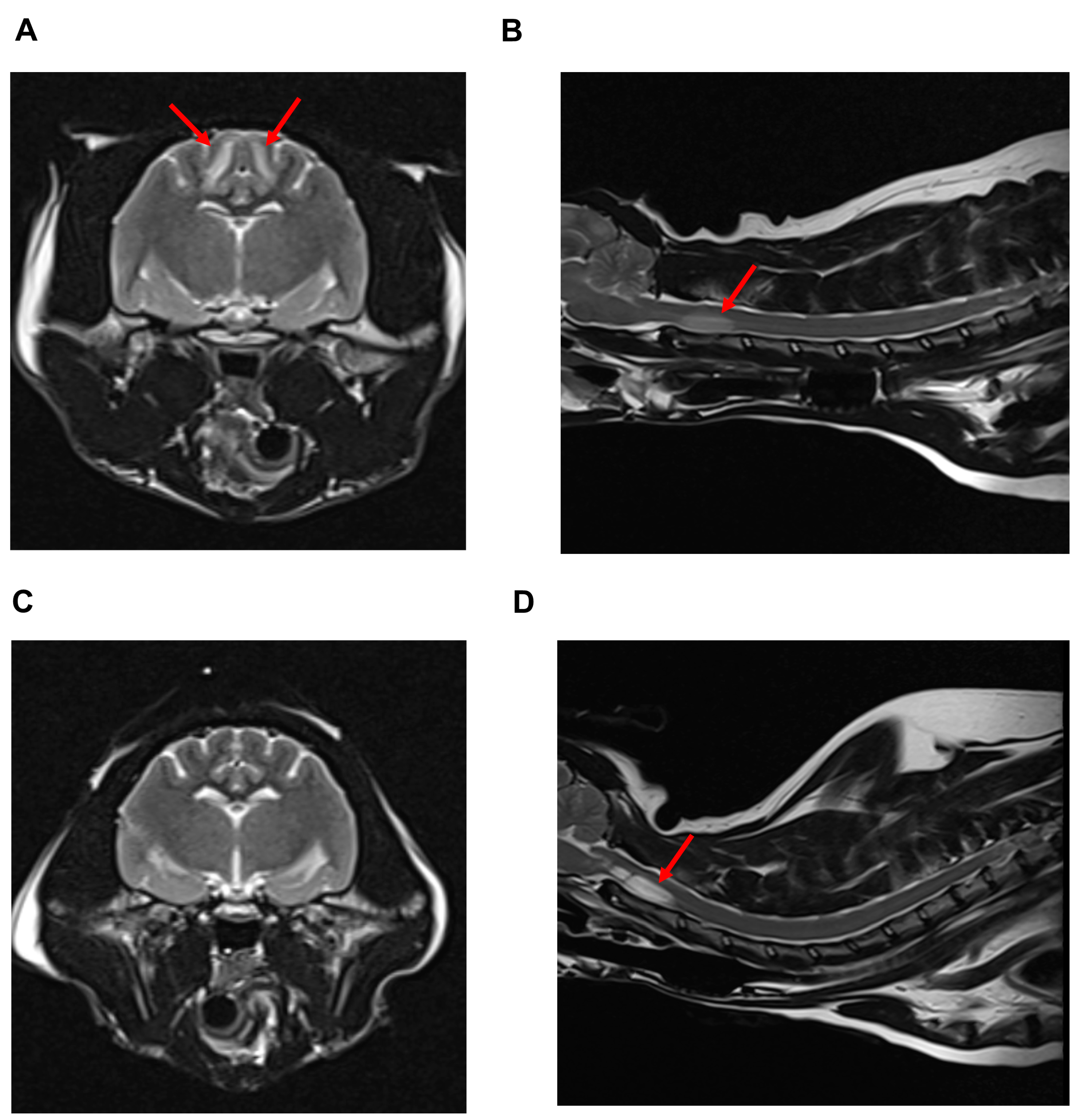
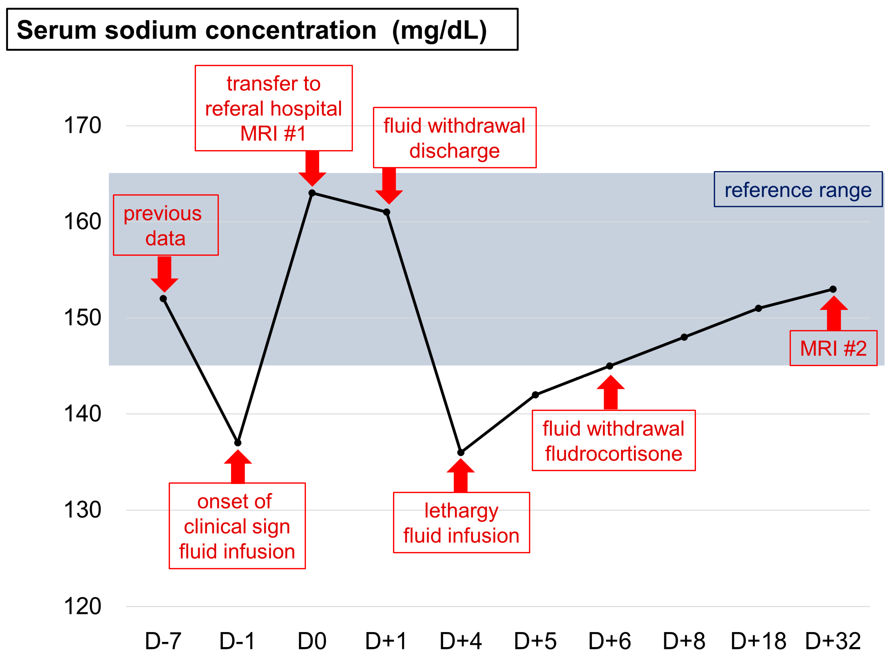
2. Case Description
3. Discussion
4. Conclusions
Author Contributions
Funding
Institutional Review Board Statement
Informed Consent Statement
Data Availability Statement
Conflicts of Interest
References
- Burton, A.G.; Hopper, K. Hyponatremia in dogs and cats. J. Vet. Emerg. Crit. Care 2019, 29, 461–471. [Google Scholar] [CrossRef] [PubMed]
- Dibartola, S.P. Hyponatremia. Vet. Clin. N. Am. Small Anim. Pract. 1998, 28, 515–532. [Google Scholar] [CrossRef] [PubMed]
- Harrigan, M. Cerebral salt wasting syndrome. Crit. Care Clin. 2001, 17, 125–138. [Google Scholar] [CrossRef] [PubMed]
- Isotani, E.; Suzuki, R.; Tomita, K.; Hokari, M.; Monma, S.; Marumo, F.; Hirakawa, K. Alterations in plasma concentrations of natriuretic peptides and antidiuretic hormone after subarachnoid hemorrhage. Stroke 1994, 25, 2198–2203. [Google Scholar] [CrossRef] [PubMed]
- Cerdà-Esteve, M.; Cuadrado-Godia, E.; Chillaron, J.J.; Pont-Sunyer, C.; Cucurella, G.; Fernández, M.; Goday, A.; Cano Pèrez, J.F.; Rodrìguez-Campello, A.; Roquer, J. Cerebral salt wasting syndrome: Review. Eur. J. Intern. Med. 2008, 19, 149–254. [Google Scholar] [CrossRef]
- Sakarcan, A.; Bocchini, J. The role of fludrocortisone in a child with cerebral salt wasting. Pediatr. Nephrol. 1998, 12, 769–771. [Google Scholar] [CrossRef]
- Nakamoto, Y.; Ozawa, T.; Mashita, T.; Mitsuda, M.; Katakabe, K.; Nakaichi, M. Clinical outcomes of suspected ischemic myelopathy in cats. J. Vet. Med. 2010, 72, 1657–1660. [Google Scholar] [CrossRef]
- Albanese, A.; Hindmarsh, P.; Stanhope, R. Management of hyponaetremia in patients with acute cerebral insults. Arch. Dis. Child 2001, 85, 246–251. [Google Scholar] [CrossRef]
- Simpson, K.M.; Risio, L.D.; Theobald, A.; Garosi, L.; Lowrie, M. Feline ischaemic myelopathy with a predilection for the cranial cervical spinal cord in older cats. J. Feline Med. Surg. 2014, 16, 1001–1006. [Google Scholar] [CrossRef]
- Fortgens, P.; Pillay, T.S. Pseudohyponatremia revisited: A modern-day pitfall. Arch. Pathol. Lab. Med. 2011, 135, 516–519. [Google Scholar] [CrossRef]
- Fried, L.F.; Palevsky, P. Hyponatremia and hypernatremia. Med. Clin. North. Am. M. 1997, 81, 585–609. [Google Scholar] [CrossRef]
- Brady, C.A.; Hughes, D.; Drobatz, K.J. Association of hyponatremia and hyperglycemia with outcome in dogs with congestive heart failure. J. Vet. Emerg. Crit. Care 2004, 14, 177–182. [Google Scholar] [CrossRef]
- Cameron, K.; Gallagher, A. Inappropriate antidiuretic hormone secretion in a cat. J. Am. Anim. Hosp. Assoc. 2010, 46, 425–432. [Google Scholar] [CrossRef]
- Maesaka, J.K.; Gupta, S.; Fishbane, S. Cerebral salt-wasting syndrome: Does it exist? Nephron 1999, 82, 100–109. [Google Scholar] [CrossRef]
- Uygun, M.A.; Özkal, E.; Acar, O.; Erongun, U. Cerebral salt wasting syndrome. Neurosurg. Rev. 1996, 19, 193–196. [Google Scholar] [CrossRef]
- Leonard, J.; Garrett, R.E.; Salottolo, K.; Slone, D.S.; Mains, C.W.; Carrick, M.M.; Bar-Or, D. Cerebral salt wasting after traumatic brain injury: A review of the literature. Scand. J. Trauma Resusc. Emerg. Med. 2015, 23, 98. [Google Scholar] [CrossRef]
- Sterns, R.H.; Silver, S.M. Cerebral salt wasting versus SIADH: What difference? J. Am. Soc. Nephrol. 2008, 19, 194–196. [Google Scholar] [CrossRef]
- Schrier, R.W.; Gross, P.; Gheorghiade, M.; Berl, T.; Verbalis, J.G.; Czerwiec, F.S.; Orlandi, C. Tolvaptan, a selective oral vasopressin V2-reveptor antagonist, for hyponatremia. N. Engl. J. Med. 2006, 355, 2099–2112. [Google Scholar] [CrossRef]
- Hasan, D.; Lindsay, K.W.; Wijdicks, E.F.M.; Murray, G.D.; Brouwers, P.J.; Bakker, W.H.; Fign, J.V.; Vermeulen, M. Effect of fludrocortisone acetate in patients with subarachnoid hemorrhage. Stroke 1989, 20, 1156–1161. [Google Scholar] [CrossRef]
- Steele, A.; Deveber, H.; Quaggin, S.E.; Scheich, J.E.; Halperin, M.L. What is responsible for the diurnal variation in potassium excretion? Am. J. Physiol. Regul. Integr. Comp. Physiol. 1994, 267, R554–R560. [Google Scholar] [CrossRef]
- Lathan, P.; Thompson, A.L. Management of hypoadrenocorticism (Addison’s disease) in dogs. Res. Vet. Sci. 2018, 9, 1–10. [Google Scholar]
- Theobald, A.; Volk, H.A.; Dennis, R.; Berlato, D.; Risio, L.D. Clinical outcome in 19 cats with clinical and magnetic resonanace imaging diagnosis of ischaemic myelopathy (2000–2011). J. Feline Med. Surg. 2013, 15, 132–141. [Google Scholar] [CrossRef]
- Silvestrini, P.; Piviani, M.; Masian, D.S. Ischaemic myelopathy in a cat with chronic kidney disease, hyperthyroidism, and hyperaldosteronism. Vet. Rec. Case Rep. 2021, 9, e11. [Google Scholar] [CrossRef]
- Cui, H.; He, G.; Yang, S.; Lv, Y.; Jiang, Z.; Gang, X.; Wang, G. Inappropriate antidiuretic hormone secretion and cerebral salt-wasting syndromes in neurological patients. Front. Neurosci. 2019, 13, 1170. [Google Scholar] [CrossRef] [PubMed]
- Bouchlarhem, A.; Haddar, L.; Berrichi, H.; Jabri, M.; Lachhab, A.; Lamassab, N.E.; Bekkaoui, S.; Mamoun, I.B.E.; Berramdane, O.; Oulali, N. Cerebral Salt Wasting Syndrome (CSW): An unusual cause of hypovolemia after spontaneous cerebral hemorrhage successfully treated with fludrocortisone. Radiol. Case Rep. 2022, 17, 106–110. [Google Scholar] [CrossRef]


| Parameters | Value | Reference Range | Unit |
|---|---|---|---|
| CBC | |||
| RBC | 8.5 | 6.54–12.2 | 1012/L |
| HCT | 34.9 | 30.3–52.3 | % |
| #Reticulocytes | 18.7 | 3–50 | 103/µl |
| WBC | 10.36 | 2.87–17.02 | 109/L |
| PLT | 169 | 151–600 | 109/L |
| Serum chemistry | |||
| Na | 137 | 150–165 | mmol/L |
| K | 2.9 | 3.5–5.8 | mmol/L |
| Total protein | 7.2 | 5.7–8.9 | g/dL |
| BUN | 27 | 16–36 | mg/dL |
| Glucose | 170 | 7–159 | mg/dL |
| SDMA | 10 | 0–14 | µg/dL |
| Total T4 | 0.8 | 0.8–4.7 | µg/dL |
| Basal cortisol | 5.91 | 0.5–10 | µg/dL |
| SAA | 41.5 | 0–5 | µg/dL |
| NT-proBNP | 478 | 0–100 | pmol/L |
| Coagulation | |||
| D-dimer | <0.1 | 0–0.1 | µg/ml |
| Hyperosmolality | Normosmolality | Hyposmolality | ||
|---|---|---|---|---|
| Hypervolemia | Normovolemia | Hypovolemia | ||
| Hyperglycemia | Hyperlipidemia | Liver function loss | Primary polydipsia | Hypo- adrenocorticism |
| Mannitol infusion | Hyperproteinemia | Kidney function loss | Hypothyroidism | Pancreatitis |
| Congestive heart failure | Iatrogenic (Diuretics) | Peritonitis | ||
| Syndrome of inappropriate antidiuretic hormone | Gastrointestinal fluid loss | |||
| Burn | ||||
| Cerebral salt wasting syndrome | ||||
| Parameters | SIADH | CSWS |
|---|---|---|
| Hydration status | Hydrated | Dehydrated |
| Urine volume | Variable | Increased |
| Plasma BNP | Variable | Increased |
| ADH | Increased | Decreased |
| Serum uric acid | Decreased | Increased |
| CVP | Increased | Decreased |
| Management | Fluid restriction | Fluid infusion |
Disclaimer/Publisher’s Note: The statements, opinions and data contained in all publications are solely those of the individual author(s) and contributor(s) and not of MDPI and/or the editor(s). MDPI and/or the editor(s) disclaim responsibility for any injury to people or property resulting from any ideas, methods, instructions or products referred to in the content. |
© 2023 by the authors. Licensee MDPI, Basel, Switzerland. This article is an open access article distributed under the terms and conditions of the Creative Commons Attribution (CC BY) license (https://creativecommons.org/licenses/by/4.0/).
Share and Cite
Kim, M.; Song, W.-J.; Park, J.; Lee, S.; Choen, S.; Kim, M.-C.; Yun, Y. Suspected Cerebral Salt Wasting Syndrome with Cervical Spinal Lesion in a Domestic Shorthair Cat. Vet. Sci. 2023, 10, 385. https://doi.org/10.3390/vetsci10060385
Kim M, Song W-J, Park J, Lee S, Choen S, Kim M-C, Yun Y. Suspected Cerebral Salt Wasting Syndrome with Cervical Spinal Lesion in a Domestic Shorthair Cat. Veterinary Sciences. 2023; 10(6):385. https://doi.org/10.3390/vetsci10060385
Chicago/Turabian StyleKim, Minkun, Woo-Jin Song, Jongjin Park, Saeyoung Lee, Sangkyung Choen, Myung-Chul Kim, and Youngmin Yun. 2023. "Suspected Cerebral Salt Wasting Syndrome with Cervical Spinal Lesion in a Domestic Shorthair Cat" Veterinary Sciences 10, no. 6: 385. https://doi.org/10.3390/vetsci10060385