Article

The Status and Risk Factors of Brucellosis in Smallholder Dairy Cattle in Selected Regions of Tanzania

by
Isaac Joseph Mengele
1,2,3,4,*,
Gabriel Mkilema Shirima
1,
Shedrack Festo Bwatota
1,
Shabani Kiyabo Motto
1,5,
Barend Mark de Clare Bronsvoort
6,7,
Daniel Mushumbusi Komwihangilo
8,
Eliamoni Lyatuu
9,
Elizabeth Anne Jessie Cook
3,4,* and
Luis E. Hernandez-Castro
6,7
1
Department of Global Health and Biomedical Sciences, School of Life Science and Bioengineering, The Nelson Mandela African Institution of Science and Technology (NM-AIST), Arusha P.O. Box 447, Tanzania
2
Tanzania Veterinary Laboratory Agency (TVLA), Central Zone Laboratory, Dodoma P.O. Box 1752, Tanzania
3
International Livestock Research Institute (ILRI), P.O. Box 30709, Nairobi 00100, Kenya
4
Center for Tropical Livestock Genetics and Health (CTLGH), International Livestock Research Institute (ILRI), P.O. Box 30709, Nairobi 00100, Kenya
5
Tanzania Veterinary Laboratory Agency (TVLA), Central Veterinary Laboratory, Dar Es Salaam P.O. Box 9254, Tanzania
6
The Epidemiology, Economics and Risk Assessment (EERA) Group, The Roslin Institute at the Royal (Dick) School of Veterinary Studies, University of Edinburgh, Midlothian EH25 9RG, UK
7
Center for Tropical Livestock Genetics and Health (CTLGH), The Roslin Institute at the Royal (Dick) School of Veterinary Studies, University of Edinburgh, Midlothian EH25 9RG, UK
8
Tanzania Livestock Research Institute (TALIRI), Dodoma P.O. Box 834, Tanzania
9
International Livestock Research Institute (ILRI), Dar Es Salaam P.O. Box 34441, Tanzania
*
Authors to whom correspondence should be addressed.
Vet. Sci. 2023, 10(2), 155; https://doi.org/10.3390/vetsci10020155
Submission received: 23 December 2022 / Revised: 16 January 2023 / Accepted: 28 January 2023 / Published: 14 February 2023
(This article belongs to the Section Veterinary Biomedical Sciences)

Abstract

:

Simple Summary

Brucellosis is a neglected, bacterial zoonotic disease that affects domesticated animals and people. Infection in cattle is caused by Brucella abortus which causes nonspecific clinical signs in female cattle including lowered milk production, infertility, and abortion in the last trimester. To determine the prevalence and the risk factors associated with brucellosis exposure, we conducted a cross-sectional study of smallholder dairy cattle in six regions of Tanzania, between July 2019 and October 2020. A total of 2048 dairy cattle blood samples were collected and tested for the presence of anti-Brucella antibodies. An overall seroprevalence of 2.39% was found with the highest seroprevalence in the Njombe Region (15.5%). The risk factors that were identified to be significantly associated with brucellosis seropositivity were having goats around dairy cattle and a history of abortion within a farm. The study findings suggest that brucellosis is still present in smallholder dairy cattle at varying levels among the studied regions. Education of smallholder dairy keepers is required regarding the disease, as well as risk and control measures for the disease. A One Health approach is required to study the role of small ruminants in the spread of the disease and to evaluate the public health risk to smallholder dairy farmers, especially in the Njombe Region.

Abstract

Bovine brucellosis is a bacterial zoonoses caused by Brucella abortus. We conducted a cross-sectional study to determine brucellosis seroprevalence and risk factors among smallholder dairy cattle across six regions in Tanzania. We sampled 2048 dairy cattle on 1374 farms between July 2019 and October 2020. Sera were tested for the presence of anti-Brucella antibodies using a competitive enzyme-linked immunosorbent assay. Seroprevalence was calculated at different administrative scales, and spatial tests were used to detect disease hotspots. A generalized mixed-effects regression model was built to explore the relationships among Brucella serostatus, animals, and farm management factors. Seroprevalence was 2.39% (49/2048 cattle, 95% CI 1.7–3.1) across the study area and the Njombe Region represented the highest percentage with 15.5% (95% CI 11.0–22.0). Moreover, hotspots were detected in the Njombe and Kilimanjaro Regions. Mixed-effects models showed that having goats (OR 3.02, 95% C 1.22–7.46) and abortion history (OR 4.91, 95% CI 1.43–16.9) were significant risk factors for brucellosis. Education of dairy farmers regarding the clinical signs, transmission routes, and control measures for brucellosis is advised. A One Health approach is required to study the role of small ruminants in cattle brucellosis and the status of brucellosis in dairy farmers in the Njombe and Kilimanjaro Regions.

1. Introduction

Brucellosis is a global, neglected bacterial zoonosis caused by an intracellular, aerobic, Gram-negative, nonencapsulated, coccobacillus bacteria of the genus Brucella [1,2,3]. Among Brucella species, B. abortus, B. melitensis, and B. suis infect cattle, small ruminants, and pigs, respectively [4,5]. In cattle, however, infection with B. melitensis or B. suis can also occur depending on disease transmission dynamics such as regular contact between dairy cattle and small ruminants or pigs at a farm or on grazing land [4,6]. Cattle transmission occurs after ingesting Brucella-contaminated feed and water from the uterine discharges, abortion materials, or fetal membranes of infected animals [7]. Bovine brucellosis causes nonspecific signs such as abortion during the last trimester, retained placenta, reduced milk production, orchitis, epididymitis, and rarely, arthritis [4,8,9]. A number of predisposing factors have been associated with brucellosis seropositivity such as older age of animal, history of abortion, large herd size, access to surface water, location, and contact with other animals [10,11,12,13,14,15].
The Tanzanian cattle production system is divided into three main sectors (e.g., pastoral, agropastoral, and dairy), which are all similarly affected by brucellosis. For instance, brucellosis seroprevalence in cattle kept by pastoral communities has been reported to be as high as 30% [16,17], whereas in agropastoral communities, seroprevalence has been reported to be up to 11.3% [14,18,19,20]. Smallholder dairy cattle are commonly crossbreeds of indigenous cattle with exotic breeds such as Friesian, Ayrshire, or Jersey.
The first brucellosis outbreak occurred in imported dairy cattle in 1927 [21], and the first brucellosis diagnosis was made in 1928 by using a serum agglutination test [22]. It was not until the 1970s that the Tanzania government started a brucellosis control programme in dairy cattle, which lasted until late 1990s. During that time, active surveillance, testing and slaughter, and calf vaccination using the B. abortus Strain 19 (S19) vaccine, were implemented which led to a reduction in brucellosis prevalence from 15.2% [21] to 2% [16]; since then, there have not been similar control programmes implemented by the government. As a result, there has been a number of reports of an increased trend of brucellosis seroprevalence in several regions, reaching up to 21% [12,13,14,19,23,24,25]. Although several studies have evidenced an increase in brucellosis within different farming systems, the factors driving brucellosis transmission in dairy systems remain unclear. Therefore, our study aims to establish the status and risk factors associated with brucellosis seroprevalence in smallholder dairy farming systems in six regions of high milk production in Tanzania.

2. Materials and Methods
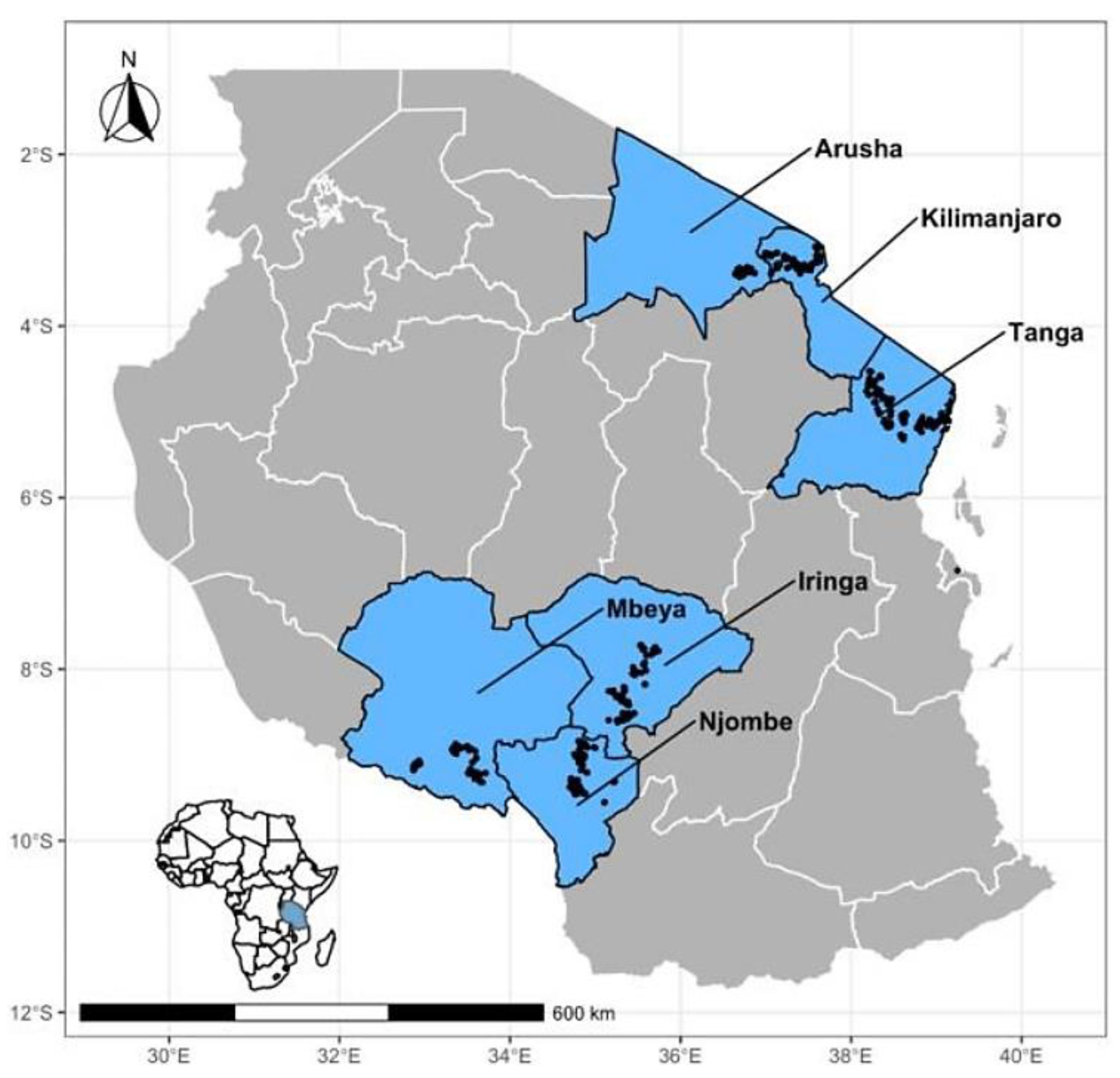
2.1. Study Area

This study was conducted from July 2019 to October 2020 across six administrative regions in Tanzania with the highest density of smallholder dairy cattle [26], i.e., the Arusha, Kilimanjaro, and Tanga Regions in the Northern Highland zone with 252,554 head of dairy cattle and the Iringa, Njombe, and Mbeya Regions in the Southern Highland zone with 103,306 head of dairy cattle (Figure 1). The 2019–2020 agricultural census reported that the nationwide top three regions with the highest number of dairy cattle (in decreasing order) were Tanga (>140,000), Arusha (>100,000), and Mbeya (>80,000). Other regions showed high densities of dairy cattle such as Kilimanjaro (>60,000), Iringa (>30,000), and Njombe (>20,000) [27].

2.2. Study Design and Sampling

Risk factors and the status of brucellosis at selected smallholder dairy farms in Tanzania were explored using a cross-sectional study. Most smallholder farms in these regions are characterized for having crossbred cattle of Friesian, Ayrshire, and Jersey with Tanzania Short Horn Zebu (TSHZ). However, Friesian crosses comprise the largest proportion (80%) of all breeds. Two main management systems are recognized in the area: (1) an intensive system in which pastures are cut and provided to livestock and (2) an extensive system in which cattle are left to graze in private or communal land.
The cattle in this study were selected from a subset of the dairy cattle registry of the Africa Dairy Genetics Gains (ADGG) program (https://data.ilri.org/portal/dataset/adgg-tanzania, accessed on 1 June 2019). The ADGG project has registered over 52,500 cattle across the regions from volunteer farmers, and approximately 4000 cattle were randomly selected for genotyping as part of genetic evaluations of this crossbred population [28]. The selected cattle had known genetic characteristics and could be identified by their preliminary information such as an ear tag number, age, and sex from the ADGG database [28].
These genotyped dairy cattle distributed across the 6 regions were the target of this study for establishing the disease phenotype. Due to small herd size in most farms, only one animal was genotyped, and therefore, sampled. However, the final sample size from each region was sufficient to estimate the seroprevalence of 5% (with 3% precision) and 95% confidence interval for the smallest region assuming simple random sampling [29,30].

2.3. Questionnaire Administration

A questionnaire tool was used to collect data on possible risk factors for brucellosis. The tool was developed and piloted before a final version was uploaded using the Open Data Kit (ODK) software platform on an International Livestock Research Institute (ILRI) server in Kenya (Supplementary Material Table S1). Then, the forms could be downloaded for completion on farm from the Google play app store onto an Android tablet (Samsung, Suwon-si, Republic of Korea).
Farmers or their representative were informed on the study aims and General Data Protection Regulation (GDPR) compliance. Farmers needed to sign a consent form before being interviewed in Kiswahili, and their cattle sampled. The questionnaire covered details of the respondent and their brucellosis knowledge, farm location, and herd management practices such as feeding and watering, disease control, abortion material handling, and cow or farm abortion history.

2.4. Blood Sampling, Pre-Analysis Processing, and Storage

Dairy cattle were manually restrained using ropes (halter) and hands, and 20 mL of blood was collected from the jugular vein using a sterile needle in two plain vacutainer tubes (BD Vacutainer®, Becton, Dickinson and Company, Franklin Lakes, NJ, USA). Tubes were barcoded (field barcode) and labeled with an animal identification number and collection date, and then stored in a cool box with ice packs before being transported to the Tanzania Veterinary Laboratory Agency (TVLA) zonal laboratories. Plasma and serum were left to separate overnight and tubes were centrifuged the next day at 3000 revolutions per minute for 5 min. Then, serum was collected and aliquoted into 2 mL cryovial tubes which were labeled and barcoded (laboratory barcode) as above before storing them at −20 °C. Tube barcodes, animal identification numbers, and collection date were recorded in a Microsoft Access® database which was later linked to the ODK questionnaire metadata. Finally, cryovials were transported at controlled temperature to the Nelson Mandela African Institution of Science and Technology (NM-AIST) in Arusha, Tanzania for storage at −20 °C until serological analysis was conducted.

2.5. Serological Analysis

All samples were tested according to the manufacturer’s instructions using a competitive enzyme-linked immunosorbent assay (cELISA) (COMPELISA 160 & 400, APHA Scientific, Weybridge, UK). The World Organisation for Animal Health (WOAH) recommends the use of cELISA for confirming prevalence of infection to Brucella in animals, furthermore, the test has high sensitivity (97.9%) and specificity (~100%) and can be used for testing poor quality serum samples [31,32]. Briefly, the test serum from the freezer and all reagents from the refrigerator (except for conjugate stored in −20 °C) were thawed at room temperature. Each test serum (20 µL) was placed in wells from columns 1 to 10 and 6 wells were left for control positive (20 µL) serum, 6 wells for control negative (20 µL) serum, and 4 wells for (20 µL) conjugate control within columns 11–12. Immediately after reconstitution, 100 µL of the conjugate was added to all 96 wells.
Plates were covered with a lid, shaken vigorously (200 revs/min) in a microtiter plate shaker for 2 min to allow mixing, and incubated at room temperature (21 ± 6 °C) for 45 min on a rotary shaker at 160 revs/min. Plate contents were shaken out, plates were washed 5 times using washing solution under low pressure, and finally, dried by taping onto a layer of absorbent towels until no more liquid was removed.
Next, 100 µL of prepared o-phenylenediamine dihydrochloride (OPD)-chromogen/substrate solution was added to each well on each plate which was incubated at room temperature for 20 min. Then, 100 µL of stopping solution was added to all wells to stop further reactions. Plate well absorbance was measured at 450 nm within 10 min using a SYNERGY|HTX multi-mode reader (BioTek Instruments, Winooski, VT, USA).

2.5.1. Plate Acceptance Criteria

The plate was considered to be valid when the mean optical density (OD) of the 6 negative control wells was greater than 0.700 (the optimal mean negative OD is 1.00), the mean OD of the 6 positive control wells was less than 0.100, the mean OD of the 4 conjugate control wells was greater than 0.700 (the optimal mean conjugate control wells is 1), and lastly, when the binding ratio (mean OD of negative control/mean OD of positive control) was greater than 10 [33,34].

2.5.2. Test Interpretation

Lack of color development indicated that the tested serum sample was positive. A positive/negative cut-off value was calculated as 60% of the mean OD of the four conjugate control wells. Any test sample giving an OD equal to or below this value was regarded as being positive.

2.6. Data Management and Analysis

Questionnaire data were downloaded from ODK and the laboratory data from the Access database were imported into RStudio, https://www.r-studio.com (accessed on 10 January 2022) and were joined and cleaned before analysis.
Animal level seroprevalences were calculated as the proportion of seropositive animals divided by the total number of animals tested or by region for both overall and regional seroprevalences. In addition, an overall adjusted seroprevalence was also estimated with a 95% exact binomial confidence interval for the reported cattle populations in each region (stratum) using the svydesign and svyciprop functions of the survey R package [35]. The stratum level 95% confidence interval was calculated using the binom.test function from R package stats.
A spatial scan statistic was used to detect statistically significant spatial clusters of seropositive animals in the Njombe and Kilimanjaro Regions only. Cluster analyses were performed using the SaTScan™ v10.1 software [36] with a Bernoulli model for binary events (i.e., seropositive/seronegative). SaTScan uses Monte Carlo hypothesis testing to obtain the p-values and SaTScan adjusts for the underlying spatial homogeneity of a background population. For each location and size of the scanning window, the alternative hypothesis was that there was an elevated risk within the window as compared with outside and a likelihood ratio test was performed. Multiple different window sizes were used and the locations were the latitude/longitude for each animal with slight jittering to avoid more than one animal being at a location. The window with the maximum likelihood was the most likely cluster, that is, the cluster least likely to be due to chance. A p-value was assigned to this cluster. For this analysis, we used 9999 Monte Carlo replications, and a cluster was considered to be statistically significant if the p-value was <0.05.
Univariable analysis was performed using the epiR and epitool packages. The multivariable model was estimated using a Firth’s adjusted logistic regression model and the logistf function in the logistf package [37] to deal with low counts for certain covariate patterns. This was used as a guide to compare with the generalized mixed-effects model fitted using the glmmTMB package with district name as a random effect (Supplementary Material Table S2). Only three regions with the highest number of seropositive animals (Kilimanjaro, Tanga and Njombe regions) were included in the multivariable model. Variables with p-values equal or less than 0.2 in the univariable analysis were included in the multivariable analysis. Animal age and breed were also included regardless of their p-value due to their inherent nature; sex was not included due to the zero value in one of the 2 by2 table cell, which could have caused errors during analysis. In multivariable analysis, the p-value for analysis of variance for dropping each value was also recorded for the Firth’s regression model with the same variables as a check. A maximum model was fitted and variables removed in a stepwise manner checking for confounding. The final best fit was based on the lowest Bayesian information criterion (BIC). Multicollinearity was checked using the Pearson correlation test on the variable pairs implemented in the ggpairs function from the GGally R package.

3. Results

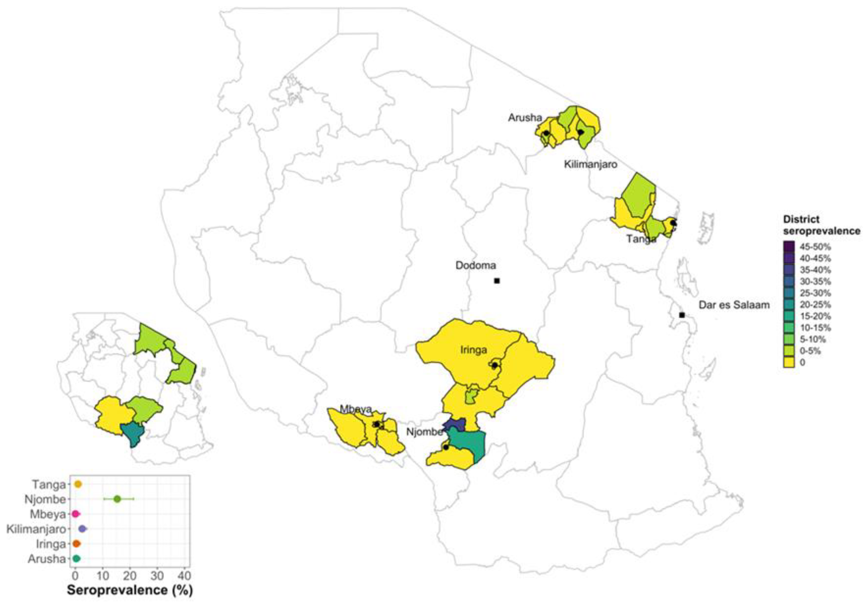
3.1. Brucellosis Seroprevalence in Smallholder Dairy Cattle in Selected Regions of Tanzania

A total of 2048 dairy cattle from 1374 smallholder farms were sampled from six regions of Tanzania and had complete test results (Table 1). Most animals (~64%) came from small farms with <4 animals (median herdsize = 2, IQR = 1) and there were four large farms in Iringa and Tanga with >90 animals. The cELISA was used to detect the circulating antibodies against Brucella species for all the samples and a total number of 49 dairy cattle were seropositive giving an unadjusted animal level seroprevalence of 2.39% (95% CI 0.017–0.31) and an adjusted animal level seroprevalence of 1.82% (95% CI 1.71–1.94) accounting for the design. Among the six regions, the Njombe Region was significantly associated with brucellosis (p < 0.05) and had the highest individual seroprevalence of 15.5% (95% CI 0.11–0.22) (Table 1 and Figure 2).

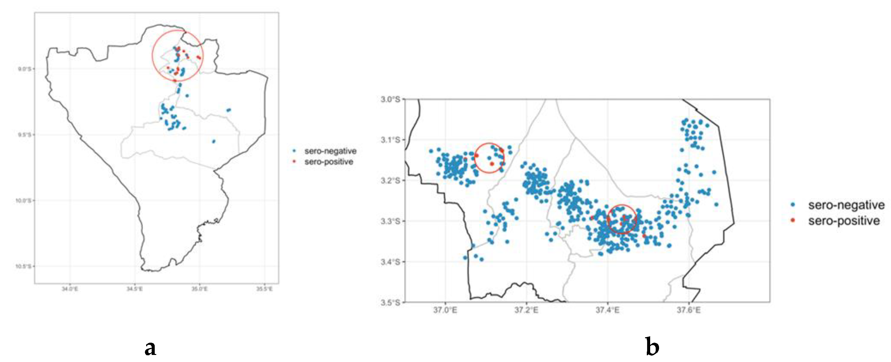
3.2. Brucellosis Hotspot Areas

The spatial choropleth map (Figure 2) shows that the seropositive animals were clustered within a small number of local authorities in the Njombe and Kilimanjaro Regions. There was a total of 29/187 seropositive animals in the Njombe Region representing 26 farms with one or more positive animals from a total of 136 farms sampled. Only three seropositives came from farms that reported an abortion in the previous 12 months and only two seropositives were cattle that the owner believed had previously had an abortion. In Kilimanjaro, there were a further 13/521 seropositive animals in the region representing 13 farms from a total of 379 farms sampled.

3.3. Brucellosis Spatial Clustering of Seropositive Animals

To explore this further, only the Njombe and Kilimanjaro Regions were mapped (Figure 3), and visually, there appeared to be a cluster of positive animals in the northern part of the Njombe Region and also in Kilimanjaro Region. This was more formally tested using the spatial clustering test (p < 0.05) which identified a cluster of 84 animals within which were all 29 seropositives with relative risk of 26.4 (95% CI 3.7–190.6) and a radius of 21.14 km in the Njombe Region (Figure 4), and a further cluster comprising 49 animals with seven positive animals with a relative risk of 11.2 (95% CI 3.9–32.1) and radius of 3.93 km was identified in the Kilimanjaro Region (Figure 3b). The other cluster had fewer seropositive animals and was not statistically significant.

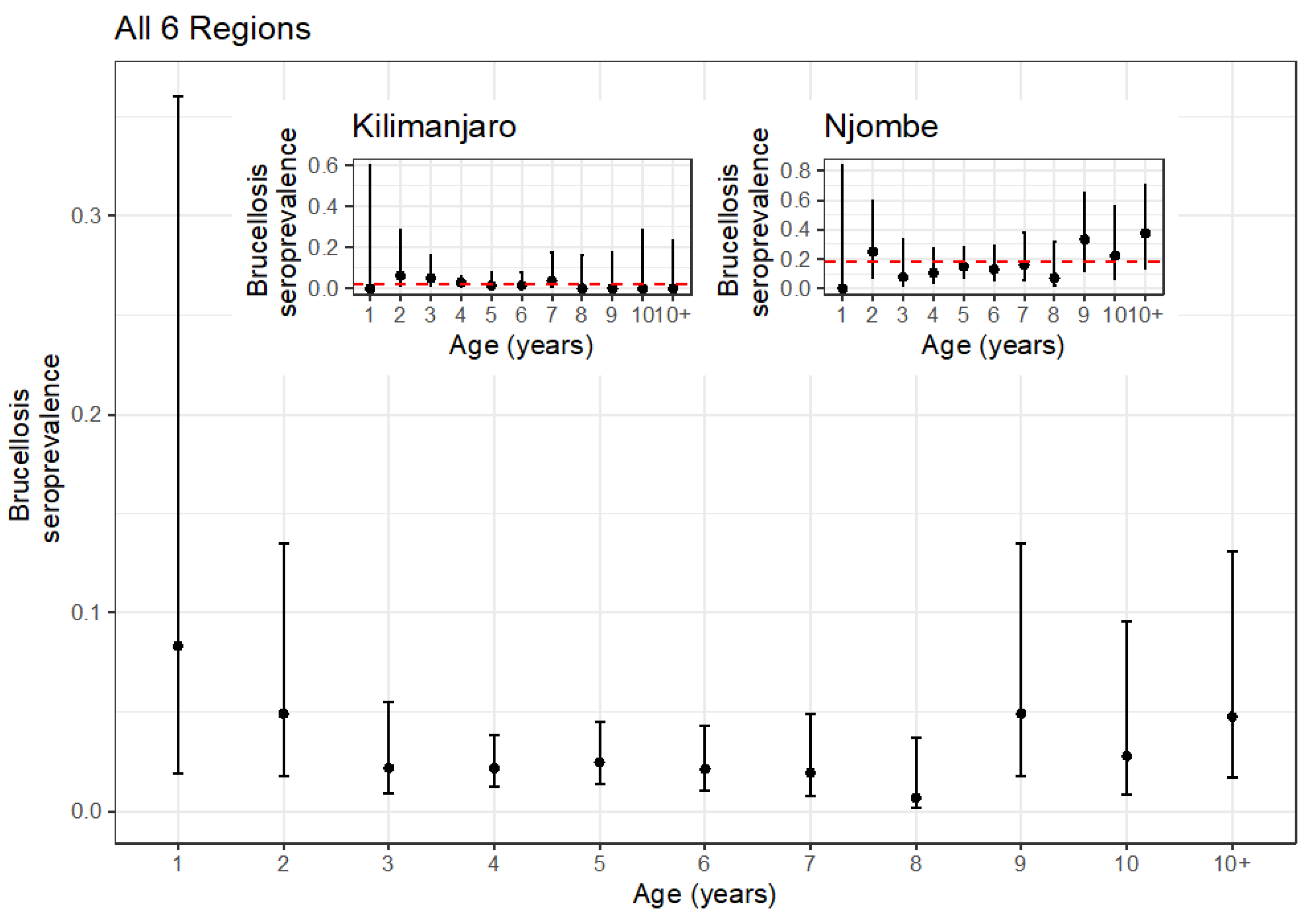
3.4. Age Stratification of Seropositive Animals

In order to try to assess if this was a single outbreak or more of an endemic expansion, the age-stratified seroprevalences were plotted (Figure 4). The overall age-stratified seroprevalences do not suggest any strong increase with age. In fact, it is very low across all ages, with the possible exception of the animals over 8 years old. However, given the apparently clustered pattern of seropositivity, the Njombe and Kilimanjaro Regions were separated out and plotted on their own (inset Figure 4). Again, the seroprevalence appears very uniform across ages in the Njombe Region at ~18% and the Kilimanjaro Region at ~2% (red dashed line).

3.5. Univariable Analysis Results

The initial univariable screening (Table 2) identified several factors at the farmer level that were associated with increased likelihood of animals being brucellosis seropositive, including level of education, knowledge and experience in dairy cattle keeping, and training and years of experience keeping cattle, which passed the threshold for inclusion in the multivariable model (p < 0.2). Cattle sex and whether the farmer had their own bull, although potentially associated (p < 0.01), had too few observations in one cell to be of use in the multivariable model. Only a small number of animals were from herds reporting routine vaccination against brucellosis, and all the animals from herds reporting vaccination were seronegative. There was some evidence supporting a breed effect, but animal age and management of feeding (zero grazed or at pasture) did not have any evidence of an association (p > 0.05). Herd size showed a decreasing risk with increasing herd size. The source of drinking water appeared to be associated with seropositivity (p < 0.05), but there were small numbers in one cell and the CI were very large and included a value of one. Distance between farms was associated (p < 0.05) with an increased risk for farms more than 100 m apart. Contact with goats, sheep, and dogs were all strongly associated with increased risk of seropositivity (p < 0.05). Correct disposal of the placenta and a history of abortion in the herd were also factors associated with an increased risk (OR > 1, p < 0.05). Finally, the clustered spatial pattern was captured by the two potential stratifying variables for region or zone with Njombe/Southern in particular showing a very strong association with being seropositive.

3.6. Final Multivariable Mixed-Effects Logistic Regression Model

The final model and the backward stepwise approach to the final model is shown in Supplementary Material Table S2. Towards the end of the selection, there were problems with large CIs for the zone variable with the glmm as compared with the Firth’s model. This was not improved by using region and, in the end, removing Zone from the glmm and leaving the random effect for the spatial component produced a much more stable model (Supplementary Material Table S2). Variables were reintroduced to check for any improvement in fit, and also an interaction between sheep and goat was tested. However, the final best fitting most parsimonious model included only the contact with goats variable and whether the farm had a recent (last 12 months) history of abortion on the farm (Table 3). Animals with contact with goats either on their own or on neighboring farms had a 3.02 (95% CI 1.22–7.46) increased odds of being seropositive, and animals from farms with a recent history of abortion had a 4.91 (95% CI 1.43–16.9) increased odds of being seropositive as compared with farms with no recent history of abortion.
Interestingly, there was no association with age, consistent with the age stratification analysis (Figure 4). In addition, the intracluster correlation coefficient (ICC) was very high at 0.641, suggesting that animals within clusters (in this case administrative districts) were very highly correlated.

4. Discussion

Brucellosis is one of the globally, neglected bacterial zoonosis [2] that continues to pose huge economic losses in LMICs including Tanzania due to lack of effective control measures and well-established surveillance systems [8,10] In Tanzania, recent studies have shown that brucellosis in dairy cattle is re-emerging [12]. Therefore, the current study aimed to determine the status and emerging risk factors for brucellosis in smallholder dairy cattle in selected high milk-producing regions of Tanzania.
The findings indicated the presence of circulating anti-Brucella antibodies in dairy cattle, suggesting the presence of brucellosis in selected regions. The population-adjusted animal level seroprevalence was 1.82% (95% CI 1.71–1.94) across all study regions. The seroprevalence was highest (15.5%) in the Njombe Region, while the Mbeya Region had no seropositive animals, and the Iringa and Arusha Regions only had one animal each. A similar study by Mathew et al. (2015) [24] provided evidence that the Njombe Region could be a brucellosis hotspot under smallholder dairy systems [24]. High seroprevalences were also reported in the Morogoro, Iringa, and Tanga Regions under smallholder dairy systems [12,14,19] but this was not observed in this study. The current findings suggest that mitigation measures for controlling brucellosis in smallholder dairy cattle must be prioritized and instituted especially in high-risk areas.
Brucellosis in smallholder dairy cattle in the Kilimanjaro Region is re-emerging. This study found that the region-specific seroprevalence in smallholder dairy cattle in the Kilimanjaro Region was 2.5%. A research study carried out in Moshi-Kilimanjaro in smallholder dairy cattle in 2000 found an animal-level seroprevalence of 12.2% [25]. However, a recent study carried out in the Kilimanjaro Region and nearby regions in dairy cattle populations found an overall animal-level seroprevalence of only 0.01% [38] suggesting that the disease was on a decreasing trend. However, the seroprevalence of 2.5% found by this study in the Kilimanjaro Region suggests that brucellosis is re-emerging, and therefore, dairy farmers must strengthen control measures for brucellosis to avoid it becoming endemic.
The spatial clustering analysis results suggest that two clusters were significantly associated with brucellosis seropositivity, one in the Njombe Region and the other in the Kilimanjaro Region, and the intracluster correlation of 0.61 suggested there was potentially a localized problem. Again, the seroprevalence appears to be very uniform across ages in the Njombe and Kilimanjaro Regions. This very clustered spatial pattern along with the very uniform age seroprevalence profile are more consistent with an outbreak of brucellosis rather than with a widespread general endemicity where you might expect to see seroprevalence increasing with age [39].
In the Mbeya Region, this study found no seropositive dairy cattle, contrary to the findings of previous studies which found seroprevalence ranging from 2.8% to 17.8% [13,15,40]. The decreasing trend of brucellosis in the Mbeya Region might be attributed to the implementation of control strategies following the findings of the previous studies.
Brucellosis in smallholder dairy cattle is also affecting neighboring countries in East and Southern Africa. Studies carried out in Rwanda, Zambia, Malawi, Burundi, and Uganda have recorded higher brucellosis seroprevalence of 23.1%, 6.0%, 7.7%, 14.7%, and 6.0%, respectively [41,42,43,44,45], than the overall findings of this study in Tanzania. Countries such as Kenya and Ethiopia have recorded seroprevalence between 1% and 1.9% which is lower than the findings of this study in Tanzania [1,10,29]. This suggested that brucellosis is present in Sub–Saharan Africa, and hence, regional efforts are required for the strategic control of brucellosis.
The univariable analysis demonstrated a number of variables significantly associated with Brucella seropositivity that were not maintained in the final model. We would like to highlight a couple of these because of their importance to control measures for Brucella in animals and people.
Dairy cattle kept by farmers who had livestock training were significantly more likely to be seropositive (OR 2.03, p < 0.05). These farmers had attended informal livestock training by mostly having meetings with livestock field officers for a few hours a day, the trainings normally focused on production and did not cover disease control extensively. Training and education on zoonotic diseases should be a focus of future programs to reduce the risks to cattle and people. The univariable analysis demonstrated that cattle on dairy farms which kept a bull were less likely to be brucellosis seropositve (OR 0.06, p < 0.05); this finding was in agreement with the findings of other studies which found that keeping or owning a bull for breeding was protective [46]. The use of brucellosis-free bulls for breeding is critical to reduce chances of spreading the disease to cows. Placenta disposal was significantly associated with seropositivity after the univariable analysis; dairy cattle on a farm which practiced incorrect disposal methods, such as feeding it to dogs, throwing it away in an open dump, or just leaving it, were 4.06 times more likely to be seropositive as compared with dairy cattle on a farm which practiced correct disposal methods such as burying, burning, or putting in toilets. Other studies have found that incorrect disposal of fetal membranes increases the chance of seropositivity [47,48]. Another study found that incorrect disposal of fetal membranes was associated with poor hygiene on farms, which led to contamination of pasture and water, and eventually, infection of cattle; however, there was no statistical significance [42]. This may also pose a risk for human exposure and appropriate disposal should be taught during training sessions.
Routine vaccination against brucellosis had a zero value in one of the 2 × 2 cells, and therefore, the analysis could not be performed. In Tanzania, the S.19 vaccine for brucellosis in cattle is produced by TVLA. However, vaccination is not widely practiced, similar to other African countries [42]. In order to promote vaccination, the Tanzanian government issued a list of strategic diseases which included controlling brucellosis by vaccination. In this study, some of the farmers claimed to have vaccinated their animals against brucellosis during interviews; however, on cross examination, there was no proof of vaccination. It is known that an effective control method for brucellosis in cattle is by vaccination [49,50]; however, some studies have not found a significant association between vaccination and brucellosis seropositivity (protection) [51,52], which may be attributed to the challenges associated with vaccination coverage, as well as vaccination and vaccine handling [52].
The multivariable mixed-effects model for brucellosis seropositivity for the three regions found that dairy farms with goats and farms which had histories of abortion during the past 12 months were significantly associated with brucellosis seropositivity.
Dairy cattle kept on farms which had goats were more likely to be seropositive (OR 3.02. 95% CI 1.22–7.46) than dairy cattle on farms which did not have goats. This finding was in agreement with the findings of other research studies that reported dairy cattle kept together with goats increased the risks of cattle contracting Brucella infection [1,46,53]. Brucella cross-species infection has been reported, B. melitensis has been identified in dairy cattle [54,55], and in Tanzania, B. abortus has been identified in goats [56]. The epidemiology of brucellosis in cattle is becoming more complex in mixed farming systems which are common practice in Tanzania and in LMICs.
In light of mixed farming practices and the cross-species infection nature of Brucella, identification of Brucella species circulating in dairy cattle populations is becoming increasingly important rather than measuring brucellosis seropositivity of cattle. Control of brucellosis by using the monovalent B. abortus S19 vaccine might be redundant if B. melitensis infects cattle. In Sub-Saharan Africa, small ruminants have been identified as being a challenge for the control of brucellosis and the primary cause of reemergence of brucellosis in cattle [57]. In Tanzania, brucellosis studies have been focused on cattle, with few studies in small ruminants. Future work should focus on understanding the roles of small ruminants in the epidemiology of brucellosis in cattle in its wider dimensions [58].
Brucellosis causes abortion in pregnant cows, and a history of abortion has been associated with brucellosis. This study found that a history of abortion was significantly associated with brucellosis seropositivity and that dairy cattle kept in a herd with a history of abortion were more likely to be brucellosis seropositive as compared with those in herds with no history of abortion (OR 4.91, 95% CI 1.43–16.9). These findings were in agreement with the findings of other studies in dairy cattle [11,41,42]. Farmers should be encouraged to report abortions in dairy cattle to the livestock officials for closer monitoring of the disease and implementation of control measures.

5. Conclusions and Recommendations

The current study on the status and risk factors for brucellosis in smallholder dairy cattle in selected regions of Tanzania confirms that brucellosis in smallholder dairy cattle is still a problem, and in some regions, it is re-emerging. The Njombe Region was identified as a hotspot and, since it is a region with emerging smallholder dairy farming, there needs to be further surveillance and control programs to manage the disease. Keeping goats or having goats around dairy cattle is an emerging risk factor for brucellosis in dairy cattle. In addition, dairy farmers should be educated about the risk and mitigation measures taken to reduce transmission between species and particularly the public health risk.

Supplementary Materials

The following supporting information can be downloaded at: https://www.mdpi.com/article/10.3390/vetsci10020155/s1, Table S1: Questionnaire; Table S2: Model selection.

Author Contributions

Conceptualization, I.J.M., G.M.S., E.A.J.C. and B.M.d.C.B.; methodology, I.J.M., G.M.S., E.A.J.C., B.M.d.C.B., S.K.M., S.F.B. and L.E.H.-C.; formal analysis, I.J.M., B.M.d.C.B., and L.E.H.-C.; resources, E.A.J.C. and B.M.d.C.B., data curation, I.J.M., S.K.M., S.F.B., B.M.d.C.B. and L.E.H.-C.; writing—original draft preparation I.J.M.; writing—review and editing, I.J.M., G.M.S., E.A.J.C., B.M.d.C.B., L.E.H.-C., E.L. and D.M.K.; supervision, G.M.S., B.M.d.C.B. and E.A.J.C.; project administration, G.M.S., B.M.d.C.B. and E.A.J.C.; funding acquisition, B.M.d.C.B. and E.A.J.C. All authors have read and agreed to the published version of the manuscript.

Funding

This study was conducted as part of the CGIAR Research Program on Livestock. ILRI is supported by contributors to the CGIAR Trust Fund. CGIAR is a global research partnership for a food-secure future. Its science is carried out by 15 Research Centres in close collaboration with hundreds of partners across the globe (www.cgiar.org, accessed on 10 December 2018). This research was funded in part by the Bill & Melinda Gates Foundation and with UK aid from the UK Foreign, Commonwealth and Development Office (Grant Agreement OPP1127286) under the auspices of the Centre for Tropical Livestock Genetics and Health (CTLGH), established jointly by the University of Edinburgh, SRUC (Scotland’s Rural College), and the International Livestock Research Institute (ILRI). This work was also supported by funding from the BBSRC (BBS/E/D/30002275). The funders had no role in study design, data collection and analysis, decision to publish, or preparation of the manuscript.

Institutional Review Board Statement

The animal study protocol was reviewed and approved by the International Livestock Research Institute Institutional Animal Care and Use Committee (ILRI-IACUC2018-27) and the research permit was granted by the Tanzania Commission for Science and Technology (COSTECH), reference (2019-207-NA-2019-95). Local authority approval was sought at all levels, from regional, district and village authorities which are under the President’s Office, Regional Administration and Local Government Authorities (PO-RALGA).

Informed Consent Statement

Consent forms were signed by cattle owners prior to the interview and sample collection.

Data Availability Statement

All relevant data are presented within the manuscript.

Acknowledgments

B.M.d.C.B.: E.A.J.C., I.J.M., S.F.B., S.K.M. and L.H.C were supported by the Bill & Melinda Gates Foundation and by a UK aid from the UK Foreign, Commonwealth, and Development Office (Grant Agreement OPP1127286) under the auspices of the Centre for Tropical Livestock Genetics and Health established jointly by the University of Edinburgh, Scotland’s Rural College, and the ILRI. Tanzania Livestock Research Institute (TALIRI) and the Local Government Authorities offices are recognized for their support. Thank you to the participants for their time. For the purpose of open access, the author has applied a Creative Commons Attribution (CC BY) license to any Author Accepted Manuscript version arising from this submission.

Conflicts of Interest

Authors declare that there are no conflict of interest.

References

  1. Asmare, K.; Sibhat, B.; Molla, W.; Ayelet, G.; Shiferaw, J.; Martin, A.D.; Skjerve, E.; Godfroid, J. The status of Bovine brucellosis in Ethiopia with special emphasis on exotic and cross bred cattle in dairy and breeding farms. Acta Trop. 2013, 126, 186–192. [Google Scholar] [CrossRef] [PubMed]
  2. Schelling, E.; Diguimbaye, C.; Daoud, S.; Nicolet, J.; Boerlin, P.; Tanner, M.; Zinsstag, J. Brucellosis and Q-fever seroprevalences of nomadic pastoralists and their livestock in Chad. Prev. Vet. Med. 2003, 61, 279–293. [Google Scholar] [CrossRef] [PubMed]
  3. Godfroid, J.; Cloeckaert, A.; Liautard, J.; Kohler, S.; Fretin, D.; Walravens, K.; Garin-Bastuji, B.; Letesson, J. From the discovery of the Malta fever’s agent to the discovery of a marine mammal reservoir, brucellosis has continuously been a re-emerging zoonosis. Vet. Res. 2005, 36, 313–326. [Google Scholar] [CrossRef] [PubMed]
  4. International Organization for Animal Health. OIE terestrial Manual Chapter 2.4.3. Bovine Brucellosis. In Manual of Diagnostic Tests and Vaccines for Terrestrial Animals; OIE: Paris, France, 2009; pp. 1–35. [Google Scholar]
  5. Pappas, G. The changing Brucella ecology: Novel reservoirs, new threats. Int. J. Antimicrob. Agents 2010, 36, S8–S11. [Google Scholar] [CrossRef] [PubMed]
  6. Muendo, E.N.; Mbatha, P.M.; Macharia, J.; Abdoel, T.H.; Janszen, P.V.; Pastoor, R.; Smits, H.L. Infection of cattle in Kenya with Brucella abortus biovar 3 and Brucella melitensis biovar 1 genotypes. Trop. Anim. Health Prod. 2012, 44, 17–20. [Google Scholar] [CrossRef]
  7. Radostits, O.M.; Gay, C.C.; Hinchcliff, K.W.; Vonstable, P.D. Veterinary Medicine. A Text Book of Disease of Cattle, Sheep, Pigs, Goats and Horses; W.B. Saunders: London, UK, 2007; pp. 963–985. [Google Scholar]
  8. McDermott, J.; Grace, D.; Zinsstag, J. Economics of brucellosis impact and control in low-income countries. Rev. Sci. Tech. 2013, 32, 249–261. [Google Scholar] [CrossRef]
  9. Currò, V.; Marineo, S.; Vicari, D.; Galuppo, L.; Galluzzo, P.; Nifosì, D.; Pugliese, M.; Migliazzo, A.; Torina, A.; Caracappa, S. The isolation of Brucella spp. from sheep and goat farms in Sicily. Small Rumin. Res. 2012, 106, S2–S5. [Google Scholar] [CrossRef]
  10. Terefe, Y.; Girma, S.; Mekonnen, N.; Asrade, B. Brucellosis and associated risk factors in dairy cattle of eastern Ethiopia. Trop. Anim. Health Prod. 2017, 49, 599–606. [Google Scholar] [CrossRef]
  11. Yanti, Y.; Sumiarto, B.; Kusumastuti, T.A.; Panus, A.; Sodirun, S. Seroprevalence and risk factors of brucellosis and the brucellosis model at the individual level of dairy cattle in the West Bandung District, Indonesia. Vet. World 2021, 14, 1–10. [Google Scholar] [CrossRef]
  12. Shirima, G.; Lyimo, B.; Kanuya, N.L. Re-emergence of Bovine Brucellosis in Smallholder Dairy Farms in Urban Settings of Tanzania. J. Appl. Life Sci. Int. 2018, 17, 1–7. [Google Scholar] [CrossRef]
  13. Sagamiko, F.; Muma, J.; Karimuribo, E.; Mwanza, A.; Sindato, C.; Hang’ombe, B. Sero-prevalence of Bovine Brucellosis and associated risk factors in mbeya region, Southern highlands of Tanzania. Acta Trop. 2018, 178, 169–175. [Google Scholar] [CrossRef] [PubMed]
  14. Swai, E.S.; Schoonman, L. The use of rose bengal plate test to asses cattle exposure to Brucella infection in traditional and smallholder dairy production systems of Tanga region of Tanzania. Vet. Med. Int. 2010, 2010, 837950. [Google Scholar] [CrossRef]
  15. Mathew, C.; Klevar, S.; Løken, T.; Mwamengele, G.; Skjerve, E.; Godfroid, J.; Stokstad, S.; Mdegela, R.H. Reproductive infections in cattle in Tanzania–lessons for control priorities. Microbiol. Infect. Dis. 2017, 5, 1–9. [Google Scholar] [CrossRef]
  16. Shirima, G.M. The Epidemiology of Brucellosis in Animals and Humans in Arusha and Manyara Regions in Tanzania. University of Glasgow. 2005. Available online: http://theses.gla.ac.uk/4826/ (accessed on 17 May 2021).
  17. Kanuya, N.L.; Matiko, M.K.; Kessy, B.M.; Mgongo, F.O.; Ropstad, E.; Reksen, O. A study on Reproductive Performance and Related Factors of Zebu cows in Pastoral Herds in a Semi-arid Area of Tanzania. Theriogenology 2006, 65, 1859–1874. [Google Scholar] [CrossRef] [PubMed]
  18. Jiwa, S.F.; Kazwala, R.R.; Tungaraza, R.; Kimera, S.I.; Kalaye, W.J. Bovine brucellosis serum agglutination test prevalence and breed disposition according to prevalent management systems in the Lake Victoria zone of Tanzania. Prev. Vet. Med. 1996, 26, 341–346. [Google Scholar] [CrossRef]
  19. Karimuribo, E.; Ngowi, H.; Swai, E.; Kambarage, D. Prevalence of brucellosis in crossbred and indigenous cattle in Tanzania. Livest. Res. Rural Dev. 2007, 19, 148–152. [Google Scholar]
  20. Mengele, I.J.; Garcia-Gonzalez, D.; Mekonnen, G.; Arenas-Gamboa, A. Bovine brucellosis seroprevalence, farmers’ awareness, practices and animal health extension services inputs in Mpwapwa district, Tanzania. Tanzan Vet. J. 2018, 36, 135–144. [Google Scholar]
  21. Mahlau, E.A. Further brucellosis surveys in Tanzania. Bull. Epizoot. Dis. Afr. 1967, 15, 373–378. [Google Scholar]
  22. Kitalyi, J. Bovine Brucellosis in Government Parastatal and Ujamaa Village Dairy Farms in the Central Zone of Tanzania: Assessment of Control Measures in Some of Farms. In Proceedings of the 2nd Tanzania Veterinary Association Scientific Conference, Arusha, Tanzania, 4–6 December 1984. [Google Scholar]
  23. Mdegela, R.; Kusiluka, L.; Kapaga, A.; Karimuribo, E.; Turuka, F.; Bundala, A.; Kivaria, F.; Kabula, B.; Manjurano, A.; Loken, T. Prevalence and determinants of mastitis and milk-borne zoonoses in smallholder dairy farming sector in Kibaha and Morogoro districts in Eastern Tanzania. J. Vet. Med. 2004, 51, 123–128. [Google Scholar] [CrossRef]
  24. Mathew, C.; Stokstad, M.; Johansen, T.; Klevar, S.; Mdegela, R.; Mwamengele, G.; Michel, P.; Escobar, L.; Fretin, D.; Godfroid, J. First isolation, identification, phenotypic and genotypic characterization of Brucella abortus biovar 3 from dairy cattle in Tanzania. BMC Vet. Res. 2015, 11, 156. [Google Scholar] [CrossRef]
  25. Swai, E.; Mshanga, D.; Sanka, N.P.; Marandu, N. Prevalence of bovine brucellosis in smallholder dairying farming area, Moshi, Tanzania. Bull. Anim. Health Prod. Afr. 2005, 53, 97–105. [Google Scholar] [CrossRef]
  26. Njombe, A.; Msanga, Y.; Mbwambo, M.; Makembe, M. Dairy Industry Status in Tanzania. Ministry of Livestock and Fisheries Development. 2011. Available online: https://dairyafrica.com/africadairyportal/wp-content/uploads/2020/06/Dairy_Industry_Status_in_Tanzania_2011.pdf (accessed on 10 May 2021).
  27. National Bureau of Statistics. National Sample Census of Agriculture 2019–2020. 2021. Available online: https://www.nbs.go.tz/index.php/en/census-surveys/agriculture-statistics/661-2019-20-national-sample-census-of-agriculture-main-report (accessed on 23 July 2022).
  28. Mrode, R.; Ojango, J.; Ekine-Dzivenu, C.; Aliloo, H.; Gibson, J.; Okeyo, M.A. Genomic prediction of crossbred dairy cattle in Tanzania: A route to productivity gains in smallholder dairy systems. J. Dairy Sci. 2021, 104, 11779–11789. [Google Scholar] [CrossRef] [PubMed]
  29. Gicheru, M.; Mwangi, E.; Mbaire, M. Prevalence and knowledge of Brucellosis in dairy cattle in Makuyu Division, Murang’a County, Kenya. Int. J. Sci. Eng. Technol. 2015, 4, 549–555. [Google Scholar] [CrossRef]
  30. Kasiulevičius, V.; Šapoka, V.; Filipavičiūtė, R. Sample size calculation in epidemiological studies. Gerontologija 2006, 7, 225–231. [Google Scholar]
  31. Stack, J.; Perrett, L.; Brew, S.; MacMillan, A. Competitive ELISA for bovine brucellosis suitable for testing poor quality samples. Vet. Rec. 1999, 145, 735–736. [Google Scholar] [CrossRef]
  32. International Organization for Animal Health. Chapter 3.1.4—Infection with B. abortus, B. mellitensis and B. suis. In Terestrial Manual; OIE: Paris, France, 2018. [Google Scholar]
  33. Ayoola, M.C.; Akinseye, V.O.; Cadmus, E.; Awosanya, E.; Popoola, O.A.; Akinyemi, O.O.; Perrett, L.; Taylor, A.; Stack, J.; Moriyon, I. Prevalence of bovine brucellosis in slaughtered cattle and barriers to better protection of abattoir workers in Ibadan, South-Western Nigeria. Pan Afr. Med. J. 2017, 28, 68. [Google Scholar] [CrossRef]
  34. Bronsvoort, B.M.; Koterwas, B.; Land, F.; Handel, I.G.; Tucker, J.; Morgan, K.L.; Tanya, V.N.; Abdoel, T.H.; Smits, H.L. Comparison of a flow assay for brucellosis antibodies with the reference cELISA test in West African Bos indicus. PLoS ONE 2009, 4, e5221. [Google Scholar] [CrossRef]
  35. Lumley, T. Complex Surveys: A Guide to Analysis Using R; John Wiley & Sons: New York, NY, USA, 2011. [Google Scholar]
  36. Kulldorff, M. Information Management Services. Software for the spatial and space-time scan statistics. SaTScanTMv8. 0 ed. 2009. Available online: https://www.satscan.org/ (accessed on 15 October 2022).
  37. Heinze, G.; Ploner, M.; Dunkler, D.; Southworth, H.; Heinze, M.G. Package ‘Logistf’. Available online: http://mirrors.nic.cz/R/web/packages/logistf/logistf.pdf (accessed on 17 October 2022).
  38. Bodenham, R.F.; Mazeri, S.; Cleaveland, S.; Crump, J.A.; Fasina, F.O.; de Glanville, W.A.; Haydon, D.T.; Kazwala, R.R.; Kibona, T.J.; Maro, V.P. Latent class evaluation of the performance of serological tests for exposure to Brucella spp. in cattle, sheep, and goats in Tanzania. PLoS Negl. Trop. Dis. 2021, 15, e0009630. [Google Scholar] [CrossRef]
  39. Awah-Ndukum, J.; Mouiche, M.; Bayang, H.N.; Ngwa, V.N.; Assana, E.; Feussom, K.; Manchang, T.K.; Zoli, P.A. Seroprevalence and associated risk factors of brucellosis among indigenous cattle in the Adamawa and north regions of Cameroon. Vet. Med. Int. 2018, 2018, 3468596. [Google Scholar] [CrossRef]
  40. Mfune, R.L. Epidemiological Study of Bovine Brucellosis in Smallholder Dairy Cattle in Lushoto and Rungwe Districts, Tanzania. Sokoine University of Agriculture (SUA). 2015. Available online: http://www.suaire.sua.ac.tz (accessed on 13 July 2022).
  41. Mfune, R.L.; Mubanga, M.; Silwamba, I.; Sagamiko, F.; Mudenda, S.; Daka, V.; Godfroid, J.; Hangombe, B.M.; Muma, J.B. Seroprevalence of bovine brucellosis in selected districts of Zambia. Int. J. Environ. Res. Public Health 2021, 18, 1436. [Google Scholar] [CrossRef]
  42. Segwagwe, B.E.; Samkange, A.; Mushonga, B.; Kandiwa, E.; Ndazigaruye, G. Prevalence and Risk Factors for Brucellosis Seropositivity in Cattle in Nyagatare District, Eastern Province, Rwanda. J. S. Afr. Vet. Assoc. 2018, 89, a1625. [Google Scholar] [CrossRef]
  43. Bernard, F.; Vincent, C.; Matthieu, L.; David, R.; James, D. Tuberculosis and brucellosis prevalence survey on dairy cattle in Mbarara milk basin (Uganda). Prev. Vet. Med. 2005, 67, 267–281. [Google Scholar] [CrossRef] [PubMed]
  44. Chagunda, M.G.; Mapemba, J.P.; Awah-Ndukum, J.; Tebug, S.F.; Wiedemann, S. Risk, knowledge and preventive measures of smallholder dairy farmers in northern Malawi with regard to zoonotic brucellosis and bovine tuberculosis. Onderstepoort J. Vet. Res. 2014, 81, a594. [Google Scholar] [CrossRef]
  45. Musallam, I.; Ndour, A.P.; Yempabou, D.; Ngong, C.C.; Dzousse, M.F.; Mouiche-Mouliom, M.; Feussom, J.M.K.; Ntirandekura, J.B.; Ntakirutimana, D.; Fane, A.; et al. Brucellosis in dairy herds: A public health concern in the milk supply chains of West and Central Africa. Acta Trop. 2019, 197, 105042. [Google Scholar] [CrossRef] [PubMed]
  46. Cárdenas, L.; Peña, M.; Melo, O.; Casal, J. Risk factors for new bovine brucellosis infections in Colombian herds. BMC Vet. Res. 2019, 15, 81. [Google Scholar] [CrossRef]
  47. Ogugua, A.J.; Akinseye, V.O.; Cadmus, E.O.; Jolaoluwa Awosanya, E.A.; Alabi, P.I.; Idowu, O.S.; Akinade, S.A.; Dale, E.J.; Perrett, L.; Taylor, A. Prevalence and risk factors associated with bovine brucellosis in herds under extensive production system in southwestern Nigeria. Trop. Anim. Health Prod. 2018, 50, 1573–1582. [Google Scholar] [CrossRef] [PubMed]
  48. Mekonnen, H.; Kalayou, S.; Kyule, M. Serological survey of bovine brucellosis in barka and arado breeds (Bos indicus) of Western Tigray, Ethiopia. Prev. Vet. Med. 2010, 94, 28–35. [Google Scholar] [CrossRef] [PubMed]
  49. Richey, E.J.; Harrell, C.D. Brucella Abortus Disease (Brucellosis) in Beef Cattle; University of Florida Cooperative Extension Service, Institute of Food and Agricultural Sciences: Gainesville, FL, USA, 1997. [Google Scholar]
  50. Muma, J.B.; Pandey, G.S.; Munyeme, M.; Mumba, C.; Mkandawire, E.; Chimana, H.M. Brucellosis among smallholder cattle farmers in Zambia. Trop. Anim. Health Prod. 2012, 44, 915–920. [Google Scholar] [CrossRef]
  51. Makita, K.; Fèvre, E.M.; Waiswa, C.; Eisler, M.C.; Thrusfield, M.; Welburn, S.C. Herd prevalence of bovine brucellosis and analysis of risk factors in cattle in urban and peri-urban areas of the Kampala economic zone, Uganda. BMC Vet. Res. 2011, 7, 60. Available online: http://www.biomedcentral.com/1746-6148/7/60 (accessed on 12 July 2022). [CrossRef]
  52. Nguna, J.; Dione, M.; Apamaku, M.; Majalija, S.; Mugizi, D.R.; Odoch, T.; Kato, C.D.; Tumwine, G.; Kabaasa, J.D.; Curtis, K. Seroprevalence of brucellosis and risk factors associated with its seropositivity in cattle, goats and humans in Iganga District, Uganda. Pan Afr. Med. J. 2019, 33, 99. [Google Scholar] [CrossRef] [PubMed]
  53. Anka, M.S.; Hassan, L.; Khairani, B.S.; Zainal, M.A.; Mohamad, R.; Salleh, A.; Adzhar, A. A case-control study of risk factors for bovine brucellosis seropositivity in Peninsular Malaysia. PLoS ONE 2014, 9, e108673. [Google Scholar] [CrossRef] [PubMed]
  54. Al-Majali, A.M.; Talafha, A.Q.; Ababneh, M.M.; Ababneh, M. Seroprevalence and risk factors for bovine brucellosis in Jordan. J. Vet. Sci. 2009, 10, 61–65. [Google Scholar] [CrossRef]
  55. Kaoud, H.; Zaki, M.; El-Dahshan, A.; Nasr, S. Epidemiology of brucellosis among farm animals. Nat. Sci. 2010, 8, 190–197. Available online: http://www.sciencepub.net/nature (accessed on 25 October 2022).
  56. Kassuku, H.A. Prevalence and risk factors for brucellosis transmission in goats in Morogoro, Tanzania. Sokoine University of Agriculture 2017. Available online: https://www.suaire.sua.ac.tz (accessed on 12 July 2022).
  57. Ducrotoy, M.; Bertu, W.J.; Matope, G.; Cadmus, S.; Conde-Álvarez, R.; Gusi, A.M.; Welburn, S.; Ocholi, R.; Blasco, J.M.; Moriyón, I. Brucellosis in Sub-Saharan Africa: Current challenges for management, diagnosis and control. Acta Trop. 2015, 165, 179–193. [Google Scholar] [CrossRef]
  58. Mengele, I.J.; Shirima, G.; Bronsvoort, B.M.; Hernandez-Castro, L.E.; Cook, E.A.J. Diagnostic challenges of brucellosis in humans and livestock in Tanzania: A thematic review. CABI One Health 2023, 2023, ohcs20230001. [Google Scholar] [CrossRef]
Figure 1. Map of Tanzania showing study regions (in light blue color) with high population of smallholder dairy cattle and unstudied regions in gray (right). Black dots indicate the locations of cattle sampled. Inset (top right corner) shows the location of Tanzania in Africa.
Vetsci 10 00155 g001
Figure 2. Choropleth map showing the regional seroprevalences (insets) and the detailed seroprevalence by local authority sampled in each region.
Vetsci 10 00155 g002
Figure 3. Map of Njombe (a) and Kilimanjaro (b) Regions showing district boundaries, the location of seropositive and seronegative animals (jittered), and the radius (red circle) of the significant clusters identified by the SaTScan analysis. In the Kilimanjaro Region, the top left cluster was not statistically significant.
Vetsci 10 00155 g003
Figure 4. Age-stratified seroprevalence of brucellosis across all six regions. Inset: Age-stratified seroprevalence for the Kilimanjaro and Njombe Regions with a red dashed line for the mean regional seroprevalence.
Vetsci 10 00155 g004
Table 1. Seroprevalence of brucellosis at the animal level in the study regions of Tanzania.
Animal Level Seroprevalence_cELISA
RegionNegativePositiveTotalPrev %95% CIPopWeights
Arusha31713180.30.00–1.7478,637247
Kilimanjaro508135212.51.65–4.241,63979
Tanga51955231.00.3–2.2161,984311
Mbeya21702170.00.0–1.672,724335
Iringa28012810.40.0–1.9708125
Njombe1582918715.511.0–22.0717738
TOTAL19994920482.391.7–3.1369,242
Pop, population; Prev, prevalence; CI, confidence interval.
Table 2. Univariable analysis based on the 3 regions with seropositive dairy animals for Brucella in Tanzania.
Number 95% CI
VariablesLevelsNegativesPositivesORLowerUpperp Value
Farmer’s genderFemale-headed farms537171
Male-headed farms648301.460.82.680.24
Livestock training attendedNo907291
Yes278182.031.113.70.02
Education level attainedBasic (none or primary only)886441
Secondary +29930.760.143.980.003
Experience in keeping dairy cattle<5 years5891
≥5 years1127380.370.180.770.006
Cattle sexFemale116447
Male210
Do you own BullNo967461
Yes21810.060.010.41<0.01
Do you routinely vaccinate for Brucella No116447
Yes210
BreedOther911
SHZxAyshire25860.210.0210.7
SHZxFriesian824390.430.0619.1
SHZxJersey9410.100.008.30.09
Age of cattle<5 years514181
5–7 years527201.080.542.2
>7 years14491.780.694.30.351
Feeding managementPasture20751
Zero-grazed978421.780.74.60.323
Herd size1–2 cows548321
3–4 cows402130.550.261.1
>4 cows23520.150.020.580.004
Water sourceRiver12121
Tap889261.770.4315.57
Well175196.541.5358.97<0.01
Distance between herds<100 m923221
≥100 m262254.02.227.22<0.01
DogsNo dog77522
Have dogs around410252.151.23.860.01
GoatsNo goats38971
Have goats around794402.81.246.310.01
SheepNo sheep979301
Have sheep around202172.751.495.07<0.01
PigsNo pigs993401
Have pigs around19270.910.402.050.811
RegionTanga5195
Kilimanjaro508132.650.889.58
Njombe1582918.957.163.8<0.01
ZoneNorthern1027181
Southern1582910.475.6819.3<0.01
Placenta disposalCorrect1102831
Incorrect36114.061.998.26<0.01
Abortion history (within herd)No1133421
Yes5252.590.996.830.06
OR, odds ratio; %, percent; CI, confidence interval.
Table 3. Final multivariable mixed-effects model for Brucella seropositivity in dairy cattle in the Tanga, Kilimanjaro and Njombe Regions of Tanzania. The intracluster correlation coefficient was 0.641.
Risk FactorOR95% CI
Goats
no goats1-
have goats around3.021.22–7.46
Abortion history
no1-
yes4.911.43–16.9
OR, odds ratio; CI, confidence interval.
Disclaimer/Publisher’s Note: The statements, opinions and data contained in all publications are solely those of the individual author(s) and contributor(s) and not of MDPI and/or the editor(s). MDPI and/or the editor(s) disclaim responsibility for any injury to people or property resulting from any ideas, methods, instructions or products referred to in the content.

