Article

β-Lactamase Producing Escherichia coli Encoding blaCTX-M and blaCMY Genes in Chicken Carcasses from Egypt

by
Elham Elsayed Abo-Almagd
1,†,
Rana Fahmi Sabala
2,†,
Samir Mohammed Abd-Elghany
2,
Charlene R. Jackson
3,
Hazem Ramadan
4,
Kálmán Imre
5,*,
Adriana Morar
5,
Viorel Herman
6 and
Khalid Ibrahim Sallam
2,*
1
Oncology Center, Faculty of Medicine, Mansoura University, Mansoura 35516, Egypt
2
Food Hygiene and Control Department, Faculty of Veterinary Medicine, Mansoura University, Mansoura 35516, Egypt
3
Bacterial Epidemiology and Antimicrobial Resistance Research Unit, US National Poultry Research Center, USDA-ARS, Athens, GA 30605, USA
4
Hygiene and Zoonoses Department, Faculty of Veterinary Medicine, Mansoura University, Mansoura 35516, Egypt
5
Department of Animal Production and Veterinary Public Health, Faculty of Veterinary Medicine, University of Life Sciences “King Mihai I” from Timișoara, 300645 Timișoara, Romania
6
Department of Infectious Diseases and Preventive Medicine, Faculty of Veterinary Medicine, University of Life Sciences “King Mihai I” from Timişoara, 300645 Timișoara, Romania
*
Authors to whom correspondence should be addressed.
These authors contributed equally to this work.
Foods 2023, 12(3), 598; https://doi.org/10.3390/foods12030598
Submission received: 25 December 2022 / Revised: 24 January 2023 / Accepted: 28 January 2023 / Published: 1 February 2023
(This article belongs to the Special Issue Advance and Future Challenges to Microbial Food Safety)

Abstract

:
Escherichia coli with multidrug resistance and β-lactamase genes may constitute a great public health hazard due to the potential for their transmission to humans through the food chain. This study determined the prevalence, antibiotic resistance profiles, phylogroups, and β-lactamase genes of E. coli isolates from chicken carcasses marketed in Mansoura, Egypt. Interestingly, E. coli was detected in 98% (98/100) of the chicken carcasses examined, which seemed among the highest contamination rates by E. coli worldwide. From the 425 genetically verified uidA gene-positive E. coli, 85 isolates were further studied for antimicrobial resistance profiles, phylogroups, and β-lactamase genes. Interestingly, 89.41% of E. coli (76/85) strains tested against 24 different antibiotics were multidrug-resistant. Of the examined 85 E. coli isolates, 22 (25.88%) isolates harbored blaCTX-M and were resistant to ampicillin, cefazoline, and ceftriaxone, while three of them were resistant to ceftazidime besides. Nine (10.59%) E. coli strains harbored AmpC- β-lactamase blaCMY and were resistant to ampicillin. One isolate co-carried blaCMY and blaCTX-M genes, though it was negative for the blaTEM gene. Of the 35 isolates that harbored either extended-spectrum β-lactamase (ESBL) and/or AmpC β-lactamase genes, six strains (17.14%) were assigned to pathogenic phylogroup F and one to phylogroup E, whereas 28 (80%) isolates belonged to commensal phylogenetic groups.

1. Introduction

Antimicrobial resistance is one of the universal threats which affect human and animal health especially in developing countries, including Egypt, due to a lack of rules controlling the usage of antimicrobials in different sectors of life (animal, agriculture, as well as human) [1,2]. Food-producing animals usually receive antimicrobials as a treatment or prophylactic courses to protect them from many bacterial infections [3,4]. Such overuse and misuse of antimicrobials results in the production of highly resistant bacteria; even commensal bacteria can acquire such resistance owing to chromosomal mutation or via gaining resistance traits from mobile genetic elements (e.g., plasmids, integrons, and transposons) [5].
The β-lactam antibiotics are one of the most important and widely used antimicrobials for the treatment of bacterial infections in both humans and animals, due to their broad antimicrobial spectrum, good safety profile, and availability of orally used formulations, in addition to their low price in comparison to other antimicrobial categories [6]. They interrupt bacterial cell-wall formation as a result of covalent binding to essential penicillin-binding proteins (PBPs), enzymes that are involved in the terminal steps of peptidoglycan cross-linking in both Gram-negative and Gram-positive bacteria. β-lactam antibiotics are classified into cephalosporins, cephamycins, monobactams, and carbapenems [7].
As a result of the excessive use of β-lactams in animals during the last decades, it is not surprising that acquired resistance to β-lactams has been detected in bacteria of animal and human health concern [8,9,10]. The resistance to β-lactam antibiotics arises as a result of drug inactivation by β-lactamases, an enzyme produced by bacteria that hydrolyze β-lactam antibiotics [11].
Extended-spectrum β-lactamases (ESBL)-producing bacteria result from mutations in the blaTEM-1 and blaSHV-1 β-lactamase genes with the global dissemination of ESBL-encoded by blaCTX-M-producing organisms [12] exhibiting resistance to penicillin, oxyiminocephalosporins, and monobactams. The ESBL-producing bacteria cannot hydrolyze cephamycins and are suppressed by clavulanic acid [13]. Meanwhile, ampicillin class C β-lactamase (AmpC) enzymes are active on cephamycins, oxyiminocephalosporins, and monobactams [13]. The genes encoding the expanded spectrum ß-lactamases are mostly located on mobile genetic elements (e.g., plasmids or transposons), which enable them to be easily mobilized and transferred by horizontal gene transfer from one bacterium to another, even between different bacterial species [5], resulting in frequent treatment failures or reduced efficacy of the therapeutic agents when employing broad-spectrum β-lactams as one of the first lines of treatments in human medicine. Egypt has been listed among the countries with a high prevalence of extended-spectrum β-lactamases (ESBL), and AmpC b-lactamases resistance, in various Gram-negative bacteria from animals and humans [14,15].
Escherichia coli are among the most important foodborne pathogens that contaminated poultry meat and implicated worldwide in public health concerns [16]. Superbug E. coli strains have been disseminated all over all kinds of samples (environment, food, water, and human). Numerous studies revealed that antimicrobial-resistant E. coli infections in humans were caused by strains of animal origin [17,18,19]. β-lactamases-producing E. coli organisms have increased worldwide over the last decades irrespective of whether the bacteria are commensal or pathogenic [20,21]. The issue about the existence of such resistance genes in organisms is that such resistance genes are plasmid-mediated and can be transferred easily from one bacterium to another [22]. This may explain the implication of E. coli in the transmission of resistance genes to humans through food sources [23,24].
In Egypt, due to the continuous administration of antimicrobials of β-lactams classes at subtherapeutic doses for growth promotion and as prophylactic measures in the poultry industry sector, ESBL and AmpC β-lactamase-producing Gram-negative bacteria, especially E. coli, have frequently been isolated from poultry clinical samples [25,26]. Therefore, this study was conducted to screen poultry meat for its contamination with such resistant strains, which is considered a critical source for public health, via the food chain, resulting in foodborne infection of strict treatment options, highlighting the need to monitor the use of such antibiotics, as well as to establish continuous epidemiological study for the rapid detection of such resistance via human food chain sources.

2. Materials and Methods

2.1. Sample Collection and Preparation

A total of 100 whole local frozen chicken carcasses were randomly collected from 25 retail shops and supermarkets distributed in Mansoura city, Egypt, during the period of December 2016 to May 2017, for isolation and molecular characterization of Escherichia coli. Each individual sample was aseptically packaged into a clean polyethylene bag, marked, and transferred in an icebox to the Laboratory of Food Hygiene and Control Department, Faculty of Veterinary Medicine, Mansoura University, wherein the microbiological analysis was performed.

2.2. Isolation and Identification of Escherichia coli

Each whole poultry carcass was separately rinsed with 225 mL of sterile tryptone soya broth (CM0989; Oxoid, Hampshire, UK) and the suspension obtained was incubated at 37 °C, for 18–24 h. Enriched cultures were plated onto sterile MacConkey agar (CM1169; Oxoid, Hampshire, UK), then the plates were incubated for 24 h at 37 °C. Up to five typical presumptive E. coli colonies were sub-cultured onto nutrient agar slopes and incubated for 24 h at 37 °C for further biochemical and molecular identification. A total of 600 typical colonies were subjected to various biochemical tests for further identification and confirmation of E. coli.

2.3. Molecular Identification of E. coli

For molecular confirmation, the biochemically positive E. coli isolates (n = 434) were subjected to PCR for amplification of the uidA gene, the gene-specific for pathogenic E coli [27]. For chromosomal DNA isolation, 5 mL from the bacterial culture in nutrient broth (CM0001; Oxoid, Hampshire, UK) was incubated overnight at 37 °C, followed by centrifugation at 3000 rpm for 15 min. The supernatant was removed and the pellet was re-suspended in 1 mL nuclease-free water, homogenized, transferred to a 1.5-mL Eppendorf tube, vortexed, and placed in a heat block at 70 °C for 15 min. The tubes containing the heated lysate were then centrifuged and the resultant supernatant was used as a DNA template.
Escherichia coli strains isolated from examined chicken carcasses were screened for the presence of the uidA gene using a primer set for the amplification of the uidA gene with the following sequence 5′-ATGCCAGTCCAGCGTTTTTGC-3′ and 5′-AAAGTGTGGGTCAATAATCAGGAAGTG-3′ for sense and antisense, respectively. The primer set can amplify a DNA of 1487 bp (Eurofins Genomics®, St. Charles, MO, USA). PCR amplification was carried out in a 20-µL reaction mixture using a ready-to-use solution of GoTaq Green Master Mix, (Promega Corporation®, Madison, WI, USA) supplied in 2× Green GoTaq reaction buffer (pH 8.5). PCR reaction mixture consisted of 10 µL GoTaq Master Mix 2×, 1 µL (10 pmol) from each of sense and antisense primers, 2 µL DNA template, and 6 µL autoclaved water. The mixture was subjected to 30 cycles of amplification in a Gene Amp PCR system 2700 (Applied Biosystems®, Foster City, CA, USA). The first cycle was preceded by denaturation at 95 °C for 5 min. Each cycle performed for the uidA gene consisted of denaturation for 45 s at 95 °C, annealing for 45 s at 63 °C, and extension at 72 °C for 1 min, followed by a final extension at 72 °C for 5 min by the end of the last cycle, then cooling at 4 °C. The PCR products were separated through running agarose gel (1.5% agarose) electrophoresis after loading 5 μL of the PCR mixture for 35 min at 100 V. The amplified DNA was then stained in an ethidium bromide solution (Sigma-Aldrich® Co., St. Louis, MO, USA), followed by visualization and photographing under ultraviolet light.

2.4. Antimicrobial Susceptibility Testing

The susceptibility testing of eighty-five selected E. coli isolates against 24 chosen antimicrobials (ampicillin, piperacillin, ampicillin/sulbactam, piperacillin/tazobactam, ticarcillin/clavulanic, aztreonam, cefazolin, ceftriaxone, ceftazidime, cefepime, levofloxacin, gentamicin, tetracycline, tobramycin, trimethoprim/sulphamethoxazole, minocycline, amikacin, ertapenem, meropenem, doripenem, imipenem, nitrofurantoin, ciprofloxacin, and tigecycline) was carried out by determining the minimum inhibitory concentration (MIC) using the Sensititre semi-automated susceptibility system (Trek Diagnostic Systems®, Inc., Westlake, OH, USA) and the Sensititre Gram-negative plate GN4F according to the manufacturer’s directions. The results were interpreted according to the Clinical and Laboratory Standards Institute (CLSI) [28], except for tigecycline, which was tested according to European Committee on Antimicrobial Susceptibility Testing (EUCAST) breakpoints [29].
Escherichia coli ATCC 25922, Pseudomonas aeruginosa ATCC 27853, Enterococcus faecalis ATCC 29212, and Staphylococcus aureus ATCC 29213 were used as the control for the determination of MIC.

2.5. PCR Screening for β-Lactamase Genes

The confirmed Escherichia coli strains isolated from examined chicken carcasses that showed resistance to beta-lactam antibiotics were screened for β-lactam resistance genes (blaTEM, blaCTX-M, blaSHV, blaOXA, blaCMY genes) using PCR amplification [30,31,32,33] The details concerning primer sequences, annealing temperatures, and amplified product sizes are summarized in Table 1.

2.6. Phylogenetic Typing of E. coli Isolates

The confirmed E. coli strains isolated from chicken carcasses were examined using the quadruplex phylotyping PCR method according to Clermont et al. [34] based on the quadruplex genotype to detect the presence/absence of the four genes (arpA, chuA, yjaA, and TspE4C2). E. coli ATCC 25922 and ATCC BAA-196 served as control positive strains. Quadruplex PCR was conducted using GoTaq Green Master Mix (Promega Corporation®, Madison, WI, USA). A reaction mixture of 20-µL volume consisted of 10 µL GoTaq Green Master Mix, 2 µL of DNA template, and 1 µL (20 pmol) for each of the sense and antisense primers of the four genes. The PCR was performed using a Perkin-Elmer GeneAmp 9600 Thermal Cycler in which the mixture was subjected to 30 cycles of amplification. The annealing temperatures for the different amplified genes were described in Table 1. The electrophoresis of PCR products was mentioned before.

3. Results and Discussion

3.1. Prevalence of E. coli in the Chicken Samples Examined

Escherichia coli was molecularly detected at a very high prevalence rate of 98% (98/100) among the 100 chicken carcasses tested. A total of 600 colonies (circular bright, pink-colored colonies) were selected from the 100 chicken carcasses, based on their morphological identification. Among these 600 colonies, 434 isolates were confirmed biochemically (production of indole from tryptophan, Methyl red test, Voges–Proskauer test, Citrate utilization test, Urease test) as E. coli and were further subjected to molecular confirmation by PCR for detection of the uidA gene: the E. coli marker gene. Interestingly, 97.92% (425/434) of the biochemically identified isolates were positive for the uidA gene (Figure 1), indicating that the biochemical tests can be used as an accurate tool for the identification of E. coli. A supplementary figure for the amplified DNA product of uidA gene for molecular verification of E. coli is presented in Figure S1.
The uidA gene is very specific to E. coli, especially the pathogenic strains associated with foodborne infection outbreaks, and is frequently identified by the redundant detection of uidA by PCR. The occurrence of E. coli at a high percentage in 98% of poultry carcass samples indicates poor hygienic measures during poultry carcass processing.
In developing countries, including Egypt, the use of conventional methods of slaughtering and evisceration resulted in poultry exposure to high contamination levels with different types of bacteria such as E. coli, which is considered a normal inhabitant of the intestinal tract of the live birds [36]. The present study is considered among those which showed the highest prevalence rate of E. coli in poultry carcasses. Likewise, high E. coli prevalence rates of 93.7% [37] and 90% [38] were reported in raw poultry carcasses in Egypt, and also by rates of 89% in Qatar [39], 87.5% in Turkey [40], and 60% in Bangladesh [41]. Such a high prevalence of E. coli could result in the production of inferior quality poultry meat with subsequent economic losses and public health hazards of great concern.
On the contrary, lower contamination rates of poultry meat by E. coli organisms had been recorded in former studies performed Egypt with an incidence of 37.7% [42], 35% [43], and 11.7% [25]. Lower E. coli contamination rates in poultry meat were also recorded worldwide. For instance, prevalence rates of 39.76%, 38.7%, and 40.82% were reported in India [44], the USA [45], and Bangladesh [46], respectively. The variations in the E. coli contamination rates among different studies depends on the hygienic facilities available through the poultry processing system. Therefore, strict hygienic measures during poultry carcass processing should be adopted.

3.2. Antimicrobial Resistance Profiles of E. coli Isolates

E. coli isolates from chicken carcasses exhibited variable degrees of resistance against the 24 antimicrobials tested (Figure 2). The highest resistance rate by more than 94% of the isolates was demonstrated against tetracycline, followed by a resistance rate of 87.06% of the isolates to ampicillin. To a lesser extent, resistance rates of isolates to trimethoprim/sulfamethoxazole, gentamicin, ampicillin/sulbactam, and cefazolin reached 51.76%, 51.76%, 45.88%, and 42.35%, respectively. On the other hand, all the isolates were susceptible to amikacin, cefepime, doripenem, piperacillin/tazobactam, ertapenem, imipenem, meropenem, nitrofurantoin, and tigecycline. Of the 85 E. coli strains tested, only one strain showed susceptibility to all antibiotics used in the study.
In the current study, the higher resistances rates of the E. coli strains isolated from poultry meat against tetracycline and ampicillin were in concordance with previous studies conducted in Egypt; in which 94.7% and 100% [47], as well as 80.9% and 71.4% [25], of E. coli isolates recovered from poultry meat were resistant to tetracycline and ampicillin, respectively. A similar pattern of antibiotic resistance of E. coli strains isolated from poultry meat had been recorded in previous studies in other countries [48,49], indicating the selective pressure of these antimicrobials in the treatment of E. coli infections in poultry due to the rational use of antimicrobial drugs in poultry production for treatment, subtherapeutic or prophylactic purposes, in addition to increasing productivity [3,50,51]. Therefore, a strict system should be established to control the use of antimicrobials not only for the animal, but also in the agricultural sectors.
Among the 85 E. coli isolates examined, 76 isolates (89.41%) were resistant to antimicrobials of three different classes or more, making them categorized as multidrug-resistant strains. Multidrug resistance (MDR) among E. coli strains isolated from poultry in previous studies exhibited a high prevalence of 69.1% [52] and 83% [53] of the E. coli isolates. The high prevalence of resistance patterns against three or more classes of antimicrobials could be related to the different antibiotic regimens used for the different livestock species [54,55]. In fact, it is nowadays accepted that the overuse of antibiotics in animals and poultry is the main driver for the dissemination of multi-drug resistance [56]. It is estimated that there is more extensive antibiotic use in livestock and poultry than in human medicine. The use of antimicrobials in animal production is associated with the emergence and spread of AMR in food-related bacteria and leads to the selection of antimicrobial resistance among pathogenic and commensal bacteria in the intestinal tract of food animals. Therefore, resistant bacteria can contaminate food products and colonize the human microbiota via the food chain by the handling and/or consumption of contaminated foods [57,58].
The present study indicated extensive contamination of chicken carcasses examined with multidrug-resistant E. coli that may constitute a tremendous threat to public health. The competent authorities should therefore enforce antimicrobial resistance (AMR) regulation to confirm the cautious use of antimicrobials to reduce the risk of transmission of antimicrobial-resistant organisms via the food chain. Farmers should be prevented from the unsystematic use of antimicrobials in poultry production and promoted to implement preventive measures by observing biosecurity in addition to good management practices.
Extended-spectrum β-lactamase (ESBL) and AmpC β-lactamase E. coli exhibited resistance against third-generation cephalosporins (ceftriaxone and ceftazidime) antibiotics detected in 35 (41.18%) E. coli isolates recovered from poultry meat in this study. Such prevalence of ESBL-producing E. coli is consistent with the prevalence rates reported in previous studies on ESBL E. coli in chicken meat in which ESBL-producing E. coli was recorded in Ghana with a prevalence rate of 52.8% [59], as well as in Germany, with a rate of 38.7% [60]. On the contrary to the findings of the current study, a lower prevalence of ESBL had been isolated from poultry meat in Tanzania at a rate of 20.1% [61] and in Pakistan by rates of 7.76% [62]. It has been indicated that among 52.9% AmpC-producing Enterobacteria, most isolates were identified as Escherichia coli [60]. It is noteworthy that many publications found that extended-spectrum β-lactamase (ESBL) and AmpC-producing E. coli present in humans and animals mostly shared identical sequence types (STs), suggesting the transmission of such resistant genes and human infections with ESBL-producing E. coli of animal source [63].

3.3. Determination of β-Lactamase Genes

The β-lactamase encoding gene blaTEM conferring resistance to penicillins was detected in 67.06% (57/85) of the E. coli isolated from the poultry carcasses. Twenty-six (25.88%) of the E. coli strains harbored blaCTX-M and were resistant to ampicillin, cefazoline, and ceftriaxone, while three of them were resistant to ceftazidime as well (Table 2). Ten (11.76%) E. coli strains harbored AmpC-β-lactamase blaCMY and were resistant to ampicillin. Six of them exhibited resistance to cefazoline and ceftriaxone harboring blaTEM, while two among these ten strains were also resistant to ceftazidime (Table 3). Six strains of the blaCMY and the blaCTX-M harboring E. coli strains were holding blaTEM genes (Table 2 and Table 3). One strain was holding both blaCMY and blaCTX-M genes and was negative for blaTEM. No E. coli strains were positive for blaSHV and blaOXA. The PCR-amplified DNA products of each of blaTEM, blaCTX-M, and blaCMY genes are presented in a supplementary figure (Figure S2).
It is well known that among the β-lactamase genes, blaTEM is the most predominant one that is widely spread in Gram-negative bacteria, encoding enzymes that hydrolyze penicillin and first-generation cephalosporins. In accordance with our findings concerning the predominance (67.06%) of blaTEM, previous studies confirmed such predominancy with prevalence rates of 92% [64] and 80% [65] in E. coli isolated from poultry. On the contrary, blaCTX-M was the predominant (58.1%) ESBL gene detected in E. coli recovered from chicken meat in the Netherlands [66].
Several studies in Egypt confirmed the spreading of blaCTX-M in different sources including food [14,25]. The blaCTX-M gene was formerly identified in ESBL-producing E. coli isolates from chicken and beef samples examined in Egypt [52]. Likewise, five ESBL E. coli strains harboring blaCTX-M were isolated from raw and ready-to-eat beef products [67], with the same blaCTX-M gene prevalence recorded in the present study. Globally, blaCTX-M was revealed as the predominant ESBL gene in ESBL-producing isolates [21], as major public health importance pathogen [68].
The AmpC β-lactamase blaCMY had been previously detected in six E. coli strains isolated from colibacillosis-infected chickens in Egypt [69], although this gene could not be detected in E. coli recovered from sound poultry meat [25]. It has been demonstrated that the first isolation of E. coli strains encoding the AmpC-resistant gene blaCMY in Egypt was recovered from hospitalized patients with urinary tract infections in three Egyptian hospitals [70]. Our study is the first that recovered E. coli strains harboring the blaCMY gene from sound (non-infected) chicken meat in Egypt.
All the ESBL and AmpC-producing E. coli strains isolated from poultry carcasses in the current study showed resistance against further antibiotics of different antimicrobial classes other than β-lactam. In this context, previous studies indicated that ESBL- and AmpC-producing bacteria are frequently cross-resistant to other antimicrobials such as aminoglycosides, chloramphenicol, tetracyclines, sulfonamides, trimethoprim, or quinolones, usually due to the existence of different resistance genes on mobile genetic elements such as plasmids, transposons, or integrons [71,72,73].

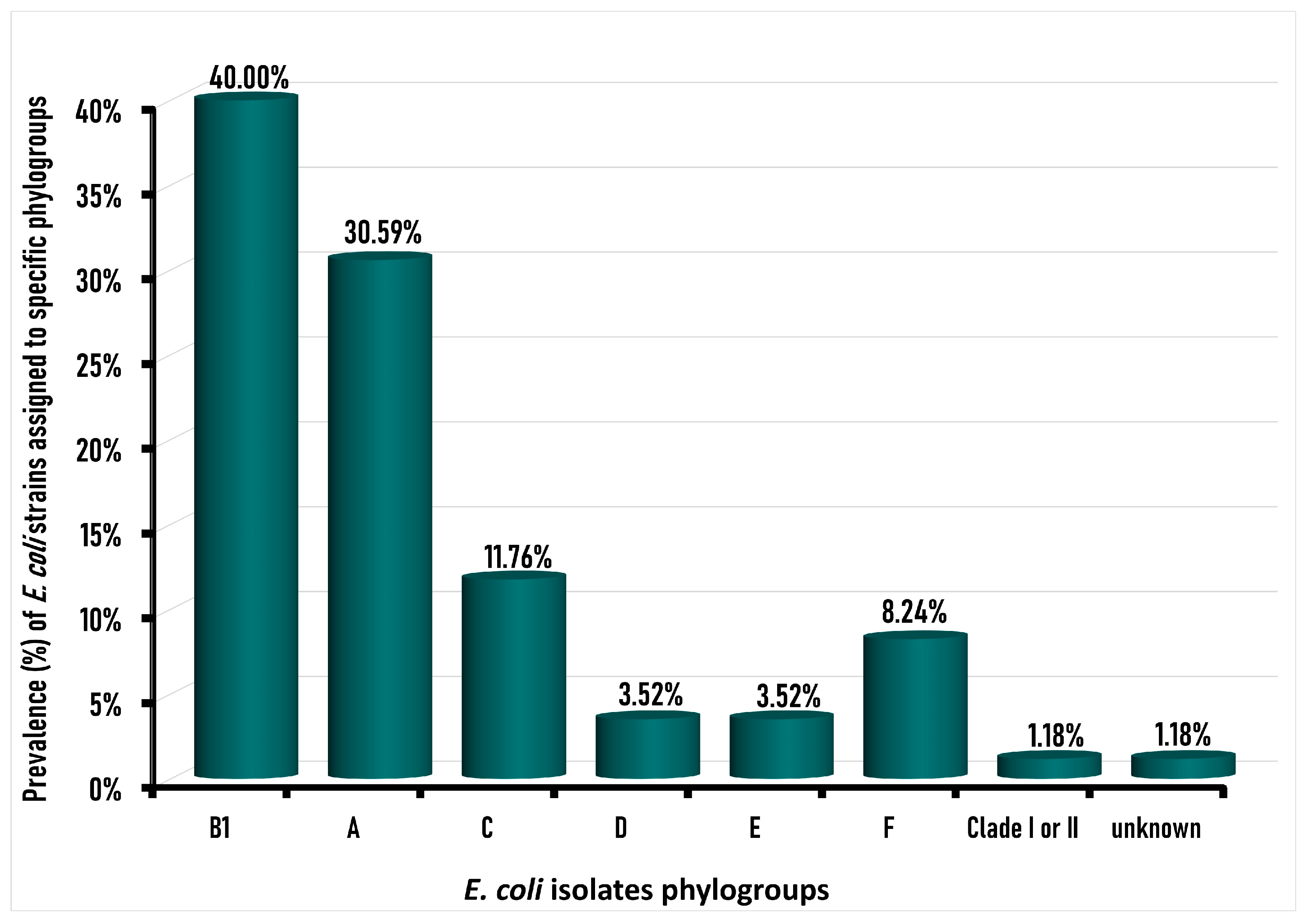
3.4. Phylogroup Characterization of Isolated E. coli Strains

The frequency distributions of different E. coli phylogroups among the tested E. coli isolates from chicken carcasses are illustrated in Figure 3.
Among the 85 E. coli isolates, the majority (70/85; 82.35%) were identified as commensal strains with a phylogroup of B1, A, or C, divided into phylogroup B1 (34/85; 40%), followed by phylogroup A (26/85; 30.59%), and phylogroup C (10/85; 11.76%). The pathogenic phylogroup E. coli was detected in 13 (15.29%) of the 85 E. coli isolates, which were distributed as 7 (8.24%) phylogroup F, 3 (3.52%) for each of D and E phylogroups, and only 1% was Clade I or II, with an absence of phylotype B2. The amplification of the genes incorporated in the phylogroup typing of the confirmed E. coli strains is presented as a supplementary figure (Figure S3) showing a representative agarose gel electrophoresis of the DNA for the arpA, chuA, yjaA, and trpA (quadruplex PCR), along with TspE4C2, trpA (group C) (singleplex), and arpA (group E).
Similar to the current study, the predominant phylogroup among the E. coli strains detected in previous studies in retail chicken samples worldwide were phylogroup B1 followed by phylogroup A, such as in Brazil [74,75], Italy [76], Ghana [51], and Pakistan [77], as well as in Egypt in poultry in which phylogroup B1 was the predominant phylogroup among the E. coli isolates (74%), followed by phylogroup C (20%), and then pathogenic phylogroup D (4%), while there were no E. coli strains possessing phylotypes A, B2, E and F [26].
Of the 35 E. coli which exhibited resistance against third-generation cephalosporin (ceftriaxone and ceftazidime) antibiotics in this study, six (17.14%) strains belonged to pathogenic phylogroup F and one to phylogroup E. However, the other 28 (80%) were considered as commensal E. coli strains belonging to commensal phylogenetic groups. Concerning our E. coli isolates that harbor blaCTX-M, 22.72% were phylogroup A, 50% belonged to phylogroup B1, while 13.63% belonged to each of phylogroup C and phylogroup F (Table 4). Similarly, 22.22% of blaCMY-positive Escherichia coli isolates were phylogroup A, while 33.33% were categorized as phylogroup B1 and F, whereas 11.11% only belonged to phylogroup C (Table 4).
It has been confirmed that the predominant phylogenetic grouping of extended-spectrum β-lactamases and AmpC-producing E. coli, which exhibited resistance against third-generation cephalosporins and were isolated from broiler chickens, were mainly type B1 and A (commensal phylogroup), followed by type D and B2 (pathogenic phylogroup) [60,78]. However, all (100%) ESBL E. coli isolated from diseased poultry samples in a previous study conducted in Egypt, belonged to phylogroup D, while most (66%) of the AmpC strains belonged to phylogroup B1 [69], which is quite opposite to that reported in our study.
The results of the current study might lead to the assumption that resistant group F isolates from chicken carcasses may contribute to the dissemination of antimicrobial resistance in humans, resulting in limited treatment of E. coli infection. It has been reported that E. coli belonging to phylogroup F that were isolated from chicken samples were closely related to the extraintestinal pathogenic E. coli causing human infections [79].
Above and beyond the fact that urinary tract infections (UTIs) are among the most common infections both in community and hospitals, they are most frequently caused by multidrug-resistant E. coli that challenge UTI treatment. Although the wide spreading of the phylogroups B2, F, and D pathogenic E. coli strains cause extraintestinal infection, the commensal E. coli strains belonging to the most newly diverged phylogroups are also responsible for severe intestinal infections.

4. Conclusions

The present study reported high contamination rates of chicken carcasses with multidrug-resistant E. coli contaminants, along with the isolation of pathogenic E. coli isolates harboring extended-spectrum β-lactamases and AmpC-β-lactamases-encoding genes blaCTX-M and blaCMY. To our knowledge, this is the first study isolating blaCMY from sound chicken meat for human consumption in Egypt. The occurrence of E. coli in almost all of chicken samples indicates poor hygiene and highlights the antimicrobial resistance problem in Egypt caused by the rational use of antimicrobials in animal husbandry and calls for a nationwide surveillance program to monitor antimicrobial resistance. These findings provide evidence that healthy broilers in Egypt could be a source for the dissemination of transmissible resistance mechanisms among foodborne pathogens brought from the unhygienic environment of the food chain during slaughtering. Although the majority of the isolated strains were commensal E. coli, about 16% of the strains were pathogenic, while 7% were pathogenic and categorized as a phylogroup F that harbored plasmid-mediated antibiotic-resistant genes, which may lead to infections via consumption of contaminated food with no treatment in humans. In addition, the commensal E. coli strains which harbor antibiotic-resistance genes, which are mainly plasmid-mediated strains, can be easily transmitted to further pathogenic bacteria that may lead to human infection with no treatment.

Supplementary Materials

The following supporting information can be downloaded at: https://www.mdpi.com/article/10.3390/foods12030598/s1, Figure S1: A representative agarose gel electrophoresis showing the amplified DNA product of the uidA gene (the marker gene for E. coli) at the expected molecular weight of 1487 bp. Genomic DNA from E. coli isolates was used as a template; Figure S2: A representative agarose gel electrophoresis for the PCR-amplified DNA products of the β-lactamase encoding genes blaCTX-M (A), blaCMY (B), and blaTEM (C), at the expected molecular weight of 550, 1007, and 1080 bp, respectively; Figure S3: A representative agarose gel electrophoresis for the PCR-amplified genes incorporated in the phylogroup typing of the confirmed E. coli strains. (A): The trpA (internal control), arpA, chuA, and yjaA were amplified as quadruplex PCR and determined at the expected molecular sizes of 489, 400, 288, and 211 bp, respectively Agarose gel electrophoresis of a singleplex-PCR for the amplification of trpA (group C), TspE4C2, and arpA (group E) which were verified at the expected molecular size of 219, 152, and 301 pb, respectively.

Author Contributions

Resources, data curation, investigation, methodology, writing—original draft, E.E.A.-A.; resources, data curation, investigation, formal analysis, writing—original draft—review and editing, project administration, R.F.S.; supervision, formal analysis, S.M.A.-E.; resources, project administration, C.R.J.; supervision, formal analysis, resources, project administration, writing—review and editing, H.R.; supervision, resources, writing—review and editing, K.I.; formal analysis, validation, visualization, data curation, A.M.; funding acquisition, software V.H.; conceptualization, project administration, investigation, writing—review and editing, K.I.S. All authors have read and agreed to the published version of the manuscript.

Funding

This research paper is supported by the project “Increasing the impact of excellence research on the capacity for innovation and technology transfer within USAMVB Timis, oara” code 6PFE, submitted in the competition Program 1—Development of the national system of research—development, Subprogram 1.2—Institutional performance, Institutional development projects—Development projects of excellence in RDI.

Data Availability Statement

Data is contained within the article or supplementary material.

Conflicts of Interest

The authors declare no conflict of interest.

References

  1. Adenipekun, E.O.; Jackson, C.R.; Oluwadun, A.; Iwalokun, B.A.; Frye, J.G.; Barrett, J.B.; Hiott, L.M.; Woodley, T.A. Prevalence and Antimicrobial Resistance in Escherichia coli from Food Animals in Lagos, Nigeria. Microb. Drug Resist. 2015, 21, 358–365. [Google Scholar] [CrossRef] [PubMed]
  2. World Health Organization. Draft Global Action Plan on Antimicrobial Resistance WHA68.7; World Health Organization: Geneva, Switzerland, 2015; pp. 1–4. Available online: http://apps.who.int/gb/ebwha/pdf_files/WHA68/A68_20-en.pdf?ua¼1 (accessed on 24 November 2022).
  3. Meena, H.R.; Kumar, V. Antimicrobial Resistance and Rational Use of Antimicrobials in Livestock: Developing Countries’ Perspective. In Livestock Health and Farming; IntechOpen: London, UK, 2020. [Google Scholar] [CrossRef]
  4. Van Boeckel, T.P.; Brower, C.; Gilbert, M.; Grenfell, B.T.; Levin, S.A.; Robinson, T.P.; Teillant, A.; Laxminarayan, R. Global trends in antimicrobial use in food animals. Proc. Natl. Acad. Sci. USA 2015, 112, 5649–5654. [Google Scholar] [CrossRef]
  5. von Wintersdorff, C.J.H.; Penders, J.; Van Niekerk, J.M.; Mills, N.D.; Majumder, S.; Van Alphen, L.B.; Savelkoul, P.H.M.; Wolffs, P.F.G. Dissemination of Antimicrobial Resistance in Microbial Ecosystems through Horizontal Gene Transfer. Front. Microbiol. 2016, 7, 173. [Google Scholar] [CrossRef]
  6. Hughes, L.A.; Pinchbeck, G.; Callaby, R.; Dawson, S.; Clegg, P.; Williams, N. Antimicrobial prescribing practice in UK equine veterinary practice. Equine Vet. J. 2013, 45, 141–147. [Google Scholar] [CrossRef] [PubMed]
  7. Bush, K.; Bradford, P.A. β-Lactams and β-lactamase inhibitors: An overview. Cold Spring Harb. Perspect. Med. 2016, 6, a025247. [Google Scholar] [CrossRef]
  8. CIPARS. Canadian Integrated Program for Antimicrobial Resistance Surveillance. Federal Organisation of CIPARS, Canada. 2006. Available online: https://www.canada.ca/en/public-health/services/surveillance/canadian-integrated-program-antimicrobial-resistance-surveillance-cipars.html (accessed on 24 December 2022).
  9. Ungureanu, V.; Corcionivoschi, N.; Gundogdu, O.; Stef, L.; Pet, I.; Pacala, N.; Madden, R.H. The emergence of β lactamase producing Escherichia coli and the problems in assessing their potential contribution to foodborne illness: A review. AgroLife Sci. J. 2019, 8, 248–260. [Google Scholar]
  10. Imre, K.; Ban-Cucerzan, A.; Herman, V.; Sallam, K.I.; Cristina, R.T.; Abd-Elghany, S.M.; Morar, D.; Popa, S.A.; Imre, M.; Morar, A. Occurrence, Pathogenic Potential and Antimicrobial Resistance of Escherichia coli Isolated from Raw Milk Cheese Commercialized in Banat Region, Romania. Antibiotics 2022, 11, 721. [Google Scholar] [CrossRef] [PubMed]
  11. Essack, S.Y. The Development of β-Lactam Antibiotics in Response to the Evolution of β-Lactamases. Pharm. Res. 2001, 18, 1391–1399. [Google Scholar] [CrossRef] [PubMed]
  12. Livermore, D.M.; Canton, R.; Gniadkowski, M.; Nordmann, P.; Rossolini, G.M.; Arlet, G.; Ayala, J.; Coque, T.M.; Kern-Zdanowicz, I.; Luzzaro, F.; et al. CTX-M: Changing the face of ESBLs in Europe. J. Antimicrob. Chemother. 2007, 59, 165–174. [Google Scholar] [CrossRef]
  13. Bush, K.; Jacoby, G.A. Updated Functional Classification of β-Lactamases. Antimicrob. Agents Chemother. 2010, 54, 969–976. [Google Scholar] [CrossRef] [Green Version]
  14. Braun, S.D.; Ahmed, M.F.E.; El-Adawy, H.; Hotzel, H.; Engelmann, I.; Weiß, D.; Monecke, S.; Ehricht, R. Surveillance of Extended-Spectrum Beta-Lactamase-Producing Escherichia coli in Dairy Cattle Farms in the Nile Delta, Egypt. Front. Microbiol. 2016, 7, 1020. [Google Scholar] [CrossRef] [PubMed]
  15. ElBaradei, A.; Maharem, D.A.; Kader, O.; Ghareeb, M.K.; Naga, I.S. Fecal carriage of ESBL-producing Escherichia coli in Egyptian patients admitted to the Medical Research Institute hospital, Alexandria University. AIMS Microbiol. 2020, 6, 422–433. [Google Scholar] [CrossRef]
  16. Panisello, P.J.; Rooney, R.; Quantick, P.C.; Stanwell-Smith, R. Application of foodborne disease outbreak data in the development and maintenance of HACCP systems. Int. J. Food Microbiol. 2000, 59, 221–234. [Google Scholar] [CrossRef] [PubMed]
  17. Johnson, J.R.; McCabe, J.S.; White, D.G.; Johnston, B.; Kuskowski, M.A.; McDermott, P. Molecular Analysis of Escherichia coli from Retail Meats (2002–2004) from the United States National Antimicrobial Resistance Monitoring System. Clin. Infect. Dis. 2009, 49, 195–201. [Google Scholar] [CrossRef]
  18. Martin, A.; Beutin, L. Characteristics of Shiga toxin-producing Escherichia coli from meat and milk products of different origins and association with food producing animals as main contamination sources. Int. J. Food Microbiol. 2011, 146, 99–104. [Google Scholar] [CrossRef]
  19. Adeyanju, G.T.; Ishola, O. Salmonella and Escherichia coli contamination of poultry meat from a processing plant and retail markets in Ibadan, Oyo State, Nigeria. Springerplus 2014, 3, 139. [Google Scholar] [CrossRef] [PubMed]
  20. Pavlickova, S.; Dolezalova, M.; Holko, I. Resistance and virulence factors of Escherichia coli isolated from chicken. J. Environ. Sci. Heal. Part B 2015, 50, 417–421. [Google Scholar] [CrossRef] [PubMed]
  21. Wu, C.; Wang, Y.; Shi, X.; Wang, S.; Ren, H.; Shen, Z.; Wang, Y.; Lin, J.; Wang, S. Rapid rise of the ESBL and mcr-1 genes in Escherichia coli of chicken origin in China, 2008–2014. Emerg. Microbes Infect. 2018, 7, 30. [Google Scholar] [CrossRef]
  22. Pfeifer, Y.; Cullik, A.; Witte, W. Resistance to cephalosporins and carbapenems in Gram-negative bacterial pathogens. Int. J. Med. Microbiol. 2010, 300, 371–379. [Google Scholar] [CrossRef]
  23. Smet, A.; Martel, A.; Persoons, D.; Dewulf, J.; Heyndrickx, M.; Herman, L.; Haesebrouck, F.; Butaye, P. Broad-spectrum β-lactamases among Enterobacteriaceae of animal origin: Molecular aspects, mobility and impact on public health. FEMS Microbiol. Rev. 2010, 34, 295–316. [Google Scholar] [CrossRef] [Green Version]
  24. Schjørring, S.; Krogfelt, K.A. Assessment of Bacterial Antibiotic Resistance Transfer in the Gut. Int. J. Microbiol. 2011, 2011, 312956. [Google Scholar] [CrossRef]
  25. Moawad, A.A.; Hotzel, H.; Awad, O.; Tomaso, H.; Neubauer, H.; Hafez, H.M.; El-Adawy, H. Occurrence of Salmonella enterica and Escherichia coli in raw chicken and beef meat in northern Egypt and dissemination of their antibiotic resistance markers. Gut Pathog. 2017, 9, 57. [Google Scholar] [CrossRef] [PubMed]
  26. Ramadan, H.H.; Jackson, C.R.; Taha, S.A.; Moawad, A.A.; Barrett, J.B.; Woodley, T.A. Contribution of Healthy Chickens to Antimicrobial-Resistant Escherichia coli Associated with Human Extraintestinal Infections in Egypt. Vector-Borne Zoonotic Dis. 2018, 18, 408–416. [Google Scholar] [CrossRef] [PubMed]
  27. Bonkoungou, I.J.O.; Lienemann, T.; Martikainen, O.; Dembelé, R.; Sanou, I.; Traoré, A.S.; Siitonen, A.; Barro, N.; Haukka, K. Diarrhoeagenic Escherichia coli detected by 16-plex PCR in children with and without diarrhoea in Burkina Faso. Clin. Microbiol. Infect. 2012, 18, 901–906. [Google Scholar] [CrossRef] [PubMed]
  28. CLSI. Clinical and Laboratory Standards Institute. In Performance Standards for Antimicrobial Susceptibility Testing, 30th ed.; CLSI Supplement M100; Clinical and Laboratory Standards Institute: Wayne, PA, USA, 2020; pp. 1–294. [Google Scholar]
  29. EUCAST. The European Committee on Antimicrobial Susceptibility Testing. Routine and Extended Internal Quality Control for MIC Determination and Disk Diffusion as Recommended by EUCAST. Version 8.0. 2018. Available online: http://www.eucast.org. (accessed on 25 November 2022).
  30. Ahmed, A.M.; Furuta, K.; Shimomura, K.; Kasama, Y.; Shimamoto, T. Genetic characterization of multidrug resistance in Shigella spp. from Japan. J. Med. Microbiol. 2006, 55, 1685–1691. [Google Scholar] [CrossRef] [PubMed]
  31. Colom, K.; PãRez, J.; Alonso, R.; Fernã¡ndez-Aranguiz, A.; LariãO, E.; Cisterna, R.; Pérez, J.; Cisterna, R. Simple and reliable multiplex PCR assay for detection of blaTEM, blaSHV and blaOXA-1 genes in Enterobacteriaceae. FEMS Microbiol. Lett. 2003, 223, 147–151. [Google Scholar] [CrossRef]
  32. Ouellette, M.; Bissonnette, L.; Roy, P.H. Precise insertion of antibiotic resistance determinants into Tn21-like transposons: Nucleotide sequence of the OXA-1. Proc. Natl. Acad. Sci. USA 1987, 84, 7378–7382. [Google Scholar] [CrossRef]
  33. Zhao, S.; Qaiyumi, S.; Friedman, S.; Singh, R.; Foley, S.L.; White, D.G.; McDermott, P.F.; Donkar, T.; Bolin, C.; Munro, S.; et al. Characterization of Salmonella enterica Serotype Newport Isolated from Humans and Food Animals. J. Clin. Microbiol. 2003, 41, 5366–5371. [Google Scholar] [CrossRef]
  34. Clermont, O.; Christenson, J.K.; Denamur, E.; Gordon, D.M. The Clermont Escherichia coli phylo-typing method revisited: Improvement of specificity and detection of new phylo-groups. Environ. Microbiol. Rep. 2013, 5, 58–65. [Google Scholar] [CrossRef]
  35. Lescat, M.; Clermont, O.; Woerther, P.L.; Glodt, J.; Dion, S.; Skurnik, D.; Djossou, F.; Dupont, C.; Perroz, G.; Picard, B.; et al. Commensal Escherichia coli strains in Guiana reveal a high genetic diversity with host-dependant population structure. Environ. Microbiol. Rep. 2012, 5, 49–57. [Google Scholar] [CrossRef]
  36. Zweifel, C.; Althaus, D.; Stephan, R. Effects of slaughter operations on the microbiological contamination of broiler carcasses in three abattoirs. Food Control. 2015, 51, 37–42. [Google Scholar] [CrossRef]
  37. El Tawab, A.A.A.; Ammar, A.M.; Nasef, S.A.; Reda, R.M. Prevalence of E. coli in diseased chickens with its antibiogram pattern. Benha Vet. Med. J. 2015, 28, 224–230. [Google Scholar] [CrossRef]
  38. Abd El Aziz, E.E.; Hassan, A.M.; Badr, J.M. In vitro efficacy of some antimicrobials on the E. coli and Mycoplasma isolates from cases of chronic respiratory disease in broilers in Egypt. Zag. Vet. J. 2007, 35, 40–49. [Google Scholar]
  39. Eltai, N.O.; Yassine, H.M.; El-Obeid, T.; Al-Hadidi, S.H.; Al Thani, A.A.; Alali, W.Q. Prevalence of Antibiotic-Resistant Escherichia coli Isolates from Local and Imported Retail Chicken Carcasses. J. Food Prot. 2020, 83, 2200–2208. [Google Scholar] [CrossRef] [PubMed]
  40. Eyý, A.; Arslan, S. Prevalence of Escherichia coli in retail poultry meat, ground beef, and beef. Med. Weter 2012, 68, 238. [Google Scholar]
  41. Hossain, M.T.; Siddique, M.P.; Hossain, F.M.A.; Zinnah, M.A.; Hossain, M.M.; Alam, M.K.; Rahman, M.T.; Choudury, K.A. Isolation, identification, toxin profile and antibiogram of Escherichia coli isolated from broilers and layers in Mymensingh district of Bangladesh. Bangladesh J. Vet. Med. 2008, 6, 1–5. [Google Scholar] [CrossRef]
  42. Samy, A.A.; Mansour, A.S.; Khalaf, D.D.; Khairy, E.A. Development of multidrug-resistant Escherichia coli in some Egyptian veterinary farms. Vet. World 2022, 15, 488–495. [Google Scholar] [CrossRef]
  43. Gwida, M.; El-Gohary, A. Prevalence and Characterization of Antibiotic Resistance Food Borne Pathogens Isolated from Locally Produced Chicken Raw Meat and their Handlers. J. Dairy Vet. Anim. Res. 2015, 2, 238–244. [Google Scholar] [CrossRef]
  44. Jana, A.; Mondal, A. Serotyping, pathogenicity and antibiogram of Escherichia coli isolated from raw poultry meat in West Bengal, India. Vet. Ital. 2013, 49, 361–365. [Google Scholar] [CrossRef]
  45. Zhao, C.; Ge, B.; De Villena, J.; Sudler, R.; Yeh, E.; Zhao, S.; White, D.G.; Wagner, D.; Meng, J. Prevalence of Campylobacter spp., Escherichia coli, and Salmonella Serovars in Retail Chicken, Turkey, Pork, and Beef from the Greater Washington, D.C., Area. Appl. Environ. Microbiol. 2001, 67, 5431–5436. [Google Scholar] [CrossRef]
  46. Bhattacharjee, P.S.; Kundu, R.L.; Mazumder, J.U.; Hossain, E.; Miah, A.H. A retrospective analysis of chicken diseases diagnosed at the Central Disease Investigation Laboratory, Dhaka, Bangladesh. Bangladesh Vet. Jr. 1996, 30, 105–113. [Google Scholar]
  47. Ibrahim, W.A.; Marouf, S.A.; Erfan, A.M.; Nasef, S.A.; El Jakee, J.K. The occurrence of disinfectant and antibiotic-resistant genes in Escherichia coli isolated from chickens in Egypt. Vet. World 2019, 12, 141–145. [Google Scholar] [CrossRef] [PubMed]
  48. Roth, N.; Käsbohrer, A.; Mayrhofer, S.; Zitz, U.; Hofacre, C.; Domig, K.J. The application of antibiotics in broiler production and the resulting antibiotic resistance in Escherichia coli: A global overview. Poult. Sci. 2019, 98, 1791–1804. [Google Scholar] [CrossRef] [PubMed]
  49. Nhung, N.T.; Chansiripornchai, N.; Carrique-Mas, J.J. Antimicrobial Resistance in Bacterial Poultry Pathogens: A Review. Front. Vet. Sci. 2017, 4, 126. [Google Scholar] [CrossRef]
  50. Hao, H.; Cheng, G.; Iqbal, Z.; Ai, X.; Hussain, H.I.; Huang, L.; Dai, M.; Wang, Y.; Liu, Z.; Yuan, Z.-H. Benefits and risks of antimicrobial use in food-producing animals. Front. Microbiol. 2014, 5, 288. [Google Scholar] [CrossRef]
  51. Rasmussen, M.M.; Opintan, J.A.; Frimodt-Møller, N.; Styrishave, B. Beta-Lactamase Producing Escherichia coli Isolates in Imported and Locally Produced Chicken Meat from Ghana. PLoS ONE 2015, 10, e0139706. [Google Scholar] [CrossRef]
  52. Ramadan, H.; Jackson, C.R.; Frye, J.G.; Hiott, L.M.; Samir, M.; Awad, A.; Woodley, T.A. Antimicrobial Resistance, Genetic Diversity and Multilocus Sequence Typing of Escherichia coli from Humans, Retail Chicken and Ground Beef in Egypt. Pathogens 2020, 9, 357. [Google Scholar] [CrossRef]
  53. Yassin, A.K.; Gong, J.; Kelly, P.; Lu, G.; Guardabassi, L.; Wei, L.; Han, X.; Qiu, H.; Price, S.; Cheng, D.; et al. Antimicrobial resistance in clinical Escherichia coli isolates from poultry and livestock, China. PLoS ONE 2017, 12, e0185326. [Google Scholar] [CrossRef]
  54. Schwarz, S.; Chaslus-Dancla, E. Use of antimicrobials in veterinary medicine and mechanisms of resistance. Vet. Res. 2001, 32, 201–225. [Google Scholar] [CrossRef]
  55. Bogaard, A.E.V.D.; London, N.; Driessen, C.; Stobberingh, E.E. Antibiotic resistance of faecal Escherichia coli in poultry, poultry farmers and poultry slaughterers. J. Antimicrob. Chemother. 2001, 47, 763–771. [Google Scholar] [CrossRef]
  56. Barton, M.D. Impact of antibiotic use in the swine industry. Curr. Opin. Microbiol. 2014, 19, 9–15. [Google Scholar] [CrossRef]
  57. Van den Bogaard, A.E.; Stobberingh, E.E. Antibiotic usage in animals: Impact on bacterial resistance and public health. Drugs 1999, 58, 589–607. [Google Scholar] [CrossRef] [PubMed]
  58. Hoelzer, K.; Wong, N.; Thomas, J.; Talkington, K.; Jungman, E.; Coukell, A. Antimicrobial drug use in food-producing animals and associated human health risks: What, and how strong, is the evidence? BMC Vet. Res. 2017, 13, 211. [Google Scholar] [CrossRef] [PubMed]
  59. Falgenhauer, L.; Imirzalioglu, C.; Oppong, K.; Akenten, C.W.; Hogan, B.; Krumkamp, R.; Poppert, S.; Levermann, V.; Schwengers, O.; Sarpong, N.; et al. Detection and Characterization of ESBL-Producing Escherichia coli From Humans and Poultry in Ghana. Front. Microbiol. 2019, 9, 3358. [Google Scholar] [CrossRef] [PubMed]
  60. Reich, F.; Atanassova, V.; Klein, G. Extended-Spectrum β-Lactamase– and AmpC-Producing Enterobacteria in Healthy Broiler Chickens, Germany. Emerg. Infect. Dis. 2013, 19, 1253–1259. [Google Scholar] [CrossRef]
  61. Chishimba, K.; Hang’ombe, B.M.; Muzandu, K.; Mshana, S.E.; Matee, M.I.; Nakajima, C.; Suzuki, Y. Detection of Extended-Spectrum Beta-Lactamase-Producing Escherichia coli in Market-Ready Chickens in Zambia. Int. J. Microbiol. 2016, 2016, 5275724. [Google Scholar] [CrossRef]
  62. Shoaib, M.; Kamboh, A.A.; Sajid, A.; Mughal, G.A.; Leghari, R.A.; Malhi, K.K.; Bughio, S.U.D.; Ali, A.; Alam, S.; Khan, S.; et al. Prevalence of Extended Spectrum Beta-Lactamase Producing Enterobacteriaceae in Commercial Broilers and Backyard Chickens. Adv. Anim. Vet. Sci. 2016, 4, 209–214. [Google Scholar] [CrossRef]
  63. Ewers, C.; Bethe, A.; Semmler, T.; Guenther, S.; Wieler, L.H. Extended-spectrum β-lactamase-producing and AmpC-producing Escherichia coli from livestock and companion animals, and their putative impact on public health: A global perspective. Clin. Microbiol. Infect. 2012, 18, 646–655. [Google Scholar] [CrossRef]
  64. Guerra, B.; Junker, E.; Schroeter, A.; Malorny, B.; Lehmann, S.; Helmuth, R. Phenotypic and genotypic characterization of antimicrobial resistance in German Escherichia coli isolates from cattle, swine and poultry. J. Antimicrob. Chemother. 2003, 52, 489–492. [Google Scholar] [CrossRef]
  65. El Tawab, A.A.A.; El Aal, S.A.A.; Mazied, E.M.; Morsy, D.A.E. Prevalence of E. coli in broiler chickens in winter and summer seasons by application of PCR with its antibiogram pattern. Benha Vet. Med. J. 2015, 29, 119–128. [Google Scholar] [CrossRef]
  66. Overdevest, I.; Willemsen, I.; Rijnsburger, M.; Eustace, A.; Xu, L.; Hawkey, P.; Heck, M.; Savelkoul, P.; Vandenbroucke-Grauls, C.; van der Zwaluw, K.; et al. Extended-Spectrum B-Lactamase Genes of Escherichia coli in Chicken Meat and Humans, the Netherlands. Emerg. Infect. Dis. 2011, 17, 1216–1222. [Google Scholar] [CrossRef]
  67. Sabala, R.F.; Usui, M.; Tamura, Y.; Abd-Elghany, S.M.; Sallam, K.I.; Elgazzar, M.M. Prevalence of colistin-resistant Escherichia coli harbouring mcr-1 in raw beef and ready-to-eat beef products in Egypt. Food Control. 2020, 119, 107436. [Google Scholar] [CrossRef]
  68. Müller, D.; Greune, L.; Heusipp, G.; Karch, H.; Fruth, A.; Tschäpe, H.; Schmidt, M.A. Identification of unconventional intestinal pathogenic Escherichia coli isolates expressing intermediate virulence factor profiles by using a novel single-step multiplex PCR. Appl. Environ. Microbiol. 2007, 73, 3380–3390. [Google Scholar] [CrossRef] [PubMed] [Green Version]
  69. El-Shazly, D.A.; Nasef, S.A.; Mahmoud, F.F.; Jonas, D. Expanded spectrum β–lactamase producing Escherichia coli isolated from chickens with colibacillosis in Egypt. Poult. Sci. 2017, 96, 2375–2384. [Google Scholar] [CrossRef] [PubMed]
  70. Helmy, M.M.; Wasfi, R. Phenotypic and Molecular Characterization of Plasmid Mediated AmpCβ-Lactamases among Escherichia coli, Klebsiella spp., and Proteus mirabilis Isolated from Urinary Tract Infections in Egyptian Hospitals. BioMed. Res. Int. 2014, 2014, 171548. [Google Scholar] [CrossRef] [PubMed]
  71. Cantón, R.; González-Alba, J.M.; Galán, J.C. CTX-M Enzymes: Origin and Diffusion. Front. Microbiol. 2012, 3, 110. [Google Scholar] [CrossRef]
  72. Machado, E.; Cantón, R.; Baquero, F.; Galaán, J.-C.; Rollán, A.; Peixe, L.; Coque, T.M. Integron Content of Extended-Spectrum-β-Lactamase-Producing Escherichia coli Strains over 12 Years in a Single Hospital in Madrid, Spain. Antimicrob. Agents Chemother. 2005, 49, 1823–1829. [Google Scholar] [CrossRef]
  73. Lupo, A.; Saras, E.; Madec, J.-Y.; Haenni, M. Emergence of blaCTX-M-55 associated with fosA, rmtB and mcr gene variants in Escherichia coli from various animal species in France. J. Antimicrob. Chemother. 2018, 73, 867–872. [Google Scholar] [CrossRef]
  74. Carlos, C.; Pires, M.M.; Stoppe, N.C.; Hachich, E.M.; Sato, M.I.Z.; Gomes, T.A.T.; Amaral, L.A.; Ottoboni, L.M.M. Escherichia coli phylogenetic group determination and its application in the identification of the major animal source of fecal contamination. BMC Microbiol. 2010, 10, 161. [Google Scholar] [CrossRef]
  75. Coura, F.M.; Diniz, S.A.; Silva, M.X.; Arcebismo, T.L.M.; Minharro, S.; Feitosa, A.C.F.; Lage, A.P.; Knöbl, T.; Mussi, J.M.S.; Heinemann, M.B. Phylogenetic Group of Escherichia coli Isolates from Broilers in Brazilian Poultry Slaughterhouse. Sci. World J. 2017, 2017, 2898701. [Google Scholar] [CrossRef]
  76. Ghodousi, A.; Bonura, C.; Di Noto, A.M.; Mammina, C. Extended-Spectrum ß-Lactamase, AmpC-Producing, and Fluoroquinolone-Resistant Escherichia coli in Retail Broiler Chicken Meat, Italy. Foodborne Pathog. Dis. 2015, 12, 619–625. [Google Scholar] [CrossRef] [PubMed]
  77. Rafique, M.; Potter, R.F.; Ferreiro, A.; Wallace, M.A.; Rahim, A.; Malik, A.A.; Siddique, N.; Abbas, M.A.; D’Souza, A.; Burnham, C.-A.D.; et al. Genomic Characterization of Antibiotic Resistant Escherichia coli Isolated From Domestic Chickens in Pakistan. Front. Microbiol. 2020, 10, 3052. [Google Scholar] [CrossRef] [PubMed]
  78. Egea, P.; López-Cerero, L.; Torres, E.; Gómez-Sánchez, M.D.C.; Serrano, L.; Sánchez-Ortiz, M.D.N.; Rodriguez-Baño, J.; Pascual, A. Increased raw poultry meat colonization by extended spectrum beta-lactamase-producing Escherichia coli in the south of Spain. Int. J. Food Microbiol. 2012, 159, 69–73. [Google Scholar] [CrossRef] [PubMed]
  79. Zhuge, X.; Zhou, Z.; Jiang, M.; Wang, Z.; Sun, Y.; Tang, F.; Xue, F.; Ren, J.; Dai, J. Chicken-source Escherichia coli within phylogroup F shares virulence genotypes and is closely related to extraintestinal pathogenic E. coli causing human infections. Transbound. Emerg. Dis. 2020, 68, 880–895. [Google Scholar] [CrossRef] [PubMed]
Figure 1. Number of E. coli isolates identified depending on colonial morphology, biochemical and molecular identification.
Foods 12 00598 g001
Figure 2. Antimicrobial resistance profiles of E. coli derived from poultry carcasses (n = 85) tested against 24 antibiotics in Egypt: ampicillin (R ≥ 32 μg/mL), ampicillin/sulbactam (R ≥ 32/16 μg/mL), doripenem (R ≥ 4 μg/mL), ticarcillin/clavulanic (R ≥ 128/2 μg/mL), aztreonam (R ≥ 16 μg/mL), cefazolin (R ≥ 32 μg/mL), ceftriaxone (R ≥ 4 μg/mL), ceftazidime (R ≥ 16 μg/mL), cefepime (R ≥ 16 μg/mL), ciprofloxacin (R ≥ 4 μg/mL), levofloxacin (R ≥ 8 μg/mL), piperacillin (R ≥ 128 μg/mL), gentamicin (R ≥ 16 μg/mL), tetracycline (R ≥ 16 μg/mL), tobramycin (R ≥ 16 μg/mL), amikacin (R ≥ 64 μg/mL), piperacillin/tazobactam (R ≥ 128/4 μg/mL), ertapenem (R ≥ 2 μg/mL), trimethoprim/sulphamethoxazole (R ≥ 4/76 μg/mL), imipenem (R ≥ 4 μg/mL), minocycline (R ≥ 16 μg/mL), meropenem (R ≥ 4 μg/mL), nitrofurantoin (R ≥ 128-μg/mL), tigecycline (R ≥ 2 μg/mL). The minimum inhibitory concentration (MIC) breakpoints were carried out according to the CLSI [28] and EUCAST [29] guidelines; R: resistant.
Foods 12 00598 g002
Figure 3. Prevalence of different phylogroups among total Escherichia coli isolates (n = 85).
Foods 12 00598 g003
Table 1. Primers used for PCR amplification and DNA sequencing.
Target GenePrimer SequencePCR Product SizeAnnealing Temperature (°C)Reference
blaCTX-MF: 5′-CGCTTTGCGATGTGCAG-3′
R: 5′-ACCGCGATATCGTTGGT-3′
550 bp55[30]
blaSHVF: 5′-AGGATTGACTGCCTTTTTG-3′
R: 5′-ATTTGCTGATTTCGCTCG-3′
795 bp64[31]
blaOXAF: 5′-TATCTACAGCAGCGCCAGTG-3′
R: 5′-CGCATCAAATGCCATAAGTG-3′
591 bp61[32]
blaTEMF: 5′-ATAAAATTCTTGAAGACGAAA-3′
R: 5′-GACAGTTACCAATGCTTAATC-3′
1080bp51[30]
blaCMYF: 5′-GACAGCCTCTTTCTCCACA-3′
R: 5′-TGGAACGAAGGCTACGTA-3′
1007 bp55[33]
arpAF: 5′-AACGCTATTCGCCAGCTTGC-3′
R: 5′-TCTCCCCATACCGTACGCTA-3′
400 bp59[34]
chuAF: 5′-ATGGTACCGGACGAACCAAC-3′
R: 5′-TGCCGCCAGTACCAAAGACA-3′
288 bp59[34]
yjaAF: 5′-CAAACGTGAAGTGTCAGGAG-3′
R: 5′-AATGCGTTCCTCAACCTGTG-3′
211 bp59[34]
TspE4C2F: 5′-CACTATTCGTAAGGTCATCC-3′
R: 5′-AGTTTATCGCTGCGGGTCGC-3′
152 bp59[34]
trpA (Group C)F: 5′-AGTTTTATGCCCAGTGCGAG-3′
R: 5′-TCTGCGCCGGTCACGCCC-3′
219 bp59[35]
arpA (Group E)F 5′-GATTCCATCTTGTCAAAATATGCC-3′
R 5′GAAAAGAAAAAGAATTCCCAAGAG-3′
301 bp57[35]
trpA (Internal control)F: 5′-CGGCGATAAAGACATCTTCAC-3′
R: 5′-GCAACGCGGCCTGGCGGAAG-3′
489 bp59[34]
Table 2. Molecular characterization and antimicrobial resistance profile of blaCTX-M-positive Escherichia coli isolates (n = 22) derived from chicken meat.
Isolate NumberblaSHVblaCTX-MblaCMYblaTEMblaOXAPhylogroupAntibiotic-Resistance Profile
5ND+NDNDNDB1AMP, ATM, CEZ, CRO, CIP, GM, MIN, LVX, PIP, TET
6ND+NDNDNDB1AMP, CEZ, CRO, GM, PIP, TET
9ND+NDNDNDB1AMP, SAM, CEZ, CRO, GM, PIP, TET
10ND+NDNDNDB1AMP, CEZ, CRO, GM, PIP, TET
13ND+NDNDNDB1AMP, CEZ, CRO, GM, PIP, TET
14ND+NDNDNDB1AMP, CEZ, CRO, GM, PIP, TET
16ND+NDNDNDFAMP, CEZ, CRO, CIP, GM, MIN, LVX, PIP, TET, SXT
17ND+NDNDNDFAMP, CEZ, CRO, CIP, GM, LVX, PIP, TET, TOB, SXT
38ND++NDNDAAMP, SAM, CEZ, CAZ, CRO, CIP, GM, MIN, PIP, TET, SXT
39ND+ND+NDB1AMP, SAM, ATM, CEZ, CAZ, CRO, GM, MIN, PIP, TET
40ND+ND+NDB1AMP, SAM, ATM, CEZ, CAZ, CRO, CIP, MIN, PIP, TET
50ND+ND+NDAAMP, SAM, CEZ, CRO, CIP, GM, MIN, LVX, PIP, TET, SXT
52ND+ND+NDAAMP, SAM, CEZ, CRO, CIP, GM, LVX, PIP, TET, SXT
53ND+NDNDNDB1AMP, CEZ, CRO, CIP, TET
65ND+ND+NDAAMP, SAM, CEZ, CRO, CIP, GM, LVX, PIP, TET, SXT
70ND+ND+NDAAMP, SAM, CEZ, CRO, CIP, GM, LVX, PIP, TET
71ND+ND+NDFAMP, SAM, CEZ, CRO, CIP, GM, PIP, TET
73ND+ND+NDB1AMP, SAM, CEZ, CRO, CIP, GM, LVX, PIP, TET, SXT
80ND+NDNDNDCAMP, SAM, CEZ, CRO, CIP, PIP, TET, SXT
86ND+NDNDNDCAMP, ATM, CEZ, CRO, CIP, GM, LVX, PIP, TET, TOB, SXT
87ND+NDNDNDCAMP, ATM, CEZ, CRO, CIP, GM, LVX, PIP
96ND+NDNDNDB1AMP, CEZ, CRO, CIP, GM, LVX, PIP, TET, SXT
Legend: AMP: ampicillin (R ≥ 32 μg/mL); SAM: ampicillin-sulbactam (R ≥ 32/16 μg/mL); CEZ: cefazolin (R ≥ 32 μg/mL); CAZ: ceftazidime (R ≥ 16 μg/mL); CRO: ceftriaxone (R ≥ 4 μg/mL); TET: tetracycline (R ≥ 16 μg/mL); GM: gentamycin (R ≥ 16 μg/mL); ATM: aztreonam (R ≥ 16 μg/mL); CIP: ciprofloxacin (R ≥ 4 μg/mL); PIP: piperacillin (R ≥ 128 μg/mL); LVX: levofloxacin (R ≥ 8 μg/mL); TOB: tobramycin (R ≥ 16 μg/mL); SXT: trimethoprim-sulfamethoxazole (R ≥ 4/76 μg/mL); MIN: minocycline (R ≥ 16 μg/mL). The minimum inhibitory concentration (MIC) breakpoints follow the CLSI [28] and EUCAST [29] guidelines. The phylogrouping was detected by the quadruplex phylotyping PCR method according to Clermont et al. [34]. R: resistant. ND: not detected.
Table 3. Molecular characterization and antimicrobial resistance profile of blaCMY-positive Escherichia coli isolates (n = 9) derived from chicken meat.
Strain NameblaSHVblaCTX-MblaCMYblaTEMblaOXAPhylogroupAntibiotic-Resistance Profile
3NDND++NDFAMP, CEZ, CAZ CRO, TET
4NDND++NDAAMP, SAM, CEZ, CAZ CRO, GM, TET, SXT
7NDND++NDFAMP, SAM, CEZ, CRO, GM, MIN, TET
35NDND+NDNDCAMP, CEZ, CRO, CIP, GM, LVX, TET, SXT
37NDND+NDNDB1CIP, GM, LVX, TET, TOB
38ND++NDNDAAMP, SAM, CEZ, CRO, CIP, GM, PIP, TET, SXT
57NDND++NDB1AMP, TET
63NDND++NDB1AMP, CEZ, TET
67NDND++NDFAMP, CEZ, CRO, TET, SXT
Legend: AMP: ampicillin (R ≥ 32 μg/mL); SAM: ampicillin-sulbactam (R ≥ 32/16 μg/mL); CEZ: cefazolin (R ≥ 32 μg/mL); CAZ: ceftazidime (R ≥ 16 μg/mL); CRO: ceftriaxone (R ≥ 4 μg/mL); TET: tetracycline (R ≥ 16 μg/mL); GM: gentamycin (R ≥ 16 μg/mL); ATM: aztreonam (R ≥ 16 μg/mL); CIP: ciprofloxacin (R ≥ 4 μg/mL); PIP: piperacillin (R ≥ 128 μg/mL); LVX: levofloxacin (R ≥ 8 μg/mL); TOB: tobramycin (R ≥ 16 μg/mL); SXT: trimethoprim-sulfamethoxazole (R ≥ 4/76 μg/mL); MIN: minocycline (R ≥ 16 μg/mL). The minimum inhibitory concentration (MIC) breakpoints follow the CLSI [28] and EUCAST [29] guidelines. The phylogrouping was detected by the quadruplex phylotyping PCR method according to Clermont et al. [34]; R: resistant; ND: not detected.
Table 4. Association of B-Lactam resistance genes (blaCTX-M, blaCMY, blaTEM) and phylotypes of Escherichia coli isolates illustrated by numbers and (%).
Gene Name Numbers of the Strains Phylogroup Numbers and (%)
AB1CDEF
blaCTX-M225 (22.72%)11(50%)3 (13.63%)--3 (13.63%)
blaCMY92 (22.22%)3 (33.33%)1 (11.11%)--3 (33.33%)
blaTEM5722 (38.59%)24 (42.10%)2 (3.5%)3 (5.26%)2 (3.5%)4 (7.01%)
Disclaimer/Publisher’s Note: The statements, opinions and data contained in all publications are solely those of the individual author(s) and contributor(s) and not of MDPI and/or the editor(s). MDPI and/or the editor(s) disclaim responsibility for any injury to people or property resulting from any ideas, methods, instructions or products referred to in the content.

