Article

Effects of Amazake Produced with Different Aspergillus on Gut Barrier and Microbiota

1
The United Graduate School of Agricultural Sciences, Kagoshima University, 1-21-24 Korimoto, Kagoshima 890-0065, Japan
2
Kirishima Shuzo Co., Ltd., 4-28-1 Shimokawahigashi, Miyazaki 885-8588, Japan
3
Faculty of Agriculture, Kagoshima University, 1-21-24 Korimoto, Kagoshima 890-0065, Japan
*
Author to whom correspondence should be addressed.
Foods 2023, 12(13), 2568; https://doi.org/10.3390/foods12132568
Submission received: 11 May 2023 / Revised: 18 June 2023 / Accepted: 28 June 2023 / Published: 30 June 2023
(This article belongs to the Special Issue Fermented Foods and Their Role in Human Health)

Abstract

:
Inflammatory bowel disease (IBD) is a chronic inflammatory disease of the gastrointestinal tract. To explore the preventive effects of dietary foods on IBD, we evaluated the effects of the traditional Japanese fermented beverage “Amazake” on gut barrier function in this study. Black koji Amazake (BA) derived from Aspergillus luchuensis MEM-C strain and yellow koji Amazake (YA) derived from Aspergillus oryzae were made in this study, and their nutrients were analyzed. Mice with mild gut barrier dysfunction induced by Western diet were administered with 10% of each Amazake for two months. Mice gut microbiota were analyzed by 16S rRNA gene sequencing. BA contained a higher amount of isomaltooligosaccharides, citric acid, and ferulic acid than YA. The animal data revealed that BA significantly induced the expressions of antioxidant factors and enzymes such as NF-E2-related factor 2 (Nfr2), heme oxygenase 1 (HO1), and superoxide dismutase-2 (SOD-2). The gut barrier protein, occludin, and fecal immunoglobulin A (IgA) were also significantly enhanced by BA. Furthermore, the levels of serum endotoxin and hepatic monocyte chemotactic protein-1 (MCP-1) were decreased in both the BA and YA groups. In gut microbiota, Lachnospiraceae was increased by BA while Akkermansia muciniphilia was increased by YA. Black koji Amazake contained a higher amount of isomaltooligosaccharides, citric acid, and ferulic acid than yellow koji Amazake and contributed to protecting gut barrier function to reduce endotoxin intrusion and inflammation.

Graphical Abstract

1. Introduction

Inflammatory bowel disease (IBD) is a chronic inflammatory disease of the gastrointestinal tract, including Crohn’s disease and ulcerative colitis (UC), which have become important health problems. IBD’s prevalence was 0.059% worldwide (2019) [1] and 0.81% in the U.K. (2022) [2]. The direct causes and pathomechanisms of IBD are unclear. It is considered that the causes of IBD include genetic factors, environmental factors such as diet and smoking, immunological factors such as decreased regulatory T cells and increased T helper 17 cells, and impaired intestinal barrier function [3]. Conventional therapies for IBD include 5-aminosalicylic acid (5-ASA), corticosteroids, and thiopurine immunomodulators. Biologic agents such as anti-tumor necrosis factor-α (TNF-α) drugs, Janus kinase (JAK) inhibitors, and anti-α4β7 integrin antibodies have recently been developed [4]. Multiple studies have reported the effects of these therapeutics on gut tight-junction structures. Treatment with 5-ASA, anti-TNF-α, and JAK inhibitors restored gut tight-junction proteins such as occludin and zonula occludens-1 (ZO-1) in mice gut inflammation induced by dextran sodium sulfate (DSS) [5,6]. On the other hand, corticosteroids, a type of anti-inflammatory agents, decreased TNF-α as t but also had side effects such as increasing intestinal epithelial permeability [7]. Unfortunately, the prolonged use of these treatments is limited because they have serious side effects, including gastrointestinal problems such as diarrhea, abdominal pain, anemia, hepatotoxicity, and nephrotoxicity [8]. Based on this background, research on the treatment/prevention of IBD using probiotics/prebiotics and polyphenols has been conducted [8]. Various models of enteritis in animal experiments have been developed and broadly classified into drug models (e.g., DSS, oxazolone, and trinitrobenzene sulfonic acid were used as inducers) to mimic acute inflammation, and dietary models (e.g., high-fat diet) to mimic chronic inflammation, which caused a decrease in Paneth cell area, goblet cell count, antimicrobial peptides, mucin, and intestinal permeability as the expression of occludin [9].
Amazake is a traditional fermented rice beverage and has been consumed in Japan for over 1000 years, as first mentioned in the second-oldest book in Japan, Nihon Shoki (Chronicles of Japan). Koji Amazake is produced from steamed rice and water with Aspergillus. Amylase and protease from Aspergillus can break down rice into about 300 components, including isomaltooligosaccharides (IMOs), free amino acids, organic acids, and vitamins [10]. So far, Amazake has been reported to increase leukocytes and neutrophils in liver cirrhosis patients [11], improve skin properties in humans [12], suppress body weight (BW) and serum triglycerides in obese mouse models [13], and have 2,2-diphenyl-1-picrylhydrazyl radical scavenging ability [14]. To our knowledge, there are no studies about the effects of Amazake on the gut barrier function and microbiome. Interestingly, the mixture of sake cake and rice koji, ingredients of Amazake, increased the amount of fecal mucin, an index of gut barrier function in mouse models [15]. The Amazake ingredients are dependent on the kind of koji used. In this study, we produced two kinds of Amazake using different types of koji and then compared their ingredients. Finally, the effects of the two Amazake on gut barrier function and microbiota were investigated in a Western-diet-induced gut disturbance mouse model.

2. Materials and Methods

2.1. Chemicals and Reagents

Japanese white rice was purchased from Matsushita rice-cleaning mill (Miyazaki, Japan). Yellow rice koji was purchased from Akita Konno Co., Ltd. (Akita, Japan). Lard and cellulose were purchased from Sigma-Aldrich Co., LLC. (Tokyo, Japan). Soybean oil, cholesterol, choline bitartrate, and methionine were purchased from Nacalai Tesque, Inc. (Kyoto, Japan). AIN-93G mineral mix and AIN-93G vitamin mix were purchased from Oriental Yeast Co., Ltd. (Tokyo, Japan). Corn starch was purchased from Sanwa Starch Co., Ltd. (Nara, Japan). Edible acid casein 30–60 Mesh was purchased from Meggle (Wasserburg am Inn., Germany). Sucrose was purchased from Hayashi Pure Chemical Ind., Ltd. (Osaka, Japan). Antibodies against occludin, NF-E2-related factor 2 (Nrf2), heme oxygenase 1 (HO1), NAD(P)H quinone dehydrogenase 1 (NQO1), and superoxide dismutase 2 (SOD2) were purchased from Santa Cruz Biotechnology (Santa Cruz, CA, USA), and β-actin was purchased from Cell Signaling Technology (Beverly, MA, USA).

2.2. Manufacture of Koji Amazake

Two kinds of Amazake were made from steamed rice and rice koji (Figure 1). All the rice used in this study was white Japanese rice. A starter called “seed koji” was added at a ratio of 0.1% steamed rice (w/w). “Seed koji” contained black koji (Aspergillus luchuensis MEM-C strain) or yellow koji (Aspergillus oryzae), respectively. The black koji was derived from A. luchuensis MEM-C strain (Kirishima Shuzo Co., Ltd., Miyazaki, Japan) and yellow koji (Akita Konno store Co., Ltd., Akita, Japan) was from A. oryzae. In detail, 2 g of black or yellow seed koji and 2 kg of steamed rice were incubated at 34–40 °C for a total of 42 h, first at 34 °C for 16 h, then at 36 °C for 8 h, and finally at 40 °C for 18 h. Next, 800 g of cooked rice, 800 g of rice koji, and 2400 mL of water were mixed in a 5 L volumetric flask and saccharified at 55 °C for 16 h. After saccharification, the mixture was filtered and pasteurized at 85 °C for 30 min. Finally, koji Amazake was diluted about five-fold with distilled water to a Brix of less than six and freeze-dried to collect Amazake powder. Rice koji and koji Amazake were stored at −20 °C. Yellow koji Amazake (YA) with A. oryzae for saccharification is the most common type of Amazake on the Japanese market. In contrast, black koji Amazake (BA) was first manufactured with the A. luchuensis MEM-C strain in this study.

2.3. Measurement of Sugars Composition in Amazake

The Amazake before lyophilization was centrifuged at 14,500 rpm for five minutes. The supernatant was accordingly diluted by water, and an equal volume of acetonitrile was added. After centrifugation at 14,500 rpm for two minutes, the supernatant was filtered through a 0.45 μm filter and used as the sample for the high-performance liquid chromatography (HPLC) system (SHIMADZU CORPORATION, Kyoto, Japan). For analyzing glucose and maltose, the separation column was Shodex Asahipak NH2P-50 4E (Showa Denko Co., Ltd., Tokyo, Japan), the column oven temperature was 40 °C, the mobile phase was 75% acetonitrile, and the flow rate was 1.0 mL/min. For analyzing isomaltose and isomaltotriose, the separation column was Shodex SUGAR SZ5532 (Showa Denko Co., Ltd.), the temperature of the column oven was 60 °C, the mobile phase was 75% acetonitrile, and the flow rate was 1.0 mL/min. The solution was detected by a differential refraction detector (RID-20A, SHIMADZU CORPORATION).

2.4. Measurement of Organic Acids Composition in Amazake

The Amazake before lyophilization was centrifuged at 14,500 rpm for 5 min and the supernatant was injected into the HPLC system after filtration through a 0.45 μm filter. The separation column was Shim-pack SCR-102H (SHIMADZU CORPORATION), the column oven temperature was 45 °C, the mobile phase was five mmol/L p-toluenesulfonic acid, the reaction reagent was a 100 μmol/L EDTA-20 mmol/L Bis-Tris, and the flow rate was 0.8 mL/min. The solution was detected by a conductivity detector (CDD-10Avp, SHIMADZU CORPORATION).

2.5. Measurement of Ferulic Acid in Amazake

The Amazake before lyophilization was centrifuged at 14,500 rpm for 2 min to obtain the supernatant. The supernatant was then two-fold diluted by 50% ethanol following centrifugation at 14,500 rpm for 2 min. Finally, the supernatant was filtered through a 0.22 μm filter and used as the sample for HPLC. The separation column was Cadenza CD-C18 (Imtakt Co., Ltd., Kyoto, Japan), the column oven temperature was 40 °C, and the mobile phase A was 0.2% formic acid; mobile phase B was acetonitrile. The gradient eluting condition was 98% of A to 60% of A in 0–16 min, and the flow rate was 1.0 mL/min. The solution was detected at 320 nm by a photodiode array detector (SPD-M30A, SHIMADZU CORPORATION).

2.6. Measurement of Dietary Fiber in Amazake

Dietary fiber in the Amazake was measured by the Prosky method [16].

2.7. Animals Experiment Design

The animal experimental protocol was drafted according to the Animal Care and Use Committee guidelines of Kagoshima University (Permission NO. A12005). Male C57BL/6N mice (five weeks of age) from Japan SLC Inc. (Shizuoka, Japan) were housed separately in cages with wood shavings bedding under controlled light (12 h night/12 h day) and temperature (24 °C) and free access to water and feed. After acclimatization for one week, the mice were randomly divided into five groups and tested for two months (n = 4): normal diet (ND) group, Western diet (WD) group, WD + 10% BA group, and WD + 10% YA group. The ND contained 3% lard and 3% soybean oil; the WD contained 30% lard, 3% soybean oil, and 1% cholesterol. The 10% Amazake in the feed was designed by subtracting 0.3% lard, 0.6% casein, and 9.6% corn starch (Table A1). Normal water was provided for the ND group and 4% fructose water for the WD group. The body weights of the mice were measured every week.

2.8. Measurement of Biochemical Indexes in the Animal Experiment

The hepatic mouse monocyte chemotactic protein-1 (MCP-1), serum endotoxin, and fecal immunoglobulin A (IgA) were measured with a kit (Thermo Fisher Scientific Inc., Waltham, MA, USA) according to the manufacturer’s instructions. Fecal mucin content was measured with the Fecal Mucin Assay Kit (Cosmo Bio Co., Ltd., Tokyo, Japan) according to the manufacturer’s instructions. Intestinal occludin protein, Nrf2, HO1, SOD2, and NQO1 were detected by a Western blot assay, as per previous our study [17]. Protein expression was quantified by the densitometry of four Western blot images with LumiVision Analyzer140 (TAITEC Co., Ltd., Saitama, Japan). These data were normalized to the ND group, which was set at 1.0.

2.9. Gut Microbiota Analysis by 16S rRNA Gene Sequencing

The feces DNA was extracted by the FastDNA SPIN kit for Feces (MP Bio Japan K. K., Tokyo, Japan). The composition of gut bacterial communities was analyzed by sequencing 16S rRNA genes, as described in our previous paper [17]. The sequences were grouped in amplicon sequence variants (ASVs) with 97% similarity by QIIME 2.0.

2.10. Statistical Analysis

Significant differences between groups were determined by Dunnett’s test by GraphPad Prism 9 (GraphPad Software Inc., Boston, MA, USA). Pearson correlations between mouse biochemical indexes and the relative abundance of the gut microbiome were calculated by GraphPad Prism 9. A probability of p < 0.05 was considered significant.

3. Results

3.1. Analysis of Amazake Ingredients

The major ingredients of both BA and YA Amazake are shown in Table 1. Compared to YA, BA Amazake contained higher amounts of ferulic acid, dietary fiber, isomaltose, isomaltotriose, total organic acids, citric acid, pyroglutamic acid, and malic acid. Notably, citric acid comprised approximately 90% of BA’s organic acids and was 20-fold higher than in YA, resulting in a lower pH.

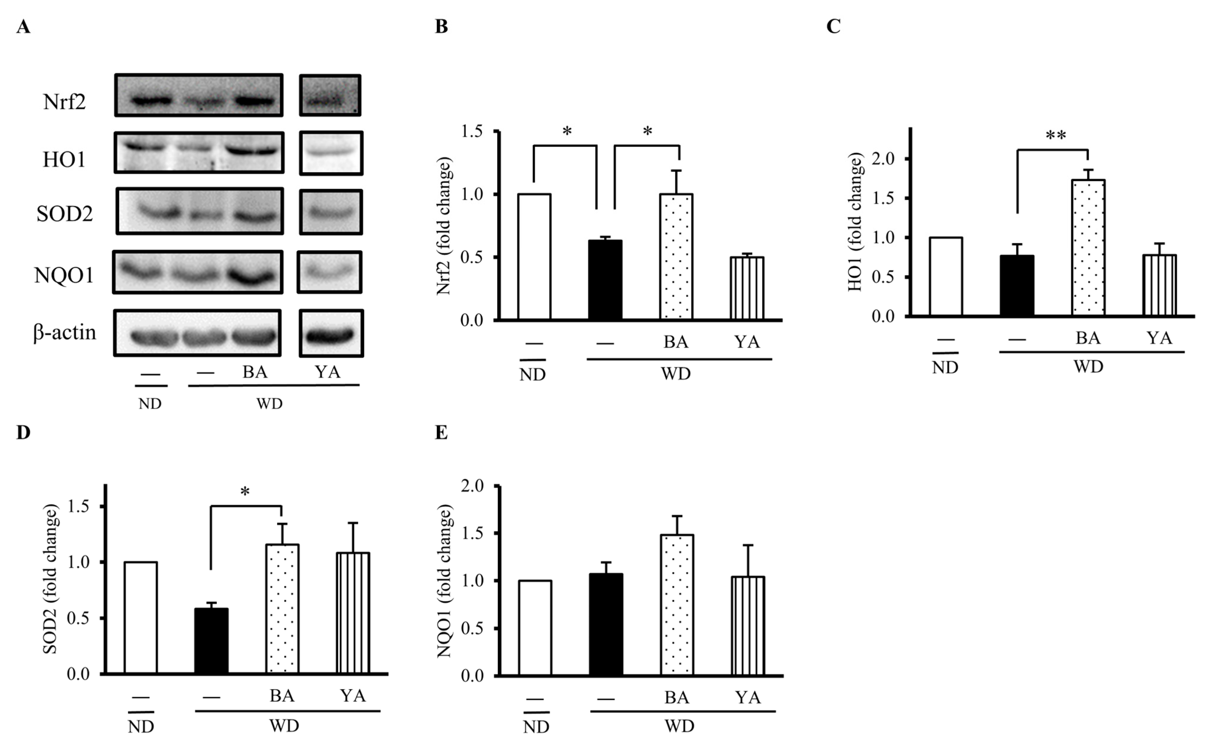
3.2. Effects of Amazake on Expressions of Gut Antioxidant Proteins

To explore the effect of Amazake on the gut, we first evaluated gut oxidative status by measuring the levels of some antioxidant factors and enzymes. The typical Western blots for protein detection are shown in Figure 2A. The level of Nrf2, a key transcription factor that regulates the expression of antioxidant proteins, was decreased 0.6-fold in the WD group compared with the ND group and recovered to a normal level by supplementing with BA (Figure 2B). The level of some typic Nrf2 downstream antioxidant enzymes such as HO1 and SOD2 were also significantly recovered from WD-reduced levels by supplementing with BA (Figure 2C,D), but NQO1 was not changed (Figure 2E). On the other hand, these indexes were not recovered in the YA group.

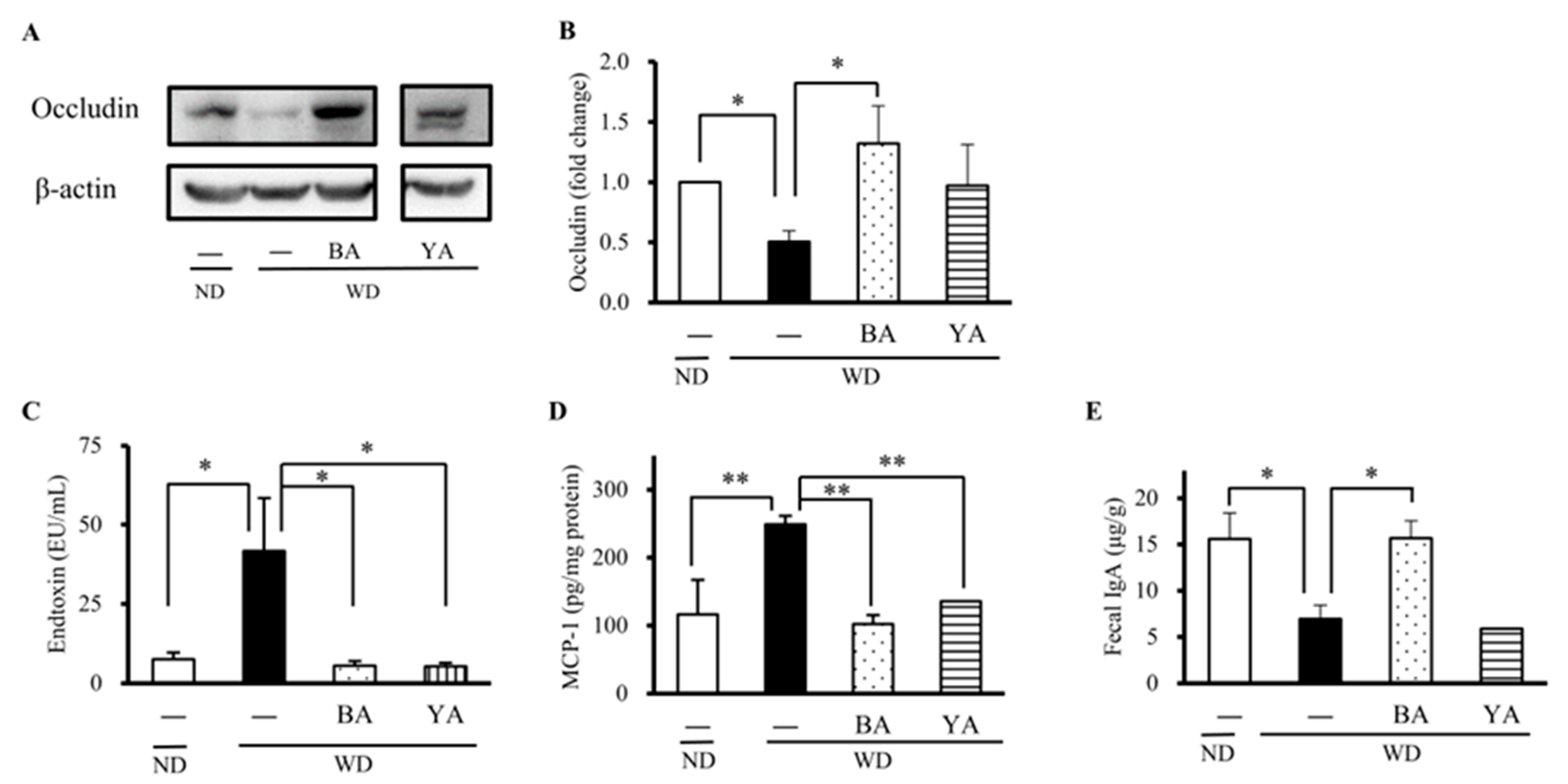
3.3. Effects of Amazake on the Expression of Gut Barrier Protein and Inflammation Factors

Next, we estimated the effects of Amazake ingredients on the gut barrier by investigating the expression of occludin, a physical barrier function. As shown in Figure 3, the level of occludin was significantly decreased in the WD group compared with the ND group and significantly increased in the BA group (Figure 3A,B). It is known that the disruption of the structure and function of the gut barrier allows bacterial toxins and other toxic substances to enter the bloodstream, amplifying systemic inflammation. Thus, we next examined serum endotoxins, which are an indicator of gut barrier function. The results revealed that the WD significantly increased serum endotoxins compared with the ND, and BA and YA significantly decreased them (p < 0.01, Figure 3C). Furthermore, the inflammatory cytokine MCP-1 in the liver was significantly increased in the WD group and significantly decreased in the BA and YA groups (Figure 3D). Fecal IgA, a chemical barrier function, was also significantly decreased in the WD group compared with the ND group and significantly increased in the BA group compared with the WD group (Figure 3E).

3.4. Effects of Amazake on Gut Microbiota Structure

Compared to the WD group, the relative abundance of Proteobacteria was increased in the BA group while Verrucomicrobia and Firmicutes were increased in the YA group (Figure 4A). At the family and species level, Lachnospiraceae was increased in the BA group and A. muciniphila was increased in YA compared with WD (Figure 4B,C). Next, we performed a Pearson correlation analysis between mice biochemical data and gut microbiome data from the ND, WD, BA, and YA groups. As shown in Figure 4D, a positive correlation (r = 0.60, p = 0.031) was observed between the relative abundance of Lachnospiraceae and fecal IgA in both the BA and YA groups. However, there was no correlation between Akkermansia and mice biochemical data (e.g., MCP-1), although A. muciniphila was 5-fold increased in the YA group compared with the WD group.

4. Discussion

4.1. Difference between the Two Types of Amazake Ingredients

In this study, we first analyzed the major ingredients of two types of Amazake manufactured with different koji (Aspergillus), then compared their effects on gut barrier function and gut microbiota. YA with yellow koji (A. oryzae) for saccharification is the most common type of Amazake on the Japanese market. In contrast, BA was first manufactured with black koji (A. luchuensis MEM-C strain) in this study. Nutrient data show that carbohydrates accounted for approximately 90%, excluding water, of the Amazake, which was almost the same as the market Amazake [18]. However, the BA Amazake showed much higher amounts of other functional ingredients compared to that of YA. It is noted that citric acid in BA Amazake was 20-fold higher than in YA, resulting in a lower pH. The reason why BA has an even higher citric acid content than YA is that the A. luchuensis MEM-C strain is an allied species of A. kawachi, which has a citrate exporter [19]. A. luchuensis is reported to possess ferulic acid esterase, which degrades rice cell wall polysaccharides to ferulic acid [20] and contributes to producing the higher ferulic acid contents of BA.
In addition to the oligosaccharides and organic acids analyzed in this study, Amazake has been reported to be rich in vitamins B2, B3, B7, and B9 [10]. Three weeks of vitamin B2 supplementation caused a reduction in systemic oxidative stress and anti-inflammatory effects in CD patients [21]. The effects of the Amazake production process on the vitamin B complex need to be investigated in a future study.

4.2. Effects of Amazake on Gut Barrier Function and Gut Microbiota

To explore the effects of Amazake on gut barrier function and gut microbiota, a mouse dietary model fed with aWD containing higher fat and fructose for 15 weeks was used to induce gut disturbance [9]. Mice’s body weight significantly increased in the WD group compared to the ND group, and BA and YA did not improve the WD-induced body weight (Figure A1). We chose occludin as the gut barrier marker in the animal experiment because gut occludin was observed to decrease in both human UCand DSS-induced UC mice [22] Occludin is a tight-junction protein acting as a physical barrier. When it was disrupted, the gut barrier allows bacterial toxins and other toxic substances enter the bloodstream (known as serum endotoxins). IgA is the most abundant antibody secreted into the gut and plays a crucial role in immune exclusion by neutralizing pathogenic toxins and preventing pathogen attachment and invasion across the gut barrier [23]. Therefore, we used gut occludin, serum endotoxin, and fecal IgA as indicators of damaged barrier function. Our data revealed that the expressions of gut occludin and fecal IgA were significantly decreased in the WD group, resulting in increased serum endotoxin, MCP-1 (Figure 3), and these events were significantly improved by BA. Therefore, we considered that BA could improve WD-caused gut barrier damage to prevent chronic inflammation.
The concern is how BA improves WD-caused gut barrier damage. It has been reported that Nrf2 working as a master regulator of cellular oxidative levels reduced gut mucosal injury by suppressing oxidative stress and affected gut tight-junction proteins and the apoptosis of cells to regulate intestinal permeability [24]. In this study, BA significantly upregulated the expressions of antioxidant factors and enzymes such as Nfr2, HO1, and SOD-2. At the same time, the expression of gut occludin barrier protein and fecal IgA were also observed to be significantly enhanced by BA. Thus, it is possible that BA enhanced gut antioxidant activities to prevent against WD-caused gut barrier damage. Future study is needed to confirm this correlation between BA antioxidants and gut damage protection in the gut.
It was also noticed that BA is rich in organic acids, notably citric acid, which has been reported to have various effects on the host and bacteria. Citric acid is widely used in foods as an acidifier and pH adjuster and as an antibacterial agent instead of antibiotics for animal feed [25]. These effects were caused by non-dissociated organic acid passing through the bacterial cell membrane and releasing hydrogen ions inside the cell, resulting in a decrease in intracellular pH and the suppression of the enzyme activity involved in bacterial metabolism, thereby exhibiting bactericidal activity [26]. Organic acids, including citric acid, were reported to improve dysbiosis as well as gut barrier function and inflammation in various animals. The administration of 3% citric acid to broiler chicks improved gut morphology, including increasing the number of goblet cells and villus width/crypt depth [27]. Citric acid of 1.5% concentration suppressed the activation of the signaling pathways involved in inflammatory cytokines and increased the gene expression of tight-junction structures such as claudin, occludin, and ZO-1 in the distal intestine of flatfish [28]. The administration of 0.5 to 1.5% citric acid to quail chicks increased Lactobacillus and decreased coliform, E.coli, and Salmonella in a concentration-dependent manner [29]. In our research, BA had the highest citric acid content (approximately 1.5% of the Amazake), suggesting that the improvement of gut barrier function by BA might be due to citric acid action.
Ferulic acid, one of the polyphenols in Amazake [10], increased occludin and Akkermansia, activated the Nrf2 pathway, and decreased endotoxins [30,31,32]. Alginate oligosaccharide and chitosan oligosaccharide activated the Nrf2 pathway and upregulated the downstream expression of HO1 and NQO1 in mice [33,34,35]. In this study, BA contained higher ferulic acid and isomaltose, which possibly contributed to activating the Nrf2 pathway-mediated expression of HO1, NQO1, and SOD2.
Ferulic acid and IMOs including isomaltose and isomaltotriose have been reported to increase A. muciniphila in mouse experiments [31,36]. In the YA group, A. muciniphila was significantly increased, although YA had fewer IMOs than BA. As shown in Table 1, BA contained high contents of ferulic acid, isomaltose, and isomaltotriose, as well as a high content of citric acid, which resulted in a pH 3.8 of BA and might also decrease gut pH. It is known that a pH under 5.6–5.9 inhibits A. muciniphila growth [37]. In fact, we also observed that the growth of A. muciniphila was inhibited in the monocultures with 125 μg/mL citric acid. Thus, the fact that BA did not increase A. muciniphila was due to its high content of citric acid. In contrast, the YA contained little citric acid, thus the growth of A. muciniphila was not inhibited.
It has been reported that A. muciniphila secretes the Amuc_1100 protein, which induces the anti-inflammatory cytokine interleukin-10 via toll-like receptor 2 [38,39] and improved colitis in mice [40]. In this study, A. muciniphila was 5-fold increased in the YA group compared with the WD group, but there was no correlation between Akkermansia and MCP-1. Similar results were also reported for Saskatoon berries, in which Akkermansia was 5-fold increased and MCP-1 was decreased in the berry administration group, but there was no correlation between the two [41]. Thus, MCP-1 and endotoxins might be regulated by many barrier factors, not only A. muciniphila.
IBD may be involved in innate or adaptive immune responses such as the activation and differentiation of T helper 17 cells (Th17) [42]. In addition, interleukin-17 (IL-17), secreted from Th17 cells, can promote the secretion of IgA and a variety of antimicrobial peptides in the gut to accelerate the healing of intestinal mucosal injury, thereby enhancing the gut barrier function [43]. However, the levels of Th17 and IL-17 were not analyzed, so it is necessary to examine gut immune cells to elucidate the mechanism of the improvement of gut barrier function in more detail.

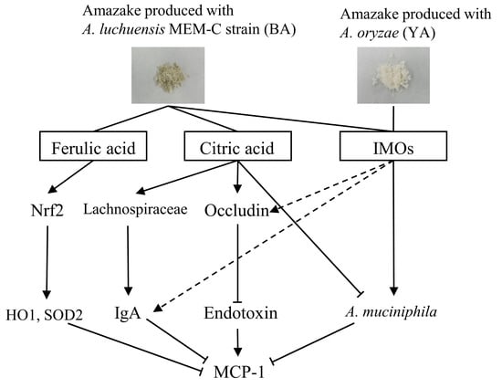
4.3. Relationship between Gut Barrier Function and Gut Microbiota

In the intestine, IgA was the most predominant compared to IgM and IgG secreted by B cells, and IgA bound to antigens such as food and microbes, avoiding direct contact with host epithelial cells [44]. In this study, BA increased IgA, which was positively correlated with the abundance of Lachnospiraceae. Superantigens expressed on the surface of commensal bacteria of the family Lachnospiraceae, such as R. gnavus, bound to IgA variable regions and stimulated potent IgA responses in mice [45]. Furthermore, diets containing organic acids, including citric acid, increased Lachnospiraceae in broilers [46], complementing the fact that the citric acid in BA increased Lachnospiraceae and IgA.

5. Conclusions

Black koji Amazake (BA) contained higher amounts of ferulic acid, isomaltooligosaccharides, and citric acid than yellow koji Amazake (YA), and contributed to protecting gut barrier function to reduce endotoxin intrusion and inflammation. In addition, BA increased Lachnospiraceae, which positively correlated with fecal IgA, while YA increased Akkermansia, which may contribute to protecting gut mucosa. The detained mechanism of gut barrier protection function by Amazake needs further study to clarify.

Author Contributions

Conceptualization, H.N., H.M. and D.-X.H.; methodology, H.N., S.S. and K.K.; software, H.N.; validation, S.S., K.S. and D.-X.H.; formal analysis, H.N., S.S. and K.K.; investigation, H.N., S.S. and K.K.; resources, S.S., K.K., H.M. and D.-X.H.; data curation, H.N., S.S. and K.K.; writing—original draft preparation, H.N. and S.S.; writing—review and editing, D.-X.H.; visualization, H.N.; supervision, D.-X.H.; project administration, D.-X.H. and H.M.; funding acquisition, D.-X.H. All authors have read and agreed to the published version of the manuscript.

Funding

This research was supported by the fund of Scholar Research of Kagoshima University to D.-X.H. (Grant No: 70030117).

Data Availability Statement

Data is contained within the article or Appendix A.

Conflicts of Interest

Author Sho Setoguchi, Kuniaki Kawano, and Hiroshi Miyagawa were employed by the company Kirishima Shuzo Co., Ltd. The remaining authors declare that the research was conducted in the absence of any commercial or financial relationships that could be construed as a potential conflict of interest.

Appendix A

Table A1. Dietary compositions of each group.
Components (%)NDWD
BAYA
Lard33029.729.7
Soybean oil3333
Corn starch43155.95.9
Casein212120.420.4
Sucrose20202020
Cellulose5555
Mineral Mix3.53.53.53.5
Vitamin Mix1111
Cholesterol0111
Choline Bitartrate0.20.20.20.2
Methionine0.30.30.30.3
Black koji Amazake (BA) 10
Yellow koji Amazake (YA) 10
Total calories (kcal/100 g)371520523523
ND: normal diet; WD: Western diet; BA: black koji Amazake; YA: yellow koji Amazake.
Figure A1. Body weight of each group at the endpoint. The data represent the mean ± SE for each group. Columns with symbols significantly differ (* p < 0.05 versus WD group by Dunnett’s test). ND: normal diet; WD: Western diet; BA: black koji Amazake; YA: yellow koji Amazake.
Foods 12 02568 g0a1

References

  1. Wang, R.; Li, Z.; Liu, S.; Zhang, D. Global, regional and national burden of inflammatory bowel disease in 204 countries and territories from 1990 to 2019: A systematic analysis based on the Global Burden of Disease Study 2019. BMJ Open 2023, 13, e065186. [Google Scholar] [CrossRef] [PubMed]
  2. Parkes, G. IBD Statistics 2022: Crohn’s and Ulcerative Colitis. Available online: https://ampersandhealth.co.uk/myibdcare/resources/ibd-statistics-2022-crohns-and-ulcerative-colitis/#:~:text=Whatpercentageofpeopleget,WorldIBDDay2022here (accessed on 3 June 2023).
  3. Jakubczyk, D.; Leszczyńska, K.; Górska, S. The Effectiveness of Probiotics in the Treatment of Inflammatory Bowel Disease (IBD)—A Critical Review. Nutrients 2020, 12, 1973. [Google Scholar] [CrossRef] [PubMed]
  4. Raine, T.; Bonovas, S.; Burisch, J.; Kucharzik, T.; Adamina, M.; Annese, V.; Bachmann, O.; Bettenworth, D.; Chaparro, M.; Czuber-Dochan, W.; et al. ECCO Guidelines on Therapeutics in Ulcerative Colitis: Medical Treatment. J. Crohn’s Colitis 2022, 16, 2–17. [Google Scholar] [CrossRef] [PubMed]
  5. He, W.-Q.; Wang, J.; Sheng, J.-Y.; Zha, J.-M.; Graham, W.V.; Turner, J.R. Contributions of Myosin Light Chain Kinase to Regulation of Epithelial Paracellular Permeability and Mucosal Homeostasis. Int. J. Mol. Sci. 2020, 21, 993. [Google Scholar] [CrossRef] [PubMed]
  6. Naydenov, N.G.; Baranwal, S.; Khan, S.; Feygin, A.; Gupta, P.; Ivanov, A.I. Novel mechanism of cytokine-induced disruption of epithelial barriers. Tissue Barriers 2013, 1, e25231. [Google Scholar] [CrossRef]
  7. Tena-Garitaonaindia, M.; Arredondo-Amador, M.; Mascaraque, C.; Asensio, M.; Marin, J.J.G.; Martínez-Augustin, O.; Sánchez de Medina, F. Modulation of intestinal barrier function by glucocorticoids: Lessons from preclinical models. Pharmacol. Res. 2022, 177, 106056. [Google Scholar] [CrossRef]
  8. do Nascimento, R.D.P.; da Fonseca Machado, A.P.; Galvez, J.; Cazarin, C.B.B.; Maróstica Junior, M.R. Ulcerative colitis: Gut microbiota, immunopathogenesis and application of natural products in animal models. Life Sci. 2020, 258, 118129. [Google Scholar] [CrossRef]
  9. Lee, J.C.; Lee, H.Y.; Kim, T.K.; Kim, M.S.; Park, Y.M.; Kim, J.; Park, K.; Kweon, M.N.; Kim, S.H.; Bae, J.W.; et al. Obesogenic diet-induced gut barrier dysfunction and pathobiont expansion aggravate experimental colitis. PLoS ONE 2017, 12, e0187515. [Google Scholar] [CrossRef]
  10. Oguro, Y.; Nishiwaki, T.; Shinada, R.; Kobayashi, K.; Kurahashi, A. Metabolite profile of koji amazake and its lactic acid fermentation product by Lactobacillus sakei UONUMA. J. Biosci. Bioeng. 2017, 124, 178–183. [Google Scholar] [CrossRef]
  11. Michio Sata, Y.N. Effect of a Late Evening Snack of Amazake in Patients with Liver Cirrhosis: A Pilot Study. J. Nutr. Food Sci. 2013, 3, 1000223. [Google Scholar] [CrossRef]
  12. Maruki-Uchida, H.; Sai, M.; Yano, S.; Morita, M.; Maeda, K. Amazake made from sake cake and rice koji suppresses sebum content in differentiated hamster sebocytes and improves skin properties in humans. Biosci. Biotechnol. Biochem. 2020, 84, 1689–1695. [Google Scholar] [CrossRef]
  13. Oura, S.; Suzuki, S.; Hata, Y.; Kawato, A.; Abe, Y. Evaluation of physiological functionalities of amazake in mice. J. Brew. Soc. JAPAN 2007, 102, 781–788. [Google Scholar] [CrossRef]
  14. Saigusa, N.; Ohba, R. Effects of koji production and saccharification time on the antioxidant activity of amazake. Food Sci. Technol. Res. 2007, 13, 162–165. [Google Scholar] [CrossRef]
  15. Kawakami, S.; Ito, R.; Maruki-Uchida, H.; Kamei, A.; Yasuoka, A.; Toyoda, T.; Ishijima, T.; Nishimura, E.; Morita, M.; Sai, M.; et al. Intake of a mixture of sake cake and rice malt increases mucin levels and changes in intestinal microbiota in mice. Nutrients 2020, 12, 449. [Google Scholar] [CrossRef]
  16. International, A.; Cunniff, P. Official Methods of Analysis of AOAC International, 16th ed.; AOAC International: Rockville, MD, USA, 1995. [Google Scholar]
  17. Nakano, H.; Wu, S.; Sakao, K.; Hara, T.; He, J.; Garcia, S.; Shetty, K.; Hou, D.X. Bilberry anthocyanins ameliorate NAFLD by improving dyslipidemia and gut microbiome dysbiosis. Nutrients 2020, 12, 3252. [Google Scholar] [CrossRef]
  18. Kurahashi, A. Ingredients, Functionality, and Safety of the Japanese Traditional Sweet Drink Amazake. J. Fungi 2021, 7, 469. [Google Scholar] [CrossRef]
  19. Kadooka, C.; Nakamura, E.; Mori, K.; Okutsu, K.; Yoshizaki, Y.; Takamine, K.; Goto, M.; Tamaki, H.; Futagami, T. LaeA Controls Citric Acid Production through Regulation of the Citrate Exporter-Encoding cexA Gene in Aspergillus luchuensis mut. kawachii. Appl. Environ. Microbiol. 2020, 86, e01950-19. [Google Scholar] [CrossRef]
  20. Koseki, T.; Ito, Y.; Furuse, S.; Ito, K.; Iwano, K. Conversion of ferulic acid into 4-vinylguaiacol, vanillin and vanillic acid in model solutions of shochu. J. Ferment. Bioeng. 1996, 82, 46–50. [Google Scholar] [CrossRef]
  21. von Martels, J.Z.H.; Bourgonje, A.R.; Klaassen, M.A.Y.; Alkhalifah, H.A.A.; Sadaghian Sadabad, M.; Vich Vila, A.; Gacesa, R.; Gabriëls, R.Y.; Steinert, R.E.; Jansen, B.H.; et al. Riboflavin Supplementation in Patients with Crohn’s Disease [the RISE-UP study]. J. Crohn’s Colitis 2020, 14, 595–607. [Google Scholar] [CrossRef]
  22. Liu, A.; Lv, H.; Wang, H.; Yang, H.; Li, Y.; Qian, J. Aging Increases the Severity of Colitis and the Related Changes to the Gut Barrier and Gut Microbiota in Humans and Mice. J. Gerontol. Ser. A 2020, 75, 1284–1292. [Google Scholar] [CrossRef]
  23. Tsuruta, T.; Muhomah, T.A.; Sonoyama, K.; Nguyen, Q.D.; Takase, Y.; Nishijima, A.; Himoto, S.; Katsumata, E.; Nishino, N. Aicda deficiency exacerbates high-fat diet-induced hyperinsulinemia but not gut dysbiosis in mice. Nutr. Res. 2021, 93, 15–26. [Google Scholar] [CrossRef] [PubMed]
  24. Wen, Z.; Liu, W.; Li, X.; Chen, W.; Liu, Z.; Wen, J.; Liu, Z. A Protective Role of the NRF2-Keap1 Pathway in Maintaining Intestinal Barrier Function. Oxid. Med. Cell Longev. 2019, 2019, 1759149. [Google Scholar] [CrossRef] [PubMed]
  25. Pearlin, B.V.; Muthuvel, S.; Govidasamy, P.; Villavan, M.; Alagawany, M.; Ragab Farag, M.; Dhama, K.; Gopi, M. Role of acidifiers in livestock nutrition and health: A review. J. Anim. Physiol. Anim. Nutr. 2020, 104, 558–569. [Google Scholar] [CrossRef] [PubMed]
  26. Suiryanrayna, M.V.A.N.; Ramana, J.V. A review of the effects of dietary organic acids fed to swine. J. Anim. Sci. Biotechnol. 2015, 6, 45. [Google Scholar] [CrossRef] [PubMed]
  27. Nourmohammadi, R.; Afzali, N. Effect of Citric Acid and Microbial Phytase on Small Intestinal Morphology in Broiler Chicken. Ital. J. Anim. Sci. 2013, 12, e7. [Google Scholar] [CrossRef]
  28. Zhao, S.; Chen, Z.; Zheng, J.; Dai, J.; Ou, W.; Xu, W.; Ai, Q.; Zhang, W.; Niu, J.; Mai, K.; et al. Citric acid mitigates soybean meal induced inflammatory response and tight junction disruption by altering TLR signal transduction in the intestine of turbot, Scophthalmus maximus L. Fish Shellfish Immunol. 2019, 92, 181–187. [Google Scholar] [CrossRef]
  29. Xue, J.J.; Huang, X.F.; Liu, Z.L.; Chen, Y.; Zhang, Y.K.; Luo, Y.; Wang, B.W.; Wang, Q.G.; Wang, C. Effects of citric acid supplementation on growth performance, intestinal morphology and microbiota, and blood parameters of geese from 1 to 28 days of age. Poult. Sci. 2023, 102, 102343. [Google Scholar] [CrossRef]
  30. Tian, B.; Geng, Y.; Wang, P.; Cai, M.; Neng, J.; Hu, J.; Xia, D.; Cao, W.; Yang, K.; Sun, P. Ferulic acid improves intestinal barrier function through altering gut microbiota composition in high-fat diet-induced mice. Eur. J. Nutr. 2022, 61, 3767–3783. [Google Scholar] [CrossRef]
  31. Ma, Y.; Chen, K.; Lv, L.; Wu, S.; Guo, Z. Ferulic acid ameliorates nonalcoholic fatty liver disease and modulates the gut microbiota composition in high-fat diet fed ApoE−/− mice. Biomed. Pharmacother. 2019, 113, 108753. [Google Scholar] [CrossRef]
  32. Mahmoud, A.M.; Hussein, O.E.; Hozayen, W.G.; Bin-Jumah, M.; Abd El-Twab, S.M. Ferulic acid prevents oxidative stress, inflammation, and liver injury via upregulation of Nrf2/HO-1 signaling in methotrexate-induced rats. Environ. Sci. Pollut. Res. 2020, 27, 7910–7921. [Google Scholar] [CrossRef]
  33. Pan, H.; Feng, W.; Chen, M.; Luan, H.; Hu, Y.; Zheng, X.; Wang, S.; Mao, Y. Alginate Oligosaccharide Ameliorates D-Galactose-Induced Kidney Aging in Mice through Activation of the Nrf2 Signaling Pathway. Biomed Res. Int. 2021, 2021, 6623328. [Google Scholar] [CrossRef]
  34. Wang, Y.; Xiong, Y.; Zhang, A.; Zhao, N.; Zhang, J.; Zhao, D.; Yu, Z.; Xu, N.; Yin, Y.; Luan, X.; et al. Oligosaccharide attenuates aging-related liver dysfunction by activating Nrf2 antioxidant signaling. Food Sci. Nutr. 2020, 8, 3872–3881. [Google Scholar] [CrossRef]
  35. Tao, W.; Wang, G.; Pei, X.; Sun, W.; Wang, M. Chitosan Oligosaccharide Attenuates Lipopolysaccharide-Induced Intestinal Barrier Dysfunction through Suppressing the Inflammatory Response and Oxidative Stress in Mice. Antioxidants 2022, 11, 1384. [Google Scholar] [CrossRef]
  36. Singh, D.P.; Singh, J.; Boparai, R.K.; Zhu, J.; Mantri, S.; Khare, P.; Khardori, R.; Kondepudi, K.K.; Chopra, K.; Bishnoi, M. Isomalto-oligosaccharides, a prebiotic, functionally augment green tea effects against high fat diet-induced metabolic alterations via preventing gut dysbacteriosis in mice. Pharmacol. Res. 2017, 123, 103–113. [Google Scholar] [CrossRef]
  37. Van Herreweghen, F.; Van den Abbeele, P.; De Mulder, T.; De Weirdt, R.; Geirnaert, A.; Hernandez-Sanabria, E.; Vilchez-Vargas, R.; Jauregui, R.; Pieper, D.H.; Belzer, C.; et al. In vitro colonisation of the distal colon by Akkermansia muciniphila is largely mucin and pH dependent. Benef. Microbes 2017, 8, 81–96. [Google Scholar] [CrossRef]
  38. Everard, A.; Belzer, C.; Geurts, L.; Ouwerkerk, J.P.; Druart, C.; Bindels, L.B.; Guiot, Y.; Derrien, M.; Muccioli, G.G.; Delzenne, N.M.; et al. Cross-talk between Akkermansia muciniphila and intestinal epithelium controls diet-induced obesity. Proc. Natl. Acad. Sci. USA 2013, 110, 9066–9071. [Google Scholar] [CrossRef]
  39. Ottman, N.; Reunanen, J.; Meijerink, M.; Pietilä, T.E.; Kainulainen, V.; Klievink, J.; Huuskonen, L.; Aalvink, S.; Skurnik, M.; Boeren, S.; et al. Pili-like proteins of Akkermansia muciniphila modulate host immune responses and gut barrier function. PLoS ONE 2017, 12, e0173004. [Google Scholar] [CrossRef]
  40. Wang, L.; Tang, L.; Feng, Y.; Zhao, S.; Han, M.; Zhang, C.; Yuan, G.; Zhu, J.; Cao, S.; Wu, Q.; et al. A purified membrane protein from Akkermansia muciniphila or the pasteurised bacterium blunts colitis associated tumourigenesis by modulation of CD8+ T cells in mice. Gut 2020, 69, 1988–1997. [Google Scholar] [CrossRef]
  41. Zhao, R.; Shen, G.X. Impact of Anthocyanin Component and Metabolite of Saskatoon Berry on Gut Microbiome and Relationship with Fecal Short Chain Fatty Acids in Diet-Induced Insulin Resistant Mice. J. Nutr. Biochem. 2022, 111, 109201. [Google Scholar] [CrossRef]
  42. Zhao, J.; Lu, Q.; Liu, Y.; Shi, Z.; Hu, L.; Zeng, Z.; Tu, Y.; Xiao, Z.; Xu, Q. Th17 Cells in Inflammatory Bowel Disease: Cytokines, Plasticity, and Therapies. J. Immunol. Res. 2021, 2021, 8816041. [Google Scholar] [CrossRef]
  43. Chen, L.; Ruan, G.; Cheng, Y.; Yi, A.; Chen, D.; Wei, Y. The role of Th17 cells in inflammatory bowel disease and the research progress. Front. Immunol. 2023, 13, 1055914. [Google Scholar] [CrossRef] [PubMed]
  44. Janzon, A.; Goodrich, J.K.; Koren, O.; Waters, J.L.; Ley, R.E. Interactions between the Gut Microbiome and Mucosal Immunoglobulins A, M, and G in the Developing Infant Gut. mSystems 2019, 4, e00612-19. [Google Scholar] [CrossRef] [PubMed]
  45. Bunker, J.J.; Drees, C.; Watson, A.R.; Plunkett, C.H.; Nagler, C.R.; Schneewind, O.; Eren, A.M.; Bendelac, A. B cell superantigens in the human intestinal microbiota. Sci. Transl. Med. 2019, 11, eaau9356. [Google Scholar] [CrossRef] [PubMed]
  46. Abdelli, N.; Pérez, J.F.; Vilarrasa, E.; Cabeza Luna, I.; Melo-Duran, D.; D’Angelo, M.; Solà-Oriol, D. Targeted-Release Organic Acids and Essential Oils Improve Performance and Digestive Function in Broilers under a Necrotic Enteritis Challenge. Animals 2020, 10, 259. [Google Scholar] [CrossRef]
Figure 1. Schemes of Amazake manufacture. The black koji was derived from A. luchuensis MEM-C strain and the yellow koji was from A. oryzae. Black and yellow rice koji were incubated at 34–40 °C for a total of 42 h, first at 34 °C for 16 h, then at 36 °C for 8 h, and finally at 40 °C for 18 h. Then, 800 g of cooked rice, 800 g of rice koji, and 2400 mL of water were mixed in a 5 L-volumetric flask and saccharified at 55 °C for 16 h. After saccharification, the mixture was filtered and pasteurized at 85 °C for 30 min and finally freeze-dried to collect Amazake powder. BA: black koji Amazake; YA: yellow koji Amazake.
Foods 12 02568 g001
Figure 2. Effects of Amazake on gut antioxidant proteins in the animal experiment. Western blotting images are shown in (A) gut Nrf2, HO1, SOD2, and NQO1 protein expressions. Fold changes of (B) Nrf2, (C) HO1, (D) SOD2, and (E) NQO1 were calculated from the intensity of (A) relative to the ND group and normalized by β-actin intensity. These data represent the mean ± SE for each group. Columns with symbols significantly differ (* p < 0.05 and ** p < 0.01 versus the WD group by Dunnett’s test). ND: normal diet; WD: Western diet; BA: black koji Amazake; YA: yellow koji Amazake; Nrf2: NF-E2-related factor 2; HO1: heme oxygenase 1; NQO1: NAD(P)H quinone dehydrogenase 1; and SOD2: superoxide dismutase 2.
Foods 12 02568 g002
Figure 3. Effects of Amazake on gut barrier function and inflammation in an animal experiment. Western blotting images are shown for (A) gut occludin. (B) Fold change of occludin calculated from the intensity of (A) relative to the ND group and normalized by β-actin intensity. (C) Serum endotoxins, (D) hepatic MCP-1, and (E) fecal IgA. Columns with symbols significantly differ (* p < 0.05 and ** p < 0.01 versus the WD group by Dunnett’s test). ND: normal diet; WD: Western diet; BA: black koji Amazake; YA: yellow koji Amazake; IgA: immunoglobulin A, MCP-1: monocyte chemotactic protein-1.
Foods 12 02568 g003
Figure 4. Effects of Amazake on relative abundance at the phylum, family, and species level of gut microbiota. (A) Relative abundance of gut microbiota at the phylum level. Relative abundance of (B) Akkermansia muciniphila, (C) Lachnospiraceae. (D) Pearson correlations between the abundance of Lachnospiraceae and the concentration of fecal IgA. These data represent the mean ± SE for each group. Columns with different letters significantly differ (p < 0.05). Columns with symbols significantly differ (* p < 0.05 and ** p < 0.01 versus the WD group by Dunnett’s test). ND: normal diet, WD: Western diet, BA: black koji Amazake, YA: yellow koji Amazake. Columns with different letters significantly changed.
Foods 12 02568 g004
Table 1. Major ingredients in two Amazake (dried weight).
ComponentsBAYA
Ferulic acid (mg/100 g)0.550.008
Dietary Fiber (g/100g)2.31.3
Isomaltose (g/100 g)9.25.2
Isomaltotriose (g/100 g)0.660.24
Total organic acids (mg/100 g)1760288
Citric acid (mg/100 g)155376
Pyroglutamic acid (mg/100 g)11123
Malic acid (mg/100 g)8.11.3
Lactic acid (mg/100 g)2.731.9
Acetic acid (mg/100 g)2.515.2
pH3.806.28
BA: black koji Amazake, YA: yellow koji Amazake.
Disclaimer/Publisher’s Note: The statements, opinions and data contained in all publications are solely those of the individual author(s) and contributor(s) and not of MDPI and/or the editor(s). MDPI and/or the editor(s) disclaim responsibility for any injury to people or property resulting from any ideas, methods, instructions or products referred to in the content.

