


Remineralization Strategies for Teeth with Molar Incisor Hypomineralization (MIH): A Literature Review
 , , , ,
Abstract
:1. Introduction
2. Search Strategy
3. Results
4. Discussion
5. Conclusions
Author Contributions
Funding
Institutional Review Board Statement
Informed Consent Statement
Data Availability Statement
Conflicts of Interest
References
- Schwendicke, F.; Elhennawy, K.; Reda, S.; Bekes, K.; Manton, D.J.; Krois, J. Global burden of molar incisor hypomineralization. J. Dent. 2018, 68, 10–18. [Google Scholar] [CrossRef] [PubMed]
- Dave, M.; Taylor, G. Global prevalence of molar incisor hypomineralisation. Evid. Based Dent. 2018, 19, 78–79. [Google Scholar] [CrossRef]
- Giuca, M.R.; Lardani, L.; Pasini, M.; Beretta, M.; Gallusi, G.; Campanella, V. State-of-the-art on MIH. Part. 1 Definition and aepidemiology. Eur. J. Paediatr. Dent. 2020, 21, 80–82. [Google Scholar]
- Weerheijm, K.L.; Duggal, M.; Mejàre, I.; Papagiannoulis, L.; Koch, G.; Martens, L.C.; Hallonsten, A.L. Judgement criteria for molar incisor hypomineralisation (MIH) in epidemiologic studies: A summary of the European meeting on MIH held in Athens, 2003. Eur. J. Paediatr. Dent. 2003, 4, 110–113. [Google Scholar]
- Lygidakis, N.A.; Garot, E.; Somani, C.; Taylor, G.D.; Rouas, P.; Wong, F.S.L. Best clinical practice guidance for clinicians dealing with children presenting with molar-incisor-hypomineralisation (MIH): An updated European Academy of Paediatric Dentistry policy document. Eur. Arch. Paediatr. Dent. 2021, 23, 3–21. [Google Scholar] [CrossRef]
- Garot, E.; Rouas, P.; Somani, C.; Taylor, G.D.; Wong, F.; Lygidakis, N.A. An update of the aetiological factors involved in molar incisor hypomineralisation (MIH): A systematic review and meta-analysis. Eur. Arch. Paediatr. Dent. 2022, 23, 23–38. [Google Scholar] [CrossRef] [PubMed]
- Hubbard, M.J.; Mangum, J.E.; Perez, V.A.; Williams, R. A breakthrough in understanding the pathogenesis of molar hypomineralisation: The mineralisation-poisoning model. Front. Physiol. 2021, 12, 802833. [Google Scholar] [CrossRef]
- Fernandes, I.C.; Forte, F.D.S.; Sampaio, F.C. Molar-incisor hypomineralization (MIH), dental fluorosis, and caries in rural areas with different fluoride levels in the drinking water. Int. J. Paediatr. Dent. 2021, 31, 475–482. [Google Scholar] [CrossRef]
- Limeback, H. Comprehensive Preventive Dentistry; John Wiley & Sons: Hoboken, NJ, USA,, 2012. [Google Scholar]
- Elhennawy, K.; Manton, D.J.; Crombie, F.; Zaslansky, P.; Radlanski, R.J.; Jost-Brinkmann, P.G.; Schwendicke, F. Structural, mechanical and chemical evaluation of molar-incisor hypomineralization-affected enamel: A systematic review. Arch. Oral Biol. 2017, 83, 272–281. [Google Scholar] [CrossRef]
- Crombie, F.A.; Manton, D.J.; Palamara, J.E.; Zalizniak, I.; Cochrane, N.J.; Reynolds, E.C. Characterisation of developmentally hypomineralised human enamel. J. Dent. 2013, 41, 611–618. [Google Scholar] [CrossRef]
- Fagrell, T.G.; Dietz, W.; Jälevik, B.; Norén, J.G. Chemical, mechanical and morphological properties of hypomineralized enamel of permanent first molars. Acta Odontol. Scand. 2010, 68, 215–222. [Google Scholar] [CrossRef] [PubMed]
- Farah, R.A.; Monk, B.C.; Swain, M.V.; Drummond, B.K. Protein content of molar-incisor hypomineralisation enamel. J. Dent. 2010, 38, 591–596. [Google Scholar] [CrossRef] [PubMed]
- Farah, R.A.; Swain, M.V.; Drummond, B.K.; Cook, R.; Atieh, M. Mineral density of hypomineralised enamel. J. Dent. 2010, 38, 50–58. [Google Scholar] [CrossRef]
- Jälevik, B. Enamel hypomineralization in permanent first molars. A clinical, histo-morphological and biochemical study. Swed. Dent. J. Suppl. 2001, 149, 1–86. [Google Scholar]
- Americano, G.C.; Jacobsen, P.E.; Soviero, V.M.; Haubek, D. A systematic review on the association between molar incisor hypomineralization and dental caries. Int. J. Paediatr. Dent. 2017, 27, 11–21. [Google Scholar] [CrossRef]
- Pitiphat, W.; Savisit, R.; Chansamak, N.; Subarnbhesaj, A. Molar incisor hypomineralization and dental caries in six- to seven-year-old Thai children. Pediatr. Dent. 2014, 36, 478–482. [Google Scholar]
- Ehlers, V.; Reuter, A.K.; Kehl, E.B.; Enax, J.; Meyer, F.; Schlecht, J.; Schmidtmann, I.; Deschner, J. Efficacy of a toothpaste based on microcrystalline hydroxyapatite on children with hypersensitivity caused by MIH: A randomised controlled trial. Oral Health Prev. Dent. 2021, 19, 647–658. [Google Scholar] [PubMed]
- Linner, T.; Khazaei, Y.; Bücher, K.; Pfisterer, J.; Hickel, R.; Kühnisch, J. Hypersensitivity in teeth affected by molar-incisor hypomineralization (MIH). Sci. Rep. 2021, 11, 17922. [Google Scholar] [CrossRef]
- Pasini, M.; Giuca, M.R.; Scatena, M.; Gatto, R.; Caruso, S. Molar incisor hypomineralization treatment with casein phosphopeptide and amorphous calcium phosphate in children. Minerva Stomatol. 2018, 67, 20–25. [Google Scholar] [CrossRef]
- Bekes, K.; Heinzelmann, K.; Lettner, S.; Schaller, H.G. Efficacy of desensitizing products containing 8% arginine and calcium carbonate for hypersensitivity relief in MIH-affected molars: An 8-week clinical study. Clin. Oral Investig. 2017, 21, 2311–2317. [Google Scholar] [CrossRef] [Green Version]
- Gevert, M.V.; Soares, R.; Wambier, L.M.; Ribeiro, A.E.; Avais, L.S.; de Souza, J.F.; Chibinski, A.C.R. How is the quality of the available evidence on molar-incisor hypomineralization treatment? An overview of systematic reviews. Clin. Oral Investig. 2022, 26, 5989–6002. [Google Scholar] [CrossRef] [PubMed]
- Crombie, F.A.; Cochrane, N.J.; Manton, D.J.; Palamara, J.E.; Reynolds, E.C. Mineralisation of developmentally hypomineralised human enamel in vitro. Caries Res. 2013, 47, 259–263. [Google Scholar] [CrossRef]
- Meyer, F.; Amaechi, B.T.; Fabritius, H.-O.; Enax, J. Overview of calcium phosphates used in biomimetic oral care. Open Dent. J. 2018, 12, 406–423. [Google Scholar] [CrossRef] [PubMed] [Green Version]
- Amaechi, B.T.; Farah, R.; Liu, J.A.; Phillips, T.S.; Perozo, B.I.; Kataoka, Y.; Meyer, F.; Enax, J. Remineralization of molar incisor hypomineralization (MIH) with a hydroxyapatite toothpaste: An in-situ study. BDJ Open 2022, 8, 33. [Google Scholar] [CrossRef] [PubMed]
- Amaechi, B.T.; AbdulAzees, P.A.; Alshareif, D.O.; Shehata, M.A.; Lima, P.P.d.C.S.; Abdollahi, A.; Kalkhorani, P.S.; Evans, V. Comparative efficacy of a hydroxyapatite and a fluoride toothpaste for prevention and remineralization of dental caries in children. BDJ Open 2019, 5, 18. [Google Scholar] [CrossRef] [PubMed] [Green Version]
- Meyer, F.; Enax, J.; Amaechi, B.T.; Limeback, H.; Fabritius, H.-O.; Ganss, B.; Pawinska, M.; Paszynska, E. Hydroxyapatite as remineralization agent for children’s dental care. Front. Dent. Med. 2022, 3, 859560. [Google Scholar] [CrossRef]
- Baroni, C.; Marchionni, S. MIH supplementation strategies: Prospective clinical and laboratory trial. J. Dent. Res. 2011, 90, 371–376. [Google Scholar] [CrossRef]
- Limeback, H.; Enax, J.; Meyer, F. Biomimetic hydroxyapatite and caries prevention: A systematic review and meta-analysis. Can. J. Dent. Hyg. 2021, 55, 148–159. [Google Scholar]
- O’Hagan-Wong, K.; Enax, J.; Meyer, F.; Ganss, B. The use of hydroxyapatite toothpaste to prevent dental caries. Odontology 2022, 110, 223–230. [Google Scholar] [CrossRef]
- Kensche, A.; Holder, C.; Basche, S.; Tahan, N.; Hannig, C.; Hannig, M. Efficacy of a mouthrinse based on hydroxyapatite to reduce initial bacterial colonisation in situ. Arch. Oral Biol. 2017, 80, 18–26. [Google Scholar] [CrossRef]
- Hannig, C.; Basche, S.; Burghardt, T.; Al-Ahmad, A.; Hannig, M. Influence of a mouthwash containing hydroxyapatite microclusters on bacterial adherence in situ. Clin. Oral Investig. 2013, 17, 805–814. [Google Scholar] [CrossRef] [PubMed]
- Harks, I.; Jockel-Schneider, Y.; Schlagenhauf, U.; May, T.W.; Gravemeier, M.; Prior, K.; Petersilka, G.; Ehmke, B. Impact of the daily use of a microcrystal hydroxyapatite dentifrice on de novo plaque formation and clinical/microbiological parameters of periodontal health. A randomized trial. PLoS ONE 2016, 11, e0160142. [Google Scholar] [CrossRef] [PubMed] [Green Version]
- Limeback, H.; Meyer, F.; Enax, J. Tooth whitening with hydroxyapatite: A systematic review. Dent. J. 2023, 11, 50. [Google Scholar] [CrossRef]
- Fabritius-Vilpoux, K.; Enax, J.; Mayweg, D.; Meyer, F.; Herbig, M.; Raabe, D.; Fabritius, H.-O. Ultrastructural changes of bovine tooth surfaces under erosion in presence of biomimetic hydroxyapatite. Bioinspired Biomim. Nanobiomaterials 2021, 10, 132–145. [Google Scholar] [CrossRef]
- Limeback, H.; Enax, J.; Meyer, F. Clinical evidence of biomimetic hydroxyapatite in oral care products for reducing dentin hypersensitivity: An updated systematic review and meta-analysis. Biomimetics 2023, 8, 23. [Google Scholar] [CrossRef]
- Ekambaram, M.; Itthagarun, A.; King, N.M. Ingestion of fluoride from dentifrices by young children and fluorosis of the teeth--a literature review. J. Clin. Pediatr. Dent. 2011, 36, 111–121. [Google Scholar] [CrossRef]
- Green, R.; Lanphear, B.; Hornung, R.; Flora, D.; Martinez-Mier, E.A.; Neufeld, R.; Ayotte, P.; Muckle, G.; Till, C. Association between maternal fluoride exposure during pregnancy and IQ scores in offspring in Canada. JAMA Pediatr. 2019, 173, 940–948. [Google Scholar] [CrossRef]
- Bashash, M.; Thomas, D.; Hu, H.; Martinez-Mier, E.A.; Sanchez, B.N.; Basu, N.; Peterson, K.E.; Ettinger, A.S.; Wright, R.; Zhang, Z.; et al. Prenatal fluoride exposure and cognitive outcomes in children at 4 and 6-12 years of age in Mexico. Environ. Health Perspect. 2017, 125, 097017. [Google Scholar] [CrossRef] [Green Version]
- Enax, J.; Meyer, F.; Schulze zur Wiesche, E.; Epple, M. On the application of calcium phosphate micro- and nanoparticles as food additive. Nanomaterials 2022, 12, 4075. [Google Scholar] [CrossRef]
- Cardoso-Martins, I.; Pessanha, S.; Coelho, A.; Arantes-Oliveira, S.; Marques, P.F. Evaluation of the efficacy of CPP-ACP remineralizing mousse in molar-incisor hypomineralized teeth using polarized Raman and scanning electron microscopy-An in vitro study. Biomedicines 2022, 10, 3086. [Google Scholar] [CrossRef]
- Cardoso-Martins, I.; Arantes-Oliveira, S.; Coelho, A.; Pessanha, S.; P, F.M. Evaluation of the efficacy of CPP-ACP remineralizing mousse in MIH white and yellow opacities-in vitro Vickers microhardness analysis. Dent. J. 2022, 10, 186. [Google Scholar] [CrossRef] [PubMed]
- Franco, S.; Cardoso-Martins, I.; Arantes-Oliveira, S.; Pessanha, S.; Marques, P.F. In vitro polarized Raman analysis for the evaluation of the efficacy of CPP-ACP remineralizing mousse in tooth hypomineralization. Results Chem. 2021, 3, 100232. [Google Scholar] [CrossRef]
- Kumar, A.; Goyal, A.; Gauba, K.; Kapur, A.; Singh, S.K.; Mehta, S.K. An evaluation of remineralised MIH using CPP-ACP and fluoride varnish: An in-situ and in-vitro study. Eur. Arch. Paediatr. Dent. 2022, 23, 79–87. [Google Scholar] [CrossRef]
- Butera, A.; Pascadopoli, M.; Pellegrini, M.; Trapani, B.; Gallo, S.; Radu, M.; Scribante, A. Biomimetic hydroxyapatite paste for molar-incisor hypomineralization: A randomized clinical trial. Oral Dis. 2022; online ahead of print. [Google Scholar] [CrossRef]
- Sezer, B.; Kargul, B. Effect of remineralization agents on molar-incisor hypomineralization-affected incisors: A randomized controlled clinical trial. J. Clin. Pediatr. Dent. 2022, 46, 192–198. [Google Scholar] [CrossRef]
- Sezer, B.; Tuğcu, N.; Calışkan, C.; Durmuş, B.; Kupets, T.; Bekiroğlu, N.; Kargül, B.; Bourgeois, D. Effect of casein phosphopeptide amorphous calcium fluoride phosphate and calcium glycerophosphate on incisors with molar-incisor hypomineralization: A cross-over, randomized clinical trial. Biomed. Mater. Eng. 2022, 33, 325–335. [Google Scholar] [CrossRef]
- Olgen, I.C.; Sonmez, H.; Bezgin, T. Effects of different remineralization agents on MIH defects: A randomized clinical study. Clin. Oral Investig. 2022, 26, 3227–3238. [Google Scholar] [CrossRef]
- Singh, K.; Goyal, A.; Gauba, K.; Rathore, M. A comparative evaluation of CPP-ACP cream and fluoride varnish in remineralization of MIH-affected teeth using laser fluorescence. J. South Asian Assoc. Pediatr. Dent. 2021, 4, 117–121. [Google Scholar]
- Singh, S.K.; Rathore, M.; Goyal, A. Assesment of remineralization of hypomineralized enamel lesions using self-assembling peptide using laser Fluorescence- a pilot study. Saudi J. Oral Dent. Res. 2021, 6, 498–501. [Google Scholar]
- Solinas, G.; Grabesu, V.; Lattari, M.; Strinna, R.; Arnould, N.; Amodeo, A.A. Management of a hypomineralisation of the enamel by applying a remineraliser based on zinc hydroxyapatite (microRepair). Case Rep. Dent. 2021, 2021, 5291858. [Google Scholar] [CrossRef]
- Bhandari, R.; Thakur, S.; Singhal, P.; Chauhan, D.; Jayam, C.; Jain, T. In vivo comparative evaluation of esthetics after microabrasion and microabrasion followed by casein phosphopeptide-amorphous calcium fluoride phosphate on molar incisor hypomineralization-affected incisors. Contemp. Clin. Dent. 2019, 10, 9–15. [Google Scholar] [CrossRef] [PubMed]
- Bakkal, M.; Abbasoglu, Z.; Kargul, B. The effect of casein phosphopeptide-amorphous calcium phosphate on molar-incisor hypomineralisation: A pilot study. Oral Health Prev. Dent. 2017, 15, 163–167. [Google Scholar]
- Biondi, A.M.; Cortese, S.G.; Babino, L.; Fridman, D.E. Comparison of mineral density in molar incisor hypomineralization applying fluoride varnishes and casein phosphopeptide-amorphous calcium phosphate. Acta Odontol. Latinoam. 2017, 30, 118–123. [Google Scholar] [PubMed]
- Restrepo, M.; Jeremias, F.; Santos-Pinto, L.; Cordeiro, R.C.; Zuanon, A.C. Effect of fluoride varnish on enamel remineralization in anterior teeth with molar incisor hypomineralization. J. Clin. Pediatr. Dent. 2016, 40, 207–210. [Google Scholar] [CrossRef] [PubMed]
- Mastroberardino, S.; Campus, G.; Strohmenger, L.; Villa, A.; Cagetti, M.G. An innovative approach to treat incisors hypomineralization (MIH): A combined use of casein phosphopeptide-amorphous calcium phosphate and hydrogen peroxide-A case report. Case Rep. Dent. 2012, 2012, 379593. [Google Scholar] [CrossRef]
- Enax, J.; Fabritius, H.-O.; Fabritius-Vilpoux, K.; Amaechi, B.T.; Meyer, F. Modes of action and clinical efficacy of particulate hydroxyapatite in preventive oral health care−state of the art. Open Dent. J. 2019, 13, 274–287. [Google Scholar] [CrossRef]
- Paszynska, E.; Pawinska, M.; Gawriolek, M.; Kaminska, I.; Otulakowska-Skrzynska, J.; Marczuk-Kolada, G.; Rzatowski, S.; Sokolowska, K.; Olszewska, A.; Schlagenhauf, U.; et al. Impact of a toothpaste with microcrystalline hydroxyapatite on the occurrence of early childhood caries: A 1-year randomized clinical trial. Sci. Rep. 2021, 11, 2650. [Google Scholar] [CrossRef]
- Amaechi, B.T.; AbdulAzees, P.A.; Enax, J.; Meyer, F. Hydroxyapatite vs fluoride gel for remineralization of caries lesion: An in vitro comparison. In Proceedings of the ORCA Congress, Cagliari, Italy, 29 June–2 July 2020. Abstract accepted. [Google Scholar]
- Available online: https://europe.gc.dental/en-GB/products/toothmousse (accessed on 20 December 2022).
- Amaechi, B.T.; AbdulAzees, P.A.; Okoye, L.O.; Meyer, F.; Enax, J. Comparison of hydroxyapatite and fluoride oral care gels for remineralization of initial caries: A pH-cycling study. BDJ Open 2020, 6, 9. [Google Scholar] [CrossRef]
- Steinert, S.; Kuchenbecker, J.; Meyer, F.; Simader, B.; Zwanzig, K.; Enax, J. Whitening effects of a novel oral care gel with biomimetic hydroxyapatite: A 4-week observational pilot study. Biomimetics 2020, 5, 65. [Google Scholar] [CrossRef]
- Sarembe, S.; Enax, J.; Morawietz, M.; Kiesow, A.; Meyer, F. In vitro whitening effect of a hydroxyapatite-based oral care gel. Eur. J. Dent. 2020, 14, 335–341. [Google Scholar] [CrossRef]
- Somani, C.; Taylor, G.D.; Garot, E.; Rouas, P.; Lygidakis, N.A.; Wong, F.S.L. An update of treatment modalities in children and adolescents with teeth affected by molar incisor hypomineralisation (MIH): A systematic review. Eur. Arch. Paediatr. Dent. 2022, 23, 39–64. [Google Scholar] [CrossRef] [PubMed]
- Loveren, C.V. Toothpastes; Karger: Berlin, Germany, 2013; Volume 23. [Google Scholar]
- Epple, M.; Enax, J. Moderne Zahnpflege aus chemischer Sicht. Chem. Unserer Zeit 2018, 52, 218–228. [Google Scholar] [CrossRef]


| No. | Paper (Year of Publication) | Condition | Tested Products and Controls | Conclusion of the Paper Abstract |
|---|---|---|---|---|
| 1 | Evaluation of the efficacy of CPP-ACP remineralizing mousse in molar-incisor hypomineralized teeth using polarized Raman and scanning electron microscopy: An in vitro study (2022) [41] | Remineralization (Raman microscopy and scanning electron microscopy) /in vitro | CPP-ACP tooth mousse | In conclusion, there was an improvement in mineral density and organization of the hypomineralized enamel after treatment with CPP-ACP tooth mousse. |
| 2 | Evaluation of the efficacy of CPP-ACP remineralizing mousse in MIH teeth with white and yellow opacities-in vitro Vickers microhardness analysis (2022) [42] | Remineralization (Vickers microhardness) /in vitro | CPP-ACP mousse | Topical application of CPP-ACP showed an increase in the physical strength of the hypomineralized and transition areas of MIH-affected enamel, likely due to an increase in mineral content. |
| 3 | In vitro polarized Raman analysis for the evaluation of the efficacy of CPP-ACP remineralizing mousse in tooth hypomineralization (2021) [43] | Remineralization (polarized Raman microscopy) /in vitro | Casein phosphopeptide amorphous calcium phosphate (CPP-ACP) | These results allowed us to conclude that there was an improvement in mineral density and organization of the hypomineralized enamel after treatment with CPP-ACP tooth mousse. |
| 4 | Mineralisation of developmentally hypomineralised human enamel in vitro (2013) [23] | Remineralization (TMR and polarised light microscopy) /in vitro | Surface layer removal ± NaOCl pre-treatment and 14-day exposure to a CPP-ACFP solution | Lesions were highly variable, but treatment with the remineralizing solution increased mineral content (1828 ± 461 vol% min · µm, %R = 17.7 ± 5.7) and porosity decreased, demonstrating the proof of concept that the mineral content of developmentally hypomineralized enamel can be improved after eruption. |
| 5 | Remineralization of molar incisor hypomineralization (MIH) with a hydroxyapatite toothpaste: an in-situ study (2022) [25] | Remineralization (microcomputed tomography) /in situ | Hydroxyapatite-toothpaste (20% hydroxyapatite) Fluoride toothpaste (1450 ppm fluoride toothpaste) | The tested toothpaste based on hydroxyapatite can remineralize MIH lesions. Pre-treating the tooth surface with acid-etchant enhanced remineralization. |
| 6 | An evaluation of remineralised MIH using CPP-ACP and fluoride varnish: An in-situ and in-vitro study (2022) [44] | Remineralization (energy-dispersive spectroscopy: calcium and phosphorus content) /in situ and in vitro | Casein phosphopeptide-amorphous calcium phosphate (CPP-ACP)-based cream Fluoride varnish | Remineralization can be achieved in MIH-affected teeth with the use of remineralizing agents. |
| 7 | Biomimetic hydroxyapatite paste for molar-incisor hypomineralization: A randomized clinical trial (2022) [45] | Various parameters: Plaque Control Record (PCR), Bleeding Index (BI), MIH Treatment Need Index (MIH-TNI), and Schiff Air Index (SAI) /in vivo | Zinc-hydroxyapatite-based paste | Biomimetic zinc-hydroxyapatite showed a desensitizing effect when used to treat MIH. |
| 5 | Effect of remineralization agents on molar-incisor hypomineralization-affected incisors: A randomized controlled clinical trial (2022) [46] | Remineralization (laser fluorescence) /in vivo | Calcium glycerophosphate (CaGP) Casein phosphopeptide amorphous calcium fluoride phosphate (CPP-ACFP) Control (1450 ppm fluoride toothpaste) | The additional use of both mineral-containing agents in MIH-affected teeth improved these hypomineralized lesions with mineral deposition. Even if both agents could be used in the hypomineralized teeth with demarcated opacities, future studies are recommended on the long-term effect of these mineral-containing agents with longer observation and larger sample size. |
| 6 | Effect of casein phosphopeptide amorphous calcium fluoride phosphate and calcium glycerophosphate on incisors with molar-incisor hypomineralization: A cross-over, randomized clinical trial (2022) [47] | Remineralization (laser fluorescence) /in vivo | Casein phosphopeptide amorphous calcium fluoride phosphate (CPP-ACFP) Calcium glycerophosphate (CaGP) | The primary outcome was CPP-ACFP and CaGP had a positive effect in decreasing hypomineralization on MIH-affected enamel for three months period. |
| 7 | Effects of different remineralization agents on MIH defects: a randomized clinical study (2022) [48] | Remineralization (ICDAS and laser fluorescence) /in vivo | Control (oral hygiene motivation only) Fluoride varnish Paste containing CPP-ACP Paste containing CPP-ACPF | Pastes containing calcium and phosphate may be recommended for the longer-term preservation of teeth with yellow-brown defects, which showed a post-eruptive breakdown in a shorter time. |
| 10 | A comparative evaluation of CPP-ACP cream and fluoride varnish in remineralization of MIH-affected teeth using laser fluorescence (2021) [49] | Remineralization (laser fluorescence) /in vivo | Professional application of fluoride varnish Daily single application of CPP-ACP cream | Both CPP-ACP cream and fluoride varnish are equally effective in achieving remineralization of MIH-affected teeth. |
| 11 | Assessment of remineralization of hypomineralized enamel lesions using self-assembling peptide using laser fluorescence- a pilot study (2021) [50] | Remineralization (laser fluorescence) /in vivo | Self-assembling peptide (SAP) | Thus, it can be concluded that the application of SAP could use as a viable treatment option. |
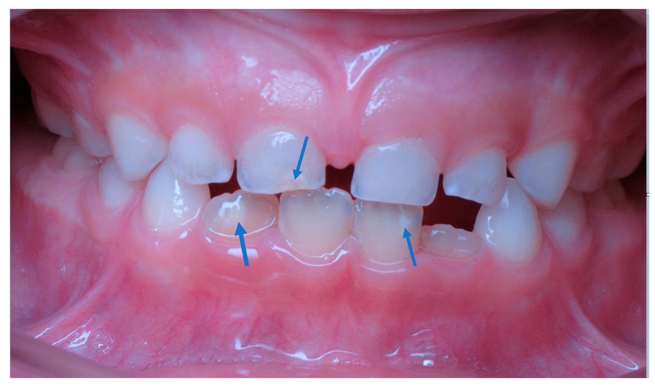
| 12 | Management of a hypomineralisation of the enamel by applying a remineraliser based on zinc hydroxyapatite (microRepair) (2021) [51] | Remineralization (photo) /in vivo | Mousse based on biomimetic nanohydroxyapatite | One year after the diagnosis, all the elements involved no longer showed any symptoms. |
| 13 | In vivo comparative evaluation of esthetics after microabrasion and microabrasion followed by casein phosphopeptide-amorphous calcium fluoride phosphate on molar incisor hypomineralization-affected incisors (2019) [52] | Tooth color (photographic evaluation) /in vivo | Microabrasion Microabrasion followed by CPP-amorphous calcium fluoride phosphate (ACFP) | Microabrasion followed by the remineralizing agent can improve the aesthetics of white tooth discoloration with time. |
| 14 | The effect of casein phosphopeptide-amorphous calcium phosphate on molar-incisor hypomineralisation: A pilot study (2017) [53] | Remineralization (laser fluorescence) /in vivo |
Paste containing 10% CPP-ACP Paste containing 10% CPP-ACP with 0.2% NaF (CPP-ACFP) | This pilot study shows that using CPP-ACP and CPP-ACFP had a positive effect in reducing hypomineralisation on enamel surfaces of MIH-diagnosed teeth for a one-month period. It is important to diagnose molar-incisor hypomineralisation at an early stage to prevent excessive caries development. Therefore, further clinical studies are necessary on the long-term application of these kinds of nanocomplexes. |
| 15 | Comparison of mineral density in molar incisor hypomineralization applying fluoride varnishes and casein phosphopeptide-amorphous calcium phosphate (2017) [54] | Remineralization (laser fluorescence) /in vivo | 5% sodium fluoride varnish (Duraphat®) 5% sodium fluoride varnish with tricalcium phosphate (Clinpro®) Casein phosphopeptide-amorphous calcium phosphate (Recaldent®) | The results obtained under the conditions used here allow concluding that Clinpro® was more effective in mild lesions, whereas Duraphat® was more effective in moderate lesions. |
| 16 | Effect of fluoride varnish on enamel remineralization in anterior teeth with molar incisor hypomineralization (2016) [55] | Remineralization (quantitative light-induced fluorescence) /in vivo |
Four applications of 5% NaF varnish, with a 1-week interval Usual home care- control | We observed no favorable effect on the remineralization of MIH lesions in anterior teeth after four applications of fluoride varnish. |
| 18 | An innovative approach to treat incisors hypomineralization (MIH): A combined use of casein phosphopeptide-amorphous calcium phosphate and hydrogen peroxide-a case report (2012) [56] | Aesthetic appearance (photographic evaluation) /in vivo | Combined use of CPP-ACP mousse and hydrogen peroxide gel | At the end of this 5-month treatment, a noticeable aesthetic improvement of the opacities was observed. |
| 19 | MIH supplementation strategies: prospective clinical and laboratory trial (2011) [28] | Mineralization, morphology, and porosity (SEM, ESEM/EDX) /in vivo | Calcium-phosphate casein | The hypothesis tested was rejected since calcium-phosphate casein improved enamel morphology in vivo. |
| No. | Paper (Year of Publication) | Condition | Tested Products and Controls | Conclusion of the Paper Abstract |
|---|---|---|---|---|
| 1 | Remineralization of molar incisor hypomineralization (MIH) with a hydroxyapatite toothpaste: an in-situ study (2022) [25] | Remineralization (microcomputed tomography) /in situ | Hydroxyapatite-toothpaste (20% hydroxyapatite) Fluoride toothpaste (1450 ppm fluoride toothpaste) | The tested toothpaste based on hydroxyapatite can remineralize MIH lesions. Pre-treating the tooth surface with acid-etchant enhanced remineralization. |
| 2 | Effect of remineralization agents on molar-incisor hypomineralization-affected incisors: A randomized controlled clinical trial (2022) [46] | Remineralization (laser fluorescence) /in vivo | Calcium glycerophosphate (CaGP) Casein phosphopeptide amorphous calcium fluoride phosphate (CPP-ACFP) Control (1450 ppm fluoride toothpaste) | The additional use of both mineral-containing agents in MIH-affected teeth improved these hypomineralized lesions with mineral deposition. Even if both agents could be used in the hypomineralized teeth with demarcated opacities, future studies are recommended on the long-term effect of these mineral-containing agents with longer observation and larger sample sizes. |
| 3 | Efficacy of a toothpaste based on microcrystalline hydroxyapatite on children with hypersensitivity caused by MIH: A randomised controlled trial (2021) [18] | Sensitivity (pain sensation in response to tactile stimulus (Wong-Baker FACES Pain Rating Scale)) /in vivo | HAP-toothpaste (10% HAP) Fluoride toothpaste (1400 ppm fluoride as amine fluoride) | Overall, non-inferiority in hypersensitivity relief of a toothpaste containing hydroxyapatite compared to amine fluoride could not be shown. However, the hydroxyapatite group tended to be less hypersensitive in both populations. Attrition of the PP population due to the COVID-19 pandemic led to the loss of statistical power. |
| 4 | Molar incisor hypomineralization treatment with casein phosphopeptide and amorphous calcium phosphate in children (2018) [20] | Sensitivity (to mechanical and thermal stimuli) /in vivo | Tooth mousse with CPP-ACP Fluoride toothpaste | The use of the remineralizing agent containing CPP-ACP resulted in a significant improvement in dental sensitivity in patients with MIH. |
| 5 | Efficacy of desensitizing products containing 8% arginine and calcium carbonate for hypersensitivity relief in MIH-affected molars: an 8-week clinical study (2017) [21] | Sensitivity (to evaporative (air) stimuli and tactile stimuli) /in vivo | Each child received a single in-office treatment with a desensitizing paste containing 8% arginine and calcium carbonate, followed by 8 weeks of brushing twice daily with a desensitizing toothpaste containing 8% arginine, calcium carbonate with 1450 ppm fluoride, using a sensitive toothbrush. Additionally, the corresponding mouthwash was used. | In conclusion, 8% arginine and calcium carbonate were able to reduce hypersensitivity successfully during this 8-week trial. |
| 6 | Effect of fluoride varnish on enamel remineralization in anterior teeth with molar incisor hypomineralization (2016) [55] | Remineralization (quantitative light-induced fluorescence) /in vivo |
Four applications of 5% NaF varnish, with a 1-week interval Usual home care- control | We observed no favorable effect on the remineralization of MIH lesions in anterior teeth after four applications of fluoride varnish. |
| Outcome | Hydroxyapatite Toothpaste | Fluoride Toothpaste |
|---|---|---|
| Combined data | 26.02 ± 20.68 | 14.64 ± 9.60 |
| Etched | 29.26 ± 22.99 | 16.83 ± 9.97 |
| Unetched | 16.62 ± 5.74 | 10.62 ± 8.13 |
| Toothpaste | ITT Population | PP Population |
|---|---|---|
| Hydroxyapatite | Mean: 2.6 [95%CI]: 1.5–3.7 | Mean: 2.6 [95%CI]: 0.9–4.3 |
| Fluoride | Mean: 3.4 [95%CI]: 2.4–4.4 | Mean: 3.1 [95%CI]: 1.7–4.5 |
Disclaimer/Publisher’s Note: The statements, opinions and data contained in all publications are solely those of the individual author(s) and contributor(s) and not of MDPI and/or the editor(s). MDPI and/or the editor(s) disclaim responsibility for any injury to people or property resulting from any ideas, methods, instructions or products referred to in the content. |
© 2023 by the authors. Licensee MDPI, Basel, Switzerland. This article is an open access article distributed under the terms and conditions of the Creative Commons Attribution (CC BY) license (https://creativecommons.org/licenses/by/4.0/).
Share and Cite
Enax, J.; Amaechi, B.T.; Farah, R.; Liu, J.A.; Schulze zur Wiesche, E.; Meyer, F. Remineralization Strategies for Teeth with Molar Incisor Hypomineralization (MIH): A Literature Review. Dent. J. 2023, 11, 80. https://doi.org/10.3390/dj11030080
Enax J, Amaechi BT, Farah R, Liu JA, Schulze zur Wiesche E, Meyer F. Remineralization Strategies for Teeth with Molar Incisor Hypomineralization (MIH): A Literature Review. Dentistry Journal. 2023; 11(3):80. https://doi.org/10.3390/dj11030080
Chicago/Turabian StyleEnax, Joachim, Bennett T. Amaechi, Rayane Farah, Jungyi Alexis Liu, Erik Schulze zur Wiesche, and Frederic Meyer. 2023. "Remineralization Strategies for Teeth with Molar Incisor Hypomineralization (MIH): A Literature Review" Dentistry Journal 11, no. 3: 80. https://doi.org/10.3390/dj11030080