Communication

Study on Cortisol Sensing Principle Based on Fluorophore and Aptamer Competitive Assay on Polymer Optical Fiber

1
DTU Electro, Department of Electrical and Photonics Engineering, Technical University of Denmark, 2800 Kongens Lyngby, Denmark
2
Pisco Group, Rønnegade 1, 3. th., 2100 Copenhagen, Denmark
*
Authors to whom correspondence should be addressed.
Photonics 2023, 10(7), 840; https://doi.org/10.3390/photonics10070840
Submission received: 15 June 2023 / Revised: 11 July 2023 / Accepted: 19 July 2023 / Published: 20 July 2023
(This article belongs to the Special Issue Editorial Board Members’ Collection Series: Photonics Sensors)

Abstract

:
In this study, we present a polymer optical fiber fluorophore/aptamer competitive assay-based cortisol sensing principle. We developed a low-cost, two-fiber perpendicular design for fluorophore-based sensing with less input light interference and high output signal intensity. The design is suitable for narrow stokes shift fluorophores. We have demonstrated the cortisol sensing principle based on the competition between tagged and normal cortisol. To date, the sensing design has exhibited a slow response, and we identified possible modifications for improvement. Our estimation shows that with miniaturization and a modified sensor assay compartment design, a less than one-hour response time can be achieved. The reported sensing principle and low-cost new design will be helpful for the future development of fluorophore-based fiber optic aptasensors that can potentially be used in a wet environment for online sensing.

1. Introduction

Cortisol is a stress hormone that plays a crucial role in humans and animals. In humans, it is an important biomarker for various diseases such as cardiovascular, immune, renal, skeletal, and endocrine system-based diseases [1,2]. Abnormal cortisol levels can cause irritation, depression, obesity, fatigue, bone fragility, and increased amino acid levels in the blood [2]. Cortisol can also affect, e.g., fish welfare, growth rate, and production of meat because stressed fish consume most of their energy for stress-related activities.
In the literature, the best limits of detection (LOD) for cortisol are 36 fg/mL [3] and 25.9 fg/mL [4]. The LOD for chromatographic techniques [2] and ordinary immunoassays like enzyme-linked immunosorbent assays (ELISA) [5] are approximately 1 pg/mL and 50 pg/mL, respectively. The concentration range of cortisol in humans and aquaculture is µg/mL and pg/mL, respectively, and therefore a broad range of LODs are of interest [1,2]. Currently used measurement techniques require time-consuming sampling and laboratory analysis. Further, available methods for cortisol sensing are expensive and cannot be used for online monitoring. Optical fiber sensors are attractive because they have several advantages, such as the feasibility of online measurements in water over long distances [1]. The glass optical fiber cortisol sensor developed by Usha et al. [4] is of high performance, but at the expense of being bulky and complicated to fabricate, requiring operation in transmission mode and feeding/removal of an analyte. In our polymer optical fiber (POF) immunosensor design, we have used, for the first time, a simpler design comprising perpendicular POFs, a hydrogel, and a luminescent competitive assay.
Aptamers are single-stranded DNA and are widely used for small molecule detection in biosensors [6]. Aptamers are developed by the systematic evolution of ligands by exponential enrichment (SELEX). In this process, a multi-round in vitro process is used to isolate aptamers from pools of single-stranded oligonucleotides with randomized sequences [7,8]. In recent years, aptamers have become the first choice for the recognition of specific molecules and sensing applications because they have several advantages over antibodies, such as low cost, easy synthesis, stability over a wide range of temperatures and pH levels, easy modification, etc. [6,7].
Fluorescence is one of the most dominant signal transductions for aptamer-based assays. This is due to its non-destructive, highly sensitive nature, good signal intensity, and ease of modification [9,10]. In recent years, various fluorescence techniques have revolutionized the field of receptor–ligand interactions and sensing. For the detection of molecules, different fluorescence-based strategies have been developed using the combination of aptamer and fluorophore, such as FRET [11,12], aptamer-based ELISA, fluorescence polarization [13,14] and quenching [15,16], competition assays, etc. In this study, we have shown the aptamer and fluorescence-based competitive binding assay principles for the sensing of cortisol. We have developed a new perpendicular fiber design to measure the signal with less input signal interference. In this study, the AF488 hydrazide fluorophore was used because it has several properties, such as high fluorescence quantum yield, low self-quenching, pH-insensitivity, high photostability, etc., which make this fluorophore an ideal candidate for robust sensing. The excitation and emission wavelengths of AF488H are 493 and 519 nm, respectively. Hence, the Stokes shift of AF488H is narrow. LED-based excitation systems have very broad spectra; therefore, expensive filters and complex instrumentation are normally required. The proposed new design provides an easy and low-cost replacement for the complex and expensive instrumentation for narrow Stokes shift fluorophores. This new design also enables the possibilities of online monitoring and remote sensing in a wet environment, which is challenging for an electronics-based sensor design. There are several challenges associated with the use of electronics-based sensors in wet environments, such as ineffective sensing in humid environments, the possibility of circuit damage, and corrosion [17,18]. Therefore, these sensors require very robust packaging, which increases their cost and complexity. Here, we successfully demonstrate a cortisol-sensing principle using a fluorophore and aptamer competitive binding assay. To the best of our knowledge, this is the first study on a fluorophore/aptamer competitive assay using a two-fiber perpendicular design. We also describe reasons for the current slow response of the sensor. To improve the response time, we discuss possible modifications in the sensing design.

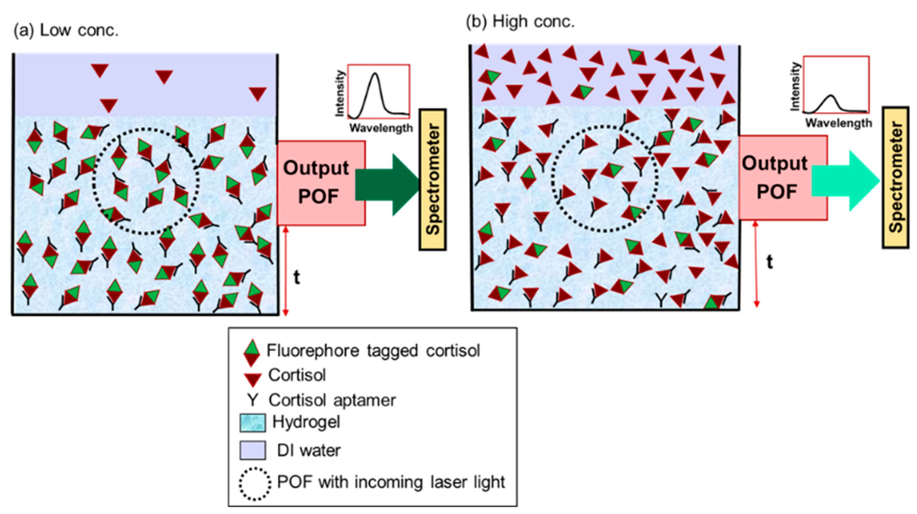
2. Sensing Principle

The sensing mechanism is based on a competitive binding assay consisting of fluorophore-tagged cortisol molecules and an aptamer for cortisol recognition (see Figure 1). A thin layer of polyacrylamide gel in which the aptamer is physically immobilized is placed inside a compartment. Both tagged and normal cortisol can diffuse in and out through the gel, and depending on the amount of normal cortisol in the surrounding liquid, the concentration of the recognized/captured cortisol or tagged cortisol will vary inside the gel just in front of the fibers [1]. This will change the fluorescence intensity. Two POFs are connected to the compartment in a perpendicular direction, one to send the excitation light and the other to receive the output fluorescence light. This perpendicular design is good when the separation between the excitation and emission wavelengths of the tagged fluorophore is small (~20 nm). The POFs are connected perpendicularly to reduce the interference of the input signal in the emission signal of the fluorescence output spectra, something that would otherwise require filtration and, therefore, intensity loss.

3. Fabrication

3.1. Materials

Cortisol 3-(O-carboxymethyl)oxime (C3CMO), hydrocortisone, N-(3-dimethylaminopropyl)-N′-ethylcarbodiimide hydrochloride (EDC), anhydrous dimethylsulfoxide (DMSO), 1-hydroxy-7-azabenzotriazole (HOAt) (0.6 M in dimethylformamide (DMF)), isopropyl alcohol (IPA), acrylamide, N,N′-methylenebis(acrylamide), phosphate-buffered saline (PBS), TLC plates, TLC saturation pads, and preparative TLC (PTLC) plates (20 cm × 20 cm × 0.2 cm silica gel on glass plates) were procured from Sigma-Aldrich, Denmark. Alexa Fluor 488 hydrazide (AF488H), 2-(N-morpholino)ethanesulfonic acid buffer (MES), and (BupH™ MES buffered saline: 0.1 M MES, 0.9% sodium chloride, pH 4.7) were purchased from Fisher Scientific, Denmark. N,N,N′,N′-Tetramethyl ethylenediamine (TEMED) and ammonium persulfate (APS) were purchased from Merck. The LED (490 nm) light source was procured from Thorlabs. A 488 nm cyan blue laser diode was procured from eBay. SMA 905 connectors and a crimp tool were purchased from FiberFin, USA. Syringe filters (0.2 micron) were purchased from GE Healthcare Life Sciences. Step- index polymer optical fiber with a PMMA core diameter of approx. 980 µm and fluorinated cladding thickness of approx. 10 µm with a black polyethylene jacket, making the total outer diameter approx. 2.2 mm, was purchased from Edmund Optics. The cortisol aptamer reported by Dalirirad et al., (ATGGGCAATGCGGGGTGGAGAATGGTTGCCGCACTTCGGC) was purchased from biomers.net GmbH [19].

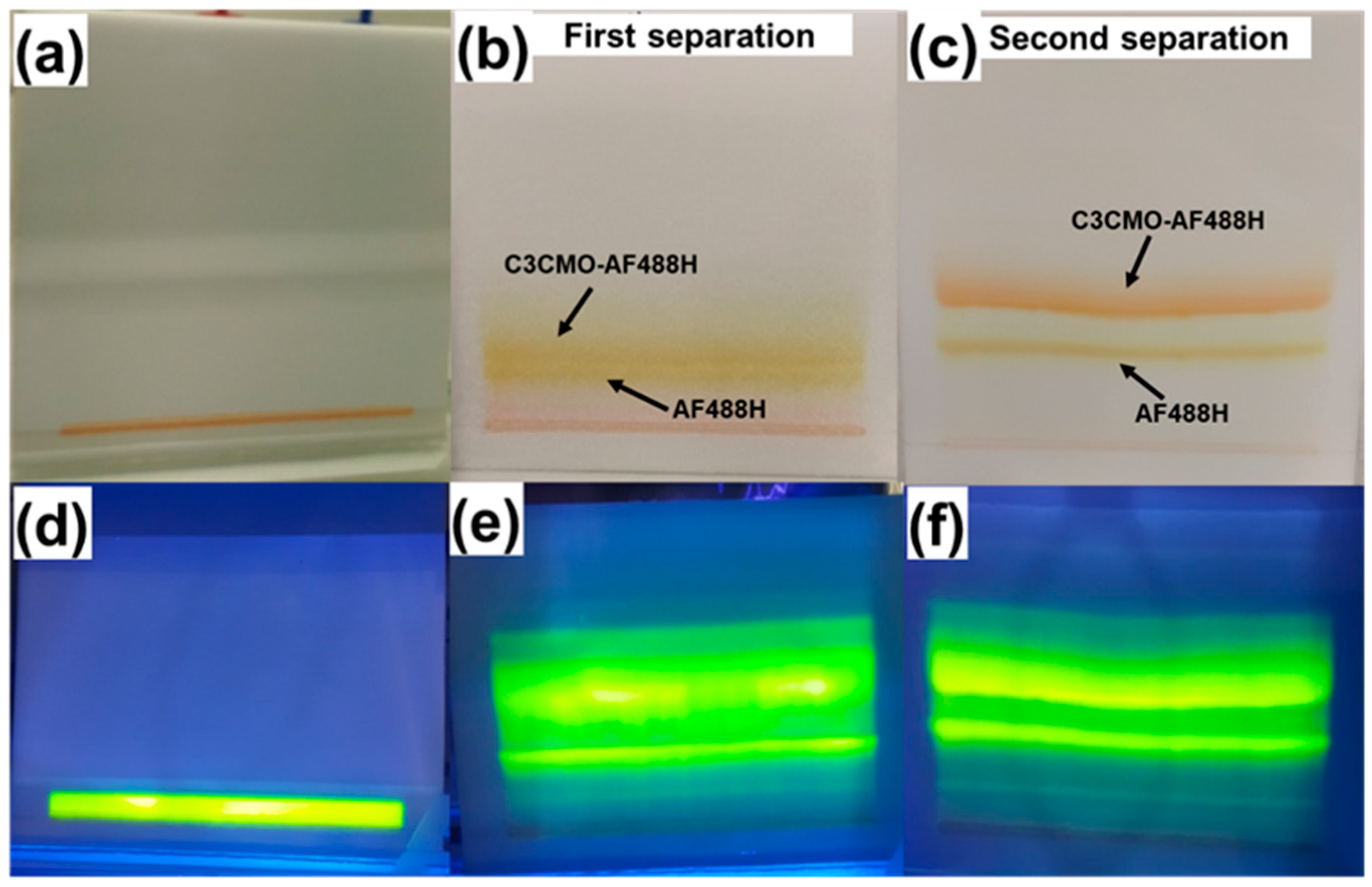
3.2. Labeling of Cortisol

The sensing mechanism is based on fluorescence; therefore, first, we labeled the cortisol with the AF488 hydrazide (AF488H) fluorophore. Further, for the conjugation, we chose the cortisol derivative cortisol 3-(O-carboxymethyl)oxime (C3CMO). The reason for choosing these two molecules is that EDC cross-linking can be used between the carboxyl group of C3CMO and the hydrazide primary amine on AF488H. The C3CMO and AF488H conjugation and chemical identification procedures are provided in detail in our previous work [1]. Since the chemical separation was modified compared to our earlier work, it is described in detail here. To separate the reactants and products, we used PTLC plates. First, we drew a horizontal line using a pencil from one vertical edge to the other 2 cm from both vertical bottom edges of the PTLC plate. Then, 200 µL of crude liquid were dispensed over the 16 cm long line, which is shown in Figure 2a,d. In Figure 2a–c, the images were captured in normal ambient light, while in Figure 2d–f they were captured with UV lamp (365 nm) illumination. The lines of liquid were dispensed as small drops on the PTLC plate using a 100 µL pipette. Since DMSO was used as a solvent in the labeling process of cortisol, it was evaporated in a vacuum oven at 45 °C and 0.02 mbar for 30 min. The same process was repeated until all of the product solution was dispensed. After that, any remaining DMSO was evaporated in a vacuum oven at 45 °C and 0.02 mbar for at least 3 h. For separation of the AF488H-tagged cortisol from the mixture, we mixed an eluent solvent consisting of 160 mL IPA and 40 mL MES buffer and poured it onto a TLC saturation pad inside a TLC separation chamber and waited for 5 min to equilibrate the solvent inside the chamber. We placed the PTLC plate inside the chamber immediately after taking it from the vacuum oven, and the chamber was closed with a glass plate and covered with aluminum foil. The separation step took 5 h for the eluent to reach the top of the PTLC plate. After that, the plate was removed. Next, the plate was kept inside the fume hood to dry overnight. In this process, the separation between the AF488H and AF488H-tagged cortisol was small and not very clear, which is shown in Figure 2b,e. Due to the limited separation, we again kept the PTLC plate inside the vacuum oven overnight, and the next day it was again kept inside the eluent chamber using the same protocol. After repeating a process like that, we observed a nice separation between the AF488H- and AF488H-linked cortisol, as shown in Figure 2c,f. Figure 2c,f shows the pictures of the PTLC plate with nicely separated lines of AF488H-tagged cortisol and AF488H. The AF488H was identified by dispensing AF488H separately on a PTLC plate placed inside the eluent chamber and measuring and comparing the height of the line, and the AF488H-tagged cortisol was identified using liquid chromatography–mass spectrometry (LC–MS) [1]. The silica gel containing AF488H-tagged cortisol was scraped off the glass plates and washed with stirring in 20 mL Milli-Q water for at least an hour before centrifugation in 2 mL vials at 13.4 krpm for 5 min. The water containing AF488H-tagged cortisol was collected from the vials. A 0.5 mL volume of water was added to the vials; they were vortexed until the silica gel was loose again and then placed on a rocking table overnight. Then the vials were centrifuged again, and the AF488H-tagged cortisol containing water was again collected. The washing step was repeated 3 times, and finally, it was filtered through the 0.2 µM syringe filter.

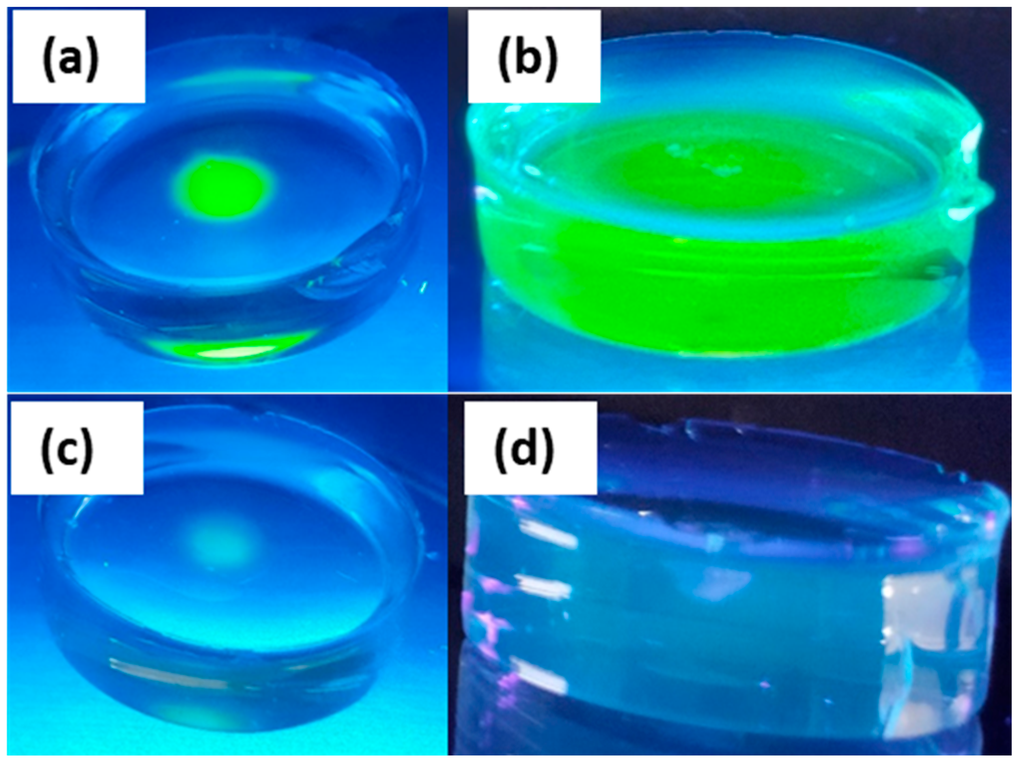
3.3. Synthesis of Hydrogel Containing Aptamer and Tagged Cortisol and Porosity Test of Hydrogel

We used polyacrylamide hydrogel [20] for sensor aptamer immobilization. The porosity and transparency of the hydrogel can be changed by varying the monomer to crosslinker ratio. Thus, it was optimized by testing ratios of 19:1, 29:1, and 37.5:1. A good porous and transparent hydrogel capable of immobilizing the aptamer while allowing in/out transport of tagged/untagged cortisol was achieved with the 37.5:1 ratio between the monomer and crosslinker. For the synthesis of polyacrylamide hydrogel containing the sensor assay, 35 μL of AF488H-tagged cortisol and 20 μL (10 μmol) of cortisol aptamer were first mixed in 80 µL of phosphate-buffered saline (PBS). The cortisol aptamer and AF488H-tagged cortisol solution was then mixed with 140 μL of 30 wt.% acrylamide/bisacrylamide (37.5:1) using a magnetic stirrer in the presence of N2 gas. The nitrogen gas was purged to remove the dissolved oxygen from the solution because it may deteriorate the polymerization reaction. The solution was then mixed with 14 μL of APS (10 wt.% in DI water) and 2 μL of TEMED. After mixing with the TEMED, the gelation started, and the gel was formed within 1 min. Porosity was tested by placing small drops of AF488H- and AF488H-tagged cortisol molecules on top of the hydrogel discs shown in Figure 3a,c, respectively. Using UV lamp (365 nm) illumination, Figure 3b,d shows the penetration of the AF488H- and AF488H-tagged cortisol molecules after 24 h inside the hydrogel, which means the synthesized polyacrylamide hydrogel was porous enough that AF488H- and AF488H-tagged cortisol molecules could diffuse inside it. As shown in Figure 3d, the intensity of the green color of AF488H-tagged cortisol inside the hydrogel was very low due to the low concentration of AF488H-tagged cortisol in that specific solution.

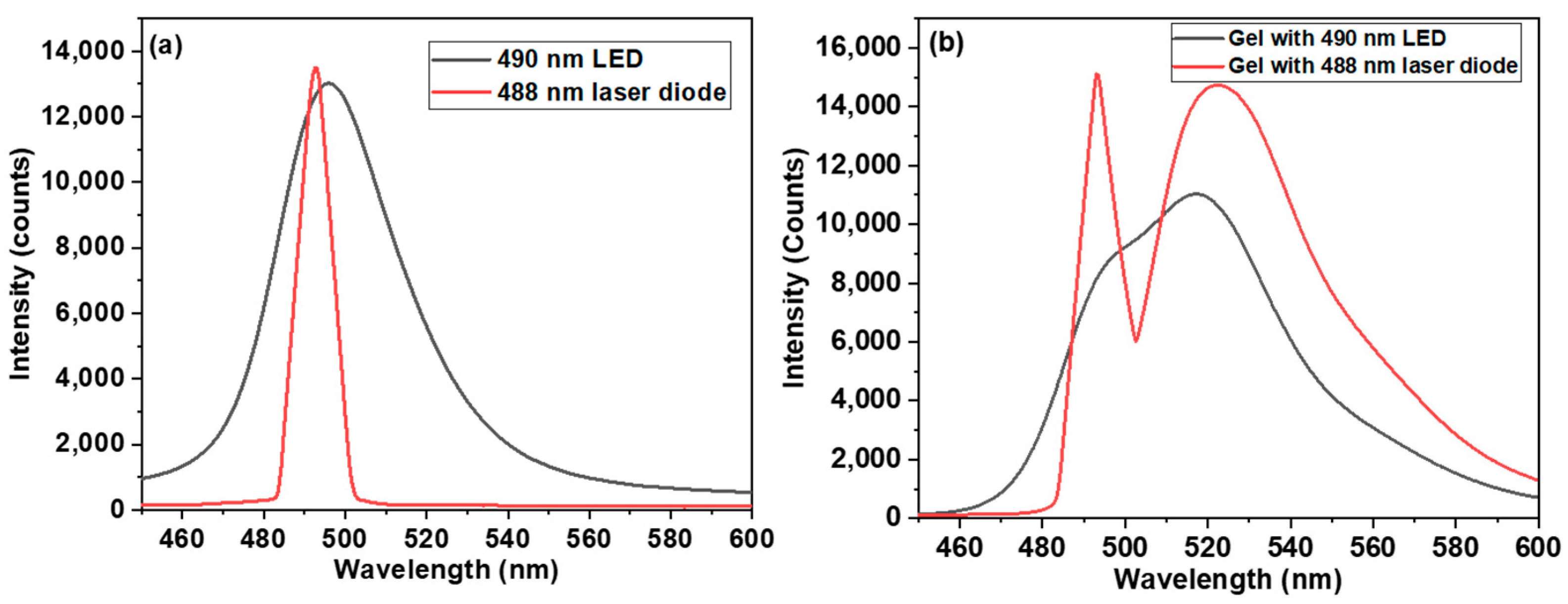
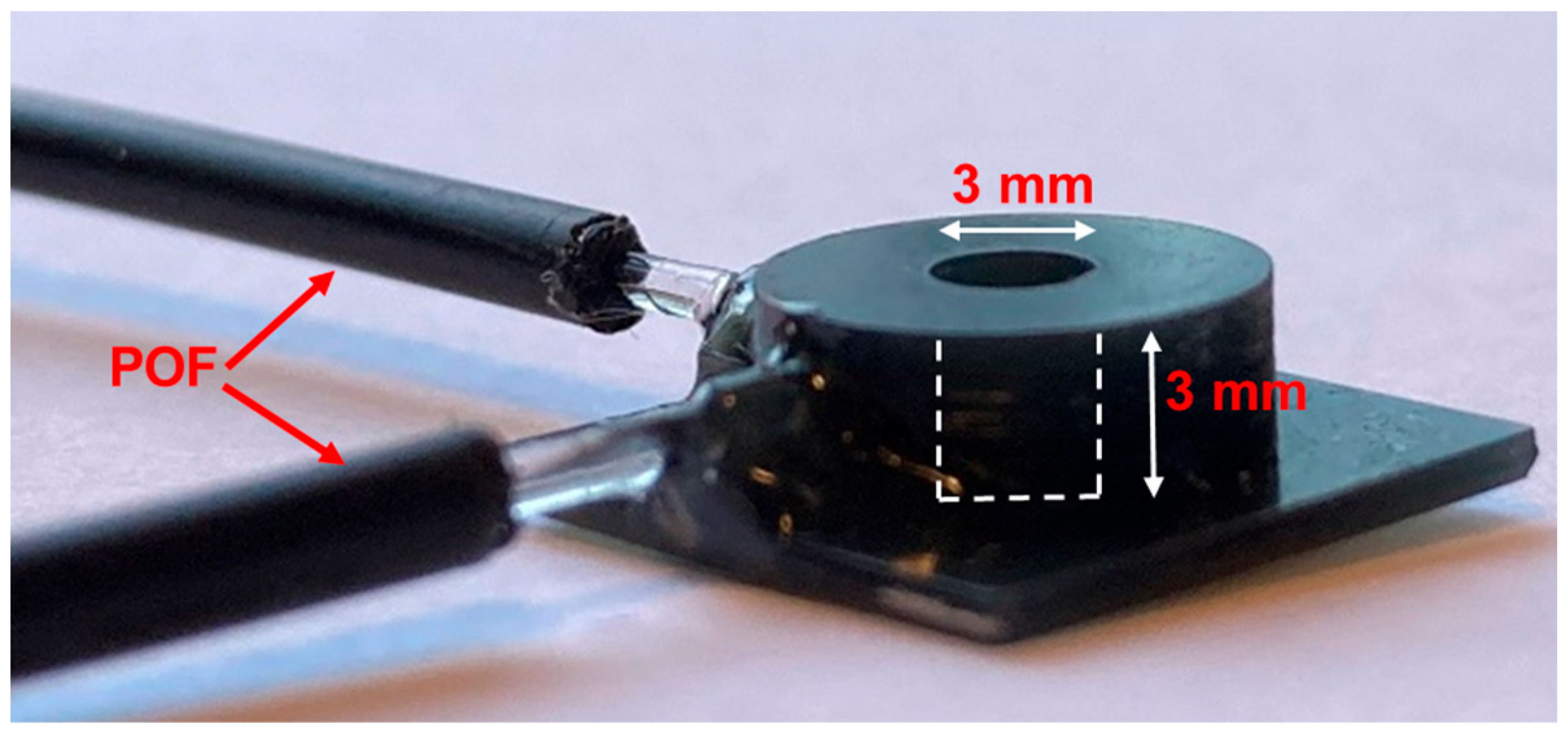
3.4. Components of Sensing Setup

The absorption and emission wavelengths of AF488H are 493 and 519 nm, respectively. Therefore, to excite this fluorophore, we tested a 490 nm LED and a 488 nm laser diode. The optical spectra of the LED and laser diode are shown in Figure 4a. The bandwidths (FWHM) of the LED and laser diode are 40 nm and 10 nm, respectively. We recorded the emission spectra of the hydrogel containing AF488H-tagged cortisol in our perpendicular fiber sensing setup, using the LED and laser diode as excitation sources. The output spectra are shown in Figure 4b. The Stokes shift between the absorption and emission wavelengths of AF488H is 26 nm, which means that the 40 nm LED spectrum covers both the absorption and emission wavelengths of AF488H. This results in a strong overlapping of the excitation and emission wavelengths when the LED is used, as clearly seen in Figure 4b. In contrast, for the laser diode, there is a clear separation between excitation and emission peaks, which means that they can easily be separated with a filter. Therefore, we used the 488 nm laser diode as an excitation source.
We note that our earlier proposed sensor design had the sensor assay compartment on the tip of the POF [1] and used 490 nm LED excitation light. The fluorescence signal was thus picked up with the same fiber using a fiber splitter and two filters (both of LED excitation and of fluorophore emission), which increased the size, complexity, and cost of the sensor. The excitation and emission filters also decreased the intensity of the output signal. Therefore, we used a new design (two perpendicular fibers) to solve all issues related to low intensity, cost, and size. The new perpendicular design was inspired by the fact that fluorophores emit light in all directions; therefore, to reduce the interference of incoming light, fluorescence can be collected from the perpendicular direction.
We fabricated a 3D-printed sensing compartment with two 1 mm cylindrical slots for POF attachment. The actual photo of the sensing assay compartment is shown in Figure 5. Two POFs are connected to the assay compartment using UV glue. The diameter and height of the cylindrical assay compartment are 3 mm. The tip of the POF was polished with P1200 and P4000 sandpaper in deionized water (DI) and then cleaned in DI water to obtain a better input and output signal. Both input and output fibers were connected to SMA 905 connectors using a crimp/swage hand tool. The hydrogel was prepared according to the aforementioned protocol in Section 3.3. The hydrogel was placed inside the cavity, and the sensing compartment was stored at room temperature for 30 min for complete polymerization of the hydrogel. After 30 min, the sensing compartment was used for measurements.

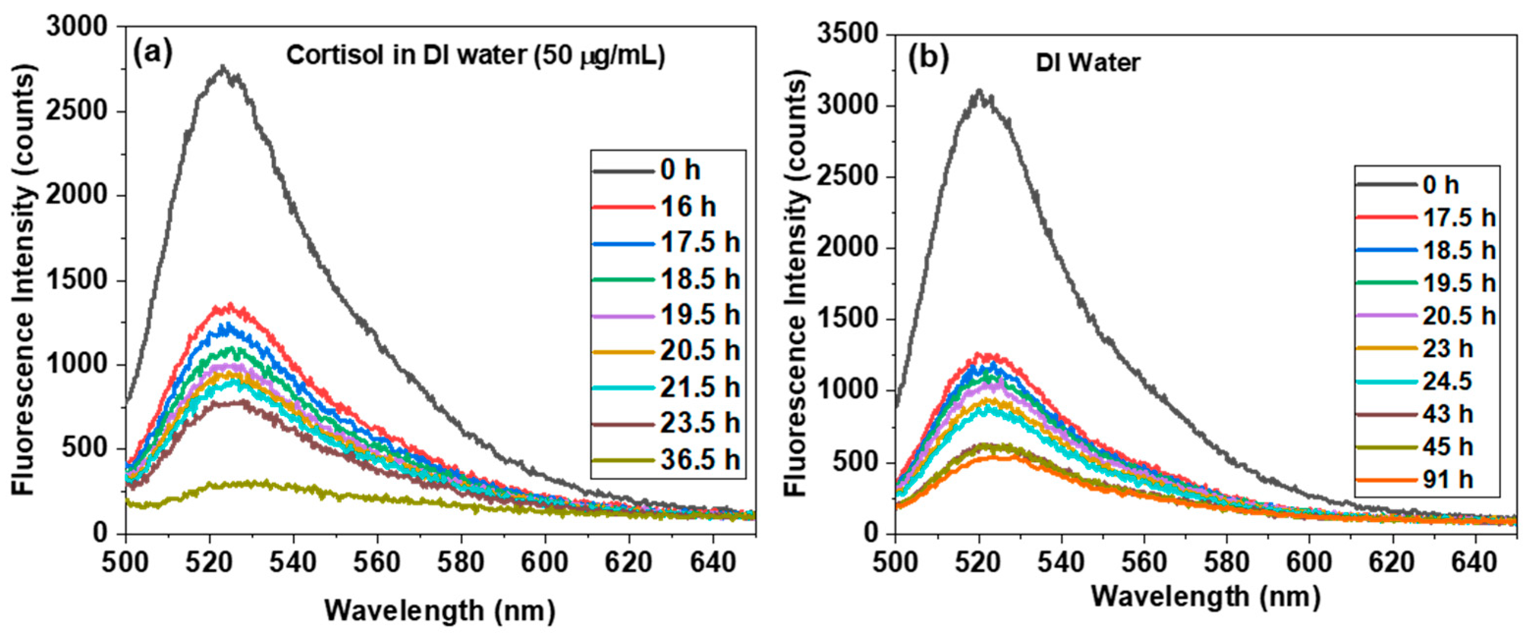
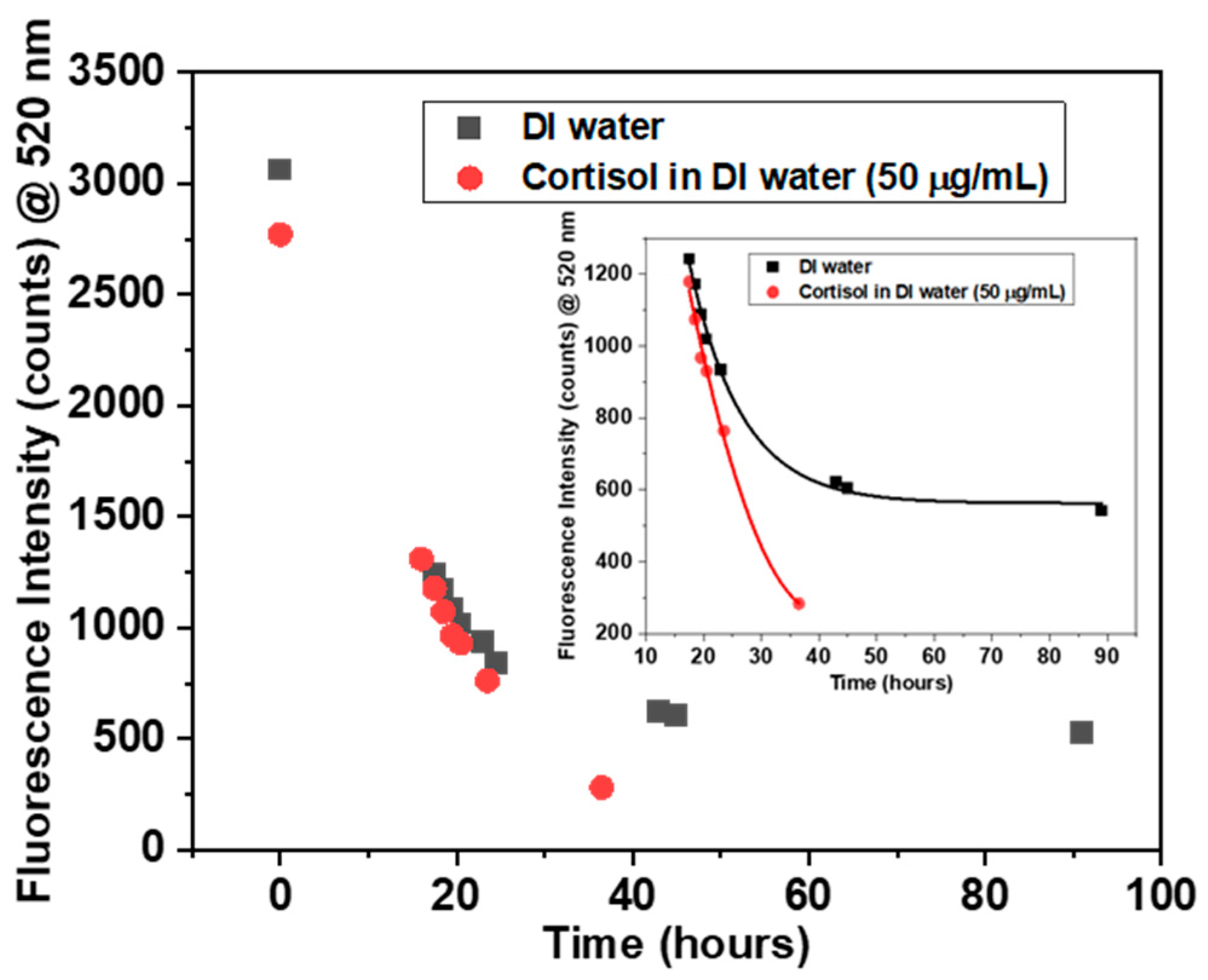
4. Results and Discussion

First, we fabricated two 3D-printed compartments connected with POF and containing gel, including aptamer and tagged cortisol. Figure 6a shows the fluorescence spectra at different times when the sensing compartment was immersed in the cortisol solution (50 µg/mL). Similarly, Figure 6b shows fluorescence spectra in the presence of pure DI water. The fluorescence signal intensity decreased with time in the cortisol solution (50 µg/mL) and DI water.
The signal intensities at different times were extracted at the emission wavelength of the AF488H (520 nm) from Figure 6 and plotted. Figure 7 shows this plot of the fluorescence intensity variation of the sensor in DI water and cortisol solution with time. Initially, in Figure 7, the rate of change of fluorescence intensity appeared similar for cortisol solution and DI water until 20 h of exposure. After 20 h, the rate of change of fluorescence intensity in the presence of cortisol solution was faster compared to that of the DI water sample. The inset of Figure 7 shows that the variation of the fluorescence intensity in the presence of cortisol solution was almost linear, while in the presence of DI water, the fluorescence variation was almost saturated after 40 h. There are two possible reasons for this kind of fluorescence variation. For the initial 20 h, the rate of change of fluorescence in the presence of DI water and cortisol solution showed a similar trend due to the leaching of the unbound AF488H-tagged cortisol molecules from the hydrogel.
These excess AF488H-tagged cortisol molecules were present in the hydrogel and were not captured by aptamers. In the presence of an aqueous environment, these unbound/free AF488H-tagged cortisol molecules could escape from the hydrogel all the way out of the compartment, but the AF488H-tagged cortisol molecules, which were captured by the aptamer, could not as easily escape from the hydrogel. These aptamer-captured AF488H-tagged cortisol molecules escaped more easily when competing cortisol was present. After 20 h, when the unbound molecules were almost leached out of the hydrogel, the aptamer-captured tagged cortisol molecules started contributing to the difference in the sensing response in DI water and cortisol solution. In other words, the aptamer-captured tagged cortisol molecules were not displaced from the hydrogel in the presence of DI water; therefore, the fluorescence intensity remained almost unchanged and saturated at a relatively high level. In the presence of the cortisol solution, the fluorescence intensity dropped quickly because the AF488H-tagged cortisol molecules were displaced by the cortisol molecules. This displacement was due to the competitive nature of this assay. These measurements show the competition between the AF488H-tagged cortisol and cortisol molecules. The reason for this competition is the affinity of the cortisol molecules to the aptamer. The affinity of the cortisol molecules towards the aptamer was higher compared to that of the AF488H-tagged cortisol molecule because the aptamer was designed specifically for the cortisol molecule. The affinity of the AF488H-tagged cortisol molecules should be lower because, after tagging with the AF488H fluorescence molecules on the cortisol, the overall molecular structure of the AF488H-tagged cortisol was changed compared to that of cortisol; therefore, due to this molecular structural change, the affinity should be lower. The reason for the slow sensor response is the following: firstly, the excess amount of AF488H-tagged cortisol in the hydrogel and, secondly, the thickness of the hydrogel. The excess amount of AF488H-tagged cortisol in the hydrogel took around 20 h to leach, and then real sensing started. That means the actual sensing started after 20 h. Due to the thick hydrogel, the cortisol molecules took a long time to enter the hydrogel and, likewise, for the displaced tagged cortisol, to exit. In Figure 3, we can see the AF488H- and AF488H-tagged cortisol took around 24 h to completely enter the 8 mm thick hydrogel disc. In our sensor, the thickness of the hydrogel was approximately 2.6 mm. That means the cortisol molecules were completely inside the hydrogel in 8–9 h. A possible way to improve the sensing response is through quantification of the AF488H-tagged cortisol and thickness optimization of the hydrogel. We needed to use the AF488H-tagged cortisol in such a way that the amount of unbound AF488H-tagged cortisol remained very small in the hydrogel, and that is possible after performing the quantification of the AF488H-tagged cortisol. Thickness optimization of the hydrogel was also needed to improve the sensing response. The thickness of the hydrogel should be a few hundred microns to reduce the response time to 1 h. If the hydrogel thickness is reduced, the fluorescence signal will decrease, which can influence the sensitivity of the sensor. Therefore, there is a trade-off between the thickness of the hydrogel and the fluorescence signal; hence, optimized thickness is required. In Figure 1, we can see that in our fabricated sensing compartment, the fibers were placed at height t above the bottom, and, therefore, we had an extra t height of hydrogel in our sensing, which was below the fiber. We needed to modify the sensing assay compartment and remove this extra hydrogel layer of thickness t from the assay compartment volume. A thin layer of hydrogel was possible with a new assay compartment and small diameter fibers. Using these suggestions, a fast response time could be achieved. In general, fluorophore/aptamer competitive assay-based sensing is performed in liquid, and narrow Stokes-shift fluorophores require complex instrumentation. We have demonstrated fluorophore/aptamer competitive assays inside the hydrogel with a new optical fiber design that enables online sensing in a wet environment, and no complex instrumentation is needed to separate the excitation and emission signals of the fluorophore. In the past, several techniques have been explored for the detection of cortisol. A comparison of different cortisol-sensing techniques is presented in Table 1.

5. Conclusions

In this study, we developed a simple and low-cost design for fluorophore- and aptamer-based optical fiber sensing. Using the laser diode and perpendicular design compartment, one can easily obtain the fluorescence signal without using any filters, splitters, or complex instruments for optical fiber sensing. The hydrogel entrapment method was used to immobilize the aptamer inside the hydrogel just in front of the fiber for excitation and emission pick-up. We have shown single-time cortisol sensing using the competition between the tagged and normal cortisol. Using the same strategies, one can label according to the analyte of interest and follow the same technique for sensing. The reported technique can also be utilized for the affinity test of the aptamers. The reported sensing design needs a few optimizations to improve the response time. We hope that the reported sensing principle and the new design will be helpful for the researcher to develop cost-effective fluorophore- and aptamer-based fiber optic sensors.

Author Contributions

Conceptualization, V.S. and J.J.; methodology, V.S. and J.J.; validation, V.S. and J.J.; formal analysis, V.S., O.B. and J.J.; investigation, V.S., O.B. and J.J.; resources, V.S., J.H., E.M., O.B. and J.J.; data curation, V.S.; writing—original draft preparation, V.S. and J.J.; writing—review and editing, V.S., O.B. and J.J.; visualization V.S. and J.J.; supervision, O.B. and J.J.; project administration, J.J.; funding acquisition, J.J. All authors have read and agreed to the published version of the manuscript.

Funding

This project was funded by the Green Development and Demonstration Programme, GUDP, Denmark. (Grant number: 34009-20-1769).

Institutional Review Board Statement

Not applicable.

Informed Consent Statement

Not applicable.

Data Availability Statement

Data underlying the results presented in this paper are not publicly available at this time but may be obtained from the authors upon reasonable request.

Conflicts of Interest

The authors declare no conflict of interest.

References

  1. Janting, J.; Inglev, R. Towards an online polymer optical fiber cortisol sensor for aquaculture. In Proceedings of the 27th International Conference on Optical Fiber Sensors, The Westin Alexandria, VA, USA, 8–12 July 2020; pp. 1–4. [Google Scholar] [CrossRef]
  2. Kaushik, A.; Vasudev, A.; Arya, S.K.; Pasha, S.K.; Bhansali, S. Recent advances in cortisol sensing technologies for point-of-care application. Biosens. Bioelectron. 2014, 53, 499–512. [Google Scholar] [CrossRef]
  3. Mota, V.C.; Martins, C.I.M.; Eding, E.H.; Canário, A.V.M.; Verreth, J.A.J. Water cortisol and testosterone in Nile tilapia (Oreochromis niloticus) recirculating aquaculture systems. Aquaculture 2017, 468, 255–261. [Google Scholar] [CrossRef]
  4. Usha, S.P.; Shrivastav, A.M.; Gupta, B.D. A contemporary approach for design and characterization of fiber-optic-cortisol sensor tailoring LMR and ZnO/PPY molecularly imprinted film. Biosens. Bioelectron. 2017, 87, 178–186. [Google Scholar] [CrossRef]
  5. Fanouraki, E.; Papandroulakis, N.; Ellis, T.; Mylonas, C.C.; Scott, A.P.; Pavlidis, M. Water Cortisol Is a Reliable Indicator of Stress in European Sea Bass, Dicentrarchus labrax. Behaviour. Bioacti. 2017, 145, 1267–1281. [Google Scholar]
  6. Song, S.; Wang, L.; Li, J.; Fan, C.; Zhao, J. Aptamer-based biosensors. TrAC—Trends Anal. Chem. 2008, 27, 108–117. [Google Scholar] [CrossRef]
  7. Ali, M.H.; Elsherbiny, M.E.; Emara, M. Updates on aptamer research. Int. J. Mol. Sci. 2019, 20, 1–23. [Google Scholar] [CrossRef] [Green Version]
  8. Tuerk, C.; Gold, L. Systematic evolution of ligands by exponential enrichment: RNA ligands to bacteriophage T4 DNA polymerase. Science 1990, 249, 505–510. [Google Scholar] [CrossRef]
  9. Pickup, J.C.; Hussain, F.; Evans, N.D.; Rolinski, O.J.; Birch, D.J.S. Fluorescence-based glucose sensors. Biosens. Bioelectron. 2005, 20, 2555–2565. [Google Scholar] [CrossRef]
  10. Zhong, W. Nanomaterials in fluorescence-based biosensing. Anal. Bioanal. Chem. 2009, 394, 47–59. [Google Scholar] [CrossRef] [PubMed] [Green Version]
  11. Jepsen, M.D.; Sparvath, S.M.; Nielsen, T.B.; Langvad, A.H.; Grossi, G.; Gothelf, K.V.; Andersen, E.S. Development of a genetically encodable FRET system using fluorescent RNA aptamers. Nat. Commun. 2018, 9, 18. [Google Scholar] [CrossRef]
  12. Pehlivan, Z.S.; Torabfam, M.; Kurt, H.; Ow-Yang, C.; Hildebrandt, N.; Yüce, M. Aptamer and nanomaterial based FRET biosensors: A review on recent advances (2014–2019). Microchim. Acta 2019, 186, 563. [Google Scholar] [CrossRef]
  13. Hendrickson, O.D.; Taranova, N.A.; Zherdev, A.V.; Dzantiev, B.B.; Eremin, S.A. Fluorescence polarization-based bioassays: New horizons. Sensors 2020, 20, 7132. [Google Scholar] [CrossRef] [PubMed]
  14. Cruz-Aguado, J.A.; Penner, G. Fluorescence polarization based displacement assay for the determination of small molecules with aptamers. Anal. Chem. 2008, 80, 8853–8855. [Google Scholar] [CrossRef] [PubMed]
  15. Trettnak, W. Optical sensors based on fluorescence quenching. In Fluorescence Spectroscopy: New Methods and Applications; Springer: Berlin/Heidelberg, Germany, 1993; pp. 79–89. [Google Scholar] [CrossRef]
  16. Geddes, C.D. Optical halide sensing using fluorescence quenching: Theory, simulations and applications—A review. Meas. Sci. Technol. 2001, 12, R53–R88. [Google Scholar] [CrossRef] [Green Version]
  17. Baylakoğlu, I.; Fortier, A.; Kyeong, S.; Ambat, R.; Conseil-Gudla, H.; Azarian, M.H.; Pecht, M.G. The detrimental effects of water on electronic devices. Adv. Electr. Electron. Eng. 2021, 1, 100016. [Google Scholar] [CrossRef]
  18. Zhang, H.; Xia, C.; Feng, G.; Fang, J. Hospitals and laboratories on paper-based sensors: A Mini Review. Sensors 2021, 21, 5998. [Google Scholar] [CrossRef]
  19. Dalirirad, S.; Steckl, A.J. Aptamer-based lateral flow assay for point of care cortisol detection in sweat. Sens. Actuators B Chem. 2019, 283, 79–86. [Google Scholar] [CrossRef]
  20. Semwal, V.; Shrivastav, A.M.; Verma, R.; Gupta, B.D. Surface plasmon resonance based fiber optic ethanol sensor using layers of silver/silicon/hydrogel entrapped with ADH/NAD. Sens. Actuators B Chem. 2016, 230, 485–492. [Google Scholar] [CrossRef]
  21. Shima, D.; Daewoo, H.; Andrew, S.J. Aptamer-Based Lateral Flow Biosensor for Rapid Detection of Salivary Cortisol. ACS Omega 2020, 5, 32890–32898. [Google Scholar]
  22. Roushani, M.; Hosseini, H.; Hajinia, Z.; Rahmati, Z. Rationally designed of hollow nitrogen doped carbon nanotubes double shelled with hierarchical nickel hydroxide nanosheet as a high performance surface substrate for cortisol aptasensing. Electrochim. Acta 2021, 388, 138608. [Google Scholar] [CrossRef]
  23. Pali, M.; Jagannath, B.; Lin, K.C.; Upasham, S.; Sankhalab, S.; Upashama, S.; Muthukumar, S.; Prasad, S. CATCH (Cortisol Apta WATCH): ‘Bio-mimic alarm’ to track Anxiety, Stress, Immunity in human sweat. Electrochim. Acta. 2021, 390, 138834. [Google Scholar] [CrossRef]
  24. Niu, C.; Ding, Y.; Zhang, C.; Liu, J. Comparing two cortisol aptamers for label-free fluorescent and colorimetric biosensors. Sens. Diagn. 2022, 1, 541–549. [Google Scholar] [CrossRef]
Figure 1. Schematic diagram of the sensing principle: (a) low cortisol concentration; (b) high cortisol concentration. (The dotted circle indicates the POF with incoming light; t is the distance between the bottom of the assay compartment and the fibers.)
Photonics 10 00840 g001
Figure 2. PTLC plates: (a) before being placed inside the eluent chamber; (b) first separation of tagged cortisol from reactants and by-products; (c) second separation of tagged cortisol from reactants and by-products; (df) are the images of (ac) in 365 nm illumination.
Photonics 10 00840 g002
Figure 3. Images of hydrogel disc under the illumination of a 365 nm UV lamp: (a) right after placing a drop of AF488H solution onto it; (b) after 24 h with AF488H; (c) right after placing a drop of AF488H- tagged cortisol onto it; and (d) after 24 h with AF488H-tagged cortisol.
Photonics 10 00840 g003
Figure 4. (a) The output spectra of the 490 nm LED and 488 nm laser diode. (b) The spectra of the 490 nm LED and 488 nm laser diode together with AF488H fluorescence after passing through the C3CMO-AF488H-containing hydrogel.
Photonics 10 00840 g004
Figure 5. Image of the fabricated sensing assay compartment. The dashed line indicates the compartment.
Photonics 10 00840 g005
Figure 6. Fluorescence intensity vs wavelength with time: (a) in cortisol (50 µg/mL) solution; (b) in DI water.
Photonics 10 00840 g006
Figure 7. Fluorescence intensity vs. time with exposure to cortisol (50 µg/mL) solution. The inset shows the fluorescence intensity vs. time from 20 h to 90 h.
Photonics 10 00840 g007
Table 1. Comparison of different cortisol sensing techniques.
TechniqueSensitivityResponse TimeMeritsLimitationsRef.
Lateral Flow assayng/L15–20 minRapid screening, disposable, good for biofluidsNot feasible for online measurements, wet environment and non-reusable.[21]
Electrochemicalng/L~hoursGood selectivityNot feasible for wet environment, complex instrumentations.[22]
Impedance spectroscopyng/mLNot mentionedReal-time, continuous monitoring, reusableNot feasible for wet environment, complex instrumentations.[23]
Colorimetricng/L20–30 minSimple, low costNot feasible for online measurements, wet environment and non-reusable. Need a lot of optimizations[24]
Fluorophore/aptamer-based competitive assayµg/mL~hoursSimple, low cost, robust, feasible for online measurements, remote sensing and wet environmentNeed gel thickness optimization, non-reusable.Present work
Disclaimer/Publisher’s Note: The statements, opinions and data contained in all publications are solely those of the individual author(s) and contributor(s) and not of MDPI and/or the editor(s). MDPI and/or the editor(s) disclaim responsibility for any injury to people or property resulting from any ideas, methods, instructions or products referred to in the content.

