
Lung Recruitment Maneuvers Assessment by Bedside Lung Ultrasound in Pediatric Acute Respiratory Distress Syndrome

 , , , , and
Abstract
:1. Introduction
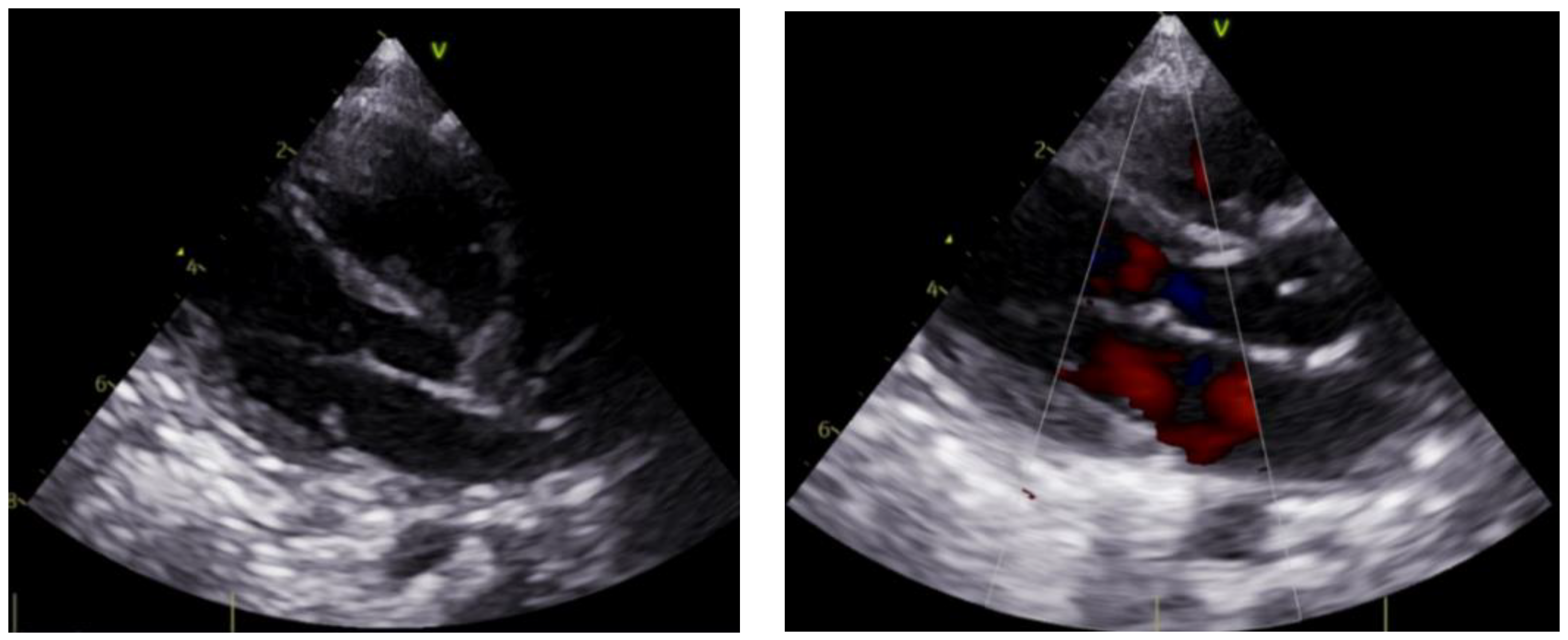
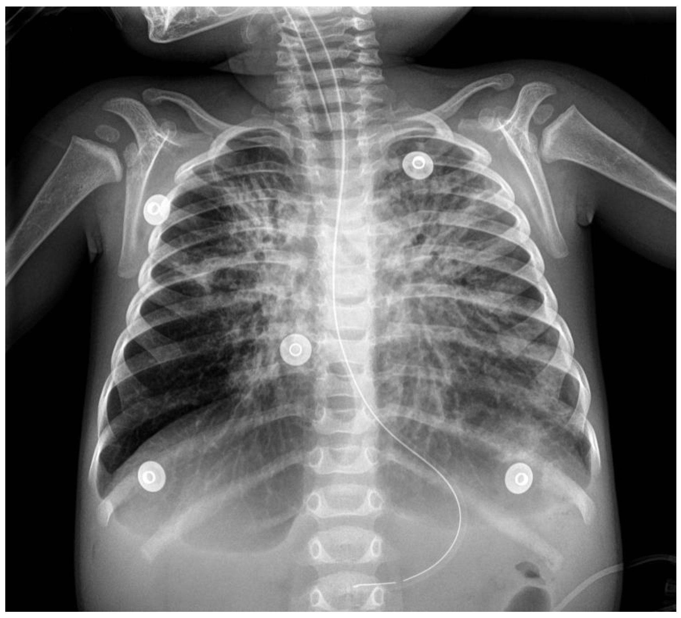
2. Case
3. Discussion
Author Contributions
Funding
Institutional Review Board Statement
Informed Consent Statement
Data Availability Statement
Conflicts of Interest
References
- Meyer, N.J.; Gattinoni, L.; Calfee, C.S. Acute Respiratory Distress Syndrome. Lancet 2021, 398, 622–637. [Google Scholar] [CrossRef]
- Ware, L.B.; Matthay, M.A. The Acute Respiratory Distress Syndrome. N. Engl. J. Med. 2000, 342, 1334–1349. [Google Scholar] [CrossRef] [PubMed]
- Meade, M.O.; Cook, D.J.; Guyatt, G.H.; Slutsky, A.S.; Arabi, Y.M.; Cooper, D.J.; Davies, A.R.; Hand, L.E.; Zhou, Q.; Thabane, L.; et al. Lung Open Ventilation Study Investigators. Ventilation strategy using low tidal volumes, recruitment maneuvers, and high positive end-expiratory pressure for acute lung injury and acute respiratory distress syndrome: A randomized controlled trial. JAMA 2008, 299, 637. [Google Scholar] [CrossRef] [PubMed] [Green Version]
- Ranieri, V.M.; Rubenfeld, G.D.; Thompson, B.T.; Ferguson, N.D.; Caldwell, E.; Fan, E.; Camporota, L.; Slutsky, A.S. Acute respiratory distress syndrome: The Berlin Definition. JAMA 2012, 307, 2526–2533. [Google Scholar] [CrossRef] [PubMed]
- The Pediatric Acute Lung Injury Consensus Conference Group. Pediatric Acute Respiratory Distress Syndrome: Consensus Recommendations From the Pediatric Acute Lung Injury Consensus Conference. Pediatric Crit. Care Med. 2015, 16, 428–439. [Google Scholar] [CrossRef] [Green Version]
- Cheifetz, I.M. Pediatric ARDS. Respir. Care 2017, 62, 718–731. [Google Scholar] [CrossRef]
- Fan, E.; Wilcox, M.E.; Brower, R.G.; Stewart, T.E.; Mehta, S.; Lapinsky, S.E.; Meade, M.O.; Ferguson, N.D. Recruitment Maneuvers for Acute Lung Injury: A Systematic Review. Am. J. Respir. Crit. Care Med. 2008, 178, 1156–1163. [Google Scholar] [CrossRef]
- Gil Cano, A.; Monge García, M.I.; Gracia Romero, M.; Díaz Monrové, J.C. Incidencia, características y evolución del barotrauma durante la ventilación mecánica con apertura pulmonar. Med. Intensiva 2012, 36, 335–342. [Google Scholar] [CrossRef]
- Briel, M.; Meade, M.; Mercat, A.; Brower, R.G.; Talmor, D.; Walter, S.D.; Slutsky, A.S.; Pullenayegum, E.; Zhou, Q.; Cook, D.; et al. Higher vs Lower Positive End-Expiratory Pressure in Patients with Acute Lung Injury and Acute Respiratory Distress Syndrome: Systematic Review and Meta-Analysis. JAMA 2010, 303, 865–873. [Google Scholar] [CrossRef]
- García-Fernández, J.; Romero, A.; Blanco, A.; Gonzalez, P.; Abad-Gurumeta, A.; Bergese, S.D. Maniobras de reclutamiento en anestesia: ¿qué más excusas para no usarlas? Rev. Española Anestesiol. Reanim. 2018, 65, 209–217. [Google Scholar] [CrossRef]
- Hodgson, C.; Goligher, E.C.; Young, M.E.; Keating, J.L.; Holland, A.E.; Romero, L.; Bradley, S.J.; Tuxen, D. Recruitment Manoeuvres for Adults with Acute Respiratory Distress Syndrome Receiving Mechanical Ventilation. Cochrane Database Syst. Rev. 2016, 11, CD006667. [Google Scholar] [CrossRef] [PubMed]
- Jones, B.P.; Tay, E.T.; Elikashvili, I.; Sanders, J.E.; Paul, A.Z.; Nelson, B.P.; Spina, L.A.; Tsung, J.W. Feasibility and Safety of Substituting Lung Ultrasonography for Chest Radiography When Diagnosing Pneumonia in Children. Chest 2016, 150, 131–138. [Google Scholar] [CrossRef] [PubMed] [Green Version]
- Conlon, T.W.; Nishisaki, A.; Singh, Y.; Bhombal, S.; De Luca, D.; Kessler, D.O.; Su, E.R.; Chen, A.E.; Fraga, M.V. Moving Beyond the Stethoscope: Diagnostic Point-of-Care Ultrasound in Pediatric Practice. Pediatrics 2019, 144, e20191402. [Google Scholar] [CrossRef] [PubMed]
- Cherian, T.; Mulholland, E.K.; Carlin, J.B.; Ostensen, H.; Amin, R.; de Campo, M.; Greenberg, D.; Lagos, R.; Lucero, M.; Madhi, S.A.; et al. Standardized Interpretation of Paediatric Chest Radiographs for the Diagnosis of Pneumonia in Epidemiological Studies. Bull. World Health Organ. 2005, 83, 353–359. [Google Scholar]
- Shah, V.P.; Tunik, M.G.; Tsung, J.W. Prospective Evaluation of Point-of-Care Ultrasonography for the Diagnosis of Pneumonia in Children and Young Adults. JAMA Pediatrics 2013, 167, 119. [Google Scholar] [CrossRef] [Green Version]
- Buonsenso, D.; Musolino, A.; Ferro, V.; De Rose, C.; Morello, R.; Ventola, C.; Liotti, F.M.; Chiaretti, A.; Biasucci, D.G.; Spanu, T.; et al. Role of lung ultrasound for the etiological diagnosis of acute lower respiratory tract infection (ALRTI) in children: A prospective study. J. Ultrasound 2021, 10, 1–13. [Google Scholar] [CrossRef]
- Singh, A.; Gupta, A.; Sen, M.; Suri, J.; Chakrabarti, S.; Bhattacharya, D. Utility of Bedside Lung Ultrasound for Assessment of Lung Recruitment in a Case of Acute Respiratory Distress Syndrome. Lung India 2019, 36, 451. [Google Scholar] [CrossRef]
- Tusman, G.; Acosta, C.M.; Costantini, M. Ultrasonography for the Assessment of Lung Recruitment Maneuvers. Crit. Ultrasound J. 2016, 8, 8. [Google Scholar] [CrossRef] [Green Version]
- Gardelli, G.; Feletti, F.; Gamberini, E.; Bonarelli, S.; Nanni, A.; Mughetti, M. Using Sonography to Assess Lung Recruitment in Patients with Acute Respiratory Distress Syndrome. Emerg. Radiol. 2009, 16, 219–221. [Google Scholar] [CrossRef]
- Bobillo-Perez, S.; Rodriguez-Fanjul, J.; Girona-Alarcon, M.; Cambra, F.J.; Jordan, I.; Balaguer, M. Ultrasound-Guided Recruitment Maneuvers in Pediatric Acute Chest Syndrome Due to Sickle Cell Disease. Med. Intensiva 2021, 45, 184–186. [Google Scholar] [CrossRef]
- Volpicelli, G.; Elbarbary, M.; Blaivas, M.; Lichtenstein, D.A.; Mathis, G.; Kirkpatrick, A.W.; Melniker, L.; Gargani, L.; Noble, V.E.; Via, G.; et al. International Liaison Committee on Lung Ultrasound (ILC-LUS) for the International Consensus Conference on Lung Ultrasound (ICC-LUS). International Evidence-Based Recommendations for Point-of-Care Lung Ultrasound. Intensive Care Med. 2012, 38, 577–591. [Google Scholar] [CrossRef] [PubMed] [Green Version]
- Bouhemad, B.; Brisson, H.; Le-Guen, M.; Arbelot, C.; Lu, Q.; Rouby, J.-J. Bedside Ultrasound Assessment of Positive End-Expiratory Pressure–Induced Lung Recruitment. Am. J. Respir. Crit. Care Med. 2011, 183, 341–347. [Google Scholar] [CrossRef] [PubMed] [Green Version]
- Patroniti, N.; Bonatti, G.; Senussi, T.; Robba, C. Mechanical Ventilation and Respiratory Monitoring during Extracorporeal Membrane Oxygenation for Respiratory Support. Ann. Transl. Med. 2018, 6, 386. [Google Scholar] [CrossRef]
- Duff, J.; Rosychuk, R.; Joffe, A. The safety and efficacy of sustained inflations as a lung recruitment maneuver in pediatric intensive care unit patients. Intensive Care Med. 2007, 33, 1778–1786. [Google Scholar] [CrossRef] [PubMed]
- Pierrakos, C.; Smit, M.R.; Hagens, L.A.; Heijnen, N.F.L.; Hollmann, M.W.; Schultz, M.J.; Paulus, F.; Bos, L.D.J. Assessment of the Effect of Recruitment Maneuver on Lung Aeration Through Imaging Analysis in Invasively Ventilated Patients: A Systematic Review. Front. Physiol. 2021, 12, 666941. [Google Scholar] [CrossRef] [PubMed]
- See, K.C.; Ong, V.; Tan, Y.L.; Sahagun, J.; Taculod, J. Chest Radiography versus Lung Ultrasound for Identification of Acute Respiratory Distress Syndrome: A Retrospective Observational Study. Crit. Care 2018, 22, 203. [Google Scholar] [CrossRef] [Green Version]
- Malbouisson, L.M.; Muller, J.-C.; Constantin, J.-M.; Lu, Q.; Puybasset, L.; Rouby, J.-J.; the CT Scan ARDS Study Group. Computed Tomography Assessment of Positive End-Expiratory Pressure-Induced Alveolar Recruitment in Patients with Acute Respiratory Distress Syndrome. Am. J. Respir. Crit. Care Med. 2001, 163, 1444–1450. [Google Scholar] [CrossRef]
- Bedetti, G.; Gargani, L.; Corbisiero, A.; Frassi, F.; Poggianti, E.; Mottola, G. Evaluation of Ultrasound Lung Comets by Hand-Held Echocardiography. Cardiovasc. Ultrasound 2006, 4, 34. [Google Scholar] [CrossRef] [Green Version]


| PEEP (cmH2O) | 20 | 25 | 30 | 25 | 20 | 15 |
|---|---|---|---|---|---|---|
| Time of RMs (min) | 0 | 2 | 4 | 6 | 8 | 10 |
| FiO2 (%) | 40 | 40 | 40 | 40 | 40 | 40 |
| FiO2 ECMO (%) | 50 | 50 | 50 | 50 | 50 | 50 |
| HbSat (%) | 98 | 96 | 96 | 95 | 95 | 96 |
| HR (bpm) | 130 | 133 | 137 | 140 | 138 | 135 |
| MBP (mmHg) | 53 | 50 | 48 | 48 | 51 | 52 |
Publisher’s Note: MDPI stays neutral with regard to jurisdictional claims in published maps and institutional affiliations. |
© 2022 by the authors. Licensee MDPI, Basel, Switzerland. This article is an open access article distributed under the terms and conditions of the Creative Commons Attribution (CC BY) license (https://creativecommons.org/licenses/by/4.0/).
Share and Cite
Mor Conejo, M.; Guitart Pardellans, C.; Fresán Ruiz, E.; Penela Sánchez, D.; Cambra Lasaosa, F.J.; Jordan Garcia, I.; Balaguer Gargallo, M.; Pons-Òdena, M. Lung Recruitment Maneuvers Assessment by Bedside Lung Ultrasound in Pediatric Acute Respiratory Distress Syndrome. Children 2022, 9, 789. https://doi.org/10.3390/children9060789
Mor Conejo M, Guitart Pardellans C, Fresán Ruiz E, Penela Sánchez D, Cambra Lasaosa FJ, Jordan Garcia I, Balaguer Gargallo M, Pons-Òdena M. Lung Recruitment Maneuvers Assessment by Bedside Lung Ultrasound in Pediatric Acute Respiratory Distress Syndrome. Children. 2022; 9(6):789. https://doi.org/10.3390/children9060789
Chicago/Turabian StyleMor Conejo, Mireia, Carmina Guitart Pardellans, Elena Fresán Ruiz, Daniel Penela Sánchez, Francisco José Cambra Lasaosa, Iolanda Jordan Garcia, Mònica Balaguer Gargallo, and Martí Pons-Òdena. 2022. "Lung Recruitment Maneuvers Assessment by Bedside Lung Ultrasound in Pediatric Acute Respiratory Distress Syndrome" Children 9, no. 6: 789. https://doi.org/10.3390/children9060789