Article

Prenatal Predictors of Neurobehavioral Outcome in Children with Fetal Growth Restriction at 6 Years of Age: A Retrospective Cohort Study

by
Mª José Benítez Marín
1,2,3,*,
Marta Blasco Alonso
2,3,4 and
Ernesto González Mesa
2,3,4
1
Obstetrics and Gynecology Service, Virgen de la Victoria University Hospital, 29010 Málaga, Spain
2
Surgical Specialties, Biochemistry and Immunology Department, Málaga University, 29071 Málaga, Spain
3
Research Group in Maternal-Fetal Medicine, Epigenetics, Women’s Diseases and Reproductive Health, Instituto de Investigación Biomédica de Málaga y Plataforma en Nanomedicina-IBIMA Plataforma Bionand, 29071 Málaga, Spain
4
Obstetrics and Gynecology Service, Regional University Hospital of Malaga, 29011 Málaga, Spain
*
Author to whom correspondence should be addressed.
Children 2023, 10(6), 997; https://doi.org/10.3390/children10060997
Submission received: 17 May 2023 / Revised: 30 May 2023 / Accepted: 31 May 2023 / Published: 1 June 2023
(This article belongs to the Special Issue 10th Anniversary of Children: Feature Papers in Neonatology)

Abstract

:
(1) Background: Fetal growth restriction (FGR) increases the risk of adverse neurodevelopmental outcomes, especially in preterm newborns. This study aims to describe the behavioral results of FGR at 6 years of age and to demonstrate the relationship of certain predictive factors with this development. (2) Methods: This retrospective cohort study included 70 children born in 2015 at the University Hospital Carlos Haya, Málaga, Spain who had been exposed to FGR during pregnancy; neonatal and infant data were recorded retrospectively. Children were assessed prospectively at 6 years of age by means of a strengths and difficulties questionnaire (SDQ) to study behavioral outcomes. (3) Results: We demonstrated that there are higher behavioral disability rates in children exposed to FGR during pregnancy and, in particular, high rates of hyperactivity or conduct problems. We also proved a negative relationship between the birth weight percentile and the total behavioral scale score, along with a positive correlation between hyperactivity and the emotional and behavioral scales. Learning difficulties were more frequent in early-onset FGR than in late-onset FGR. (4) Conclusions: Our study of behavioral development has demonstrated higher behavioral disability rates in children with FGR at 6 years of age; specifically, high rates of hyperactivity or conduct problems. At the same time, we have proved a negative relationship between the birth weight percentile and the total behavioral scale score.

1. Introduction

Fetal growth restriction (FGR) has varied greatly in terms of its management over time. Nowadays, FGR management techniques have improved the standard treatment guidelines. As a result, restriction severity and prematurity issues now have established follow-up and completion dates to reduce perinatal morbidity and mortality [1,2,3]. Thanks to improvements in pregnancy management, perinatal care, and neonatal techniques, perinatal mortality has decreased considerably, especially in extremely premature infants. However, these improvements may not appreciably reduce perinatal morbidity [4].
Neonatal outcomes in FGR have been exhaustively researched, and prematurity has been strongly associated with short-term outcomes [2]. Childhood development may also be influenced by FGR [5] and interest in long-term outcomes, specifically regarding neurodevelopment, is growing. A considerable number of studies in human and animal models have demonstrated changes to the nervous system’s structure, affecting brain volume [6,7,8], grey matter volume and structure [8,9,10], and white matter structure and myelinization [11,12], as well as influencing the gyrification process [13] in FGR conditions. These changes may affect motor, cognitive, adaptive, and behavioral development.
However, the results of these studies are often heterogeneous and contradictory. In most cases, assessments may be carried out at very early stages before the development of a given research object’s cognitive deficits or behavioral changes. The studies also include a heterogeneous group of children, both those who are small for their gestational age and those with FGR; in many cases, the authors did not take into account the issue of prematurity, which could alter the results. Despite these heterogeneities and contradictions, outcomes such as poor results in intelligence coefficients, poor academic results, cognitive and emotional alterations, and attention and hyperactivity disorders, as well as behavioral disabilities have been described with early-onset FGR. The most severe cases have been linked with motor disorders and cerebral palsy [6,14,15,16,17,18,19,20]. These results do not shed much light on late-onset FGR when reaching the gestational term [21,22,23,24,25,26,27].
Similarly, various research groups have attempted to show the relationship between different prenatal markers and motor, cognitive, and behavioral development. In our previous systematic review, we were able to associate brain sparing with poorer cognitive development. However, the link between brain sparing effect and behavioral skills development was difficult to establish [28].
In our previous research, we assessed FGR children by means of the Battelle Developmental Inventory (BDI), evaluating milestones in different areas. We found a high rate of poor global development, with motor and communication skills being the main areas affected. Conversely, we were able to associate the brain-sparing effect with worse coefficients of global development. However, the cognitive delay rate was low [29].
It has been shown that early attention and early stimulation in children that are born preterm can have a positive effect on motor neurodevelopment, and that this positive effect on cognition continues into school age [30]. Therefore, the early detection of FGR in neurocognitive risk could allow for the implementation of early stimulation strategies to improve their deficits for a longer period. In the same way, cognitive development and emotional intelligence could be improved, and better resilience and future personal relationships obtained, by identifying and improving the influencing factors (both positive and negative).
We believe that children who have been exposed to FGR during pregnancy have a higher risk of behavioral disorders. This study aims to describe the behavioral results of FGR in children at 6 years of age. Secondly, we demonstrate the relationship of certain predictive factors with this developmental issue, which might help us to select a population that is at risk of FGR, in order to assist with early childhood support or with psychological assessment and management.

2. Methods

2.1. Population

This cohort study had a retrospective design, in which we selected a group of FGR children born in 2015 at University Hospital Carlos Haya, Málaga, Spain. This hospital is a specialized center for the diagnosis and treatment of this pathology. Table 1 describes our inclusion and exclusion criteria, based on the definition of FGR. We define the brain-sparing effect as a cerebroplacental ratio (CPR) below the 5th percentile. When FGR is diagnosed at or below 32 weeks of gestational age, it is defined as early-onset FGR. In the event that this diagnosis is beyond 32 weeks, it is defined as late-onset FGR. Recruitment started in 2021, following approval from the regional ethics committee, and parental consent was obtained before child assessment began. Medical and sociodemographic data were collected from clinical records and parents’ reports. This study protocol was previously described in our most recent publication [29].

2.2. Parents’ Reports and Data Collection

We examined the pregnancy and neonatal care information given in the medical records and recorded all the variables related to pregnancy care, FGR characteristics, Doppler measurements, and delivery episodes. After birth, we recorded the variables related to neonatal anthropometric measurements, adverse neonatal outcomes, and, if applicable, admission days in the neonatal intensive care unit (NICU).
Parents completed a survey to provide information on sociodemographic items and childhood problems, such as academic difficulties, the need for early child support, kindergarten attendance, or any major health problems.

2.3. Behavioral Assessment

The Spanish version of the strengths and difficulties questionnaire (SDQ) was completed prospectively when the subjects were 6 years of age [31]. This questionnaire provides a brief emotional and behavioral screening test for children that is completed by their parents. It can be used in both low- and high-risk populations [32]. This test consists of 25 items, divided into 5 scales. The first four scales assess negative symptoms (emotional symptoms, conduct problems, hyperactivity and inattention symptoms, and peer relationship problems), while the last scale evaluates positive social relationships (prosocial behavior) [33]. Depending on the scores obtained, children are classified as normal, borderline, or abnormal. For those classified as borderline or abnormal, more in-depth studies are required to diagnose a behavioral problem. We selected this test because it is effective in differentiating between psychiatric and non-psychiatric populations, with the advantage of being shorter than other behavioral tests [34].
At the same time, we assessed the children using a BDI screening test. These results are shown in an earlier article published by our research group [29].

2.4. Statistical Analysis

A descriptive analysis was carried out to detail the frequency distribution of the different variables in the cohort, along with the distribution of the behavioral classifications in each scale. We examined the relationship between the sociodemographic variables, Doppler measurements and FGR characteristics, and the behavioral problems in each scale using the chi-squared test.
Finally, we conducted a multiple linear regression analysis to investigate the effect of (1) sociodemographic variables, (2) FGR characteristics and age at delivery, and (3) neonatal and child outcomes on the score from the total difficulties scale. Normality was tested using a Kolmogorov–Smirnov analysis. Pearson’s coefficient was used for the correlations when we confirmed the normal distribution. For all analyses, we considered a p-value below 0.05 to be significant. All data were processed and analyzed with the support of the Statistical Package for the Social Sciences (SPSS), version 22.0 (SPSS Inc., Chicago, IL, USA).

3. Results

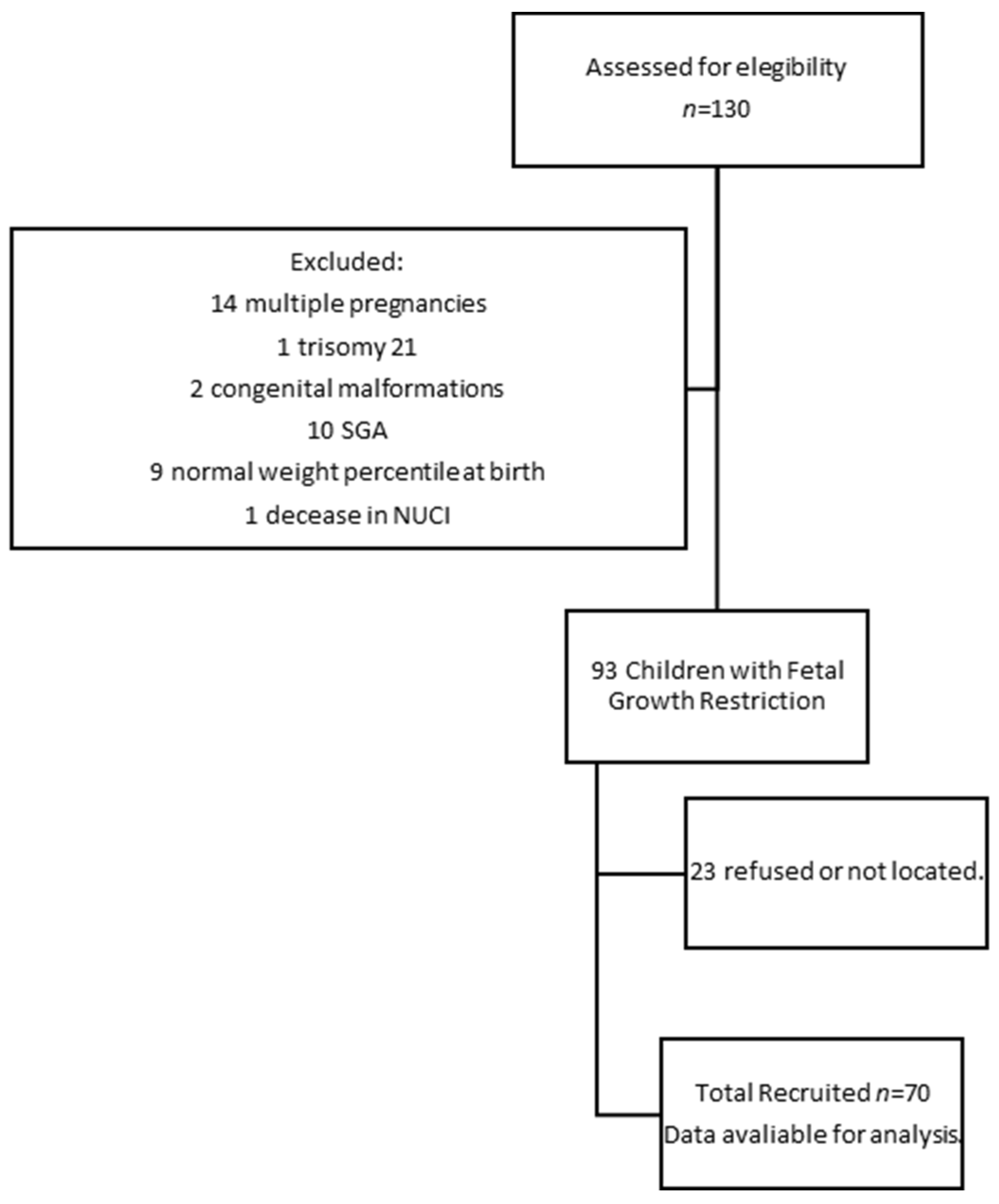
3.1. Population and Characteristics of Population Participants

One hundred and thirty children diagnosed with FGR during pregnancy who were born in 2015 were initially located. Figure 1 describes our flow diagram of the participants in the research. The study was carried out in a single medical facility; therefore, no sampling of cases was carried out. Consequently, all diagnosed cases of FGR that met the inclusion criteria were considered for the study. Seventy 6-year-old children were recruited, representing the initial population with a confidence level of 95%, a type-II error of 0.6, and a statistical power of 94% [29].
The characteristics of the population participants were described in our previous paper [29]. Table 2 summarizes the sociodemographics, delivery characteristics, and adverse neonatal outcomes of the population.

3.2. Behavioral Outcomes at 6 Years of Age

The children were aged 70–84 months at the time of the behavioral test. The average age of the children at the time of assessment was 76.20 months old (SD = 3.70). The learning disabilities rate was 27.1%. Additionally, 36.8% of the children needed early child support, and 16.7% of the children are currently following this program. Parents reported two cases of children who were diagnosed as having an autistic spectrum disorder. One of these children exhibited difficulties in performing adaptive skills, while the other child exhibited a global development disorder. These data were published in our previous paper [29].
Table 3 summarizes the percentages of children with behavioral problems in different areas. We were able to record 30% of the global behavioral disabilities. In the case of hyperactivity symptoms, 30% of the children had an abnormal classification, and 8.6% had a borderline classification. Hyperactivity symptoms and conduct problems were the most common issues.
We found a positive correlation between the total score and the scores obtained on different scales: emotional scale (r = 0.655, p ≤ 0.001), behavior scale (r = 0.655, p ≤ 0.001), hyperactivity (r = 0.843, p ≤ 0.001), and peer problems (r = 0.465, p ≤ 0.001). Similarly, a positive correlation was found between the hyperactivity and emotional scale (r = 0.387, p = 0.001) and the behavior scale (r = 0.549, p ≤ 0.001). Therefore, hyperactive children tended to present more emotional and behavioral problems.

3.3. Bivariant Analyses

Table 4 and Table 5 summarize the percentages of children with behavioral problems according to the diagnosis variables. We found no differences in the classification according to the onset time of FGR. However, with the exception of the peer problems scale, there was a tendency of poor results in each scale for the early-onset FGR diagnosis group. Learning difficulties were more frequent in the early-onset FGR group (43.5%) than in the late-onset FGR group (19.6%), reaching statistical significance (χ2(1, N = 69) = 4.39, p = 0.036).
Regarding the Doppler measurement variables, we found that a pathological CPR measurement was related to poor results on the full scale. When pathological UA, MCA, and CPR measurements were detected, there was a tendency towards poor classifications in the remaining behavioral scales (no significance).
Gestational age at delivery or birth characteristics were not related to behavioral outcomes. A positive correlation was found between the birth-weight percentile at delivery and the different scales: total score (r = −0.310, p = 0.009), emotional scale (r = −0.280, p = 0.019), and hyperactivity scale (r = −0.246, p = 0.040). Head circumference was not related to the results, although we were able to negatively correlate the birth-weight percentile with the score of the total scale (r = −0.310, p = 0.009), emotional scale (r = −0.280, p = 0.019), and hyperactivity scale (r = −0.246, p = 0.040). Neonatal outcome was not associated with behavioral problems.

3.4. Multivariate Analyses

In order to analyze the mediating factors, we performed a multiple linear regression analysis. Table 6 summarizes the variables included in each model. We included sociodemographic factors in the first model. Maternal employment status (ES) was positively related to global behavioral score (F(1,62) = 5.15, p = 0.027). In this regard, unemployed mothers had children with higher scores and, consequently, worse classifications. The R2 value was 0.077, showing that 7% of the effect is explained by differences in the mother’s ES.
In the second model, the birth-weight percentile was negatively related to the total score (F(1,59) = 5.58, p = 0.022) in the second model. The R2 value was 0.089; therefore, this variable could explain the 8.9% effect.
In the third model, we found that the need for early child support was negatively related to the total score of behavioral problems (F(1,59) = 5.22, p = 0.026). These findings could be explained by the higher rates of severe FGR and extreme prematurity among these children, making them more prone to behavioral disabilities. Table 7 sums up these models.

4. Discussion

We conducted a study to assess the behavioral development of children with FGR at 6 years of age. We have demonstrated higher behavioral disability rates in these children, specifically, high rates of hyperactivity or conduct problems. At the same time, we have proved a negative relationship between the birth-weight percentile and the total behavioral scale score.
FGR is a cause for concern among obstetricians due to the perinatal morbidities it generates, which are secondary to prematurity or the hemodynamic process itself. In recent decades, the neurodevelopmental deficits of these children, especially behavioral or cognitive impairments, have attracted the attention of both clinicians and researchers.

4.1. Regarding the Prevalence of Behavioral Disorders

Our study assessed the possible behavioral problems at the age of 6 (70–84 months) of children previously diagnosed with FGR. At this stage, the children’s attentional capacity has matured to the point where they are able to maintain a state of alertness, their ability to resolve conflicts or problems has increased, and psychosocial relationships with their environment are well established.
Multiple studies have shown that the neurodevelopment of children with FGR during childhood is not comparable to that of children with a normal birth weight. In our study, we found a high prevalence of possible behavioral problems. In fact, 14.3% of the children had abnormal scores on the questionnaire, while 15.7% had borderline scores. Therefore, 30% of the children assessed (abnormal and borderline scores) should undergo more specific evaluations to establish a correct diagnosis. In particular, within the areas studied, the areas of conduct (borderline: 25.7%, abnormal: 11.4%) and hyperactivity (borderline: 8.6%, abnormal: 30%) were the most affected (externalizing problems), while emotional areas and peer problems were less strongly affected (internalizing difficulties). Parents reported that 27.1% of the children had experienced learning difficulties at school.
When evaluating these differences based on the time of FGR diagnosis, early-onset FGR presented worse scores in terms of externalizing problems (behavior and hyperactivity), while late-onset FGR presented worse scores in terms of peer problems and prosocial areas. Similarly, we found a higher percentage of academic difficulties in the subgroup of children diagnosed with early-onset FGR (43.5%) compared to children diagnosed with late-onset FGR (19.6%).
Several studies have shown similar results to ours. Guellec et al. (2011) assessed children at 8 years of age using the SDQ. They identified 33.3% of behavioral problems in FGR infants that were born before 28 weeks of gestational age and 19.1% of behavioral problems in those born between 29 and 32 weeks of gestational age. Similarly, the learning difficulties rates were similar to ours, with 35.5% in those born before 28 weeks of gestational age and 28% in those born between 29 and 32 weeks of gestational age. However, their hyperactivity problem rates varied between 19.1 and 23.5%, while we found 39.1% of hyperactivity disabilities in those children diagnosed before 32 weeks of gestation (data not shown). It is important to note that they classified and evaluated infants by gestational age in their study, without determining which subjects presented real growth restrictions [19].
A previous meta-analysis evaluated behavioral and executive function problems in infants born before 33 weeks of gestation or weighing less than 1500 g. Compared to those born at term or at an appropriate weight, the infants displayed worse results in terms of academic achievement, attention, internalizing problems, and executive function. However, the analysis did not differentiate between growth restriction and growth that was appropriate for the subjects’ gestational age [14]. We must note that executive function is a cognitive process and has significant implications for behavioral skills such as self-regulation and inhibition [35].
These findings have also been supported by studies on late-onset FGR children. Geva et al. (2006) showed that FGR children presented a higher incidence of learning disabilities, memory problems, low creativity, and executive function problems compared to children with normal growth [16]. Similarly, Kulseng et al. (2007) found that children born with a very low birth weight at term (less than 1500 gr) presented worse results in terms of attention tasks and executive function at 14 years of age, at around 25% [23]. However, in another study, attention deficit and hyperactivity disorders were slightly higher in underweight children than in the normal-weight population [36]. The authors did not perform a correct discrimination of growth restriction, so it is impossible to compare their findings with those in our study. On the other hand, we did not conduct a specific evaluation of the attentional network. Despite the fact that we found a high rate of attention and hyperactivity problems in our population, a diagnostic test is needed to confirm this finding.
We did not find a relationship between the behavior assessment results and gestational age at delivery, although we did find that children with a lower birth weight had poor results regarding the total, emotional, and hyperactivity scores. This may be due to our sample size. Nevertheless, a previous meta-analysis found that being underweight and premature may lead to attention-deficit hyperactivity disorders [37].
Behavioral disabilities have significant consequences since problems in this area in the preschool years may be associated with a higher prevalence of externalizing and internalizing problems at the age of 10–14 years, leading to learning and adaptation problems [38].

4.2. Regarding the Prediction of Doppler Markers and Prematurity

Doppler markers have proven useful in monitoring FGR fetuses to reduce perinatal morbidity and mortality. The umbilical artery (UA) has been the major protagonist in predicting short-term adverse outcomes in this group of children [39,40,41]. However, associations between the changes in UA waves and neurodevelopment, specifically psychosocial and academic development, are controversial [42,43]. In our case, when we evaluated the predictive capacity of the UA in neurodevelopment, we found no association between its percentile and the results on the SDQ, so we could not relate it to behavioral effects. Nonetheless, children with a pathological pulsatility index (PI) for the UA tended to present worse outcomes. These results must be interpreted with caution since we have only a few cases of extreme prematurity and pathological UA waves.
Traditionally, brain sparing has been considered a protective factor to maintain proper brain function in more critical hemodynamic situations. However, this fact is controversial, and multiple studies have shown that it may not entirely be a protective factor. Regarding behavior, Richter et al. (2020) found that the presence of brain sparing was associated with better behavioral outcomes, specifically externalized behavioral profiles (conduct and hyperactivity) [44]. Other studies have failed to link this phenomenon to behavioral issues when assessing children later [45,46]. However, the parents reported more socialization and attention problems in children that had shown brain sparing, although this was not verified with diagnostic tests [45]. In our case, we observed in the bivariate analysis that those children with a pathological CPR had significantly worse scores on the total behavior scale. Emotional, conduct, and hyperactivity areas were also negatively affected by brain sparing, although the values did not reach significance.
The brain-sparing effect has been confirmed by several studies as a hierarchy process, such that when vasodilation is detected in the middle cerebral artery (MCA), this process has already occurred in the anterior cerebral artery (ACA). This hierarchy has been associated with poorer cognitive outcomes, specifically in terms of attention, social interaction, and adaptation abilities [47]. In our case, due to the design of our study, we were not able to verify this hierarchy. However, we observed that children who presented a brain-sparing process (whether by a pathological MCA or CPR) had a higher rate of psychosocial and hyperactivity symptoms.
Despite the importance of redistribution, gestational age at delivery remains the main factor regarding neurological development during childhood [45,48,49]. Nevertheless, the severity of growth restriction may be a risk marker for the appearance of behavioral problems during childhood and adolescence, and, thus, may inform future screening and prevention strategies.
Our study has several strengths. The most important of these is the strict definition of the FGR condition. This allows us not to underestimate our results. Moreover, we have carried out an assessment of children at a late age (6 years of age). At this age, social skills and behavioral problems are well established. Finally, we assessed the children using the SDQ, a validated screening test that allows for the correct discrimination between pathological and non-pathological cases [32,34].
Our main limitation was the sample size. As a single-center study, we were unable to locate a large number of children with a history of FGR. Of those cases that were found, a small proportion of parents could not be located or refused to be assessed. Because of our small sample size, the R-square coefficient obtained was low, this being a limitation in our multiple linear regression model. It would explain the low percentage of variability obtained. Furthermore, we could not evaluate a control group without growth restriction to compare the results obtained. Although the behavioral assessment was prospective, the cohort design was retrospective. This fact only provides an association between behavioral outcomes and FGR characteristics. A multicenter study with a larger sample size and a control group is needed in order to verify our findings.

5. Conclusions

We found high behavioral disability rates, particularly in the hyperactivity or conduct areas, in children with FGR. At the same time, we found a negative relationship between the birth-weight percentile and the score of the total behavioral scale. Regarding FGR onset, early-onset FGR children presented poor scores for externalizing problems (behavior and hyperactivity), while late-onset FGR children presented worse scores regarding problems with peers and prosocial areas. No relationship was found between gestational age at delivery and behavioral disabilities. Behavior was not affected by gestational age at delivery, although it was influenced by birth weight. However, the hyperactivity scale was not affected by either birth weight or gestational age at delivery. We observed that children exhibiting brain sparing presented significantly worse results in the total behavior scale, in addition to a negative effect in the emotional, conduct, and hyperactivity areas.

Author Contributions

Conceptualization, M.J.B.M. and E.G.M.; methodology, M.J.B.M. and E.G.M.; software, M.J.B.M.; validation, E.G.M.; formal analysis, M.J.B.M. and E.G.M.; investigation, M.J.B.M. and E.G.M.; resources, M.B.A.; data curation, M.J.B.M. and E.G.M.; writing—original draft preparation, M.J.B.M. and M.B.A.; writing—review and editing, M.J.B.M., M.B.A. and E.G.M.; visualization, M.J.B.M., M.B.A. and E.G.M.; supervision, E.G.M. All authors have read and agreed to the published version of the manuscript.

Funding

This research received no external funding. Málaga University supported publication charges.

Institutional Review Board Statement

The study was conducted in accordance with the Declaration of Helsinki and approved by the Ethics Committee of the “Comité de Etica de Investigación Provincial de Málaga” (28 January 2021).

Informed Consent Statement

Informed consent was obtained from all subjects involved in the study.

Data Availability Statement

Not applicable.

Acknowledgments

The authors would like to thank all the participants in this study and Malaga University for their support.

Conflicts of Interest

The authors declare no conflict of interest.

References

  1. Figueras, F.; Gratacós, E. Update on the Diagnosis and Classification of Fetal Growth Restriction and Proposal of a Stage-Based Management Protocol. Fetal Diagn. Ther. 2014, 36, 86–98. [Google Scholar] [CrossRef]
  2. Lees, C.; Marlow, N.; Arabin, B.; Bilardo, C.M.; Brezinka, C.; Derks, J.B.; Duvekot, J.; Frusca, T.; Diemert, A.; Ferrazzi, E.; et al. Perinatal morbidity and mortality in early-onset fetal growth restriction: Cohort outcomes of the trial of randomized umbilical and fetal flow in Europe (TRUFFLE). Ultrasound Obstet. Gynecol. 2013, 42, 400–408. [Google Scholar] [CrossRef] [PubMed]
  3. Thornton, J.G.; Hornbuckle, J.; Vail, A.; Spiegelhalter, D.J.; Levene, M.; GRIT study group. Infant wellbeing at 2 years of age in the Growth Restriction Intervention Trial (GRIT): Multicentred randomised controlled trial. Lancet 2004, 364, 513–520. [Google Scholar] [PubMed]
  4. Costeloe, K.L.; Hennessy, E.M.; Haider, S.; Stacey, F.; Marlow, N.; Draper, E.S. Short term outcomes after extreme preterm birth in England: Comparison of two birth cohorts in 1995 and 2006 (the EPICure studies). BMJ 2012, 345, e7976. [Google Scholar] [CrossRef] [PubMed]
  5. Simeoni, U.; Armengaud, J.-B.; Siddeek, B.; Tolsa, J.-F. Perinatal Origins of Adult Disease. Neonatology 2018, 113, 393–399. [Google Scholar] [CrossRef]
  6. Padilla, N.; Perapoch, J.; Carrascosa, A.; Acosta-Rojas, R.; Botet, F.; Gratacós, E. Twelve-month neurodevelopmental outcome in preterm infants with and without intrauterine growth restriction. Acta Paediatr. 2010, 99, 1498–1503. [Google Scholar] [CrossRef]
  7. Samuelsen, G.B.; Pakkenberg, B.; Bogdanović, N.; Gundersen, H.J.G.; Larsen, J.F.; Græm, N.; Laursen, H. Severe cell reduction in the future brain cortex in human growth–restricted fetuses and infants. Am. J. Obstet. Gynecol. 2007, 197, 56.e1–56.e7. [Google Scholar] [CrossRef]
  8. Tolsa, C.B.; Zimine, S.; Warfield, S.K.; Freschi, M.; Rossignol, A.S.; Lazeyras, F.; Hanquinet, S.; Pfizenmaier, M.; Hüppi, P.S. Early Alteration of Structural and Functional Brain Development in Premature Infants Born with Intrauterine Growth Restriction. Pediatr. Res. 2004, 56, 132–138. [Google Scholar] [CrossRef]
  9. Mallard, C.; Loeliger, M.; Copolov, D.; Rees, S. Reduced number of neurons in the hippocampus and the cerebellum in the postnatal guinea-pig following intrauterine growth-restriction. Neuroscience 2000, 100, 327–333. [Google Scholar] [CrossRef]
  10. Bruno, C.J.; Bengani, S.; Gomes, W.A.; Brewer, M.; Vega, M.; Xie, X.; Kim, M.; Fuloria, M. MRI Differences Associated with Intrauterine Growth Restriction in Preterm Infants. Neonatology 2017, 111, 317–323. [Google Scholar] [CrossRef]
  11. Fischi-Gómez, E.; Vasung, L.; Meskaldji, D.-E.; Lazeyras, F.; Borradori-Tolsa, C.; Hagmann, P.; Barisnikov, K.; Thiran, J.-P.; Hüppi, P.S. Structural Brain Connectivity in School-Age Preterm Infants Provides Evidence for Impaired Networks Relevant for Higher Order Cognitive Skills and Social Cognition. Cereb. Cortex 2015, 25, 2793–2805. [Google Scholar] [CrossRef] [PubMed]
  12. Padilla, N.; Junqué, C.; Figueras, F.; Sanz-Cortes, M.; Bargalló, N.; Arranz, A.; Donaire, A.; Figueras, J.; Gratacos, E. Differential vulnerability of gray matter and white matter to intrauterine growth restriction in preterm infants at 12 months corrected age. Brain Res. 2014, 1545, 1–11. [Google Scholar] [CrossRef] [PubMed]
  13. Dubois, J.; Benders, M.; Borradori-Tolsa, C.; Cachia, A.; Lazeyras, F.; Ha-Vinh Leuchter, R.; Sizonenko, S.V.; Warfield, S.K.; Mangin, J.F.; Hüppi, P.S. Primary cortical folding in the human newborn: An early marker of later functional development. Brain 2008, 131, 2028–2041. [Google Scholar] [CrossRef] [PubMed]
  14. Aarnoudse-Moens, C.S.H.; Weisglas-Kuperus, N.; van Goudoever, J.B.; Oosterlaan, J. Meta-Analysis of Neurobehavioral Outcomes in Very Preterm and/or Very Low Birth Weight Children. Pediatrics 2009, 124, 717–728. [Google Scholar] [CrossRef]
  15. Arpino, C.; Compagnone, E.; Montanaro, M.L.; Cacciatore, D.; De Luca, A.; Cerulli, A.; Di Girolamo, S.; Curatolo, P. Preterm birth and neurodevelopmental outcome: A review. Child’s Nerv. Syst. 2010, 26, 1139–1149. [Google Scholar] [CrossRef]
  16. Geva, R.; Eshel, R.; Leitner, Y.; Valevski, A.F.; Harel, S. Neuropsychological Outcome of Children with Intrauterine Growth Restriction: A 9-Year Prospective Study. Pediatrics 2006, 118, 91–100. [Google Scholar] [CrossRef]
  17. Geva, R.; Eshel, R.; Leitner, Y.; Fattal-Valevski, A.; Harel, S. Memory functions of children born with asymmetric intrauterine growth restriction. Brain Res. 2006, 1117, 186–194. [Google Scholar] [CrossRef]
  18. Gortner, L.; van Husen, M.; Thyen, U.; Gembruch, U.; Friedrich, H.-J.; Landmann, E. Outcome in preterm small for gestational age infants compared to appropriate for gestational age preterms at the age of 2 years: A prospective study. Eur. J. Obstet. Gynecol. Reprod. Biol. 2003, 110, S93–S97. [Google Scholar] [CrossRef]
  19. Guellec, I.; Lapillonne, A.; Renolleau, S.; Charlaluk, M.-L.; Roze, J.-C.; Marret, S.; Vieux, R.; Monique, K.; Ancel, P.-Y.; The EPIPAGE Study Group. Neurologic Outcomes at School Age in Very Preterm Infants Born with Severe or Mild Growth Restriction. Pediatrics 2011, 127, e883–e891. [Google Scholar] [CrossRef]
  20. Morsing, E.; Asard, M.; Ley, D.; Stjernqvist, K.; Marsal, K. Cognitive Function After Intrauterine Growth Restriction and Very Preterm Birth. Pediatrics 2011, 127, e874–e882. [Google Scholar] [CrossRef]
  21. Arcangeli, T.; Thilaganathan, B.; Hooper, R.; Khan, K.S.; Bhide, A. Neurodevelopmental delay in small babies at term: A systematic review. Ultrasound Obstet. Gynecol. 2012, 40, 267–275. [Google Scholar] [CrossRef]
  22. De Bie, H.M.A.; Oostrom, K.J.; Delemarre-van de Waal, H.A. Brain Development, Intelligence and Cognitive Outcome in Children Born Small for Gestational Age. Horm. Res. Paediatr. 2010, 73, 6–14. [Google Scholar] [CrossRef]
  23. Kulseng, S.; Jennekens-Schinkel, A.; Naess, P.; Romundstad, P.; Indredavik, M.; Vik, T.; Brubakk, A.M. Very-low-birthweight and term small-for-gestational-age adolescents: Attention revisited. Acta Paediatr. 2007, 95, 224–230. [Google Scholar] [CrossRef]
  24. Paz, I.; Laor, A.; Gale, R.; Harlap, S.; Stevenson, D.K.; Seidman, D.S. Term infants with fetal growth restriction are not at increased risk for low intelligence scores at age 17 years. J. Pediatr. 2001, 138, 87–91. [Google Scholar] [CrossRef]
  25. Savchev, S.; Sanz-Cortes, M.; Cruz-Martinez, R.; Arranz, A.; Botet, F.; Gratacos, E.; Figueras, F. Neurodevelopmental outcome of full-term small-for-gestational-age infants with normal placental function. Ultrasound Obstet. Gynecol. 2013, 42, 201–206. [Google Scholar] [CrossRef] [PubMed]
  26. Sommerfelt, K. Cognitive development of term small for gestational age children at five years of age. Arch. Dis. Child. 2000, 83, 25–30. [Google Scholar] [CrossRef] [PubMed]
  27. Theodore, R.F.; Thompson, J.M.D.; Waldie, K.E.; Becroft, D.M.O.; Robinson, E.; Wild, C.J.; Clark, P.M.; Mitchell, E.A.; Wild, C. Determinants of cognitive ability at 7 years: A longitudinal case–control study of children born small-for-gestational age at term. Eur. J. Pediatr. 2009, 168, 1217–1224. [Google Scholar] [CrossRef] [PubMed]
  28. Benítez-Marín, M.J.; Marín-Clavijo, J.; Blanco-Elena, J.A.; Jiménez-López, J.; González-Mesa, E. Brain Sparing Effect on Neurodevelopment in Children with Intrauterine Growth Restriction: A Systematic Review. Children 2021, 8, 745. [Google Scholar] [CrossRef]
  29. Benítez Marín, M.J.; Blanco Elena, J.A.; Marín Clavijo, J.; Jiménez López, J.; Lubián López, D.M.; González Mesa, E. Neurodevelopment Outcome in Children with Fetal Growth Restriction at Six Years of Age: A Retrospective Cohort Study. Int. J. Environ. Res. Public Health 2022, 19, 11043. [Google Scholar] [CrossRef]
  30. Spittle, A.; Orton, J.; Anderson, P.J.; Boyd, R.; Doyle, L.W. Early developmental intervention programmes provided post hospital discharge to prevent motor and cognitive impairment in preterm infants. Cochrane Database Syst. Rev. 2015, 2015, CD005495. [Google Scholar] [CrossRef]
  31. Robert Goodman. Available online: https://www.sdqinfo.org/a0.html (accessed on 1 December 2020).
  32. Goodman, R. The Strengths and Difficulties Questionnaire: A Research Note. J. Child Psychol. Psychiatry 1997, 38, 581–586. [Google Scholar] [CrossRef] [PubMed]
  33. Goodman, R. The Extended Version of the Strengths and Difficulties Questionnaire as a Guide to Child Psychiatric Caseness and Consequent Burden. J. Child Psychol. Psychiatry 1999, 40, 791–799. [Google Scholar] [CrossRef] [PubMed]
  34. Español-Martín, G.; Pagerols, M.; Prat, R.; Rivas, C.; Sixto, L.; Valero, S.; Artigas, M.S.; Ribasés, M.; Ramos-Quiroga, J.A.; Casas, M.; et al. Strengths and Difficulties Questionnaire: Psychometric Properties and Normative Data for Spanish 5- to 17-Year-Olds. Assessment 2021, 28, 1445–1458. [Google Scholar] [CrossRef]
  35. Duncan, A.F.; Matthews, M.A. Neurodevelopmental Outcomes in Early Childhood. Clin. Perinatol. 2018, 45, 377–392. [Google Scholar] [CrossRef] [PubMed]
  36. Indredavik, M.S.; Vik, T.; Heyerdahl, S.; Kulseng, S.; Fayers, P.; Brubakk, A.M. Psychiatric symptoms and disorders in adolescents with low birth weight. Arch. Dis. Child-Fetal Neonatal Ed. 2004, 89, F445–F450. [Google Scholar] [CrossRef] [PubMed]
  37. Franz, A.P.; Bolat, G.U.; Bolat, H.; Matijasevich, A.; Santos, I.S.; Silveira, R.C.; Procianoy, R.S.; Rohde, L.A.; Moreira-Maia, C.R. Attention-Deficit/Hyperactivity Disorder and Very Preterm/Very Low Birth Weight: A Meta-analysis. Pediatrics 2018, 141, e20171645. [Google Scholar] [CrossRef]
  38. Bornstein, M.H.; Hahn, C.-S.; Haynes, O.M. Social competence, externalizing, and internalizing behavioral adjustment from early childhood through early adolescence: Developmental cascades. Dev. Psychopathol. 2010, 22, 717–735. [Google Scholar] [CrossRef]
  39. Valcamonico, A.; Danti, L.; Frusca, T.; Soregaroli, M.; Zucca, S.; Abrami, F.; Tiberti, A. Absent end-diastolic velocity in umbilical artery: Risk of neonatal morbidity and brain damage. Am. J. Obstet. Gynecol. 1994, 170, 796–801. [Google Scholar] [CrossRef]
  40. Voßbeck, S.; de Camargo, O.K.; Grab, D.; Bode, H.; Pohlandt, F. Neonatal and neurodevelopmental outcome in infants born before 30 weeks of gestation with absent or reversed end-diastolic flow velocities in the umbilical artery. Eur. J. Pediatr. 2001, 160, 128–134. [Google Scholar] [CrossRef]
  41. Soregaroli, M.; Bonera, R.; Danti, L.; Dinolfo, D.; Taddei, F.; Valcamonico, A.; Frusca, T. Prognostic role of umbilical artery Doppler velocimetry in growth-restricted fetuses. J. Matern. Fetal Neonatal Med. 2002, 11, 199–203. [Google Scholar] [CrossRef]
  42. Kirsten, G.F.; van Zyl, J.I.; van Zijl, F.; Maritz, J.S.; Odendaal, H.J. Infants of Women with Severe Early Pre-Eclampsia: The Effect of Absent End-Diastolic Umbilical Artery Doppler Flow Velocities on Neurodevelopmental Outcome. Obstet. Gynecol. Surv. 2001, 56, 124–125. [Google Scholar] [CrossRef]
  43. Schreuder, A.M. Outcome at school age following antenatal detection of absent or reversed end diastolic flow velocity in the umbilical artery. Arch. Dis. Child-Fetal Neonatal Ed. 2002, 86, F108–F114. [Google Scholar] [CrossRef] [PubMed]
  44. Richter, A.E.; Salavati, S.; Kooi, E.M.W.; Heijer, A.E.d.; Foreman, A.B.; Schoots, M.H.; Bilardo, C.M.; Scherjon, S.A.; Tanis, J.C.; Bos, A.F. Fetal Brain-Sparing, Postnatal Cerebral Oxygenation, and Neurodevelopment at 4 Years of Age Following Fetal Growth Restriction. Front. Pediatr. 2020, 8, 225. [Google Scholar] [CrossRef] [PubMed]
  45. Beukers, F.; Aarnoudse-Moens, C.S.H.; van Weissenbruch, M.M.; Ganzevoort, W.; van Goudoever, J.B.; van Wassenaer-Leemhuis, A.G. Fetal Growth Restriction with Brain Sparing: Neurocognitive and Behavioral Outcomes at 12 Years of Age. J. Pediatr. 2017, 188, 103–109. [Google Scholar] [CrossRef] [PubMed]
  46. Van den Broek, A.J.M.; Kok, J.H.; Houtzager, B.A.; Scherjon, S.A. Behavioural problems at the age of eleven years in preterm-born children with or without fetal brain sparing: A prospective cohort study. Early Hum. Dev. 2010, 86, 379–384. [Google Scholar] [CrossRef]
  47. Hernandez-Andrade, E.; Figueroa-Diesel, H.; Jansson, T.; Rangel-Nava, H.; Gratacos, E. Changes in regional fetal cerebral blood flow perfusion in relation to hemodynamic deterioration in severely growth-restricted fetuses. Ultrasound Obstet. Gynecol. 2008, 32, 71–76. [Google Scholar] [CrossRef]
  48. Scherjon, S.A.; Oosting, H.; Smolders-DeHaas, H.; Zondervan, H.A.; Kok, J.H. Neurodevelopmental outcome at three years of age after fetal ‘brain-sparing’. Early Hum. Dev. 1998, 52, 67–79. [Google Scholar] [CrossRef]
  49. Stampalija, T.; Arabin, B.; Wolf, H.; Bilardo, C.M.; Lees, C.; Brezinka, C.; Derks, J.B.; Diemert, A.; Duvekot, J.J.; Ferrazzi, E.; et al. Is middle cerebral artery Doppler related to neonatal and 2-year infant outcome in early fetal growth restriction? Am. J. Obstet. Gynecol. 2017, 216, 521.e1–521.e13. [Google Scholar] [CrossRef]
Figure 1. Flow diagram detailing the participants in the research.
Children 10 00997 g001
Table 1. Inclusion and exclusion criteria.
Inclusion CriteriaExclusion Criteria
Birth weight less than the 3rd percentileStructural and chromosomal abnormalities
Multiple pregnancies
Small for gestational age
Birth weight less than the 10th percentile and abnormal Doppler study:
-
UA-PI above the 95th percentile
-
MCA-PI below the 5th percentile
-
CPR below the 5th percentile, or
-
Uterine arteries-PI above the 95th percentile
CPR = MCA-PI/UA-PI
UA-PI: umbilical artery pulsatility index, MCA-PI: middle cerebral artery pulsatility index, CPR: cerebroplacental ratio, PI: pulsatility index.
Table 2. Sociodemographics, delivery characteristics, and neonatal outcome frequencies.
Variablesn (%)Mean ± SD
Separated parents21 (30)
Maternal educational level
Primary school7 (10)
Secondary school38 (54.3)
Bachelor’s degree23 (32.9)
Paternal educational level
Primary school18 (25.7)
Secondary school36 (51.4)
Bachelor’s degree13 (18.6)
Maternal Unemployed status25 (35.7)
Paternal Unemployed status8 (11.4)
Socioeconomic status
Low10 (14.3)
Middle54 (77.1)
High4 (5.7)
Smoker in pregnancy16 (22.9)
Postpartum depression14 (20)
Pre-eclampsia24 (34.3)
Gestational age at diagnosis of FGR (mean) 33.14 ± 4.31
Early onset23 (32.9)
Late onset46 (65.7)
Fetal weight at diagnosis 1616.38 g ± 660.25
UA PI Percentile (mean) 61.52 ± 27.94
Pathologic14 (20)
MCA PI Percentile (mean) 15.69 ± 22.68
Pathologic29 (41.4)
CPR percentile (mean) 14.18 ± 22.74
Pathologic35 (50)
Vaginal delivery25 (35.7)
Cesarean section45 (64.3)
Gestational age at delivery 35.61 ± 3.21
<28 weeks2 (2.9)
28–32 weeks10 (14.3)
32–37 weeks28 (40)
>37 weeks30 (42.9)
Pathological non-stressant test31 (44.3)
Arterial blood cord pH 7.27 ± 0.09
Birthweight (grams) 1848.30 ± 589.74
Gender (female)37 (52.9)
Head circumference at delivery (cm) 30.26 ± 3.37
Breastfeeding52 (74.3)
Days of NICU admission 127 ± 26.78
Neonatal outcomes
ARDS22 (31.4)
Neonatal sepsis14 (20)
ROP6 (8.6)
BPD4 (5.7)
GMH3 (4.3)
PDA6 (8.6)
NEC3 (4.3)
Intestinal perforation2 (2.9)
Acute kidney failure2 (2.9)
FGR: fetal growth restriction, UA PI: umbilical artery pulsatility index, MCA PI: middle cerebral artery pulsatility index, CPR: cerebroplacental ratio, SD: standard deviation, NICU: neonatal intensive care unit, ARDS: acute respiratory distress syndrome, ROP: retinopathy of prematurity, BDP: bronchopulmonary dysplasia, GMH: germinal matrix hemorrhage, PDA: patent ductus arteriosus, NEC: necrotizing enterocolitis.
Table 3. Percentages of children with behavioral problems.
SDQ ScalesNormal (%)Borderline (%)Abnormal (%)
Total difficulties7015.714.3
Emotional problems 72.91017.1
Conduct problems 62.925.711.4
Hyperactivity 61.48.630
Peer problems 78.611.410
Prosocial95.72.91.4
Table 4. Percentages of children with behavior problems, depending on FGR onset.
FGR OnsetNormalBorderlineAbnormal
TotalE-O65.217.417.4n/s
L-O71.715.213
EmotionalE-O65.221.713n/s
L-O76.14.319.6
ConductE-O60.934.84.3n/s
L-O6321.715.2
HyperactivityE-O56.54.339.1n/s
L-O6318.926.1
Peer problemsE-O82.6134.3n/s
L-O76.110.913
ProsocialE-O10000n/s
L-O93.54.32.2
E-O: Early-onset, L-O: Late-onset, n/s: not significant.
Table 5. Percentages of children with behavioral problems, depending on Doppler measurements.
UANormal (%)Borderline (%)Abnormal (%)
TotalPathological64.314.321.4n/s
Normal74.513.711.8
EmotionalPathological64.314.321.4n/s
Normal74.59.815.7
ConductPathological64.328.67.1n/s
Normal64.723.511.8
HyperactivityPathological5014.335.7n/s
Normal66.77.825.5
Peer problemsPathological78.614.37.1n/s
Normal80.47.811.8
ProsocialPathological10000n/s
Normal94.13.92
MCA
TotalPathological6917.213.8n/s
Normal8013.36.7
EmotionalPathological6913.817.2n/s
Normal806.713.3
ConductPathological62.1316.9n/s
Normal7016.713.3
HyperactivityPathological65.56.927.6n/s
Normal66.71023.3
Peer problemsPathological75.910.313.8n/s
Normal83.3106.7
ProsocialPathological93.16.90n/s
Normal96.703.3
CPR
TotalPathological6022.917.1χ2(2, N = 70) = 6.36, p = 0.042
Normal88.511.50
EmotionalPathological65.714.320n/s
Normal88.53.87.7
ConductPathological6025.714.3n/s
Normal69.226.93.8
HyperactivityPathological57.111.431.4n/s
Normal73.13.823.1
Peer problemsPathological82.98.68.6n/s
Normal73.115.411.5
ProsocialPathological97.102.9n/s
Normal92.37.70
UA: umbilical artery; MCA: middle cerebral artery; CPR: cerebroplacental ratio; n/s: not significant.
Table 6. Variables included in each model.
ModelVariables
1Separated parents
Maternal and paternal educational level
Maternal and paternal employment status
Socioeconomic status
2Doppler measurements:
UA-PI percentile, MCA-PI percentile, and CPR percentile
Birthweight percentile at delivery
Gestational age at delivery
3Gender
Adverse neonatal outcomes
Early child support
Academic difficulties
Nursery assistance
UA-PI: umbilical artery pulsatility index; MCA-PI: middle cerebral artery pulsatility index; CPR: cerebroplacental ratio.
Table 7. Multiple linear regression values of the different models.
ModelVariablesΒStandard ErrorT95% CIpR2
LowerUpper
1Maternal employment status−2.971.31−2.26−5.60−0.3550.0270.077
(Constant)12.951.0612.1810.8215.08<0.001
2Birth-weight percentile−0.6590.279−2.36−1.21−0.1000.0220.089
(Constant)11.260.69716.159.8612.66<0.001
3Need for early child support3.281.432.280.4096.160.0260.081
(Constant)9.60.78112.38.0411.16<0.001
Variables with significant value in the different models: maternal employment status (unemployed or active worker), birth-weight percentile, and early child support (yes or no). B: beta standardized coefficient, T: t-value, p: p-value, R2: R-squared value, CI: confidence interval.
Disclaimer/Publisher’s Note: The statements, opinions and data contained in all publications are solely those of the individual author(s) and contributor(s) and not of MDPI and/or the editor(s). MDPI and/or the editor(s) disclaim responsibility for any injury to people or property resulting from any ideas, methods, instructions or products referred to in the content.

