
Atherosclerotic Plaque Fissuration and Clinical Outcomes in Pre-Diabetics vs. Normoglycemics Patients Affected by Asymptomatic Significant Carotid Artery Stenosis at 2 Years of Follow-Up: Role of microRNAs Modulation: The ATIMIR Study

 ,
,  , ,
, ,  ,
,
Abstract
:1. Background
2. Research Design and Methods
2.1. Study Inclusion and Exclusion Criteria
2.2. Laboratory Analysis
2.3. MicroRNAs Isolation and Reverse-Transcription Real Time PCR (qRT-PCR)
2.4. Carotid Ultrasound
2.5. Intervention: Endarterectomy and Carotid Revascularization
2.6. Clinical Visits, Data Collection and Analysis
2.7. Major Adverse Cardiac Events Definition
2.8. Study Endpoints
2.9. Statistical Analysis
3. Results
4. Discussion
5. Conclusions
Author Contributions
Funding
Informed Consent Statement
Data Availability Statement
Conflicts of Interest
References
- Spagnoli, L.G.; Mauriello, A.; Sangiorgi, G.; Fratoni, S.; Bonanno, E.; Schwartz, R.S.; Piepgras, D.G.; Pistolese, R.; Ippoliti, A.; Holmes, D.R. Extracranial thrombotically active carotid plaque as a risk factor for ischemic stroke. JAMA 2004, 292, 1845–1852. [Google Scholar] [CrossRef] [PubMed] [Green Version]
- Marfella, R.; D’Amico, M.; Di Filippo, C.; Baldi, A.; Siniscalchi, M.; Sasso, F.C.; Portoghese, M.; Carbonara, O.; Crescenzi, B.; Sangiuolo, P.; et al. Increased activity of the ubiquitin-proteasome system in patients with symptomatic carotid disease is associated with enhanced inflammation and may destabilize the atherosclerotic plaque: Effects of rosiglitazone treatment. J. Am. Coll. Cardiol. 2006, 47, 2444–2455. [Google Scholar] [CrossRef] [PubMed] [Green Version]
- Andreou, I.; Sun, X.; Stone, P.H.; Edelman, E.R.; Feinberg, M.W. miRNAs in atherosclerotic plaque initiation, progression, and rupture. Trends Mol. Med. 2015, 21, 307–318. [Google Scholar] [CrossRef] [PubMed]
- Balestrieri, M.L.; Rizzo, M.R.; Barbieri, M.; Paolisso, P.; D’Onofrio, N.; Giovane, A.; Siniscalchi, M.; Minicucci, F.; Sardu, C.; D’Andrea, D.; et al. Sirtuin 6 expression and inflammatory activity in diabetic atherosclerotic plaques: Effects of incretin treatment. Diabetes 2015, 64, 1395–1406. [Google Scholar] [CrossRef] [PubMed] [Green Version]
- Sardu, C.; Paolisso, P.; Sacra, C.; Mauro, C.; Minicucci, F.; Portoghese, M.; Rizzo, M.R.; Barbieri, M.; Sasso, F.C.; D’Onofrio, N.; et al. Effects of Metformin Therapy on Coronary Endothelial Dysfunction in Patients with Prediabetes with Stable Angina and Nonobstructive Coronary Artery Stenosis: The CODYCE Multicenter Prospective Study. Diabetes Care 2019, 42, 1946–1955. [Google Scholar] [CrossRef] [PubMed] [Green Version]
- Sardu, C.; D’Onofrio, N.; Torella, M.; Portoghese, M.; Loreni, F.; Mureddu, S.; Signoriello, G.; Scisciola, L.; Barbieri, M.; Rizzo, M.R.; et al. Pericoronary fat inflammation and Major Adverse Cardiac Events (MACE) in prediabetic patients with acute myocardial infarction: Effects of metformin. Cardiovasc. Diabetol. 2019, 18, 126. [Google Scholar] [CrossRef] [PubMed]
- Maitrias, P.; Metzinger-Le Meuth, V.; Nader, J.; Reix, T.; Caus, T.; Metzinger, L. The Involvement of miRNA in Carotid-Related Stroke. Arterioscler. Thromb. Vasc. Biol. 2017, 37, 1608–1617. [Google Scholar] [CrossRef] [PubMed] [Green Version]
- Dolz, S.; Górriz, D.; Tembl, J.I.; Sánchez, D.; Fortea, G.; Parkhutik, V.; Lago, A. Circulating MicroRNAs as Novel Biomarkers of Stenosis Progression in Asymptomatic Carotid Stenosis. Stroke 2017, 48, 10–16. [Google Scholar] [CrossRef] [PubMed]
- Aboyans, V.; Ricco, J.B.; Bartelink, M.L.; Björck, M.; Brodmann, M.; Cohnert, T.; Collet, J.-P.; Czerny, M.; de Carlo, M.; Debus, S.; et al. 2017 ESC Guidelines on the Diagnosis and Treatment of Peripheral Arterial Diseases, in collaboration with the European Society for Vascular Surgery (ESVS): Document covering atherosclerotic disease of extracranial carotid and vertebral, mesenteric, renal, upper and lower extremity arteries. The Task Force for the Diagnosis and Treatment of Peripheral Arterial Diseases of the European Society of Cardiology (ESC) and of the European Society for Vascular Surgery (ESVS). Eur. Heart J. 2018, 39, 763–816. [Google Scholar] [PubMed] [Green Version]
- North American Symptomatic Carotid Endarterectomy Trial (NASCET) Steering Committee. North American symptomatic carotid endarterectomy trial: Methods, patient characteristics, and progress. Stroke 1991, 22, 711–720. [Google Scholar] [CrossRef] [PubMed] [Green Version]
- American Diabetes Association. 2. Classification and Diagnosis of Diabetes: Standards of Medical Care in Diabetes-2020. Diabetes Care 2020, 43 (Suppl. 1), S14–S31. [Google Scholar] [CrossRef] [PubMed] [Green Version]
- Sardu, C.; Gatta, G.; Pieretti, G.; Viola, L.; Sacra, C.; Di Grezia, G.; Musto, L.; Minelli, S.; La Forgia, D.; Capodieci, M.; et al. Pre-Menopausal Breast Fat Density Might Predict MACE During 10 Years of Follow-Up: The BRECARD Study. JACC Cardiovasc. Imaging 2021, 14, 426–438. [Google Scholar] [CrossRef] [PubMed]
- D’Onofrio, N.; Pieretti, G.; Ciccarelli, F.; Gambardella, A.; Passariello, N.; Rizzo, M.R.; Barbieri, M.; Marfella, R.; Nicoletti, G.; Balestrieri, M.L.; et al. Abdominal Fat SIRT6 Expression and Its Relationship with Inflammatory and Metabolic Pathways in Pre-Diabetic Overweight Patients. Int. J. Mol. Sci. 2019, 20, 1153. [Google Scholar] [CrossRef] [PubMed] [Green Version]
- Jiang, H.; Toscano, J.F.; Song, S.S.; Schlick, K.H.; Dumitrascu, O.M.; Pan, J.; Lyden, P.D.; Saver, J.L.; Gonzalez, N.R. Differential expression of circulating exosomal microRNAs in refractory intracranial atherosclerosis associated with antiangiogenesis. Sci. Rep. 2018, 9, 19429. [Google Scholar] [CrossRef] [PubMed]
- Cipollone, F.; Felicioni, L.; Sarzani, R.; Ucchino, S.; Spigonardo, F.; Mandolini, C.; Malatesta, S.; Bucci, M.; Mammarella, C.; Santovito, D.; et al. A unique microRNA signature associated with plaque instability in humans. Stroke 2011, 42, 2556–2563. [Google Scholar] [CrossRef] [PubMed] [Green Version]
- Huang, Y.; Cai, X.; Chen, P.; Mai, W.; Tang, H.; Huang, Y.; Hu, Y. Associations of pre-diabetes with all-cause and cardiovascular mortality: A meta-analysis. Ann. Med. 2014, 46, 684–692. [Google Scholar] [CrossRef] [PubMed]
- Mozos, I.; Malainer, C.; Horbańczuk, J.; Gug, C.; Stoian, D.; Luca, C.T.; Atanasov, A.G. Inflammatory Markers for Arterial Stiffness in Cardiovascular Diseases. Front. Immunol. 2017, 8, 1058. [Google Scholar] [CrossRef] [PubMed] [Green Version]
- Tibaut, M.; Caprnda, M.; Kubatka, P.; Sinkovič, A.; Valentova, V.; Filipova, S.; Gazdikova, K.; Gaspar, L.; Mozos, I.; Egom, E.E.; et al. Markers of Atherosclerosis: Part 1—Serological Markers. Heart Lung Circ. 2019, 28, 667–677. [Google Scholar] [CrossRef] [PubMed]


| Patients Characteristics | NG n (125) | PDNMU n (73) | PDMU n (36) | p |
|---|---|---|---|---|
| Age, years | 66.7 ± 6.5 | 68.8 ± 7.2 | 69.7 ± 9.2 | 0.104; 0.130; 0.891 |
| Males, n (%) | 87 (69.6) | 52 (71.2) | 25 (69.4) | 0.752; 0.562; 0.412 |
| Patients characteristics | ||||
| Hypertension, n (%) | 86 (64.8) | 61 (83.6) | 31 (86.1) | 0.05 *; 0.011 **; 0.256 |
| Hypercholesterolemia, n (%) | 84 (67.2) | 58 (79.5) | 29 (80.5) | 0.001 *; 0.004 **; 0.150 |
| Cigarette smoking, n (%) | 17 (13.6) | 10 (13.6) | 5 (13.9) | 0.859; 0.870; 0.451 |
| Ischemic heart disease, n (%) | 45 (36) | 35 (47.9) | 18 (50) | 0.05 *; 0.04 **; 0.120 |
| BMI (kg/m2) | 26.8 ± 1.9 | 28.4 ± 1.8 | 28.0 ± 1.7 | 0.001 *; 0.001 **; 0.145 |
| Systolic blood pressure (mmHg) | 124.7 ± 10.1 | 129.4 ± 12.8 | 129.5 ± 9.1 | 0.018 *; 0.007 **;0.530 |
| Diastolic blood pressure (mmHg) | 72.1 ± 6.7 | 77.7 ± 7.1 | 76.1 ± 7.9 | 0.035 *; 0.05 **;0.189 |
| Heart rate | 84 ± 8 | 83 ± 6 | 84 ± 6 | 0.175; 0.321; 0.128 |
| HbA1C (%) | 5.3 ± 0.4 | 6.2 ± 0.8 | 5.8 ± 0.4 | 0.010 *; 0.022 **; 0.050 *** |
| Blood glucose (mmol/L) | 86.8 ± 15.3 | 127.8 ± 18.1 | 105.6 ± 15.5 | 0.001*; 0.05 **; 0.032 *** |
| HOMA-IR | 4.1 ± 0.25 | 5.1 ± 0.69 | 5.0 ± 0.72 | 0.045*; 0.05 **; 0.05 *** |
| Total cholesterol, mg/dL | 178 ± 32 | 206 ± 44 | 187 ± 25 | 0.001 *; 0.018 **; 0.001 *** |
| HDL cholesterol, mg/dL | 46.8 ± 10.6 | 40.2 ± 15.8 | 42.9 ± 12.6 | 0.005 *; 0.012 **; 0.05 *** |
| LDL cholesterol, mg/dL | 103.9 ± 35.1 | 128.1 ± 48.4 | 114.3 ± 27.5 | 0.035 *; 0.048 **; 0.050 *** |
| Triglycerides, mg/dL | 131.3 ± 23.2 | 167.9 ± 44.9 | 134.5 ± 40.7 | 0.004 *; 0.133; 0.05 *** |
| Creatinine, mg/dL | 1.0 ± 0.1 | 1.1 ± 0.2 | 1.0 ± 0.1 | 0.082; 0.141; 0.520 |
| Plaque characteristics | ||||
| Stenosis severity, % | 75.8 ± 4.1 | 76.8 ± 4.8 | 75.9 ± 4.4 | 0.207; 0.315; 0.135 |
| Plaque morphology: | ||||
| -Fibrous, n (%) | 37 (29.6) | 16 (21.9) | 9 (25) | 0.168; 0.171; 0.180 |
| -Fibro atheromatous, n (%) | 43 (34.4) | 21 (28.8) | 11 (30.6) | 0.208; 0.121; 0.560 |
| -Atheromatous, n (%) | 45 (36) | 36 (49.3) | 16 (44.4) | 0.05 *; 0.05 **; 0.120 |
| Active therapy (%) | ||||
| Aspirin, n (%) | 107 (85.6) | 65 (89) | 33 (91.6) | 0.120; 0.132; 0.891 |
| Warfarin, n (%) | 3 (2.4) | 2 (2.7) | 1 (2.8) | 0.252; 0.230; 0.158 |
| β-Blockers, n (%) | 66 (52.8) | 39 (53.4) | 19 (52.7) | 0.270; 0.260; 0.713 |
| Calcium-channel blockers, n (%) | 3 (2.4) | 4 (5.5) | 2 (5.5) | 0.05 *; 0.03 **; 0.301 |
| Statins, n (%) | 112 (89.6) | 67 (91.8) | 32 (91.6) | 0.001 *; 0.002 **; 0.253 |
| ACE inhibitors, n (%) | 41 (32.8) | 36 (49.3) | 17 (47.2) | 0.001 *; 0.001 **; 0.320 |
| Diuretic agents, n (%) | 6 (4.8) | 8 (10.9) | 4 (11.1) | 0.05 *; 0.03 **; 0.301 |
| ARBs, n (%) | 21 (16.8) | 21 (28.8) | 10 (27.7) | 0.001 *; 0.001 **; 0.289 |
| NG n (125) | PDNMU n (73) | PDMU n (36) | p Value | |
|---|---|---|---|---|
| Baseline miRs’ expression | ||||
| miR-24, A.U. | 182.15 ± 57.86 | 1120,04 ± 206.64 | 458,97 ± 167.74 | <0.05 *,**,*** |
| miR-27, A.U. | 0.91 ± 0.04 | 2.06 ± 0.21 | 1.54 ± 0.30 | <0.05 *,**,*** |
| miR-100, A.U. | 0.77 ± 0.04 | 1.76 ± 0.22 | 0.80 ± 0.12 | <0.05 *,*** |
| miR-126, A.U. | 45.64 ± 23.49 | 108.65 ± 42.71 | 134.22 ± 70.58 | >0.05 |
| miR-133, A.U. | 10.82 ± 1.44 | 14.96 ± 2.22 | 9.14 ± 2.16 | >0.05 |
| miRs’ expression at 2 years of follow up | ||||
| miR-24, A.U. | 11.21 ± 6.36 | 98.25 ± 21.08 | 51.24 ± 17.88 | <0.05 *,**,*** |
| miR-27, A.U. | 0.13 ± 0.02 | 0.95 ± 0.10 | 0.58 ± 0.09 | <0.05 *,**,*** |
| miR-100, A.U. | 0.028 ± 0.0004 | 0.089 ± 0.006 | 0.059 ± 0.001 | <0.05 *,**,*** |
| miR-126, A.U. | 2.79 ± 1.98 | 29.55 ± 5.89 | 10.43 ± 5.84 | <0.05 *,**,*** |
| miR-133, A.U. | 0.30 ± 0.06 | 1.74 ± 0.28 | 0.94 ± 0.28 | <0.05 *,**,*** |
| Inflammatory markers at baseline | ||||
| CRP (mmol/L) | 0.82 ± 0.31 | 1.13± 0.49 | 1.10 ± 0.46 | <0.05 *,** |
| IL6 (pg/mL) | 3.49 ± 0.38 | 4.41 ± 0.52 | 4.36 ± 0.43 | <0.05 *,** |
| TNFα (pg/mL) | 5.56 ± 0.92 | 6.96 ± 0.56 | 6.92 ± 0.54 | <0.05 *,** |
| Inflammatory markers at 2 years of follow up | ||||
| CRP (mmol/L) | 0.41 ± 0.10 | 0.92 ± 0.22 | 0.73± 0.28 | <0.05 *,**,*** |
| IL6 (pg/mL) | 3.10 ± 0.31 | 4.19 ± 0.22 | 3.52 ± 0.31 | <0.05 *, **,*** |
| TNFα (pg/mL) | 4.72 ± 0.71 | 6.52 ± 0.49 | 5.54 ± 0.38 | <0.05 *,**,*** |
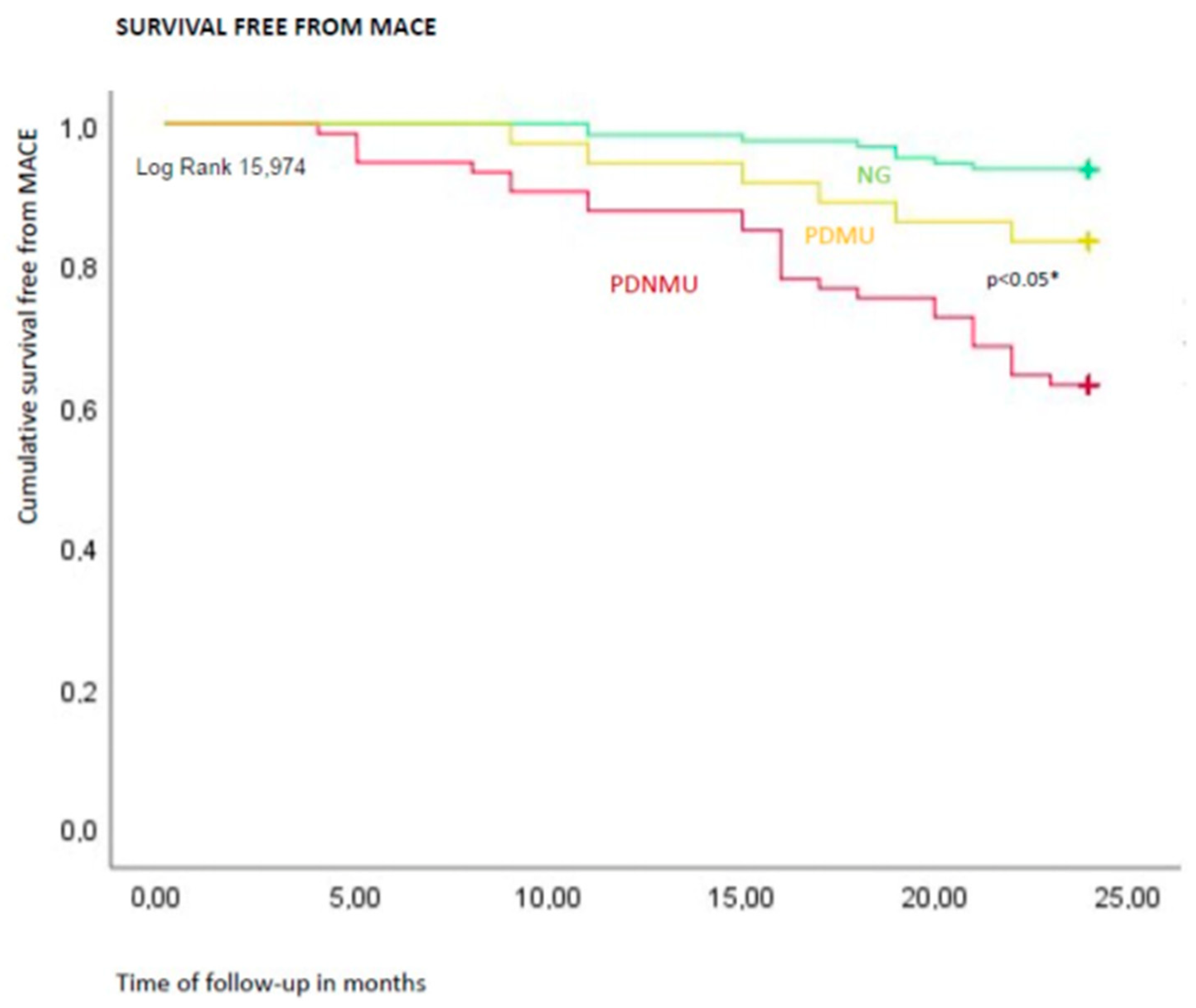
| Number of MACE, n (%) | 8 (6.4) | 27 (36.9%) | 6 (16.6) | <0.05 *,**,*** |
| Risk Factors | HR | CI 95% | p Value | HR | CI 95% | p Value |
|---|---|---|---|---|---|---|
| Age | 0.939 | 0.876–1.006 | 0.075 | 0.970 | 0.930–1.013 | 0.169 |
| BMI | 0.970 | 0.926–1.261 | 0.327 | 1.070 | 0.809–1.414 | 0.637 |
| Hypertension | 1.188 | 0.643–2.196 | 0.582 | 0.620 | 0.176–2.187 | 0.457 |
| Dyslipidemia | 1.707 | 0.921–3.163 | 0.089 | 1.425 | 0.560–3.629 | 0.457 |
| Metformin therapy | 0.185 | 0.085–0.401 | 0.001 | 0.386 | 0.053–2.823 | 0.348 |
| Glucose blood values | 1.014 | 1.009–1.020 | 0.001 | 1.020 | 1.001–1.038 | 0.029 * |
| Cholesterol blood values | 1.012 | 1.004–1.019 | 0.002 | 1.013 | 0.999–1.027 | 0.074 |
| ACEi | 1.325 | 0.650–2.702 | 0.439 | 0.583 | 0.159–2.137 | 0.416 |
| Statins | 0.883 | 0.450–1.730 | 0.716 | 0.501 | 0.166–1.503 | 0.217 |
| Pre-diabetes | 3.003 | 1.531–5.869 | 0.001 | 1.021 | 0.688–1.201 | 0.195 |
| Atheromatous carotid plaque | 6.388 | 2.949–13.838 | 0.001 | 5.373 | 1.251–11.079 | 0.024 * |
| IL 6 | 6.246 | 3.195–12.210 | 0.001 | 1.283 | 0.020–4.003 | 0.350 |
| miR-24 | 5.001 | 1.781–6.122 | 0.001 | 3.842 | 1.768–19.222 | 0.011 * |
Publisher’s Note: MDPI stays neutral with regard to jurisdictional claims in published maps and institutional affiliations. |
© 2021 by the authors. Licensee MDPI, Basel, Switzerland. This article is an open access article distributed under the terms and conditions of the Creative Commons Attribution (CC BY) license (https://creativecommons.org/licenses/by/4.0/).
Share and Cite
Sardu, C.; Modugno, P.; Castellano, G.; Scisciola, L.; Barbieri, M.; Petrella, L.; Fanelli, M.; Macchia, G.; Caradonna, E.; Massetti, M.; et al. Atherosclerotic Plaque Fissuration and Clinical Outcomes in Pre-Diabetics vs. Normoglycemics Patients Affected by Asymptomatic Significant Carotid Artery Stenosis at 2 Years of Follow-Up: Role of microRNAs Modulation: The ATIMIR Study. Biomedicines 2021, 9, 401. https://doi.org/10.3390/biomedicines9040401
Sardu C, Modugno P, Castellano G, Scisciola L, Barbieri M, Petrella L, Fanelli M, Macchia G, Caradonna E, Massetti M, et al. Atherosclerotic Plaque Fissuration and Clinical Outcomes in Pre-Diabetics vs. Normoglycemics Patients Affected by Asymptomatic Significant Carotid Artery Stenosis at 2 Years of Follow-Up: Role of microRNAs Modulation: The ATIMIR Study. Biomedicines. 2021; 9(4):401. https://doi.org/10.3390/biomedicines9040401
Chicago/Turabian StyleSardu, Celestino, Pietro Modugno, Gaetano Castellano, Lucia Scisciola, Michelangela Barbieri, Lella Petrella, Mara Fanelli, Gabriella Macchia, Eugenio Caradonna, Massimo Massetti, and et al. 2021. "Atherosclerotic Plaque Fissuration and Clinical Outcomes in Pre-Diabetics vs. Normoglycemics Patients Affected by Asymptomatic Significant Carotid Artery Stenosis at 2 Years of Follow-Up: Role of microRNAs Modulation: The ATIMIR Study" Biomedicines 9, no. 4: 401. https://doi.org/10.3390/biomedicines9040401