Review

Combined Proteotranscriptomic-Based Strategy to Discover Novel Antimicrobial Peptides from Cone Snails

1
Bioinformatic Team, Département Agriculture et Ressource Animales, UMRI 28, Institut National Polytechnique Félix Houphouët-Boigny, Yamoussoukro BP 1093, Ivory Coast
2
Plateforme de Biologie Moléculaire, Institut Pasteur de Côte d’Ivoire, Abidjan BP 490, Ivory Coast
3
Institut des Biomolécules Max Mousseron, UMR 5247, Université de Montpellier, CNRS, ENSCM, 34095 Montpellier, France
*
Authors to whom correspondence should be addressed.
Biomedicines 2021, 9(4), 344; https://doi.org/10.3390/biomedicines9040344
Submission received: 15 January 2021 / Revised: 22 March 2021 / Accepted: 23 March 2021 / Published: 29 March 2021
(This article belongs to the Special Issue Biomedicine from the Sea)

Abstract

:
Despite their impressive diversity and already broad therapeutic applications, cone snail venoms have received less attention as a natural source in the investigation of antimicrobial peptides than other venomous animals such as scorpions, spiders, or snakes. Cone snails are among the largest genera (Conus sp.) of marine invertebrates, with more than seven hundred species described to date. These predatory mollusks use their sophisticated venom apparatus to capture prey or defend themselves. In-depth studies of these venoms have unraveled many biologically active peptides with pharmacological properties of interest in the field of pain management, the treatment of epilepsy, neurodegenerative diseases, and cardiac ischemia. Considering sequencing efficiency and affordability, cone snail venom gland transcriptome analyses could allow the discovery of new, promising antimicrobial peptides. We first present here the need for novel compounds like antimicrobial peptides as a viable alternative to conventional antibiotics. Secondly, we review the current knowledge on cone snails as a source of antimicrobial peptides. Then, we present the current state of the art in analytical methods applied to crude or milked venom followed by how antibacterial activity assay can be implemented for fostering cone snail antimicrobial peptides studies. We also propose a new innovative profile Hidden Markov model-based approach to annotate full venom gland transcriptomes and speed up the discovery of potentially active peptides from cone snails.

1. Introduction

In 1928, Alexander Fleming discovered the now widely known Penicillin from a Penicillium mold, later identified as Penicillium rubens [1]. Sixteen years later, Selman Waksman isolated Streptomycin from Streptomyces griseus [2], and together with Penicillin, both these discoveries undoubtedly revolutionized modern medicine. Indeed, such antibiotics were produced in large quantities during the Second World War to successfully treat infectious diseases [3]. Since then, antibiotics have become an integral part of the lives of millions of people as a primary medication for multiple microbial infections. Unfortunately, the misuse and overuse of antibiotics led to the emergence of antimicrobial resistance, which has become one of the major threats to global human health, as pointed out by the World Health Organization [4]. In 2016, 700,000 deaths were reported to be directly imputable to resistant infections [5]. In the same way, a study on global antimicrobial resistance has estimated that by 2050, over 10 million deaths each year and a cumulative USD 100 trillion of economic losses will be directly imputable to drug resistance [6].
At the beginning of the 21st century, the pharmaceutical industry appeared to reach a critical juncture caused by the low number of novel drugs entering the clinical phases [7]. Nevertheless, in the same couple of years, the advent of new sequencing technologies, higher computational capacities, and advances in biological research led the industry and researchers to turn more deeply towards natural extracts like animal venoms as a source of novel drug leads [8]. Back in the 17th century, Francesco Redi led some studies on toxins, especially from the “poisonous” snakes. Some years later, the discovery of snake venom glands set the stage for the birth of modern toxicology [9]. Since then, the iconic scorpions, spiders, and snakes, but also more recently lesser-known invertebrates like cone snails, have been investigated for their venom toxins.
Conus sp. is a major group of marine invertebrates, with about 761 described species according to the world register of marine species (WoRMS) [10]. The major component of the venom gland of cone snails is made of small peptides known as conotoxins, which often have neurotoxic activities. It has been conservatively estimated that more than 80,000 biologically active conotoxins are yet to be discovered [11]. The studies of conotoxins have already led to the discovery of the well-known drug ziconotide (ω-MVIIA conotoxin) from Conus magus used to treat chronic pain [12,13]. Each species of cone snail produces hundreds of conotoxins, which are the result of years of evolution and coevolution in the same environment as their prey and predators [14]. Venom compounds can be classified based on their molecular targets and mode of action, like peptides with antimicrobial activity. Antimicrobial peptides (AMPs) are biomolecules that act on bacteria, viruses, or fungi. They are usually short polypeptides with a net positive charge that comes from their rich composition in lysine, arginine, and histidine [15].
Compared to other animals like scorpions or spiders, the antimicrobial peptides from cone snails have received little attention [16,17,18]. For example, the investigation of scorpion venoms led to the discovery of Hadrurin, Scorpine, Vejovine, StCT2, Pandinin 1, and Pandinin 2, which are effective antimicrobial peptides [19,20,21,22,23]. Similar studies also found antimicrobial peptides in snakes, spiders, bees, ants, centipedes, lugworms, wasps, and cuttlefish [15,24,25,26,27,28,29,30,31,32].
To fill this gap in cone snail venom studies, we show here the need for novel compounds in the battle against antimicrobial resistance, update the knowledge on cone snails as a source of antimicrobial peptides and present the analytical methods to decipher the venom of cone snail for antimicrobial peptides.

2. Global Antimicrobial Resistance and the Need for Novel Compounds

The word “antibiotic” was used by Selman Waksman, in 1941, to describe any small molecule made by a microorganism that antagonizes the growth of other microorganisms [33]. An antibiotic interferes with bacteria growth via a specific mode of action, at concentrations that present minimal toxicity and are sufficiently potent to be effective against the infection. Despite a 36% increase in human use of antibiotics from 2000 to 2010, approximately 20% of deaths worldwide remain related to infectious diseases because of antibiotic resistance [34]. This development of resistance is a normal evolutionary process for microorganisms. Nevertheless, it is worsened by the selective pressure exerted by the widespread use of antibacterial drugs, rendering them largely ineffective on bacterial infections.
In response to exposure to antibiotics, susceptible bacteria are killed. However, there is a resistant portion that recolonizes the infection site. Bacteria use two major mechanisms to “resist” antibiotics, which are mutational adaptation and the acquisition of genetic material through horizontal gene transfer. The latter is one of the most important drivers of bacterial evolution. In general, mutations alter antibiotic action via the structural modification of the antibiotic target site, preventing the molecule from reaching its target by decreasing penetration or actively extruding the antimicrobial compound [35]. Resistance to antibiotics can usually be achieved through multiple biochemical pathways. For example, resistant bacteria can produce enzymes like carbapenemase or beta-lactamases, which can degrade or destroy antibiotic molecules. An analysis of 83 studies conducted in Africa showed a prevalence of carbapenemase-producing bacteria isolated in hospitals ranging from 3% to 67.7%, with a mortality rate of up to 67% [36]. Beta-lactamases-producing bacteria and quinolone-resistant bacteria have been detected in humans, but also animals, and the environment [37,38]. Up to 35% of isolates from animals and 93.2% from humans have shown resistance to quinolones [37].
The increase in drug-resistant pathogens is the consequence of various factors. It is the result of biochemical and genetic modifications, as pointed out above, but also the result of the over-reliance on antibiotics. For example, antibiotics are largely used as growth promoters in livestock farming [39]. Resistant bacteria are, then, transmitted to humans through direct contact with animals [40,41,42], exposure to animal manure, and by the consumption of undercooked meat [43]. High rates of prescriptions of antibiotics are another critical factor in the evolution of resistance. In some countries, antibiotics are available without prescriptions. When they are prescribed, most prescriptions are unsuitable. Broad-spectrum antibiotics are favored to treat infections caused by several species of bacteria or those for which diagnosis is time-consuming. Additionally, some physicians are inclined to prescribe multiple antibiotics for the same condition [44]. Patients’ failure to comply with dosages is also a major contributor to the development of resistance. Indeed, patients miss doses, either by mistake or deliberately. Finally, exposure of surviving microorganisms to subtherapeutic concentrations of the drug also increases the chances of developing resistance [45]. Due to increasing resistance to antibiotics, a report has predicted that by 2050 10 million deaths will occur annually due to resistant pathogens. Without control action, by 2050 the cost of antimicrobial resistance will be approximately USD 100 trillion, with a 3.5% decline in global gross domestic product [6]. As the world is heading towards a postantibiotic era, there is an urgent need for novel compounds that are able to meet current needs, such as antimicrobial peptides.
Antimicrobial peptides (AMPs) are active molecules produced by a broad range of organisms ranging from microbes to mammals to ensure their self-defense against microbes [46]. AMPs are multifaceted molecules that kill microbes via a pleiotropic mechanisms of action, such as the destruction of the barrier function of the cellular membranes and the inhibition of macromolecule synthesis making them efficient even against multiresistant bacteria [47,48]. The efficiency of AMPs is due to rapid killing kinetics, reduced toxicity, and reduced microbial resistance [49] as demonstrated by many studies [50,51,52]. AMPs are therefore a promising alternative to conventional antibiotics.

3. Cone snails as a Source of Novel Antimicrobial Peptides

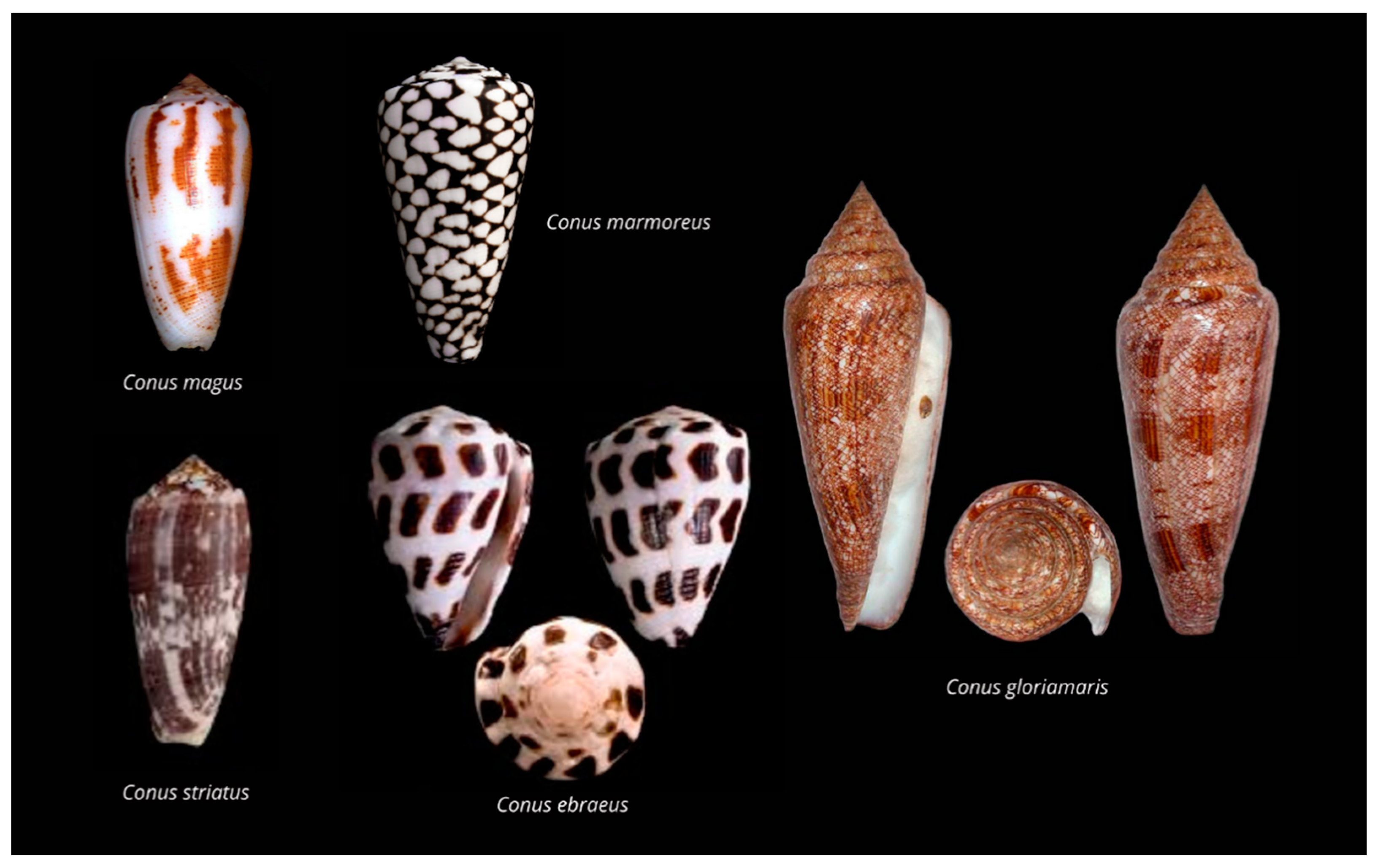
3.1. Cone Snails Diversity

The Conidae family is composed of 138 genera from which Conus sp. is the type taxon. Cone snails contain around 761 recognized species [53,54] from which approximately 118 have been studied to date (http://conoserver.org/?page=stats&tab=organisms (accessed on 28 January 2021)). However, it should be noted that although this is a rapidly evolving field, especially with the advent of transcriptomics, not all sequences deposited in Conoserver correspond to proteomic-verified conopeptides. The shell patterns of cone snails (Figure 1), clearly recognizable, have made them precious collection items through centuries [55].
Cone snails inhabit tropical and subtropical coastal zones and oceans up to 1000 m of depth but are mostly found in coral reefs hiding in the sand, under the coral shelf, or in shallow waters [56]. The greatest diversity of cone snail species is found in the Indo-Pacific ocean, but they become rarer beyond the 40° N or S parallel [57,58]. Cone snails are active at night as they leave their hiding place to go hunting for prey. They are highly specialized predators and can be classified based on their feeding habits: worm hunters (vermivorous), mollusk hunters (molluscivorous), fish hunters (piscivorous), and generalist feeders. They first detect prey using chemosensors, then crawl softly towards the prey, and generally extend their proboscis and fire their venom-loaded harpoon upon contact. Venom injection into a prey animal induces rapid immobilization, which, in the case of fish hunters, can take less than a few hundred milliseconds [59].

3.2. Cone Snail’s Venom Composition and Conotoxins Classification

Conotoxins are the disulfide-rich peptides found in the venom of cone snails responsible for their toxicity [17,60]. They are produced in the venom gland as precursors, which generally contain a signal, a pro, and a mature sequence. The cone snail’s venom is a remarkably complex mixture of peptides that has drawn the attention of biomedical researchers for their high unprecedented potency and selectivity for their target. Conotoxins are classified following three different criteria: the precursor’s endoplasmic reticulum signal sequence, the cysteine pattern of the mature peptide region, and the pharmacological targets. Based on their signal peptide, conotoxins are further divided into 28 gene superfamilies: A, B1, B2, B3, C, D, E, F, G, I1, I2, I3, J, K, L, M, N, O1, O2, O3, P, Q, R, S, T, V, Y. The most expressed peptides from cone snail venom glands studied to date are from A, M, and O superfamilies [61].
Conotoxins can also be divided according to the cysteine pattern or the pharmacological target [62]. Conotoxins have been classified into cysteine frameworks (pattern of cysteine residues) following the arrangement of cysteine along the mature sequence or the disulfide connectivities. The updated cysteine arrangement and disulfide connectivities definition of known conotoxins is presented in Table 1.
Conotoxins exhibit a large range of pharmacological targets. Considering the receptor specificity, we can classify conotoxins into 12 families: α (alpha), γ (gamma), δ (delta), ε (epsilon), ι (iota), κ (kappa), µ (mu), ρ (rho), ς (sigma), τ (tau), χ (chi) and ω (omega) (Table 2). A more detailed description of pharmacological families is provided by Kaas et al. [62].

3.3. Antimicrobial Activity of Conidae’s Conopeptides

Although conotoxins have a wide array of unique structures, the most common use of conotoxins is focused on pain management [63]. However, some conotoxins have proved to be highly effective against pathogens with little resistance due to their membrane-disruptive mechanisms [64]. The antimicrobial effects of conotoxins are seldomly documented and remain underexplored. An example of the in vitro antiparasitic activity of conotoxins was demonstrated on the tachyzoite form of Toxoplasma gondii [18]. The tachyzoite is the mobile, invasive, and intracellular obligate form of T. gondii. The synthetic conotoxin s-cal14.1a derived from Californiconus californicus, at micromolar concentration, lowers down to half the viability of extracellular tachyzoites and inhibits host cell invasion by 61%. In toxoplasma infections, no drugs have shown effectiveness when tachyzoites were localized in cytoplasmic parasitophorous vacuoles. However, s-cal14.1a inhibits not only the establishment of the infection but also the intracellular proliferation of T. gondii by 50%. This conotoxin potentially disrupts the replication machinery of the parasite by passing the membrane of the host cell, parasitophorous vacuoles, and parasite membranes without the host cell being affected.
Other studies have shown mitigated antibacterial effects of conotoxins. Conotoxin O1_cal29b, isolated from C. californicus, inhibited in vitro the growth of the multidrug-resistant Mycobacterium tuberculosis. O1_cal29b was effective at low concentration. The inhibition occurred at a minimal inhibitory concentration (MIC) of 0.22–3.52 µM [61]. In parallel, peptide Lo6/7a, a 24-residue conotoxin isolated from the venom of Conasprella longurionis, exhibited low and extremely specific activity against Bacillus megaterium at an exceedingly high concentration (1 mM) [65]. Additionally, the conolysin-Mt, a disulfide-poor conopeptide from Conus mustelinus, showed a low antimicrobial activity with a MIC greater than 50 µM against two Escherichia coli strains. Its MIC for the Gram-positive Staphylococcus aureus is in a range of 25–50 µM [66].
Interestingly, a study has shown that macrocyclization can convert a conotoxin into an effective antimicrobial peptide [67]. MVIIA is a linear cystine-knot peptide with multiple basic amino acids at both termini, but up to 500 µM, it was inactive against E. coli, Pseudomonas aeruginosa, and Staphylococcus aureus. Meanwhile, it exhibited moderate antifungal activity against Candida kefyr and Candida tropicalis with MICs of 28.8 µM and 39.8 µM, respectively. Likewise, the linear analog MVIIA-GS, the MVIIA conotoxin with a Gly Ser linker, gave similar results, indicating that the linker did not affect the antimicrobial activity. The authors then considered ligating the two ends of MVIIA using a linker peptide. This ligation allowed the formation of epitopes that gain membranolytic activity on microbes. Indeed, all ten cyclic conotoxins derived from MVIIA were active against the selected three bacterial strains with MICs ranging from 3.3 to 90.2 µM. Most of these analogs exhibited improved antifungal activity with MICs, from up to 18.2 µM against C. kefyr and up to 11.4 µM against C. tropicalis. Additionally, the conversion of the disulfide bonds to aminobutyric acids improved the antimicrobial activity of the cyclic analogs. These results demonstrated that the end-to-end cyclization of a linear peptide improves its biological activity and confers antimicrobial properties that were not found in the linear form. The mimetics of conopeptides may serve as more promising candidates for the further development of therapeutically useful agents for the treatment of infections.
Although Conolysin-Mt and conotoxin MVIIA target the lipid bilayer of bacteria, in addition to the lipid bilayer, transpeptidase enzymes were explored as novel targets for antimicrobial conopeptides. For example, sortases that anchor surface proteins were investigated as attractive targets due to their prevalence in the cell wall of Gram-positive bacteria [16]. Their inhibition compromises the pathogenesis and the virulence of the bacteria. Based on structural studies, M2-conotoxin and contryphan-R were found to mediate the inhibition of SrtA and SrtB by obstructing the assembly of iron acquisition, immune evasion, complement pathway inhibition, clumps, biofilm formation, and host matrix attachment proteins within the cell wall of S. aureus. Therefore, more studies are required to validate the efficacy of M2-conotoxin and contryphan-R in bacterial cultures.

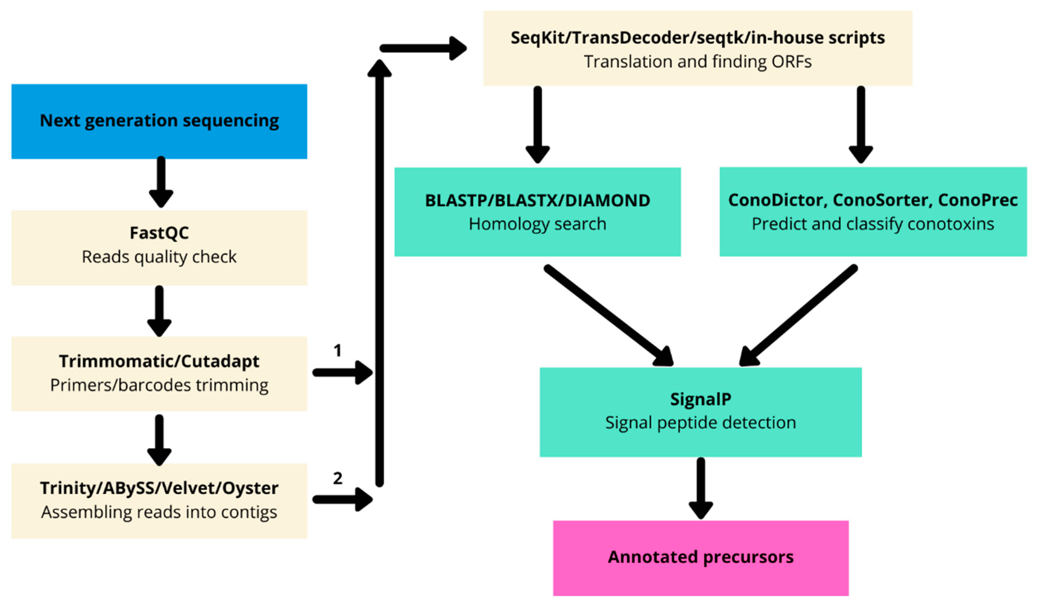
4. Bioinformatics-Aided Proteotranscriptomics

4.1. Cone Snail Venom Extraction

Cone snail venom can be obtained either by the dissection of the venom duct or by milking. Venom gland tissue extraction implies the availability of tens to hundreds of specimens to collect enough material for the discovery process. Besides the ethical concerns, this method has many disadvantages (cellular debris, unmatured and degraded products in the reconstituted venom) compared to venom “milking” [56]. Although dissection and tissue extraction has proven to be successful in many cases [68], venom milking should be considered whenever possible, as it provides a soluble fraction that contains fully mature conotoxins intended for a particular ecological role, as demonstrated by many studies [69,70,71,72,73,74,75,76]. Nevertheless, if possible, a combination of both strategies (milking and dissection) should be considered as all Conidae venoms tested to date for AMPs have not been milked but rather extracted. Indeed, it was found that cone snails can deploy different combinations of conotoxins depending on the stimulus (predatory vs. defense) [70].
To collect the predation-evoked venom, the procedure usually begins with a lure, which is a live prey according to the cone snail’s feeding habit [77]. The cone extends its proboscis toward the lure that is placed in front of a microtube. The venom is injected into the tube through a fine piece of prey tegument (fish fin for instance). Therefore, milking provides a more soluble venom, free of cellular debris and degraded products, that is ideally suited for biological and proteomic assays [56,78].

4.2. Next-Generation Transcriptomics Sequencing and Bioinformatics

Before the advent of next-generation sequencing, biologically active peptides from cone snails were discovered using “bioactivity-guided fractionation”. This method was limited because it required large amounts of crude venom and turned out to be time-consuming [79]. Next-generation sequencing revolutionized the field as the venom duct transcriptome can be completely sequenced and the conotoxin sequences recovered in one single experiment. From the next-generation sequencing data, bioinformatics tools are required to correctly identify conotoxin precursors from the raw reads and/or assembled contigs (Figure 2).
The raw reads obtained from Illumina or 454 sequencings are controlled for quality using FastQC v.0.11.9 (http://www.bioinformatics.babraham.ac.uk/projects/fastqc (accessed on 24 March 2021)). FastQC helps researchers to be aware of some issues that can arise during sequencing by providing statistics on reads like k-mer content, GC content, or graph of quality per base. The validated reads are then trimmed for barcodes and primers using Trimmomatic version 0.39 [80] or Cutadapt version 3.3 [81]. Short Illumina reads obtained after RNA sequencing rarely contain the full-length conotoxin precursor, although some active peptides from cone snails can be as short as 10 amino acids [82]. Therefore, they need to be assembled in longer contigs. De novo assembly can be performed using software like Trinity version 2.12 [83], Velvet version 1.2.10 [84], and ABySS v.2.3.0 [85] to obtain the desired contigs. Another emerging method is the use of multiassembling tools like the Oyster river protocol [86] for a better recovery of contigs, as one method alone is not always able to properly identify all desired contigs.
The contigs are then translated in silico into amino acids. The tools used at this step should be well tested [87]. At this point, the traditional method consists of the homology search using BLAST [88] against a specialized database of conotoxins like ConoServer [89], followed by a second homology search against a larger database like the NCBI nonredundant (https://www.ncbi.nlm.nih.gov/refseq/about/nonredundantproteins accessed on 28 January 2021) [90] or the Uniprot/SwissProt [91] databases for validation of conopeptides and housekeeping genes identification.

4.3. Venom Gland Transcriptome Annotation Based on Profile Hidden Markov Models

A second way to elucidate transcriptome composition is by using ConoDictor version 2 [92] (new version in preparation), or ConoSorter [93] to predict putative conotoxins using methods such as hidden Markov models (HMMs). Using specialized predictive tools is helpful because they are exclusively designed to detect putative conopeptides from RNA-seq data and are way speedier compared to the traditional BLAST. Functional and structural annotations like superfamily belonging, signal peptide presence, or cysteine framework can then be predicted using signalP version 5 [94] or previously mentioned tools.
Nevertheless, currently, only a few HMMs are available on PFAM [95] to annotate conotoxins (Table 3).
To accelerate the annotation of cone snail transcriptome, we propose here an up-to-date exhaustive list of profile HMMs (pHMMs) that allow the annotation and classification of the major conotoxins superfamilies (available on our GitHub https://github.com/koualab/cono-amp-review/blob/main/conohmm.txt (accessed on 18 March 2021)). A sequence logo is associated with each of the newly built pHMM (Table 4).

4.4. Venom Proteomics

The complex cone snail venom mixture made of polypeptides, peptides, inorganic salt, and amines accounts for most of the molecular and functional diversity as well as the observed behavioral and biochemical modulations [70,76,97]. For the high propensity of post-translational modifications (PTMs) alone, the conopeptides cannot be simply deciphered from transcriptomic data without proteomic evidence. The introduction in the field of mass spectrometry-based techniques has drastically changed the landscape of conotoxin studies [98]. This technique allowed us not only to correctly estimate the number of conopeptides expressed by a cone snail but also improved the resolution of the identified peptides. For instance, LC-MS of milked (injected) venom can help to reveal dramatic intraspecific variation like in some recent studies [97,99,100]. Furthermore, using deep venomics, Dutertre et al., were able to increase the estimation of the number of peptides expressed by Conus marmoreus (around 8000 peptides) originated from only 105 conotoxins peptide precursors [69].

4.5. Proteotranscriptomics

Proteotranscriptomic studies combine the best of both worlds: de novo deduced sequences of the digested peptides are directly mapped to the RNAseq generated peptide database using dedicated tools. The data obtained through bottom-up proteomics are generally compared to a sequence database using tools like ConoMass [89], MASCOT (Matrix Science, Boston, MA, USA; www.matrixscience.com version 2.5 ProteinPilot (SCIEX, Framingham, MA, USA), v.4.4 or PEAKS studio (Bioinformatics Solutions, Waterloo, ON, Canada) version 8.5. The top-down bioinformatic analyses are more difficult due to the nature of the obtained data. Such data often leads to false-positive identification due to the newness of the sequence variants and PTMs that they represent. To overcome this issue, new and less stringent algorithms have been developed, such as Byonic [101] and ProSight version 2 [102]. Both can be used to identify sequences and PTMs from top-down proteomic data.

4.6. In Silico AMPs Structure Determination

Structure determination of AMPs is usually conducted by NMR. However, the potentially large number of sequences identified by transcriptomics, bioinformatics, and proteomics prevent the generalized use of NMR, as it will be too expensive and time-consuming. A better approach involves the in silico determination of the predicted AMPs. With the development of new technologies and algorithms in biophysics, in silico prediction of the structure is becoming more and more precise. Rosetta has proven to be successful in predicting the three-dimensional structure of proteins ab initio from their amino acid sequence. By incorporating new energy functions, Rosetta has been able to predict a completely new protein fold [72,73]. Following a similar strategy, QUARK [103] has shown the highest scores in CASP9 (http://predictioncenter.org/casp9/CD/data/html/groups.server.fm.html (accessed on 28 January 2021)) and CASP10 (http://predictioncenter.org/casp10/groups_analysis.cgi?type=server&tbm=on&tbm_hard=on&tbmfm=on&fm=on&submit=Filter (accessed on 28 January 2021)) challenges. Additionally, the 14th CASP experiment has crowned the i-TASSER web portal (https://zhanglab.ccmb.med.umich.edu/I-TASSER (accessed on 28 January 2021)) as the first protein structure prediction. AlphaFold in CASP13 has shown the greatest advance in the resolution of the protein folding challenges using machine learning methods and, once available, could be used for AMPs structure prediction [104,105].

5. Antibacterial Activity Assays

Microbiological bioassays involve the detection and characterization of the antibacterial activity of newly discovered AMPs. Usually, microdilution [66], radial diffusion assay [67], and spot diffusion [65] were used to determine the minimal inhibitory concentration of the tested conopeptide. Quantitative susceptibility testing is performed by making 2-fold dilutions of the tested AMP in a liquid culture medium inoculating it with a standard number of microorganisms (10.5 to 10.6 colony-forming unit (cfu)/mL) and incubating it at 35–37 °C for 18–24 h. Mueller–Hinton Broth is the recommended medium for the susceptibility testing of commonly isolated, rapidly growing aerobic or aeroanaerobic organisms because it supports the satisfactory growth of most pathogens [106]. However, LB medium and Tryptic Soy Broth can also be used [66,67]. The amount of AMP that inhibits the visible growth of the microorganism is called the minimal inhibitory concentration [107]. Beyond MIC, minimal bactericidal concentration (MBC) can be determined to characterize the type of activity performed by the conopeptide. Subcultures of the samples obtained from the clear tubes or wells are plated on a solid medium and reincubated for an additional 18–24 h. The MBC represents the lowest concentration that either revealed no visible bacterial growth after subculturing or resulted in a 3-log10 reduction in colony-forming units (cfu) per mL on subculture. Briefly, a 3-log10 (or 99.9%) reduction in viable bacterial count in an 18–24 h period is the accepted definition of bactericidal activity [108]. The bacteriostatic activity has been defined as a ratio of MBC to MIC > 4 [81].
In case of low activity, different peptide mixtures can be tested in combination to increase the bactericidal activity and the synergistic antimicrobial effect evaluated. The fractional inhibitory concentration (FIC) index is defined as the inhibitory concentration of the antimicrobial combination divided by that of the single antimicrobial component [109]. The following equation (Equation (1)) represents the method for determining the FIC index:
F I C i n d e x = M I C o f d r u g X c o m b i n a t i o n M I C o f d r u g X a l o n e + M I C o f d r u g Y c o m b i n a t i o n M I C o f d r u g Y a l o n e
The FIC indices are interpreted as follows: ≤0.5 = synergism; 0.5–1 = additivity; 1–4 = indifference; and >4 = antagonism.
Lastly, to distinguish if the antibacterial activity is concentration and/or time-dependent, time-kill curves can be performed. Viable colony counts are determined at different time points up to 24 h. The cfu of the organisms is to be determined and a graph of the log cfu/mL is plotted against time. This kinetics can also be related to the stages of the growth of the bacteria (lag, exponential, stationary phase). Concentration-dependent bactericidal activity occurs when the rate of killed-bacteria increases with progressively higher AMP concentrations corresponding to multiples of the MIC (i.e., 1×, 5×, 10×). Time-dependent bactericidal activity occurs when bacterial killing does not change with increasing AMPs concentrations to more than the MIC.

6. Conclusions

Cone snail venom has already been the source of approved toxin-based drugs, and these mixtures are also potential reservoirs for antimicrobial peptides. We first reported the standard methods in venom drug-based research for transcriptome sequencing and proteomics. We further showed a new strategy based on a dedicated predictive approach focused on up-to-date profile Hidden Markov models that should then make it easy to quickly identify potentially interesting putative peptides from cone snail venom gland transcriptomes. We also pointed out the proteomics and proteotranscriptomics methods to confirm conotoxin transcriptome-based prediction. Furthermore, a point on the in silico prediction of conotoxins’ three-dimensional structure has been made. We conclude our review by mentioning the antibacterial activity assay methods for the proper assay of conotoxins AMPs. We hope that this review will help researchers and industry to lead projects on cone snail antimicrobial peptides to tackle the increasing global antimicrobial resistance.

Author Contributions

Conceptualization, A.E., D.K. and A.A.; writing—original draft preparation, A.E., A.A.; writing—review and editing, D.K., S.K.-N., S.D. All authors have read and agreed to the published version of the manuscript.

Funding

This research received no external funding.

Institutional Review Board Statement

Not applicable.

Informed Consent Statement

Not applicable.

Data Availability Statement

Not applicable.

Conflicts of Interest

The authors declare no conflict of interest.

References

  1. Gaynes, R. The Discovery of Penicillin—New Insights After More than 75 Years of Clinical Use. Emerg. Infect. Dis. 2017, 23, 849–853. [Google Scholar] [CrossRef]
  2. Woodruff, H.B.; Selman, A. Waksman, Winner of the 1952 Nobel Prize for Physiology or Medicine. Appl. Environ. Microbiol. 2014, 80, 2–8. [Google Scholar] [CrossRef] [Green Version]
  3. Penicillin, S. CDC Control of Infectious Diseases, 1900–1999. JAMA 1999, 282, 1029. [Google Scholar] [CrossRef] [Green Version]
  4. Zhang, L.; Gallo, R.L. Antimicrobial Peptides. Curr. Biol. 2016, 26, R14–R19. [Google Scholar] [CrossRef]
  5. O’Neil, J. Tackling Drug-Resistant Infections Globally: Final Report and Recommendations 2016; Government of the United Kingdom: London, UK, 2016.
  6. Humphreys, G.; Fleck, F. United Nations Meeting on Antimicrobial Resistance. Bull. World Health Organ. 2016, 94, 638–639. [Google Scholar] [CrossRef]
  7. Prashanth, J.R.; Brust, A.; Jin, A.-H.; Alewood, P.F.; Dutertre, S.; Lewis, R.J. Cone Snail Venomics: From Novel Biology to Novel Therapeutics. Future Med. Chem. 2014, 6, 1659–1675. [Google Scholar] [CrossRef]
  8. Bordon, K.D.C.F.; Cologna, C.T.; Fornari-Baldo, E.C.; Pinheiro-Júnior, E.L.; Cerni, F.A.; Amorim, F.G.; Anjolette, F.A.P.; Cordeiro, F.A.; Wiezel, G.A.; Cardoso, I.A.; et al. From Animal Poisons and Venoms to Medicines: Achievements, Challenges and Perspectives in Drug Discovery. Front. Pharmacol. 2020, 11, 1132. [Google Scholar] [CrossRef] [PubMed]
  9. Utkin, Y.N. Animal Venom Studies: Current Benefits and Future Developments. World J. Biol. Chem. 2015, 6, 28–33. [Google Scholar] [CrossRef]
  10. Worms, E.B. World Register of Marine Species. Available online: http://www.marinespecies.org7813 (accessed on 28 January 2021).
  11. Gao, B.; Peng, C.; Yang, J.; Yi, Y.; Zhang, J.; Shi, Q. Cone Snails: A Big Store of Conotoxins for Novel Drug Discovery. Toxins 2017, 9, 397. [Google Scholar] [CrossRef] [Green Version]
  12. Eisapoor, S.S.; Jamili, S.; Shahbazzadeh, D.; Mostafavi, P.G.; Bagheri, K.P. A New, High Yield, Rapid, and Cost-Effective Protocol to Deprotection of Cysteine-Rich Conopeptide, Omega-Conotoxin MVIIA. Chem. Biol. Drug Des. 2016, 87, 687–693. [Google Scholar] [CrossRef]
  13. Rigo, F.K.; Dalmolin, G.D.; Trevisan, G.; Tonello, R.; Silva, M.A.; Rossato, M.F.; Klafke, J.Z.; Cordeiro, M.D.N.; Junior, C.J.C.; Montijo, D.; et al. Effect of ω-Conotoxin MVIIA and Phα1β on Paclitaxel-Induced Acute and Chronic Pain. Pharmacol. Biochem. Behav. 2013, 114, 16–22. [Google Scholar] [CrossRef] [Green Version]
  14. Herzig, V.; Cristofori-Armstrong, B.; Israel, M.R.; Nixon, S.A.; Vetter, I.; King, G.F. Animal Toxins—Nature’s Evolutionary-Refined Toolkit for Basic Research and Drug Discovery. Biochem. Pharmacol. 2020, 181, 114096. [Google Scholar] [CrossRef]
  15. Fratini, F.; Cilia, G.; Turchi, B.; Felicioli, A. Insects, Arachnids and Centipedes Venom: A Powerful Weapon against Bacteria. A Literature Review. Toxicon 2017, 130, 91–103. [Google Scholar] [CrossRef] [PubMed]
  16. Younis, S.; Taj, S.; Rashid, S. Structural Studies of Staphylococcus Aureus Sortase Inhibiton via Conus Venom Peptides. Arch. Biochem. Biophys. 2019, 671, 87–102. [Google Scholar] [CrossRef] [PubMed]
  17. Bernáldez-Sarabia, J.; Figueroa-Montiel, A.; Dueñas, S.; Cervantes-Luévano, K.; Beltrán, J.; Ortiz, E.; Jiménez, S.; Possani, L.; Paniagua-Solís, J.; Gonzalez-Canudas, J.; et al. The Diversified O-Superfamily in Californiconus Californicus Presents a Conotoxin with Antimycobacterial Activity. Toxins 2019, 11, 128. [Google Scholar] [CrossRef] [PubMed] [Green Version]
  18. de León-Nava, M.A.; Romero-Núñez, E.; Luna-Nophal, A.; Bernáldez-Sarabia, J.; Sánchez-Campos, L.N.; Licea-Navarro, A.F.; Morales-Montor, J.; Muñiz-Hernández, S. In Vitro Effect of the Synthetic Cal14.1a Conotoxin, Derived from Conus Californicus, on the Human Parasite Toxoplasma Gondii. Mar. Drugs 2016, 14, 66. [Google Scholar] [CrossRef] [PubMed] [Green Version]
  19. Ahmed, U.; Malik Mujaddad-ur-Rehman, N.K.; Fawad, S.A.; Fatima, A. Antibacterial Activity of the Venom of Heterometrus Xanthopus. Indian J. Pharmacol. 2012, 44, 509. [Google Scholar] [CrossRef] [PubMed] [Green Version]
  20. Cao, L.; Li, Z.; Zhang, R.; Wu, Y.; Li, W.; Cao, Z. StCT2, a New Antibacterial Peptide Characterized from the Venom of the Scorpion Scorpiops Tibetanus. Peptides 2012, 36, 213–220. [Google Scholar] [CrossRef] [PubMed]
  21. Conde, R.; Zamudio, F.Z.; Rodríguez, M.H.; Possani, L.D. Scorpine, an Anti-Malaria and Anti-Bacterial Agent Purified from Scorpion Venom. FEBS Lett. 2000, 471, 165–168. [Google Scholar] [CrossRef] [Green Version]
  22. Corzo, G.; Escoubas, P.; Villegas, E.; Barnham, K.J.; He, W.; Norton, R.S.; Nakajima, T. Characterization of Unique Amphipathic Antimicrobial Peptides from Venom of the Scorpion Pandinus Imperator. Biochem. J. 2001, 359, 35–45. [Google Scholar] [CrossRef]
  23. Hernández-Aponte, C.A.; Silva-Sanchez, J.; Quintero-Hernández, V.; Rodríguez-Romero, A.; Balderas, C.; Possani, L.D.; Gurrola, G.B. Vejovine, a New Antibiotic from the Scorpion Venom of Vaejovis Mexicanus. Toxicon 2011, 57, 84–92. [Google Scholar] [CrossRef] [PubMed]
  24. Benoist, L.; Houyvet, B.; Henry, J.; Corre, E.; Zanuttini, B.; Zatylny-Gaudin, C. In-Depth In Silico Search for Cuttlefish (Sepia Officinalis) Antimicrobial Peptides Following Bacterial Challenge of Haemocytes. Mar. Drugs 2020, 18, 439. [Google Scholar] [CrossRef] [PubMed]
  25. Canelli, A.P.; Dos Santos Rodrigues, T.F.; de Goes, V.F.F.; Caetano, G.F.; Mazzi, M.V. Evaluation of the Effectiveness of Crotoxin as an Antiseptic against Candida spp. Biofilms. Toxins 2020, 12, 532. [Google Scholar] [CrossRef] [PubMed]
  26. Leandro, L.F.; Mendes, C.A.; Casemiro, L.A.; Vinholis, A.H.; Cunha, W.R.; de Almeida, R.; Martins, C.H. Antimicrobial Activity of Apitoxin, Melittin and Phospholipase A2 of Honey Bee (Apis Mellifera) Venom against Oral Pathogens. An. Acad. Bras. Ciênc. 2015, 87, 147–155. [Google Scholar] [CrossRef] [PubMed] [Green Version]
  27. Melo-Braga, M.N.; Almeida, F.D.M.; Dos Santos, D.M.; de Avelar Júnior, J.T.; Dos Reis, P.V.M.; de Lima, M.E. Antimicrobial Peptides from Lycosidae (Sundevall, 1833) Spiders. Curr. Protein Pept. Sci. 2020, 21, 527–541. [Google Scholar] [CrossRef] [PubMed]
  28. Nie, Y.; Zeng, X.-C.; Yang, Y.; Luo, F.; Luo, X.; Wu, S.; Zhang, L.; Zhou, J. A Novel Class of Antimicrobial Peptides from the Scorpion Heterometrus Spinifer. Peptides 2012, 38, 389–394. [Google Scholar] [CrossRef]
  29. Primon-Barros, M.; Macedo, A.J. Animal Venom Peptides: Potential for New Antimicrobial Agents. Curr. Top. Med. Chem. 2017, 17, 1119–1156. [Google Scholar] [CrossRef]
  30. Santos, D.M.; Verly, R.M.; Piló-Veloso, D.; de Maria, M.; de Carvalho, M.A.R.; Cisalpino, P.S.; Soares, B.M.; Diniz, C.G.; Farias, L.M.; Moreira, D.F.F.; et al. LyeTx I, a Potent Antimicrobial Peptide from the Venom of the Spider Lycosa Erythrognatha. Amino Acids 2010, 39, 135–144. [Google Scholar] [CrossRef] [Green Version]
  31. Schneider, R.; Primon-Barros, M.; von Borowski, R.G.; Chat, S.; Nonin-Lecomte, S.; Gillet, R.; Macedo, A.J. Pseudonajide Peptide Derived from Snake Venom Alters Cell Envelope Integrity Interfering on Biofilm Formation in Staphylococcus Epidermidis. BMC Microbiol. 2020, 20, 237. [Google Scholar] [CrossRef]
  32. Yan, L.; Adams, M.E. Lycotoxins, Antimicrobial Peptides from Venom of the Wolf SpiderLycosa Carolinensis. J. Biol. Chem. 1998, 273, 2059–2066. [Google Scholar] [CrossRef] [Green Version]
  33. Clardy, J.; Fischbach, M.; Currie, C. The Natural History of Antibiotics. Curr. Biol. 2009, 19, R437–R441. [Google Scholar] [CrossRef] [PubMed] [Green Version]
  34. Da Cunha, B.R.; Fonseca, L.P.; Calado, C.R.C. Antibiotic Discovery: Where Have We Come from, Where Do We Go? Antibiotics 2019, 8, 45. [Google Scholar] [CrossRef] [PubMed] [Green Version]
  35. Munita, J.M.; Arias, C.A. Mechanisms of Antibiotic Resistance. Microbiol. Spectr. 2016, 4, 481–511. [Google Scholar] [CrossRef] [Green Version]
  36. Manenzhe, R.I.; Zar, H.J.; Nicol, M.P.; Kaba, M. The Spread of Carbapenemase-Producing Bacteria in Africa: A Systematic Review. J. Antimicrob. Chemother. 2015, 70, 23–40. [Google Scholar] [CrossRef] [Green Version]
  37. Baguy, O.M.; Nathalie, G.K.; David, C.N.G.; Daniel, S.N.G.; Julien, C.K.; Rose, K.N.; Djénéba, O.G.; Valérie, G.; Bertin, T.K.; Mireille, D. First Report of Qnr Genes in Multidrugs Resistant (ESBL) Enterobacteria Isolated from Different Ecosystems in Abidjan, Ivory Coast. Int. J. Biol. Sci. Appl. 2014, 21, 170–175. [Google Scholar]
  38. Carole, G.M.V.; Kouadio, G.N.; Baguy, O.M.; Djeneba, O.G.; Ayayi, A.; Bertin, T.K.; Anatole, T.A.; Innocent, K.K.; Kpoda, D.S.; Eric, T.; et al. Antimicrobial Resistance Profile and Molecular Characterization of Extended-Spectrum Beta-Lactamase Genes in Enterobacteria Isolated from Human, Animal and Environment. J. Adv. Microbiol. 2018, 10, 1–9. [Google Scholar] [CrossRef] [Green Version]
  39. Collignon, P.; Wegener, H.C.; Braam, P.; Butler, C.D. The Routine Use of Antibiotics to Promote Animal Growth Does Little to Benefit Protein Undernutrition in the Developing World. Clin. Infect. Dis. 2005, 41, 1007–1013. [Google Scholar] [CrossRef] [Green Version]
  40. Addis, Z.; Kebede, N.; Sisay, Z.; Alemayehu, H.; Wubetie, A.; Kassa, T. Prevalence and Antimicrobial Resistance of Salmonella Isolated from Lactating Cows and in Contact Humans in Dairy Farms of Addis Ababa: A Cross Sectional Study. BMC Infect. Dis. 2011, 11, 222. [Google Scholar] [CrossRef] [Green Version]
  41. Fortini, D.; Fashae, K.; Garcia-Fernandez, A.; Villa, L.; Carattoli, A. Plasmid-Mediated Quinolone Resistance and -Lactamases in Escherichia Coli from Healthy Animals from Nigeria. J. Antimicrob. Chemother. 2011, 66, 1269–1272. [Google Scholar] [CrossRef]
  42. Kikuvi, G.M.; Ombui, J.N.; Mitema, E.S. Serotypes and Antimicrobial Resistance Profiles of Salmonella Isolates from Pigs at Slaughter in Kenya. J. Infect. Dev. Ctries. 2010, 4, 243–248. [Google Scholar] [CrossRef] [Green Version]
  43. Martin, M.J.; Thottathil, S.E.; Newman, T.B. Antibiotics Overuse in Animal Agriculture: A Call to Action for Health Care Providers. Am. J. Public Health 2015, 105, 2409–2410. [Google Scholar] [CrossRef]
  44. Reynolds, L.; McKee, M. Factors Influencing Antibiotic Prescribing in China: An Exploratory Analysis. Health Policy 2009, 90, 32–36. [Google Scholar] [CrossRef]
  45. Ayukekbong, J.A.; Ntemgwa, M.; Atabe, A.N. The Threat of Antimicrobial Resistance in Developing Countries: Causes and Control Strategies. Antimicrob. Resist. Infect. Control 2017, 6, 47. [Google Scholar] [CrossRef] [PubMed]
  46. Zasloff, M. Antimicrobial Peptides of Multicellular Organisms. Nature 2002, 415, 389–395. [Google Scholar] [CrossRef] [PubMed]
  47. Haney, E.F.; Petersen, A.P.; Lau, C.K.; Jing, W.; Storey, D.G.; Vogel, H.J. Mechanism of Action of Puroindoline Derived Tryptophan-Rich Antimicrobial Peptides. Biochim. Biophys. Acta Biomembr. 2013, 1828, 1802–1813. [Google Scholar] [CrossRef] [PubMed] [Green Version]
  48. Roy, R.N. The Mechanism of Inhibition of Protein Synthesis by the Proline-Rich Peptide Oncocin. Mol. Biol. 2015, 22, 466–469. [Google Scholar] [CrossRef] [PubMed] [Green Version]
  49. Wang, S.; Thacker, P.A.; Watford, M.; Qiao, S. Functions of Antimicrobial Peptides in Gut Homeostasis. Curr. Protein Pept. Sci. 2015, 16, 582–591. [Google Scholar] [CrossRef] [PubMed]
  50. Magana, M.; Pushpanathan, M.; Santos, A.L.; Leanse, L.; Fernandez, M.; Ioannidis, A.; Giulianotti, M.A.; Apidianakis, Y.; Bradfute, S.; Ferguson, A.L.; et al. The Value of Antimicrobial Peptides in the Age of Resistance. Lancet Infect. Dis. 2020, 20, e216–e230. [Google Scholar] [CrossRef]
  51. Tan, X.W.; Goh, T.W.; Saraswathi, P.; Nyein, C.L.; Setiawan, M.; Riau, A.; Lakshminarayanan, R.; Liu, S.; Tan, D.; Beuerman, R.W.; et al. Effectiveness of Antimicrobial Peptide Immobilization for Preventing Perioperative Cornea Implant-Associated Bacterial Infection. Antimicrob. Agents Chemother. 2014, 58, 5229–5238. [Google Scholar] [CrossRef] [Green Version]
  52. Zhang, K.; Zhang, H.; Gao, C.; Chen, R.; Li, C. Antimicrobial Mechanism of PBD2 against Staphylococcus Aureus. Molecules 2020, 25, 3513. [Google Scholar] [CrossRef]
  53. Kumar, P.S. A Perspective on Toxicology of Conus Venom Peptides. Asian Pac. J. Trop. Med. 2015, 8, 337–351. [Google Scholar] [CrossRef] [Green Version]
  54. Eric, M.; Loïc, L.; Christophe, R.; Alain, R. A Taxonomic Iconography of Living Conidae; ConchBooks: Harxheim, Germany, 2018; Volume 1, ISBN 978-3-939767-91-6. [Google Scholar]
  55. Olivera, B.M.; Corneli, P.S.; Watkins, M.; Fedosov, A. Biodiversity of Cone Snails and Other Venomous Marine Gastropods: Evolutionary Success Through Neuropharmacology. Annu. Rev. Anim. Biosci. 2014, 2, 487–513. [Google Scholar] [CrossRef] [PubMed]
  56. Dutertre, S.; Lewis, R. Cone Snail Biology, Bioprospecting and Conservation. In Snails: Biology, Ecology and Conservation; Nova Science Publishers, Inc.: New York, NY, USA, 2013; pp. 85–105. [Google Scholar]
  57. Gao, B.; Peng, C.; Lin, B.; Chen, Q.; Zhang, J.; Shi, Q. Screening and Validation of Highly-Efficient Insecticidal Conotoxins from a Transcriptome-Based Dataset of Chinese Tubular Cone Snail. Toxins 2017, 9, 214. [Google Scholar] [CrossRef]
  58. Schulz, J.R.; Norton, A.G.; Gilly, W.F. The Projectile Tooth of a Fish-Hunting Cone Snail: Conus Catus Injects Venom into Fish Prey Using a High-Speed Ballistic Mechanism. Biol. Bull. 2004, 207, 77–79. [Google Scholar] [CrossRef] [PubMed] [Green Version]
  59. Duque, H.M.; Dias, S.C.; Franco, O.L. Structural and Functional Analyses of Cone Snail Toxins. Mar. Drugs 2019, 17, 370. [Google Scholar] [CrossRef] [Green Version]
  60. Robinson, S.D.; Norton, R.S. Conotoxin Gene Superfamilies. Mar. Drugs 2014, 12, 6058–6101. [Google Scholar] [CrossRef] [Green Version]
  61. Akondi, K.B.; Muttenthaler, M.; Dutertre, S.; Kaas, Q.; Craik, D.J.; Lewis, R.J.; Alewood, P.F. Discovery, Synthesis, and Structure—Activity Relationships of Conotoxins. Chem. Rev. 2014, 114, 5815–5847. [Google Scholar] [CrossRef]
  62. Kaas, Q.; Westermann, J.-C.; Craik, D.J. Conopeptide Characterization and Classifications: An Analysis Using ConoServer. Toxicon 2010, 55, 1491–1509. [Google Scholar] [CrossRef]
  63. Lewis, R.J.; Dutertre, S.; Vetter, I.; Christie, M.J. Conus Venom Peptide Pharmacology. Pharmacol. Rev. 2012, 64, 259–298. [Google Scholar] [CrossRef]
  64. Yacoub, T.; Rima, M.; Karam, M.; Sabatier, J.-M.; Fajloun, Z. Antimicrobials from Venomous Animals: An Overview. Molecules 2020, 25, 2402. [Google Scholar] [CrossRef]
  65. Lebbe, E.K.M.; Ghequire, M.G.K.; Peigneur, S.; Mille, B.G.; Devi, P.; Ravichandran, S.; Waelkens, E.; D’Souza, L.; de Mot, R.; Tytgat, J. Novel Conopeptides of Largely Unexplored Indo Pacific Conus sp. Mar. Drugs 2016, 14, 199. [Google Scholar] [CrossRef] [PubMed] [Green Version]
  66. Biggs, J.S.; Rosenfeld, Y.; Shai, Y.; Olivera, B.M. Conolysin-Mt: A Conus Peptide That Disrupts Cellular Membranes. Biochemistry 2007, 46, 12586–12593. [Google Scholar] [CrossRef]
  67. Hemu, X.; Tam, J.P. Macrocyclic Antimicrobial Peptides Engineered from ω-Conotoxin. Curr. Pharm. Des. 2017, 23, 2131–2138. [Google Scholar] [CrossRef] [PubMed] [Green Version]
  68. Olivera, B.M. Conus Peptides: Biodiversity-Based Discovery and Exogenomics. J. Biol. Chem. 2006, 281, 31173–31177. [Google Scholar] [CrossRef] [PubMed] [Green Version]
  69. Dutertre, S.; Jin, A.; Kaas, Q.; Jones, A.; Alewood, P.F.; Lewis, R.J. Deep Venomics Reveals the Mechanism for Expanded Peptide Diversity in Cone Snail Venom. Mol. Cell. Proteom. 2013, 12, 312–329. [Google Scholar] [CrossRef] [Green Version]
  70. Dutertre, S.; Jin, A.-H.; Vetter, I.; Hamilton, B.; Sunagar, K.; Lavergne, V.; Dutertre, V.; Fry, B.G.; Antunes, A.; Venter, D.J.; et al. Evolution of Separate Predation- and Defence-Evoked Venoms in Carnivorous Cone Snails. Nat. Commun. 2014, 5, 3521. [Google Scholar] [CrossRef] [Green Version]
  71. Robinson, S.D.; Safavi-Hemami, H.; Raghuraman, S.; Imperial, J.S.; Papenfuss, A.T.; Teichert, R.W.; Purcell, A.W.; Olivera, B.M.; Norton, R.S. Discovery by Proteogenomics and Characterization of an RF-Amide Neuropeptide from Cone Snail Venom. J. Proteom. 2015, 114, 38–47. [Google Scholar] [CrossRef] [Green Version]
  72. Bergeron, Z.L.; Chun, J.B.; Baker, M.R.; Sandall, D.W.; Peigneur, S.; Yu, P.Y.C.; Thapa, P.; Milisen, J.W.; Tytgat, J.; Livett, B.G.; et al. A ‘Conovenomic’ Analysis of the Milked Venom from the Mollusk-Hunting Cone Snail Conus Textile—The Pharmacological Importance of Post-Translational Modifications. Peptides 2013, 49, 145–158. [Google Scholar] [CrossRef]
  73. Jin, A.-H.; Vetter, I.; Himaya, S.W.A.; Alewood, P.F.; Lewis, R.J.; Dutertre, S. Transcriptome and Proteome of Conus Planorbis Identify the Nicotinic Receptors as Primary Target for the Defensive Venom. Proteomics 2015, 15, 4030–4040. [Google Scholar] [CrossRef]
  74. Prashanth, J.R.; Dutertre, S.; Jin, A.H.; Lavergne, V.; Hamilton, B.; Cardoso, F.C.; Griffin, J.; Venter, D.J.; Alewood, P.F.; Lewis, R.J. The Role of Defensive Ecological Interactions in the Evolution of Conotoxins. Mol. Ecol. 2016, 25, 598–615. [Google Scholar] [CrossRef]
  75. Jin, A.-H.; Dutertre, S.; Dutt, M.; Lavergne, V.; Jones, A.; Lewis, R.J.; Alewood, P.F. Transcriptomic-Proteomic Correlation in the Predation-Evoked Venom of the Cone Snail, Conus Imperialis. Mar. Drugs 2019, 17, 177. [Google Scholar] [CrossRef] [PubMed] [Green Version]
  76. Prator, C.A.; Murayama, K.M.; Schulz, J.R. Venom Variation during Prey Capture by the Cone Snail, Conus Textile. PLoS ONE 2014, 9, e98991. [Google Scholar] [CrossRef] [PubMed] [Green Version]
  77. Hopkins, C.; Grilley, M.; Miller, C.; Shon, K.-J.; Cruz, L.J.; Gray, W.R.; Dykert, J.; Rivier, J.; Yoshikami, D.; Olivera, B.M. A New Family of Conus Peptides Targeted to the Nicotinic Acetylcholine Receptor. J. Biol. Chem. 1995, 270, 22361–22367. [Google Scholar] [CrossRef] [Green Version]
  78. Bingham, J.-P.; Mitsunaga, E.; Bergeron, Z.L. Drugs from Slugs—Past, Present and Future Perspectives of ω-Conotoxin Research. Chem. Biol. Interact. 2010, 183, 1–18. [Google Scholar] [CrossRef]
  79. Prashanth, J.R.; Lewis, R.J.; Dutertre, S. Towards an Integrated Venomics Approach for Accelerated Conopeptide Discovery. Toxicon 2012, 60, 470–477. [Google Scholar] [CrossRef]
  80. Bolger, A.M.; Lohse, M.; Usadel, B. Trimmomatic: A Flexible Trimmer for Illumina Sequence Data. Bioinformatics 2014, 30, 2114–2120. [Google Scholar] [CrossRef] [Green Version]
  81. Martin, M. Cutadapt Removes Adapter Sequences from High-Throughput Sequencing Reads. EMBnet. J. 2011, 17, 10–12. [Google Scholar] [CrossRef]
  82. Hu, H.; Bandyopadhyay, P.K.; Olivera, B.M.; Yandell, M. Characterization of the Conus Bullatus Genome and Its Venom-Duct Transcriptome. BMC Genom. 2011, 12, 60. [Google Scholar] [CrossRef] [Green Version]
  83. Grabherr, M.G.; Haas, B.J.; Yassour, M.; Levin, J.Z.; Thompson, D.A.; Amit, I.; Adiconis, X.; Fan, L.; Raychowdhury, R.; Zeng, Q.; et al. Full-Length Transcriptome Assembly from RNA-Seq Data without a Reference Genome. Nat. Biotechnol. 2011, 29, 644–652. [Google Scholar] [CrossRef] [Green Version]
  84. Zerbino, D.R. Using the Velvet de Novo Assembler for Short-Read Sequencing Technologies. Curr. Protoc. Bioinform. 2010, 31, 11–15. [Google Scholar] [CrossRef] [Green Version]
  85. Simpson, J.T.; Wong, K.; Jackman, S.D.; Schein, J.E.; Jones, S.J.M.; Birol, İ. ABySS: A Parallel Assembler for Short Read Sequence Data. Genome Res. 2009, 19, 1117–1123. [Google Scholar] [CrossRef] [PubMed] [Green Version]
  86. MacManes, M.D. The Oyster River Protocol: A Multi-Assembler and Kmer Approach for de Novo Transcriptome Assembly. PeerJ 2018, 6, e5428. [Google Scholar] [CrossRef]
  87. Rubio, A.; Mier, P.; Andrade-Navarro, M.A.; Garzón, A.; Jiménez, J.; Pérez-Pulido, A.J. CRISPR Sequences Are Sometimes Erroneously Translated and Can Contaminate Public Databases with Spurious Proteins Containing Spaced Repeats. Database 2020, 2020, baaa088. [Google Scholar] [CrossRef] [PubMed]
  88. Camacho, C.; Coulouris, G.; Avagyan, V.; Ma, N.; Papadopoulos, J.; Bealer, K.; Madden, T.L. BLAST+: Architecture and Applications. BMC Bioinform. 2009, 10, 421. [Google Scholar] [CrossRef] [Green Version]
  89. Kaas, Q.; Yu, R.; Jin, A.-H.; Dutertre, S.; Craik, D.J. ConoServer: Updated Content, Knowledge, and Discovery Tools in the Conopeptide Database. Nucleic Acids Res. 2012, 40, D325–D330. [Google Scholar] [CrossRef] [PubMed]
  90. Acland, A.; Agarwala, R.; Barrett, T.; Beck, J.; Benson, D.A.; Bollin, C.; Bolton, E.; Bryant, S.H.; Canese, K.; Church, D.M.; et al. Database Resources of the National Center for Biotechnology Information. Nucleic Acids Res. 2014, 42, D7–D17. [Google Scholar] [CrossRef] [Green Version]
  91. Consortium, T.U. UniProt: A Worldwide Hub of Protein Knowledge. Nucleic Acids Res. 2019, 47, D506–D515. [Google Scholar] [CrossRef] [PubMed] [Green Version]
  92. Koua, D.; Brauer, A.; Laht, S.; Kaplinski, L.; Favreau, P.; Remm, M.; Lisacek, F.; Stöcklin, R. ConoDictor: A Tool for Prediction of Conopeptide Superfamilies. Nucleic Acids Res. 2012, 40, W238–W241. [Google Scholar] [CrossRef]
  93. Lavergne, V.; Dutertre, S.; Jin, A.; Lewis, R.J.; Taft, R.J.; Alewood, P.F. Systematic Interrogation of the Conus Marmoreus Venom Duct Transcriptome with ConoSorter Reveals 158 Novel Conotoxins and 13 New Gene Superfamilies. BMC Genom. 2013, 14, 708. [Google Scholar] [CrossRef] [Green Version]
  94. Armenteros, J.J.A.; Tsirigos, K.D.; Sønderby, C.K.; Petersen, T.N.; Winther, O.; Brunak, S.; von Heijne, G.; Nielsen, H. SignalP 5.0 Improves Signal Peptide Predictions Using Deep Neural Networks. Nat. Biotechnol. 2019, 37, 420–423. [Google Scholar] [CrossRef]
  95. Mistry, J.; Chuguransky, S.; Williams, L.; Qureshi, M.; Salazar, G.A.; Sonnhammer, E.L.L.; Tosatto, S.C.E.; Paladin, L.; Raj, S.; Richardson, L.J.; et al. Pfam: The Protein Families Database in 2021. Nucleic Acids Res. 2021, 49, D412–D419. [Google Scholar] [CrossRef]
  96. Crooks, G.E.; Hon, G.; Chandonia, J.-M.; Brenner, S.E. WebLogo: A Sequence Logo Generator. Genome Res. 2004, 14, 1188–1190. [Google Scholar] [CrossRef] [Green Version]
  97. Rodriguez, A.M.; Dutertre, S.; Lewis, R.J.; Marí, F. Intraspecific Variations in Conus Purpurascens Injected Venom Using LC/MALDI-TOF-MS and LC-ESI-TripleTOF-MS. Anal. Bioanal. Chem. 2015, 407, 6105–6116. [Google Scholar] [CrossRef]
  98. Jones, A.; Bingham, J.-P.; Gehrmann, J.; Bond, T.; Loughnan, M.; Atkins, A.; Lewis, R.J.; Alewood, P.F. Isolation and Characterization of Conopeptides by High-Performance Liquid Chromatography Combined with Mass Spectrometry and Tandem Mass Spectrometry. Rapid Commun. Mass Spectrom. 1996, 10, 138–143. [Google Scholar] [CrossRef]
  99. Romeo, C.; Francesco, L.D.; Oliverio, M.; Palazzo, P.; Massilia, G.R.; Ascenzi, P.; Polticelli, F.; Schininà, M.E. Conus Ventricosus Venom Peptides Profiling by HPLC-MS: A New Insight in the Intraspecific Variation. J. Sep. Sci. 2008, 31, 488–498. [Google Scholar] [CrossRef]
  100. Abdel-Rahman, M.A.; Abdel-Nabi, I.M.; El-Naggar, M.S.; Abbas, O.A.; Strong, P.N. Intraspecific Variation in the Venom of the Vermivorous Cone Snail Conus Vexillum. Comp. Biochem. Physiol. Part C Toxicol. Pharmacol. 2011, 154, 318–325. [Google Scholar] [CrossRef]
  101. Bern, M.; Cai, Y.; Goldberg, D. Lookup Peaks: A Hybrid of de Novo Sequencing and Database Search for Protein Identification by Tandem Mass Spectrometry. Anal. Chem. 2007, 79, 1393–1400. [Google Scholar] [CrossRef] [PubMed]
  102. Zamdborg, L.; LeDuc, R.D.; Glowacz, K.J.; Kim, Y.-B.; Viswanathan, V.; Spaulding, I.T.; Early, B.P.; Bluhm, E.J.; Babai, S.; Kelleher, N.L. ProSight PTM 2.0: Improved Protein Identification and Characterization for Top down Mass Spectrometry. Nucleic Acids Res. 2007, 35, W701–W706. [Google Scholar] [CrossRef]
  103. Xu, D.; Zhang, Y. Toward Optimal Fragment Generations for Ab Initio Protein Structure Assembly: Ab Initio Fragment Generation. Proteins Struct. Funct. Bioinform. 2013, 81, 229–239. [Google Scholar] [CrossRef] [PubMed] [Green Version]
  104. Senior, A.W.; Evans, R.; Jumper, J.; Kirkpatrick, J.; Sifre, L.; Green, T.; Qin, C.; Žídek, A.; Nelson, A.W.R.; Bridgland, A.; et al. Protein Structure Prediction Using Multiple Deep Neural Networks in the 13th Critical Assessment of Protein Structure Prediction (CASP13). Proteins Struct. Funct. Bioinform. 2019, 87, 1141–1148. [Google Scholar] [CrossRef] [Green Version]
  105. Senior, A.W.; Evans, R.; Jumper, J.; Kirkpatrick, J.; Sifre, L.; Green, T.; Qin, C.; Žídek, A.; Nelson, A.W.R.; Bridgland, A.; et al. Improved Protein Structure Prediction Using Potentials from Deep Learning. Nature 2020, 577, 706–710. [Google Scholar] [CrossRef]
  106. Clinical and Laboratory Standards Institute. Methods for Dilution Antimicrobial Susceptibility Tests for Bacteria That Grow Aerobically: M07-A10; Approved Standard, 10th ed.; Committee for Clinical Laboratory Standards: Wayne, PA, USA, 2015; ISBN 978-1-56238-987-1. [Google Scholar]
  107. Luna-Ramirez, K.; Tonk, M.; Rahnamaeian, M.; Vilcinskas, A. Bioactivity of Natural and Engineered Antimicrobial Peptides from Venom of the Scorpions Urodacus Yaschenkoi and U. Manicatus. Toxins 2017, 9, 22. [Google Scholar] [CrossRef] [PubMed] [Green Version]
  108. Gumerova, N.I.; Al-Sayed, E.; Krivosudský, L.; Čipčić-Paljetak, H.; Verbanac, D.; Rompel, A. Antibacterial Activity of Polyoxometalates Against Moraxella Catarrhalis. Front. Chem. 2018, 6, 336. [Google Scholar] [CrossRef]
  109. Almaaytah, A.; Qaoud, M.T.; Abualhaijaa, A.; Al-Balas, Q.; Alzoubi, K.H. Hybridization and Antibiotic Synergism as a Tool for Reducing the Cytotoxicity of Antimicrobial Peptides. Infect. Drug Resist. 2018, 11, 835–847. [Google Scholar] [CrossRef] [PubMed] [Green Version]
Figure 1. Shells of representative species of Conus sp., including fish hunters (C. magus and C. striatus), mollusk hunters (C. marmoreus and C. gloriamaris), and a worm hunter (C. ebraeus).
Biomedicines 09 00344 g001
Figure 2. Transcriptomic bioinformatic pipeline for putative conopeptide prediction. Both assembly free (1) and assembly (2) methods are possible. Sequencing: Blue, Preprocessing: Pale brown, Sequence annotation: Pale green, Result: Pink.
Biomedicines 09 00344 g002
Table 1. Definition of conotoxins cysteine frameworks along with cysteine spacing and disulfide connectivity. Adapted from Kaas et al. [62].
Name.Number of CysteinesCysteine PatternDisulfide Connectivity
I4CC-C-CI–III, II–IV
II6CCC-C-C-C
III6CC-C-C-CC(I–IV, II–V, III–VI), (I–VI, II–IV, III–V), (I–V, II–IV, III–VI)
IV6CC-C-C-C-CI–V, II–III, IV–VI
V4CC-CCI–III, II–IV
VI/VII6C-C-CC-C-CI–IV, II–V, III–VI
VIII10C-C-C-C-C-C-C-C-C-C
IX6C-C-C-C-C-CI–IV, II–V, III–VI
X4CC-C-CI–IV, II–III
XI8C-C-CC-CC-C-CI–IV, II–VI, III–VII, V–VIII
XII8C-C-C-C-CC-C-C
XIII8C-C-C-CC-C-C-C
XIV4C-C-C-CI–III, II–IV
XV8C-C-CC-C-C-C-C
XVI4C-C-CC
XVII8C-C-CC-C-CC-C
XVIII6C-C-CC-CC
XIX10C-C-C-CCC-C-C-C-C
XX10C-CC-C-CC-C-C-C-C
XXI10CC-C-C-C-CC-C-C-C
XXII8C-C-C-C-C-C-C-C
XXIII6C-C-C-CC-C
XXIV4C-CC-C
XXV6C-C-C-C-CC
XXVI8C-C-C-C-CC-CC
XXVII8C-C-C-CCC-C-C
XXVIII10C-C-C-CC-C-C-C-C-C
XXIX8CCC-C-CC-C-C
XXX10C-C-CCC-C-C-C-CC
XXXII6C-CC-C-C-C
XXXIII12C-C-C-C-C-C-C-C-C-C-C-C
Table 2. Pharmacological families of conotoxins. The UniProt version 2021_1 (https://uniprot.org) accession number of the representative conotoxins is indicated in the parenthesis. Adapted from Kaas et al. [62].
Pharmacological FamilyDefinitionConotoxin Representative
α (alpha)Nicotinic acetylcholine receptorsAlpha-conotoxin GIA (P01519)
γ (gamma)Neuronal pacemaker cation currents (inward cation current)Gamma-conotoxin PnVIIA (P56711)
δ (delta)Voltage-gated Na channels (agonist, delay inactivation)Delta-conotoxin TxVIA (Q9U655)
ε (epsilon)Presynaptic Ca channels or G protein-coupled presynaptic receptorsEpsilon-conotoxin TxVA (P81755)
ι (iota)Voltage-gated Na channels (agonist, no delayed inactivation)Iota-conotoxin RXIA (Q7Z094)
κ (kappa)Voltage-gated K channels (blocker)Kappa-conotoxin PVIIA (P56633)
µ (mu)Voltage-gated Na channels (antagonist, blocker)Mu-conotoxin GIIIA (P01523)
ρ (rho)Alpha1-adrenoceptors (GPCR)Rho-conotoxin TIA (P58811)
ς (sigma)Serotonin-gated ion channels 5-HT3Sigma-conotoxin GVIIIA (P58924)
τ (tau)Somatostatin receptorTau-conotoxin CnVA (P0DJL6)
χ (chi)Neuronal noradrenaline transporterChi-conotoxin MrIA (P58808)
ω (omega)Voltage-gated Ca channels (blocker)Omega-conotoxin GVIA (P01522)
Table 3. Current conotoxins HMMs profiles available in PFAM.
AccessionIDDescription
PF16981Chi-conotoxinchi-Conotoxin or t superfamily
PF02950ConotoxinConotoxin
PF17557Conotoxin_I2I2-superfamily conotoxins
PF05374Mu-conotoxinMu-Conotoxin
PF07473Toxin_11Spasmodic peptide gm9a; conotoxin from Conus species
PF07829Toxin_14Alpha-A conotoxin PIVA-like protein
PF08087Toxin_18Conotoxin O-superfamily
PF08088Toxin_19Conotoxin I-superfamily
PF08094Toxin_24Conotoxin TVIIA/GS family
PF08097Toxin_26Conotoxin T-superfamily
PF07365Toxin_8Alpha conotoxin precursor
Table 4. Updated profile HMM (pHMM) of major conotoxins superfamily sequence signal and their associated sequence logo [96].
Conotoxin SuperfamilypHMMCysteine FrameworkSequence Logo
ACN_AI, II, IV, VI/VII, XIV, XXII Biomedicines 09 00344 i001
BCN_B(conantokins, disulfide-poor) Biomedicines 09 00344 i002
DCN_DXXVIII, IV, XIV, XV, XX, XXIV Biomedicines 09 00344 i003
HCN_HVI/VII Biomedicines 09 00344 i004
I1CN_I1VI/VII, XI, XXII Biomedicines 09 00344 i005
I2CN_I2VI/VII, XI, XII, XIII, XIV Biomedicines 09 00344 i006
I3CN_I3VI/VII, XI Biomedicines 09 00344 i007
JCN_JXIV Biomedicines 09 00344 i008
KCN_KXXIII Biomedicines 09 00344 i009
LCN_LXIV, XXIV Biomedicines 09 00344 i010
MCN_MXXXII, I, II, III, IV, VI/VII, IX, XIV, XVI Biomedicines 09 00344 i011
NCN_NXV Biomedicines 09 00344 i012
O1CN_O1XXIX, I, VI/VII, IX, XII, XIV, XVI Biomedicines 09 00344 i013
O2CN_O2VI/VII, XIV, XV, XVI Biomedicines 09 00344 i014
O3CN_O3VI/VII, XVI Biomedicines 09 00344 i015
PCN_PIX, XIV Biomedicines 09 00344 i016
RCN_RXIV Biomedicines 09 00344 i017
SCN_SXXXIII, VIII Biomedicines 09 00344 i018
TCN_TI, V, X, XVI Biomedicines 09 00344 i019
Publisher’s Note: MDPI stays neutral with regard to jurisdictional claims in published maps and institutional affiliations.

