Article

Different Trajectories for Diabetes Mellitus Onset and Recovery According to the Centralized Aerobic–Anaerobic Energy Balance Compensation Theory

by
Alexandre A. Vetcher
1,2,*,
Kirill V. Zhukov
1,
Bagrat A. Gasparyan
1,
Pavel I. Borovikov
3,
Arfenia S. Karamian
2,
Dovlet T. Rejepov
2,
Maria N. Kuznetsova
2 and
Alexander Y. Shishonin
1
1
Complementary and Integrative Health Clinic of Dr. Shishonin, 5 Yasnogorskaya Str., 117588 Moscow, Russia
2
Institute of Biochemical Technology and Nanotechnology, Peoples’ Friendship University of Russia, n.a. P. Lumumba (RUDN), 6 Miklukho-Maklaya St., 117198 Moscow, Russia
3
FSBI National Medical Research Center for Obstetrics, Gynecology and Perinatology n.a. V. I. Kulakov of the Ministry of Healthcare of the Russian Federation, 4, Oparina Str., 117997 Moscow, Russia
*
Author to whom correspondence should be addressed.
Biomedicines 2023, 11(8), 2147; https://doi.org/10.3390/biomedicines11082147
Submission received: 11 July 2023 / Revised: 27 July 2023 / Accepted: 28 July 2023 / Published: 30 July 2023

Abstract

:
We recently reported that the restoration of cervical vertebral arterial blood flow access (measured as systolic peak (PS)) to the rhomboid fossa leads to the recovery of the HbA1c level in the case of patients with a pre-Diabetes Mellitus (pre-DM) condition. The theory of centralized aerobic–anaerobic energy balance compensation (TCAAEBC) provides a successful theoretical explanation for this observation. It considers the human body as a dissipative structure. Reported connections between arterial hypertension (AHT) and the level of HbA1c are linked through OABFRH. According to the TCAAEBC, this delivers incorrect information about blood oxygen availability to the cerebellum. The restoration of PS normalizes AHT in 5–6 weeks and HbA1c in 12–13 weeks. In the current study, we demonstrate the model which fits the obtained experimental data. According to the model, pathways of onset and recovery from pre-DM are different. The consequence of these differences is discussed. The great significance of the TCAAEBC for medical practice forces the creation of an appropriate mathematical model, but the required adjustment of the model needs experimental data which can only be obtained from an animal model(s). The essential part of this study is devoted to the analysis of the advantages and disadvantages of widely available common mammalian models for TCAAEBC cases.

1. Introduction

Nowadays, the association of DM with AHT is ultimately considered a fact [1,2,3,4]. DM is a well-studied and exhaustively described disorder where the body either does not generate enough or respond normally to insulin, which causes blood glucose levels to be abnormally high, as well as its connections to cardiovascular diseases [5]. This is also correct for AHT—the very common health condition at which arterial blood pressure (BP) either in systolic, diastolic, or both points exceed the thresholds accepted by different medical communities [6,7,8]. Related to this, it needs to be underlined that there are different explanations for such connections, as well as proposals of different candidates on the roles of causes and consequences [9,10]. One of the most promising ways to explain this link is the opportunity provided by the TCAAEBC [11,12]. The main idea of it is the consideration of the patient as a system and the employment of a systematic approach to recovery. Some can remind that this idea was originally announced by Hippocrates in the form of “Treating the Patient, Not Just the Disease” [13]. Unfortunately, for the last four centuries, medicine has been developing rather under the influence of René Descartes’s theory of mind–body dualism [14,15]. Now, there is enough reason to create long-waiting reverence towards the systemic approach [16] which is offered by the TCAAEBC [17].
The TCAAEBC currently considers the human body from the point of the thermodynamics of irreversible processes, considering “good” and “bad” health conditions as positions on the map which describes the energetic behavior of the body. Since, according to the TCAAEBC, the body acquires NGSs as a result of adaptation to continuous misinformation due to OABFRH [18,19].
The typical example is the issue of the way a living organism balances between two methods of glycolysis—aerobic (AE) and anaerobic (AN)—to cover energetic requests. How does it work? A century ago, Ervin Bauer summarized that “All living organisms are characterized by being a system that is not in equilibrium in its environment and is so organized that it transforms the sources and forms of energy taken up from its environment into such state that acts against the establishment of equilibrium in the given environment. All the energy taken up by the organism from the environment must be fully used to deviate from the equilibrium state” [20,21]. This theory states that the organism within itself must possess available energy to prevail the equilibrium. It is required to maintain the necessary order. Otherwise, the organism is eventually destroyed by the external environment. Physics interprets this as an action of the first law of thermodynamics. It can be reflected, therefore, with the following equation:
Q = U + W,
where Q is the heat, U is the internal energy of the system, and W is the work carried out by the system.
The living body should keep a certain constant level of internal energy, ECONST. Then, for the living systems:
E = ECONST + W,
where E is the sum of all energies flowing through the biosystem, ECONST is the abovementioned constant total internal energy of the biosystem, and W is the total work done by the biosystem in the external environment. It looks that to maintain a structure through which the constantly changing energy flow is passing on a constantly adjusted level of energy, we need feedback that regulates the consumption level.
According to the TCAAEBC, to keep energy metabolism in the brain stem at a constant level of ECONST, the cellular and microcirculatory levels of the AN (glucose, lipoproteins, etc.—EAN) and AE (oxygen—EAE) molecules involved in it are under control to fulfill the equation:
ECONST = EAE + EAN
The decrement in EAE causes two types of centralized adaptation reactions, which occur to maintain the level of ECONST. Such changes appear due to certain reasons, e.g., a decrement in oxygen content in the microcirculatory bed and brain stem cells. These are reactions of CAAEBC to maintain the level of ECONST. Since the reactions of AN compensation are less energy efficient, they are onset only after the complete exhaustion of the reserves of AE compensation reactions. The latter are neurogenic cardiovascular reactions, which are manifested as a steady increase in systolic BP (an increment in the cardiac output force), a constriction of the peripheral capillaries, and an increment in cardiac rate. The aim of the AE compensation reactions is an urgent increment in the brain stem blood perfusion and an eventual recovery of the EAE level.
From the other side, AN compensation reactions appear to be neurohumoral which cause an increment in the AN metabolism of sugars, phospholipids, and other energetically rich biochemical compounds. The aim of these reactions is an increment in EAN to maintain, in the case of reduced EAE, the level of ECONST.
These reactions of the organism manifest phenotypic adaptation (PA) [22]. The PA of a living being to any changes in the environment starts with the aid of small forces through a less difficult itinerary. In the first step, oxygen saturation of the brain takes place, and then the reflex system which causes compensatory AHT is interrupted. If the brain encounters a lack of oxygen for an extended time, then according to the PA concept, changes take place at the biochemical level, i.e., the biochemical balance shifts, in other words, the AN part is increased and AE—decreased.
The brain, experiencing the lack of oxygen, considers its decrement as a decrement in the level of oxygen in the environment and tries to adapt the performance of the organism to the changed AE condition [23,24], i.e., the brain tries to accommodate the already changed, according to the information from the oxygen detector, conditions. But they have remained the same. Then, the brain starts to receive signals about the heart’s critical condition, and it should start to behave according to its designation as a control center. To save the cardiac resource, it adjusts the biochemical system to accommodate the reduced partial pressure of oxygen. Such an offset in the AE–AN equilibrium to AN maintains the overall balance of energy that is necessary to fulfill Bauer’s universal principle of biology and to balance the effect on the body of the second law of thermodynamics [20,21].
The best clinical example of the “fast” and “slow” compensatory mechanisms is squeezing the vessels of the neck to provoke a person’s short-term hypoxia (oxygen deprivation). The same can be obtained with tough physical exercise (sudden rise in oxygen consumption). Both cases instantly result in a reflexive increment in BP and heart rate [23,24]. After deprivation is over, all vital signs quickly return to normal. This is the case of “quick” adaptation.
The case of “slow” adaptation appears in the case of a long-term obstruction of the vessels due to, e.g., a narrowing of the lumen of the vessels due to the atherosclerotic process or cervical osteochondrosis, etc. Here, we observe the shift in the AE–AN balance. The best example is the development of the metabolic syndrome, in particular, DM.
It needs to be underlined that the TCAAEBC introduces to the description of AHT onset such terms as ECONST, which allows us to structure the balance of body energies, and EAE and EAN to describe AE and AN contribution to the overall energy flow of the body. In the TCAAEBC, we connect the partial pressure of oxygen in the brain stem as an index causing the brain to regulate these energy processes.
The idea that some cases of AHT are results of OABFRH by intervertebral disc compression with hernias and protrusions of the cervical spine leads to certain consequences. The anatomical features of the cervical vertebrae are such that veins and arteries pass through the holes in their transverse processes (arteria vertebralis, venae vertebralis). Due to the offset of the vertebrae (or disc), a deep muscle spasm around them appears. It then causes OABFRH. It is the PS reduction of up to five times which results in the dramatic reduction in the amount of delivered oxygen to the oxygen detectors in the brain stem. Since the detectors’ signal causes the brain to acknowledge the lack of oxygen, it takes emergency measures and forces the heart to increase its strength and/or heart rate so that blood, through all the blocks and obstacles, is still able to reach the brain and provide much-needed oxygen. The increment in pressure and/or heart rate which is needed to protect the brain from hypoxia is being developed. Vice versa, after unlocking the vertebral arteries, the pressure and heart rate should return to normal. Therefore, since patients with AHT have been healed through the restoration of vertebral arteries PS, then the TCAAEBC should be considered as confirmed. This is easy to register using measurements of BP and PS. The elimination of OABFRH during the correction of deep neck muscles leads to the measurable stable restoration of PS of sinistra and dextra arteria vertebralis to the normal values. To prove this, we need to compare BP and PS data on the animal model both before the introduction of OABFRH and after removal.
So far, this approach has been successfully employed in the explanation of AHT [14], DM [11], left ventricular hypertrophy [25], sudden cardiac death [26], vertebral cartilage issues [27], and problems with posture [28] (Figure 1).
Let us underline that the TCAAEBC provides the view on the way of interconnection of multiple NCOs through the acknowledgement of the main role of the central nervous system (CNS). From this perspective, the recent reports in connection(s) through the CNS of, e.g., autoimmune (one of the examples of which is DM) and neurodegenerative diseases [29] can be united on the level of physiology until molecular level is under clarification.
The visible success of the logical explanation leads to the next step—mathematical modeling, which allows testing and demonstrating critical points of verification [30]. To compile a model, it is necessary to start from the list of parameters to evaluate [31,32]. The most logical parameter, approved by WHO since 2006, to describe the degree of DM is hemoglobin-glycated hemoglobin (also known as glycosylated hemoglobin, HbA1c, and hemoglobin A1c (A1C)) [33,34]. The American Diabetes Association (ADA) 2015 chose the HbA1c level (H) ≥ 6.5% (48 mol/mol) as the diagnostic criterion for DM. For pre-DM, it should be from 5.7 to 6.4% [35].
H allows the performance of long-term glycemic control since it reflects the cumulative glycemic history of the preceding 3 months or so. It characterizes the average glucose level and demonstrates high reproducibility even with different methods of analysis [36]. H allows the measurement of chronic hyperglycemia and estimates the risk of long-term diabetes complications. In addition, H is the most abundant (>90%) glycated hemoglobin.
H is calculated according to Equation (1):
H = HbA 1 c HbA 1 c + HbA 0 × 100 %
where HbA0 is the rest of the hemoglobin.
The current study is devoted to the modeling of H trajectories during onset and recovery from pre-DM in the case when it is caused by the events described by the TCAAEBC.

2. Materials and Methods
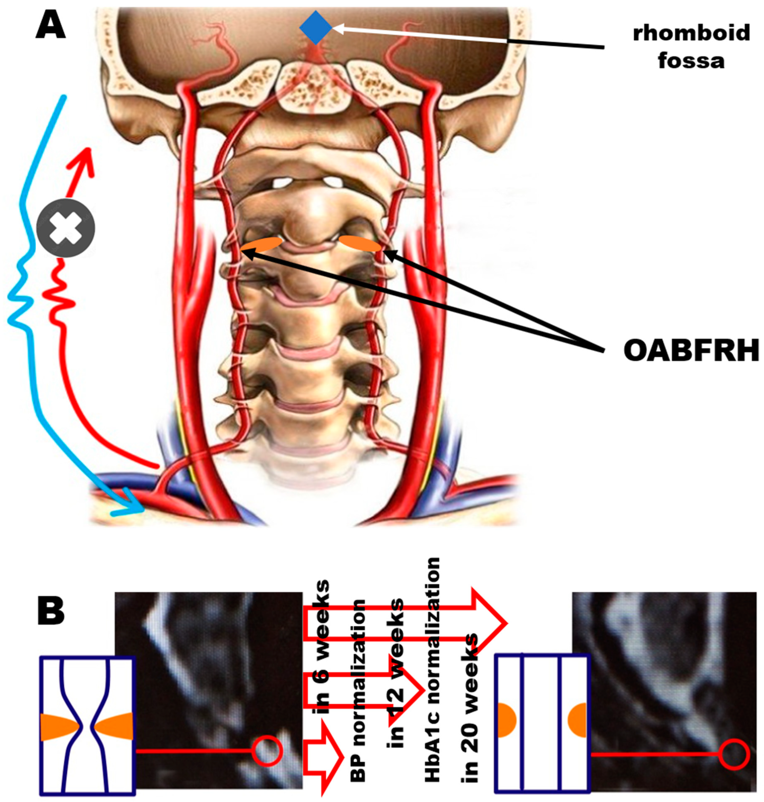
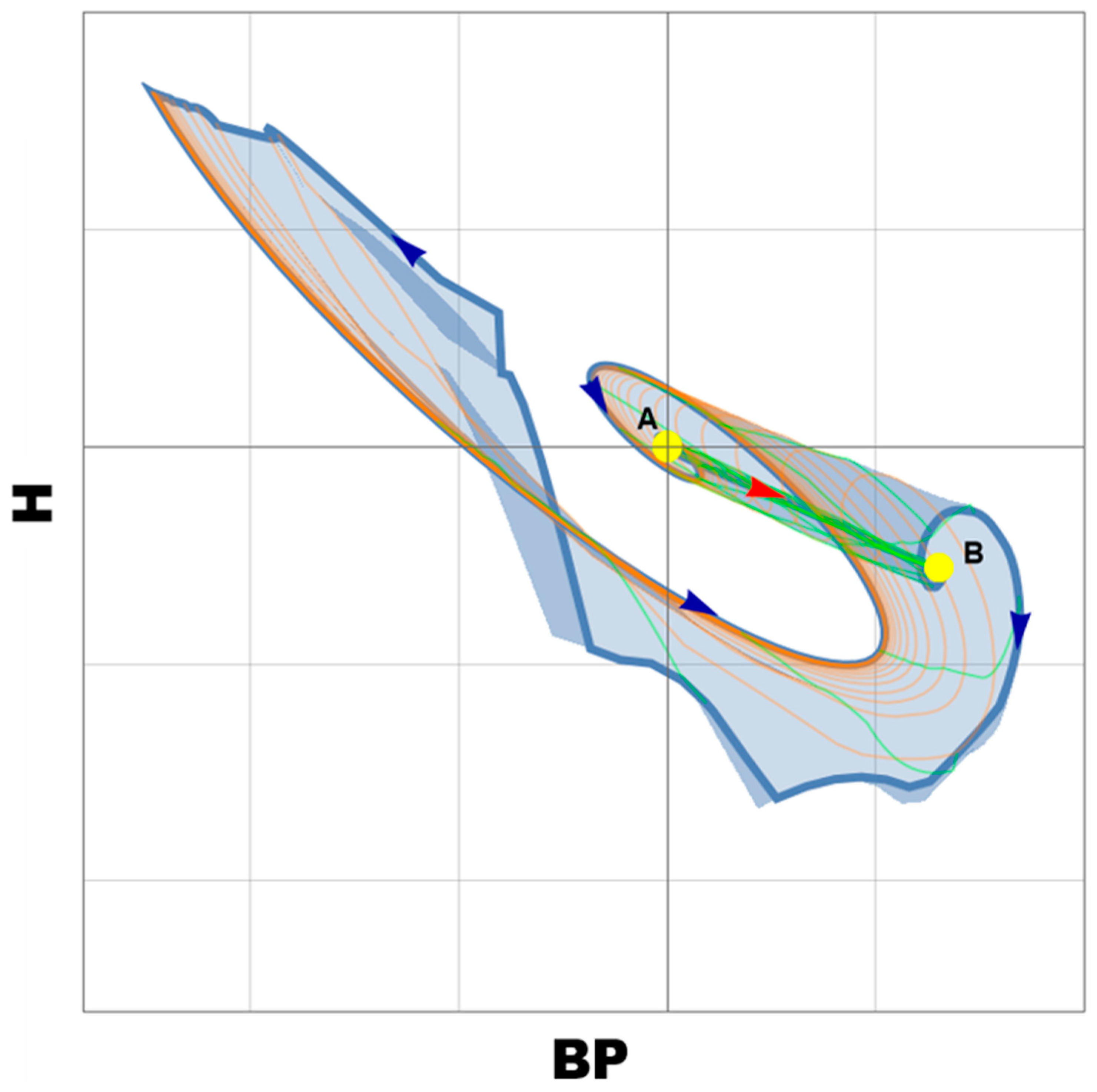
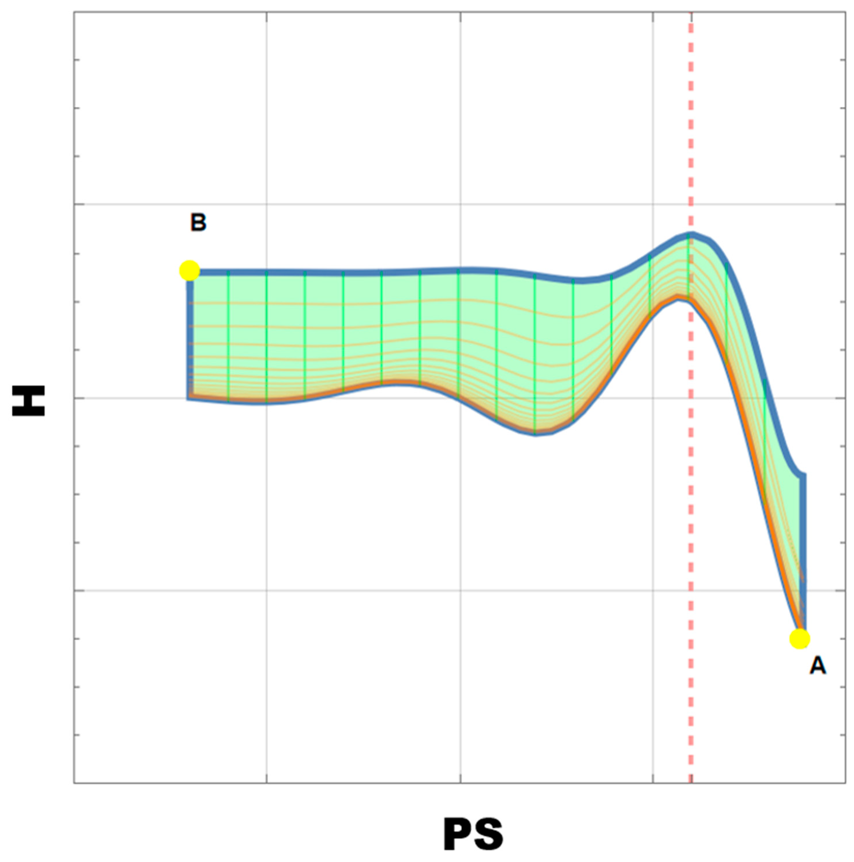
For our modeling, we used the previously reported data shown in Table 1, which demonstrates the absence of significant gender differences. Therefore, we used parameters from the combined sample.

3. Modeling

The mathematical model of the recovery from the adaptive OABFRH state of the body during the restoration of PS provides additional issues yet to be considered. As mentioned above, long-term adaptation occurs with repeated or ongoing OABFRH. It is a combination of structural, functional, and metabolic adaptive reactions. Therefore, the main feature of the model is taking into account a large number of such reactions that have different effects and effects over time. In model building, we used an approach similar to [39].

Model Explanation

The adaptation processes are formed continuously as a result of repeated activation of the mechanisms of urgent adaptation to OABFRH. Homeostasis on each step generates conditions for optimal vitality of the organism. Eventually, a dissipative structure is formed with at least two stable points (attractors of the unstable focus type). The first point is the basic state of the body in the absence of OABFRH (Figure 2). The second point demonstrates the state of the body in the case of adaptation to OABFRH. The most important observation is that the onset and recovery itineraries are different.
In the real space of values, we define the functions of apparent OABFRH H (t, PS) and BP (t, PS), where t is time. Consider the dynamical system of evolution ∂/∂t of the functions H and BP; we denote r = PS for brevity. To account for the contribution to the general system of the cumulative effect of the action of each term H and BP, we interpret the coefficients DH and DBP as the values of their “range” with diffusion term 2 r 2 functions, according to the Equations (2) and (3):
H t = a 11 H + a 12 B P + F ( H , B P ) + D H 2 H r 2 ,
B P t = a 21 H + a 22 B P + G ( H , B P ) + D B P 2 B P r 2
The terms F and G have the meanings of the regression dependencies that may appear during field experiments (below, F = G = 0). The initial boundary conditions are BP (t,20) = 130 and H (0,r) = 100(1 − r/20).
The values are taken as typical parameters of the patient’s ultrasonography. The maximum PS does not exceed 45 cm/s and the time is 12 weeks. Later, the system is reduced to a dimensionless form and numerically solved using the FEM method implemented in the Wolfram Mathematica 13.0 environment. In Figure 3, one can see the relationships between H and PS. The most important observation from it is that the itinerary consists of two parts with a sharp change in the slope. This can be checked by more regular PS measurements in further studies on animal models that allow data collection with the desired periodicity. We especially left the axes without any scale since it should be defined after experimental data collection.
The modeling of DM-related and AHT-related parameter interactions opens a new page in TCAAEBC studies. There is a wide variety of blood flow parameters which are associated with arterial blood access to the rhomboid fossa that are relatively easy to determine during triplex sonography [40,41,42] and their association with H(t) is easy to evaluate. Our experience suggests that it is still possible to find the parameter that could be optimal for the characterization of OABFRH according to the TCAAEBC. Finding such a parameter could allow the control of recovery to be easier and faster. We are sure that the search should be continued. Moreover, nowadays there are additional methods proposed for quantitative blood flow measurements such as pulsed wave Doppler studies, high-frame-rate ultrasound vector flow imaging, phase contrast MRI, and several others [42,43,44]. The long list of proposed approaches looks endless, even if the number of proposed measurement parameters is much shorter.
The same is true with the understanding of the parameters that could be offered instead of HbA1c for better characterization of DM [45]. Since its acceptance by WHO as a major index for DM, its usefulness is regularly questioned [46,47]. A similar situation is with the parameters to characterize AHT [48,49,50].

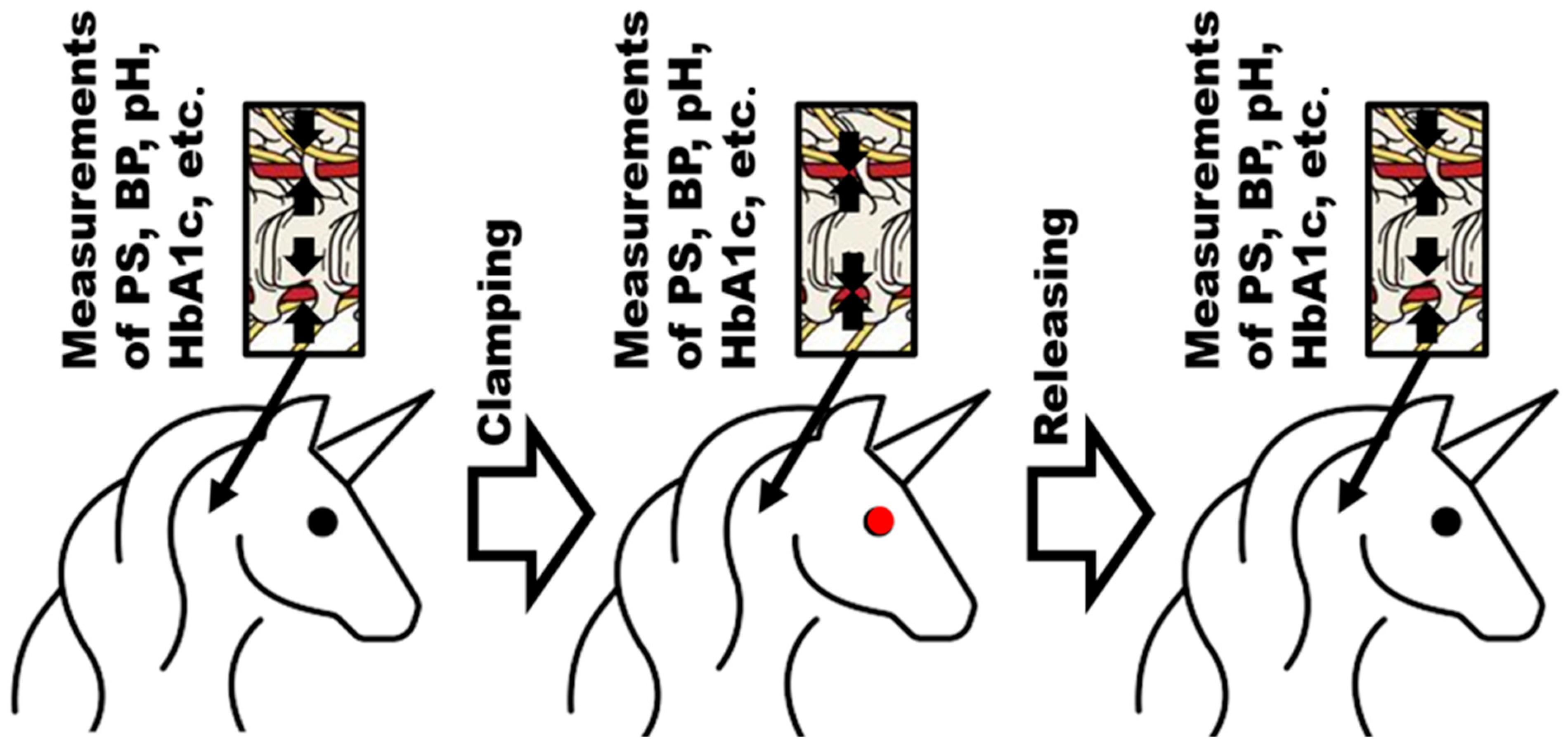
4. Appropriate Animal Model(s)

The overview on the planned experimental setup is demonstrated in Figure 4.
Let us list the available and acceptable options from the medical community’s animal models with the features that are essential for the TCAAEBC verification (Table 2). Acceptance of the model is extremely important, since otherwise we could step into the same trap as Mendel, who was proposed to repeat his experiments on Hieracium because his model—Pisum sativum—looked less acceptable to his pen pal [51,52]. Let us underline that neither of these were the animal model.
From the very beginning, we would like to exclude primate models from consideration due to:
The absence of defined knowledge that required data could be obtained only from them;
The existing restrictions on the involvement of such models in nonobligatory experiments [53,54].
Table 2. Comparison of mammalian models on required-to-check CAAEBC parameters [55,56,57,58,59,60].
Model AnimalResults Are Transferrable to
Human Clinical Situation(s)
Lack of Reserve Arterial Way to Rhomboid FossaGeneral Convenience to Measure BPBlood Boost
for Biochemical Analysis
Simplicity of Brachycephalic Arterial Blood Flow
Measurement
Availability of Reported
Positive Experience with the Reverse Brachycephalic
Arteries Blocking
Mice ++++
Rats+++++
Rabbits++++
Minipigs+++++
Goats+++
Sheep++-
Guinea pigs+++
Cats+++++
Dogs+++++
There are some general points about aspects that should be taken into consideration to choose an appropriate animal model. Some experimentalists even underline that there is no ideal model for every clinical situation, but they can be more or less accurate to represent them [61]. Importantly, the contemporary knowledge of the detailed characteristics of each animal model is unavailable. Therefore, experiments often need to be designed on pure guess. This can be a problem later in the translation stage of the acquired data to human clinical situations. In the majority of the convenient animal models, the small diameter and tangled geometry often close the door to certain models [62].
The fundamental advantage of the animal model is the ease with which the results may be applied to clinical settings including humans [63,64]. It is frequently so difficult to identify the best model for a given situation that a pilot experiment is required [65,66].
The arterial blood supply in rats exhibits heterosegmental organization [59]. This fact is not in favor and has the greatest difference from real-world clinical scenarios of any model on the list. However, this is so far the only model where the elevation in BP was demonstrated after implementation of the plastic implants, causing atlantoaxial misalignment. The removal of the implant reversed BP to normal values. The evaluation of PS was not performed [24].
Let us think about the (mini)pig model’s drawbacks. Although there is no evidence on cervical vertebral arteries, there are reports that this model can create collateral blood flow to duplicate vertebral arteries in the situation of occlusion [67]. The fundamental benefit of using a minipig model to test CAAEBC is that the physiology and vascular anatomy are similar to those of people [68]. This model’s popularity is primarily due to similar clinical conditions.
When it comes to dogs and cats, these models show some similarities to the human artery system, which is helpful for our purposes. For example, rabbits are used for various cases of cardiovascular modeling [69,70,71]. However, the public’s attitude is significantly less positive about these models in comparison with the two listed above [72]. Therefore, we consider them as the last resort.
The abdominal aorta of the rabbit, used as a model, provides homosegmental blood flow. This model has a significant benefit in that it nearly completely lacks an intraspinal collateral arterial system [71]. The key benefits are cheaper maintenance expenses and simpler manipulation.
More segmental blood is present in the guinea pig model. The small segmental arteries are to blame for this [71]. This model is the least advantageous to model CAAEBC because of the inability of blocking blood passage to the rhomboid fossa due to the larger segmental arterial supply.
The mouse model is comparable to those using other rodents [73]. Despite all of this model’s benefits, including how quickly experimental symptoms can appear, how inexpensive it is, and how simple it is to manipulate [61], we believe it is flawed for the same reason as the rat model. Detailed information on the anatomy of the cervical part of the spinal cord vascular organization is extremely important to plan the experiment properly [74].
It looks like even if the rat model is already confirmed as a primary candidate because of its reported performance, the minipig and rabbit models could be considered as the second and third options.

5. Gaps in Evidence and Future Directions Instead of Conclusions

Even if the primary parameter for DM characterization is HbA1c (according to WHO), which does not allow for characterization faster than 90 days (according to its nature), to allow daily modeling, we need to collect data on daily glucose profile.
The observation of the behavior of the model describing the recovery from DM as a function of the restoration of the arterial blood flow access to the rhomboid fossa leads to the following outcomes:
  • The onset and recovery itineraries from pre-DM are different;
  • The slope of the HbA1c dependence on PS demonstrates two different areas. Each of them should have different health condition situations, which should be verified on animal models. These preliminary conclusions should be preceded by the optimization of the list of parameters to obtain a further description of OABFRH’s influence on DM. The collected data could be used in the evaluation of the set of proper parameters for the aging index(es). This became critically important in 2022, when WHO (in ICD-11) eventually started to consider aging as a health condition and not as a normal stage of development [75]; therefore, AHT and DM, which were considered aging satellites, could experience a change in mankind’s attitude toward them.

Author Contributions

Conceptualization, A.Y.S. and A.A.V.; methodology, A.Y.S.; software, B.A.G.; validation, B.A.G. and K.V.Z.; formal analysis, A.Y.S. and A.A.V.; investigation, A.Y.S. and K.V.Z.; resources, A.Y.S.; data curation, P.I.B. and B.A.G.; writing—original draft preparation, A.S.K. and A.Y.S., A.A.V., D.T.R., and M.N.K.; writing—review and editing, A.Y.S. and A.A.V.; visualization, A.A.V.; supervision, A.Y.S. and A.A.V.; project administration K.V.Z.; funding acquisition, A.Y.S. All authors have read and agreed to the published version of the manuscript.

Funding

This paper was supported by the RUDN University Strategic Academic Leadership Program (recipient A.A.V.).

Institutional Review Board Statement

Not applicable.

Informed Consent Statement

Not applicable.

Data Availability Statement

Not applicable.

Acknowledgments

Alexandre A. Vetcher is grateful to the RUDN University Strategic Academic Leadership Program for the support.

Conflicts of Interest

The authors declare no conflict of interest.

References

  1. Bower, J.K.; Appel, L.J.; Matsushita, K.; Young, J.H.; Alonso, A.; Brancati, F.L.; Selvin, E. Glycated Hemoglobin and Risk of Hypertension in the Atherosclerosis Risk in Communities Study. Diabetes Care 2012, 35, 1031–1037. [Google Scholar] [CrossRef] [PubMed] [Green Version]
  2. Selvin, E.; Steffes, M.W.; Zhu, H.; Matsushita, K.; Wagenknecht, L.; Pankow, J.; Coresh, J.; Brancati, F.L. Glycated hemoglobin, diabetes, and cardiovascular risk in nondiabetic adults. N. Engl. J. Med. 2010, 362, 800–811. [Google Scholar] [CrossRef] [PubMed]
  3. Adams, R.J.; Appleton, S.L.; Hill, C.L.; Wilson, D.H.; Taylor, A.W.; Chittleborough, C.R.; Gill, T.K.; Ruffin, R.E. Independent association of HbA(1c) and incident cardiovascular disease in people without diabetes. Obes. (Silver Spring) 2009, 17, 559–563. [Google Scholar]
  4. Song, J.; Wei, N.; Zhao, Y.; Jiang, Y.; Wu, X.; Gao, H. Elevated glycosylated hemoglobin levels and their interactive effects on hypertension risk in nondiabetic Chinese population: A cross-sectional survey. BMC Cardiovasc. Disord. 2020, 20, 218. [Google Scholar] [CrossRef]
  5. Cosentino, F.; Grant, P.J.; Aboyans, V.; Bailey, C.J.; Ceriello, A.; Delgado, V.; Federici, M.; Filippatos, G.; Grobbee, D.E.; Hansen, T.B.; et al. ESC Scientific Document Group. 2019 ESC Guidelines on diabetes, pre-diabetes, and cardiovascular diseases developed in collaboration with the EASD. Eur Heart J. 2020, 41, 255–323. [Google Scholar] [CrossRef] [PubMed] [Green Version]
  6. Williams, B.; Mancia, G.; Spiering, W.; Agabiti Rosei, E.; Azizi, M.; Burnier, M.; Clement, D.L.; Coca, A.; de Simone, G.; Dominiczak, A.; et al. ESC Scientific Document Group. 2018 ESC/ESH Guidelines for the management of arterial hypertension. Eur. Heart J. 2018, 39, 3021–3104. [Google Scholar] [CrossRef] [Green Version]
  7. Perrone, V.; Veronesi, C.; Gambera, M.; Nati, G.; Perone, F.; Tagliabue, P.F.; Degli Esposti, L.; Volpe, M. Treatment with Free Triple Combination Therapy of Atorvastatin, Perindopril, Amlodipine in Hypertensive Patients: A Real-World Population Study in Italy. High Blood Press. Cardiovasc. Prev. 2019, 26, 399–404. [Google Scholar] [CrossRef]
  8. Mancia, G.; Kreutz, R.; Brunström, M.; Burnier, M.; Grassi, G.; Januszewicz, A.; Muiesan, M.L.; Tsioufis, K.; Agabiti-Rosei, E.; Algharably, E.A.E.; et al. 2023 ESH Guidelines for the management of arterial hypertension The Task Force for the management of arterial hypertension of the European Society of Hypertension Endorsed by the European Renal Association (ERA) and the International Society of Hypertension (ISH). J Hypertens. 2023. Online ahead of print. [Google Scholar] [CrossRef]
  9. Mancia, G. The association of hypertension and diabetes: Prevalence, cardiovascular risk and protection by blood pressure reduction. Acta Diabetol. 2005, 42, 17–25. [Google Scholar] [CrossRef]
  10. Petrie, J.; Guzik, T.J.; Touyz, R.M. Diabetes, Hypertension, and Cardiovascular Disease: Clinical Insights and Vascular Mechanisms. Can. J. Cardiol. 2017, 34, 575–584. [Google Scholar] [CrossRef] [Green Version]
  11. Vetcher, A.A.; Zhukov, K.V.; Gasparyan, B.A.; Shishonin, A.Y. Hypothetical reason for the restoration of HbA1c level for pre-diabetic patients through the restoration of arterial blood flow access to rhomboid fossa. Diabetology 2022, 3, 470–476. [Google Scholar] [CrossRef]
  12. Zhukov, K.V.; Gasparuan, B.A.; Vetcher, A.A.; Shishonin, A.Y. Centralized aerobic-anaerobic energy balance compensation theory perspective in biomedicine. Arch. Biotechnol. Biomed. 2022, 6, 7–9. [Google Scholar] [CrossRef]
  13. King, H. The Holistic Hippocrates: Treating the Patient, Not Just the Disease. In Hippocrates Now: The ‘Father of Medicine’ in the Internet Age; Bloomsbury Academic: London, UK, 2020; pp. 133–154. [Google Scholar] [CrossRef] [Green Version]
  14. Correll, J. Descartes’ Dualism and Its Influence on Our Medical System. SUURJ Seattle Univ. Undergrad. Res. J. 2022, 6, 47–55. [Google Scholar]
  15. Alex, S. Rene Descartes: Colossus of binaries—Real or perceived? Arch. Med. Health Sci. 2017, 5, 126–128. [Google Scholar] [CrossRef]
  16. Billman, G.E. Homeostasis: The Underappreciated and Far Too Often Ignored Central Organizing Principle of Physiology. Front. Physiol. 2020, 11, 200–2011. [Google Scholar] [CrossRef] [Green Version]
  17. Vetcher, A.A.; Zhukov, K.V.; Gasparuan, B.A.; Shishonin, A.Y. Time to take the Thermodynamics of Irreversible processes into consideration to describe in vivo biodegradation. Res. Dev. Polym. Sci. 2022, 1, 1004–1006. [Google Scholar] [CrossRef]
  18. Vetcher, A.A.; Zhukov, K.V.; Gasparuan, B.A.; Shishonin, A.Y. The cerebellum role in arterial hypertension. Med. Hypotheses 2022, 162, 10835. [Google Scholar] [CrossRef]
  19. Vetcher, A.A.; Zhukov, K.V.; Gasparuan, B.A.; Shishonin, A.Y. The Role of Cervical Vertebral Arteries Blood Flow in Centralized Aerobic- Anaerobic Energy Balance Compensation: When Hypothesis Becomes a Theory. Antioxid. Cardiovasc. Dis. 2021, 5, 1027–1032. [Google Scholar]
  20. Braukmann, S. The Organism and the Open System: Ervin Bauer and Ludwig von Bertalanffy. Ann. New York Acad. Sci. 2006, 901, 291–300. [Google Scholar]
  21. Elek, G.; Muller, M. The living matter according to Ervin Bauer (1890–1938), (on the 75th anniversary of his tragic death). Acta Physiol. Hung. 2013, 100, 124–132. [Google Scholar] [CrossRef]
  22. Gumenyuk, V.I.; Dobroborsky, B.S.; Fedosovsky, M.E. Determination of the effectiveness of training of personnel of energy facilities by quantitatively accounting for the human factor. Glob. Energy 2012, 154, 308–312. (In Russian) [Google Scholar]
  23. Curtelin, D.; Morales-Alamo, D.; de Torres-Peralta, R.S.; Rasmussen, P.; Martin-Rincon, M.; Perez-Valera, M.; Siebenmann, C.; Perez-Suarez, I.; Cherouveim, E.; Sheel, A.W.; et al. Cerebral blood flow, frontal lobe oxygenation and intra-arterial blood pressure during sprint exercise in normoxia and severe acute hypoxia in humans. J. Cereb. Blood Flow Metab. 2018, 38, 136–150. [Google Scholar] [CrossRef] [PubMed] [Green Version]
  24. He, Z.B.; Lv, Y.-K.; Li, H.; Yao, Q.; Wang, K.; Song, X.; Wu, Z.; Qin, X. Atlantoaxial Misalignment Causes High Blood Pressure in Rats: A Novel AHT Model. BioMed Res. Int. 2017, 2017, 5986957. [Google Scholar] [PubMed] [Green Version]
  25. Zhukov, K.V.; Gasparyan, B.A.; Vetcher, A.A.; Shishonin, A.Y. Left ventricular hypertrophy linked with arterial hypertension through centralized aerobic-anaerobic energy balance compensation theory. Ann. Clin. Hypertens. 2022, 6, 012–014. [Google Scholar] [CrossRef]
  26. Vetcher, A.A.; Zhukov, K.V.; Gasparyan, B.A.; Shishonin, A.Y. How the theory of centralized aerobic-anaerobic energy balance compensation explains sudden cardiac death in young athletes. Eur. J. Intern. Med. 2022, 107, 118–119. [Google Scholar] [CrossRef]
  27. Zhukov, K.V.; Vetcher, A.A.; Gasparuan, B.A.; Shishonin, A.Y. Alteration of Relative Rates of Biodegradation and Regeneration of Cervical Spine Cartilage through the Restoration of Arterial Blood Flow Access to Rhomboid Fossa: A Hypothesis. Polymers 2021, 13, 4248. [Google Scholar] [CrossRef]
  28. Zhukov, K.V.; Gasparyan, B.A.; Vetcher, A.A.; Shishonin, A.Y. The theory of centralized aerobic-anaerobic energy balance compensation and the right posture. Open J. Orthop. Rheumatol. 2022, 7, 011–012. [Google Scholar] [CrossRef]
  29. Bulatov, E.; Khaiboullina, S.; dos Reis, H.J.; Palotás, A.; Venkataraman, K.; Vijayalakshmi, M.; Rizvanov, A. Ubiquitin-Proteasome System: Promising Therapeutic Targets in Autoimmune and Neurodegenerative Diseases. Bionanoscience 2016, 6, 341–344. [Google Scholar] [CrossRef]
  30. Banwarth-Kuhn, M.; Sindi, S. How and why to build a mathematical model: A case study using prion aggregation. J. Biol. Chem. 2020, 295, 5022–5035. [Google Scholar] [CrossRef] [Green Version]
  31. Porgo, T.V.; Norris, S.L.; Salanti, G.; Johnson, L.F.; Simpson, J.A.; Low, N.; Egger, M.; Althaus, C.L. The use of mathematical modeling studies for evidence synthesis and guideline development: A glossary. Res. Synth. Methods 2019, 10, 125–133. [Google Scholar] [CrossRef] [Green Version]
  32. Menshikov, Y. Criteria for Adequacy Estimation of Mathematical Descriptions of Physical Processes. In Computational Models in Engineering; Volkov, K., Ed.; IntechOpen: London, UK, 2019; pp. 1–19. [Google Scholar] [CrossRef] [Green Version]
  33. American Diabetes Association. Diagnosis and classification of diabetes mellitus. Diabetes Care 2010, 33 (Suppl. 1), S62–S69. [Google Scholar] [CrossRef] [PubMed] [Green Version]
  34. WHO. Definition and Diagnosis of Diabetes Mellitus and Intermediate Hyperglycemia; Report of a WHO/IDF Consultation; WHO Document Production Services: Geneva, Switzerland, 2006; p. 50. [Google Scholar]
  35. American Diabetes Association. Standards of medical care in diabetes-2015 abridged for primary care providers. Clin. Diabetes 2015, 33, 97–111. [Google Scholar] [CrossRef] [PubMed] [Green Version]
  36. Genc, S.; Gurdol, F.; Kanmaz-Ozer, M.; Ince, N.; Ozcelik, F.; Omer, B. The Analytical Performances of Four Different Glycated Hemoglobin Methods. Med. Chem. 2014, 4, 501–505. [Google Scholar] [CrossRef] [Green Version]
  37. Kobalava, Z.D.; Konradi, A.O.; Nedogoda, S.V.; Shlyakhto, E.V. Arterial hypertension in adults. Clinical guidelines 2020. Russ. J. Cardiol. 2020, 25, 149–218. [Google Scholar]
  38. Schoning, M.; Walter, J.; Scheel, P. Estimation of cerebral blood flow through color duplex sonography of the carotid and vertebral arteries in healthy adults. Stroke 1994, 25, 17–22. [Google Scholar] [CrossRef] [Green Version]
  39. Kholin, A.M.; Muminova, K.T.; Balashov, I.S.; Khodzhaeva, Z.S.; Borovikov, P.I.; Ivanets, T.Y.; Gus, A.I. First-trimester prediction of preeclampsia: Validation of screening algorithms in a Russian population. Akusherstvo I Ginekol. Obstet. Gynecol. 2017, 8, 74–84. (In Russian) [Google Scholar] [CrossRef]
  40. Vetcher, A.A.; Zhukov, K.V.; Gasparuan, B.A.; Shishonin, A.Y. The cervical blood flow parameters with the best correlation from arterial blood pressure in hypertension cases. Int. J. Recent Sci. Res. 2021, 9, 42957–42958. [Google Scholar] [CrossRef]
  41. Baselli, G.; Fasani, F.; Pelizzari, L.; Cazzoli, M.; Baglio, F.; Laganà, M.M. Real-Time Phase-Contrast MRI to Monitor Cervical Blood and Cerebrospinal Fluid Flow Beat-by-Beat Variability. Biosensors 2022, 12, 417. [Google Scholar] [CrossRef]
  42. Du, Y.; Ding, H.; He, L.; Yiu, B.Y.S.; Deng, L.; Yu, A.C.H.; Zhu, L. Quantitative Blood Flow Measurements in the Common Carotid Artery: A Comparative Study of High-Frame-Rate Ultrasound Vector Flow Imaging, Pulsed Wave Doppler, and Phase Contrast Magnetic Resonance Imaging. Diagnostics 2022, 12, 690. [Google Scholar] [CrossRef]
  43. Gewirtz, H.; Dilsizian, V. Integration of quantitative positron emission tomography absolute myocardial blood flow measurements in the clinical management of coronary artery disease. Circulation 2016, 133, 2180–2196. [Google Scholar]
  44. Li, D.; Liu, Y.; Zeng, X.; Xiong, Z.; Yao, Y.; Liang, D.; Qu, H.; Xiang, H.; Yang, Z.; Nie, L.; et al. Quantitative study of the changes in cerebral blood flow and iron deposition during progression of Alzheimer’s disease. J. Alzheimer’s Dis. 2020, 78, 439–452. [Google Scholar] [CrossRef] [PubMed]
  45. Munk, J.K.; Lind, B.S.; Jørgensen, H.L. Change in HbA1c concentration as decision parameter for frequency of HbA1c measurement. Scand. J. Clin. Lab. Investig. 2019, 79, 320–324. [Google Scholar] [CrossRef] [PubMed]
  46. Higgins, T.N.; Tran, D.; Cembrowski, G.S.; Shalapay, C.; Steele, P.; Wiley, C. Is HbA1c a good screening test for diabetes mellitus? Clin. Biochem. 2011, 44, 1469–1472. [Google Scholar] [CrossRef] [PubMed]
  47. Karnchanasorn, R.; Huang, J.; Ou, H.-Y.; Feng, W.; Chuang, L.-M.; Ken, C.; Chiu, K.C.; Samoa, R. Comparison of the Current Diagnostic Criterion of HbA1c with Fasting and 2-Hour Plasma Glucose Concentration. J. Diabetes Res. 2016, 2016, 6195494. [Google Scholar] [CrossRef] [Green Version]
  48. Resende, E.A.M.R.; Silva, V.J.D.; Correia, D. Comparison of pulse wave analysis parameters by oscillometry in hypertensive diabetic and nondiabetic patients in a Brazilian outpatient care. Medicine 2019, 98, e18100. [Google Scholar] [CrossRef]
  49. Tąpolska, M.; Spałek, M.; Szybowicz, U.; Domin, R.; Owsik, K.; Sochacka, K.; Skrypnik, D.; Bogdański, P.; Owecki, M. Arterial Stiffness Parameters Correlate with Estimated Cardiovascular Risk in Humans: A Clinical Study. Int. J. Environ. Res. Public Health 2019, 16, 2547. [Google Scholar] [CrossRef] [Green Version]
  50. Murkamilov, I.T.; Aitbaev, K.A.; Fomin, V.V.; Murkamilova, Z.A.; Redzhapova, N.A.; Yusupov, F.A. Clinical and functional characteristics of arterial stiffness in elderly. Syst. Hypertens. 2021, 18, 31–36. [Google Scholar] [CrossRef]
  51. Iltis, H. Gregor Mendel and His Work. Sci. Mon. 1943, 56, 414–423. [Google Scholar]
  52. Nogler, G.A. The lesser-known Mendel: His experiments on Hieracium. Genetics 2006, 172, 1–6. [Google Scholar] [CrossRef]
  53. Phillips, K.A.; Bales, K.L.; Capitanio, J.P.; Conley, A.; Czoty, P.W.; ‘t Hart, B.A.; Hopkins, W.D.; Hu, S.-L.; Miller, L.A.; Nader, M.A.; et al. Why primate models matter. Am. J. Primatol. 2014, 76, 801–827. [Google Scholar] [CrossRef] [Green Version]
  54. Aguilera, B.; Perez Gomez, J.; DeGrazia, D. Should biomedical research with great apes be restricted? A systematic review of reasons. BMC Med. Ethics 2021, 22, 15. [Google Scholar] [CrossRef]
  55. Casals, J.B.; Pieri, N.C.G.; Feitosa, M.L.T.; Ercolin, A.C.M.; Roballo, K.C.S.; Barreto, R.S.N.; Bressan, F.F.; Martins, D.S.; Miglino, M.A.; Ambrósio, C.E. The use of animal models for stroke research: A review. Comp. Med. 2011, 61, 305–313. [Google Scholar]
  56. Jia, T.; Wang, C.; Han, Z.; Wang, X.; Ding, M.; Wang, Q. Experimental Rodent Models of Cardiovascular Diseases. Front. Cardiovasc. Med. 2020, 7, 588075. [Google Scholar] [CrossRef] [PubMed]
  57. Zaragoza, C.; Gomez-Guerrero, C.; Martin-Ventura, J.L.; Blanco-Colio, L.; Lavin, B.; Mallavia, B.; Tarin, C.; Mas, S.; Ortiz, A.; Egido, J. Animal Models of Cardiovascular Diseases. J. Biomed. Biotechnol. 2011, 2011, 497841. [Google Scholar] [CrossRef] [PubMed]
  58. Leong, X.F.; Ng, C.Y.; Jaarin, K. Animal Models in Cardiovascular Research: Hypertension and Atherosclerosis. BioMed Res. Int. 2015, 2015, 528757. [Google Scholar] [CrossRef] [Green Version]
  59. Mazensky, D.; Flesarova, S.; Sulla, I. Arterial Blood Supply to the SpinalCord in Animal Models of Spinal CordInjury: A Review. Anat. Rec. 2017, 300, 2091–2106. [Google Scholar] [CrossRef] [Green Version]
  60. Li, Y.; Zhang, J. Animal models of stroke. Anim. Model. Exp. Med. 2021, 4, 204–219. [Google Scholar] [CrossRef] [PubMed]
  61. Simon, F.; Oberhuber, A. Ischemia and reperfusion injury ofthe spinal cord: Experimental strategies to examine postischemicparaplegia. Neural Regen. Res. 2016, 11, 414–415. [Google Scholar] [CrossRef]
  62. Melissano, G.; Civilini, E.; Bertoglio, L.; Calliari, F.; Campos, M.; Amato, A.; Chiesa, R. Angio-CT imaging of the spinal cordvascularisation: A pictorial essay. Eur. J. Vasc. Endovasc. Surg. 2010, 39, 436–440. [Google Scholar] [CrossRef] [Green Version]
  63. Ramer, L.M.; Ramer, M.S.; Bradbury, E.J. Restoring function afterspinal cord injury: Towards clinical translation of experimental strategies. Lancet Neurol. 2014, 13, 1241–1256. [Google Scholar] [CrossRef]
  64. Courtine, G.; Bunge, M.B.; Fawcett, J.W.; Grossman, R.G.; Kaas, J.H.; Lemon, R.; Maier, I.; Martin, J.; Nudo, R.J.; Ramon-Cueto, A.; et al. Can experiments in nonhuman primates expedite the translation of treatments for spinal cord injury in humans? Nat. Med. 2007, 13, 561–566. [Google Scholar] [CrossRef] [PubMed]
  65. Geissler, S.A.; Schmidt, C.E.; Schallert, T. Rodent models and behavioral outcomes of cervical spinal cord injury. J. Spine 2013, S4, 001. [Google Scholar] [CrossRef] [PubMed]
  66. Simon, F.; Erhart, P.; Vcelar, B.; Scheuerle, A.; Schelzig, H.; Oberhuber, A. Erythropoietin preconditioning improves clinical and histo-logic outcome in an acute spinal cord ischemia and reperfusionrabbit model. J. Vasc. Surg. 2015, 64, 1797–1804. [Google Scholar] [CrossRef] [Green Version]
  67. Uezu, T.; Koja, K.; Kuniyoshi, Y.; Miyagi, K.; Shimoji, M.; Arakaki, K.; Yamashiro, S.; Mabuni, K.; Senaha, S. Blood distribution tothe anterior spinal artery from each segment of intercostal andlumbar arteries. J. Cardiovasc. Surg. 2003, 44, 637–645. [Google Scholar]
  68. Singh, V.K.; Thrall, K.D.; Hauer-Jensen, M. Minipigs as models in drug discovery. Expert Opin. Drug Discov. 2016, 11, 1131–1134. [Google Scholar] [CrossRef] [Green Version]
  69. Maršala, M. Spinal cord blood flow and metabolism in transient spinal ischemia. In Spinal Cord Monitoring; Stålberg, E., Sharma, H.S., Olsson, Y., Eds.; Springer: New York, NY, USA, 1998; pp. 5–25. [Google Scholar]
  70. Pais, D.; Casal, D.; Arantes, M.; Casimiro, M.; O’Neill, J.G. 2007. Spinal cord arteries in Canis familiaris and their variations: Implications in experimental procedures. Braz. J. Morphol. Sci. 2007, 24, 224–228. [Google Scholar]
  71. De Girolami, U.; Zivin, J.A. Neuropathology of experimental spinal cord ischemia in the rabbit. J. Neuropathol. Exp. Neurol. 1982, 41, 129–149. [Google Scholar] [CrossRef]
  72. National Research Council (US) Committee on Scientific and Humane Issues in the Use of Random Source Dogs and Cats in Research. Scientific and Humane Issues in the Use of Random Source Dogs and Cats in Research; Use of Dogs and Cats in Research: Public Perception and Evolution of Laws and Guidelines; National Academies Press (US): Washington, DC, USA, 2009; pp. 1–9. [Google Scholar]
  73. Scremin, O.U. The spinal cord blood vessels. In The Spinal Cord: A Christopher and Dana Reeve Foundation Text and Atlas; Watson, C., Paxinos, C., Kayalioglu, G., Eds.; Elsevier: San Diego, CA, USA, 2009; pp. 57–63. [Google Scholar]
  74. Amato, A.C.M.; Stolf, N.A.G. Anatomy of spinal blood supply. J. Vasc. Bras. 2015, 14, 248–252. [Google Scholar] [CrossRef] [Green Version]
  75. Available online: https://icd.who.int (accessed on 27 July 2023).
Figure 1. (A) Schematic of the TCAAEBC view on OABFRH. (B) Itinerary of patients’ recovery during the treatment according to [27].
Biomedicines 11 02147 g001
Figure 2. Point B is an unstable focus that appears during OABFRH and point A is an unstable focus in the absence of OABFRH of the dynamic system. The pathways from A to B are marked with dark arrows which show the direction.
Biomedicines 11 02147 g002
Figure 3. Function H (t,PS). A and B as described above.
Biomedicines 11 02147 g003
Figure 4. The animal model to check the CAAEBC applicability to AHT.
Biomedicines 11 02147 g004
Table 1. Changes in BP, PS, and H from the above-described treatment (edited from [11]).
ParameterMFM + FNormal
Sample size192948
Age, years63.1 ± 11.765.5 ± 12.264.3 ± 12.0
BP before treatment, torr159.5 ± 18.3163.5 ± 17.9161.9 ± 18.1
BP after treatment, torr132.3 ± 19.2131.7 ± 16.6132.9 ± 17.3<140 [37]
PS before treatment, cm/s22.5 ± 8.121.9 ± 9.322.2 ± 7.5
PS after treatment, cm/s41.7 ± 6.743.2 ± 7.442.5 ± 7.848 ± 10 [38]
H before treatment, %6.03 ± 0.346.11 ± 0.456.08 ± 0.41
H after treatment, %5.7 ± 0.635.73 ± 0.515.72 ± 0.585.7–6.4 [35]
Disclaimer/Publisher’s Note: The statements, opinions and data contained in all publications are solely those of the individual author(s) and contributor(s) and not of MDPI and/or the editor(s). MDPI and/or the editor(s) disclaim responsibility for any injury to people or property resulting from any ideas, methods, instructions or products referred to in the content.

