Review

Collagens Regulating Adipose Tissue Formation and Functions

by
Iida Jääskeläinen
1,†,
Tiina Petäistö
1,†,
Elahe Mirzarazi Dahagi
2,
Mahdokht Mahmoodi
3,
Taina Pihlajaniemi
1,
Mari T. Kaartinen
2,3,4 and
Ritva Heljasvaara
1,*
1
ECM-Hypoxia Research Unit, Faculty of Biochemistry and Molecular Medicine, University of Oulu, 90014 Oulu, Finland
2
Department of Anatomy and Cell Biology, Faculty of Medicine and Health Sciences, McGill University, Montréal, QC H3A 0C7, Canada
3
Faculty of Dental Medicine and Oral Health Sciences, McGill University, Montréal, QC H3A 0C7, Canada
4
Division of Experimental Medicine, Faculty of Medicine and Health Sciences, McGill University, Montréal, QC H3A 0C7, Canada
*
Author to whom correspondence should be addressed.
These authors contributed equally to this work.
Biomedicines 2023, 11(5), 1412; https://doi.org/10.3390/biomedicines11051412
Submission received: 9 March 2023 / Revised: 28 April 2023 / Accepted: 3 May 2023 / Published: 10 May 2023
(This article belongs to the Special Issue ECM Code in Physiological and Pathological Processes)

Abstract

:
The globally increasing prevalence of obesity is associated with the development of metabolic diseases such as type 2 diabetes, dyslipidemia, and fatty liver. Excess adipose tissue (AT) often leads to its malfunction and to a systemic metabolic dysfunction because, in addition to storing lipids, AT is an active endocrine system. Adipocytes are embedded in a unique extracellular matrix (ECM), which provides structural support to the cells as well as participating in the regulation of their functions, such as proliferation and differentiation. Adipocytes have a thin pericellular layer of a specialized ECM, referred to as the basement membrane (BM), which is an important functional unit that lies between cells and tissue stroma. Collagens form a major group of proteins in the ECM, and some of them, especially the BM-associated collagens, support AT functions and participate in the regulation of adipocyte differentiation. In pathological conditions such as obesity, AT often proceeds to fibrosis, characterized by the accumulation of large collagen bundles, which disturbs the natural functions of the AT. In this review, we summarize the current knowledge on the vertebrate collagens that are important for AT development and function and include basic information on some other important ECM components, principally fibronectin, of the AT. We also briefly discuss the function of AT collagens in certain metabolic diseases in which they have been shown to play central roles.

1. Adipose Tissues

Adipose tissue (AT) is a type of loose connective tissue that functions as an energy reservoir and protects other tissues and organs from lipotoxicity. AT is also an important endocrine system that secretes adipokines (adipo-cytokines), making it a key tissue that regulates whole-body lipid and glucose metabolism as well as general health [1,2]. There are at least three types of ATs: white AT (WAT), brown AT (BAT), and beige or brite (brown-in-white) AT [1]. Furthermore, bone marrow adipose tissue (BMAT) has been suggested to form its own distinct type of AT, while lactating breast tissue is described to include pink adipocytes [2]. In humans, BAT depots localize mainly in the cervical-supraclavicular and perirenal regions of the body and are most prominent in infants and young adults, and decrease upon aging. The largest AT, the WAT, can be divided into two main depots based on its anatomical location, function, and signaling. Subcutaneous AT (SAT) is located directly under the skin and visceral AT (VAT) localizes around internal organs in the abdominal cavity [2,3].
Morphologically, white adipocytes are characterized by one large lipid droplet which presses the nucleus and cell organelles against the cell membrane, which reflects their obvious function of fat storage [4]. The properties of white adipocytes can differ depending on their location. VAT adipocytes are larger and metabolically more active, more prone to lipogenesis and lipolysis, and release more pro-inflammatory cytokines. In contrast, SAT produces higher levels of favorable adipokines, which include adiponectin and leptin, and is more vascularized and less vulnerable to the adverse effects of obesity. While both types of depots expand in obesity, an increase in VAT around internal organs is more detrimental to metabolic health. Increased VAT also correlates with metabolic dysfunctions [5].
Brown adipocytes are functionally and morphologically distinct from white adipocytes. They are smaller in size than white adipocytes and have a multilocular lipid reservoir [4]. While WAT stores lipids, BAT consumes fatty acids for heat production (thermogenesis) by disengaging the respiratory chain from energy production in the mitochondria via a brown-adipocyte-specific protein called uncoupling protein 1 (UCP1) [2,4,6]. Beige adipocytes are spread amongst WATs, and, upon activating signals, such as a cold temperature, they upregulate the production of brown-adipocyte-specific proteins and begin to resemble brown-like cells [7]. After the identification of physiologically active BAT in adult humans [8,9,10], its activation as a therapeutic strategy for the treatment of obesity and related disorders has attracted substantial interest. In cell and animal models, the transdifferentiation or browning of white adipocytes, for example via cold-, nutrient-, or noradrenaline-stimulated upregulation of UPC1, promotes energy expenditure, reduces adiposity, and protects against diet-induced obesity and insulin resistance. In humans, the pharmacological activation of BAT combined with physical exercise and a healthy diet is the most promising strategy to control weight gain [1,4,11,12,13,14].

1.1. Pathological Conditions in AT

In this review, we focus on the roles of collagens (Figure 1) in the pathological conditions that primarily affect WAT. We performed a search of the literature on collagen family members in AT and adipogenesis, and in metabolic disorders including obesity, metabolic syndrome, T2D, AT fibrosis, and dyslipidemias. We summarize the current findings in the following chapters. In addition to these disorders, many other metabolic diseases are associated with obesity and dysfunctions of AT. For example, ectopic lipid accumulation in the liver, which causes non-alcoholic fatty liver disease, as well as cardiovascular diseases, renal dysfunction, infertility, and many types of cancer, are often consequences or comorbidities of problems in AT functions [15]. The roles of ECM and collagens in these diseases are discussed in several other review articles [16,17,18,19,20,21], and, therefore, are not addressed in this article.

1.1.1. Obesity and Type 2 Diabetes

Obesity is characterized by abnormal and excessive fat accumulation and is a risk factor for many pathological conditions and diseases, such as insulin resistance, type 2 diabetes (T2D), and cardiovascular diseases [15]. AT accommodates to the increased requirement for fat storage in obesity via adipocyte differentiation (adipogenesis and hyperplasia) and enlargement (hypertrophy) [22]. The expanding WAT accumulates many types of immune cells, which, together with hypertrophic adipocytes, secrete pro-inflammatory cytokines that sustain persistent low-grade inflammation. This leads to harmful changes in the metabolism and gene expression profiles of adipocytes [15]. Chronic inflammation drives the development of T2D, where adipocytes do not respond to insulin signaling and, as a result, the uptake of glucose from the circulation via insulin-sensitive glucose transporter 4 (GLUT4) is reduced [23]. Insulin inhibits lipolysis in healthy WAT; however, in T2D, this pathway is dysregulated. Reduced insulin sensitivity leads to the increased breakdown of stored lipids, increases the lipid supply to circulation, and enhances the accumulation of fat to internal organs such as liver, which further complicates metabolic problems [24].

1.1.2. AT Fibrosis

AT expansion involves an acute remodeling of the extracellular matrix (ECM) to allow for larger adipocytes and vascular growth in the tissue (angiogenesis) [25,26]. In obesity, the rapid growth of AT mass is associated with limited oxygen supply due to defects in tissue vascularization. The resulting hypoxic tissue environment activates the hypoxia-inducible factor-1 (HIF-1) pathway, which induces transcriptional programs that sustain AT inflammation and fibrosis. Infiltrated macrophages, as well as adipocytes, secrete pro-inflammatory chemokines and growth factors, such as tumor necrosis factor alfa (TNF-α), interleukin 6 (IL-6), and transforming growth factor beta (TGF-β), which further drive the immune progenitors toward pro-inflammatory phenotypes. Hypoxia and inflammation upregulate the expression of ECM genes, including collagens, fibronectin (FN), and hyaluronan, as well as ECM crosslinking enzymes lysyl oxidase (LOX) and transglutaminases, which leads to the excessive accumulation and rigidity of ECM elements in obese AT [25,26,27,28,29,30,31,32,33]. The stiff, fibrotic tissue environment in AT is believed to limit adipocyte size growth [34]. Therefore, the excess lipids may accumulate as ectopic fat depots elsewhere in the body, including in the liver, skeletal muscle, and other organs, as well as in the bloodstream [15]. AT fibrosis also promotes insulin resistance, and AT ECM has been suggested to play a major role in defining how severely obesity impacts the metabolic health of an individual [35,36].
Fibrosis manifests as large collagen I and III bundles which are deposited in AT, replacing the functional AT parenchyma, as well as pericellular fibrosis gathering around the adipocytes [37]. Furthermore, fibrotic bundles can also concentrate around blood vessels, leading to compromised angiogenesis [37,38]. The suppression of angiogenesis is associated with impaired tissue oxygenation and, thus, with an increase in the negative effects of AT fibrosis on metabolic health [30,39,40]. In mice, a high-fat diet (HFD) induces a transcription signature in VAT adipocytes, characterized by the upregulation of genes for ECM and cytoskeletal proteins which, together, cause the cells to experience mechanical stress from both inside and outside [41]. This leads to the promotion of fibrotic processes, and at the same time, the suppression of adipocyte programs, including many lipolytic genes and mitochondrial genes [41]. In fact, AT fibrosis involves a switch in the adipocyte phenotype towards a more fibrotic gene expression profile [41,42]. For example, macrophage-derived platelet-derived growth factor receptor α (PDGFRα) is a known pro-fibrotic signal which directly activates adipocytes and control ECM dynamics [40,43,44]. Recently, the Hippo pathway has been indicated as a key regulator of AT fibrosis in mice. In cooperation with TGF-β activity, inactivation of the Hippo pathway appears to induce AT fibrosis by promoting a shift in the adipocyte gene expression profile toward a myofibroblast-like gene expression [45].

1.1.3. Lipodystropy

Lipodystrophies, also known as lipoatrophies, refer to diseases characterized by a loss of AT [46]. Lipoatrophy can be generalized, taking place in all parts of the body, or can be a local phenomenon that occurs in a certain fat depot or depots. The disease can be either genetic or, sometimes, acquired as a consequence of, e.g., autoimmune disease or HIV infection. The severity of lipodystrophies can vary depending on the type of condition and how much AT has been lost. Often, they are accompanied by severe metabolic complications such as dyslipidemias, in which the circulating lipid balance is disturbed, and insulin resistance and diabetes ensue [46]. The fact that obesity and lipodystrophy can cause similar metabolic complications highlights the importance of maintaining a balance where an appropriate amount of properly functioning AT maintains an individual’s metabolic health.

2. Extracellular Matrix in AT

Comparisons between lean, healthy AT and obese, unhealthy AT have revealed that healthy AT is characterized by a high gene expression and abundance of ECM elements such as collagens and ECM remodeling enzymes. In contrast, metabolically unhealthy AT presents a significant upregulation of factors that increase the rigidity of the tissue, including the collagen-crosslinking LOX, the transglutaminases TG2 and Factor XIII-A [47,48,49,50,51,52], and the cell adhesion-related TSP-1 [53]. Interestingly, in an in vitro setting, the decellularized ECM of non-diabetic AT was shown to retain the functions of the adipocytes of T2D patients, and vice versa: ECM extracted from diabetic AT had adverse effects on the adipocytes of non-diabetic individuals, indicating that ECM plays a key role in adipocyte function in diabetes [54]. Moreover, different AT depots, specifically SAT and VAT depots, have their own composition of ECM molecules which are differently up- or downregulated in obesity [53,54,55]. For example, in mice fed with a HFD, the expression of genes for collagen I and IV α chains are upregulated specifically in SAT, whereas collagen III and FN are upregulated in both SAT and VAT [54]. The fact that these depots have very different functional roles in whole-body metabolism suggests that the influence of ECM on AT functions is fundamental.
The cellular components of AT consist of multiple cell types, including preadipocytes, mesenchymal stromal cells, macrophages, immune cells, and fibroblasts. AT cells, as well as tissue structure and function, require an appropriate composition of the ECM, which is achieved through dynamic ECM remodeling, involving both its removal and the de novo synthesis of new ECM components. ECM modification is accomplished by regulating the synthesis and assembly of the ECM components themselves or by influencing the factors associated with their degradation, such as matrix metalloproteinases (MMPs) and their inhibitors. The ECM in ATs consists mainly of collagens, especially collagen types I, IV, and VI, as well as FN and laminins [34,56]. Many ECM molecules are produced by adipocytes themselves, but a major portion of the collagens are synthesized by the cells in the stromal vascular fraction [40,41].
For adipocytes, the remodeling of the ECM constitutes an important process for preadipocyte maturation. When adipocytes achieve maturity, they surround themselves with a basal lamina, or basement membrane (BM), which is a sheet-like specialized form of the ECM. The main components of the BM are laminins and collagen IV, which both form their own network and are attached to each other by various linking molecules such as nidogen [57,58]. The basal lamina helps to keep the cells intact when they are subjected to the mechanical stretch caused by their large lipid-storage droplets. In addition to providing this structural support, ECM molecules are also essential regulators of many AT functions such as energy metabolism via storing and releasing various growth factors [58,59,60]. The roles of major AT collagens in adipogenesis and AT-related disorders are further discussed in subsequent chapters.
Other important ECM proteins in the AT include FN, which is found abundantly together with fibrillar collagen ECM in the interstitial matrix as well as in the ECM of the BMs [61,62], and whose presence has been described in human and mouse WAT. The FN matrix can regulate numerous cellular functions in tissues, including cell adhesion, migration, proliferation, and differentiation [61,63,64], as well as the activity and assembly of other ECM components such as LOX [65], bone morphogenetic protein 1 (BMP-1) [66,67], collagens I and III [68,69], thrombospondin-1 (TSP-1) [68], and fibrillin-1 [70,71], at least in vitro. FN produced by tissue-resident cells is referred to as cellular FN (cFN) and contains extra domains A (EDA) and B (EDB) as a result of the alternative splicing of the FN1/Fn1 gene [72,73]. The soluble plasma FN (pFN) is a product of the hepatocytes in the liver, from where it is secreted into the blood and circulates at a high concentration. pFN can affect several tissues and organ ECMs [74].
The FN matrix inhibits preadipocyte differentiation to mature adipocytes by maintaining cellular adhesions and fibroblastic preadipocyte morphology [75,76,77]. In preadipocyte cultures, FN is expressed at the early pre-adipocyte stage, and its role is associated with the maintenance of the pre-adipocyte phenotype via the preadipocyte factor 1 (Pref-1) [78]; however, it is not established if the form involved in pre-adipocyte maintenance is pFN or cFN. pFN is a well-known substrate for Factor XIII-A transglutaminase, which promotes its self-assembly and crosslinking to collagen type I [79,80]. This covalent modification stabilizes the pFN matrix (but not the cFN matrix) in 3T3-E1 preadipocytes, which, in turn, modulates insulin signaling [52]. Knockout mice of FXIII-A, which are protected from HFD-induced insulin resistance and inflammation, have less pFN and a less collagenous matrix in their WAT compared to their wild type controls [51]. Obese HFD-fed mice show a significant increase in the circulating EDA-FN (cFN), which, possibly through interaction with toll-like receptor 4, may mediate the development of insulin resistance in mice [81].
The SAT and VAT of obese humans were shown to have decreased FN1 mRNA expression compared to lean control tissues and presented a negative correlation with the body mass index (BMI). However, in a lean–obese monozygotic twin study, it was also shown that FN1 mRNA was increased in isolated SAT adipocytes in the heavier, obese twin and correlates with pre-diabetic markers [47]. The circulating pFN was found to be higher in obese individuals with normal or fatty livers [82,83].
Another notable matrix molecule in the AT is the ECM glycoprotein osteopontin (OPN). In AT, it is produced by mature adipocytes as well as by stromal cells: both macrophages and senescent T cells [84,85,86]. OPN expression is significantly increased in obese and overweight patients, and even moreso in patients with obese-induced T2D, as compared with lean subjects [85]. In mice fed with a HFD, OPN deficiency leads to numerous beneficial outcomes including lower body weight, better insulin sensitivity, decreased hepatosteatosis, decreased AT fibrosis, and improved BAT function [87,88]. In addition, in the absence of OPN, both systemic and AT inflammation are decreased. The mechanisms of OPN action in AT are related to ECM remodeling, as demonstrated by reduced MMP and TGF-β production in AT, as well as to the regulation of inflammation, shown by a reduced infiltration of macrophages and monocytes into AT deficient of OPN [87,88]. These findings suggest that OPN has a role in linking obesity and the development of insulin resistance.
OPN mediates signals via integrins, and via the CD44 receptor, which can also bind another common ECM molecule, the glycosaminoglycan hyaluronan (HA) [89]. HA affects, for example, monocyte recruitment into AT, as well as the process of adipogenesis [90,91]. It has been reported to inhibit the differentiation of preadipocytes in vitro, either by knocking down or overexpressing HA synthases in the culture system [92,93,94]. In in vivo mouse models, HA accumulated in AT with HFD, and exogenous HA-degrading hyaluronidase or HA synthesis inhibitor reduced VAT accumulation and hepatosteatosis and increased insulin sensitivity [91,94]. Worth mentioning as an example of an ECM molecule of VAT is also TSP-1, a glycoprotein that contributes to obesity and insulin resistance [95,96]. In addition to these, MMPs and their inhibitors, tissue inhibitors of metalloproteinases (TIMPs) that control the degradation and turnover of collagens and other ECM components, play important and variable roles in AT formation and function [59].

3. AT Collagens and Their Roles in Adipogenic Differentiation and Dysfunctional AT

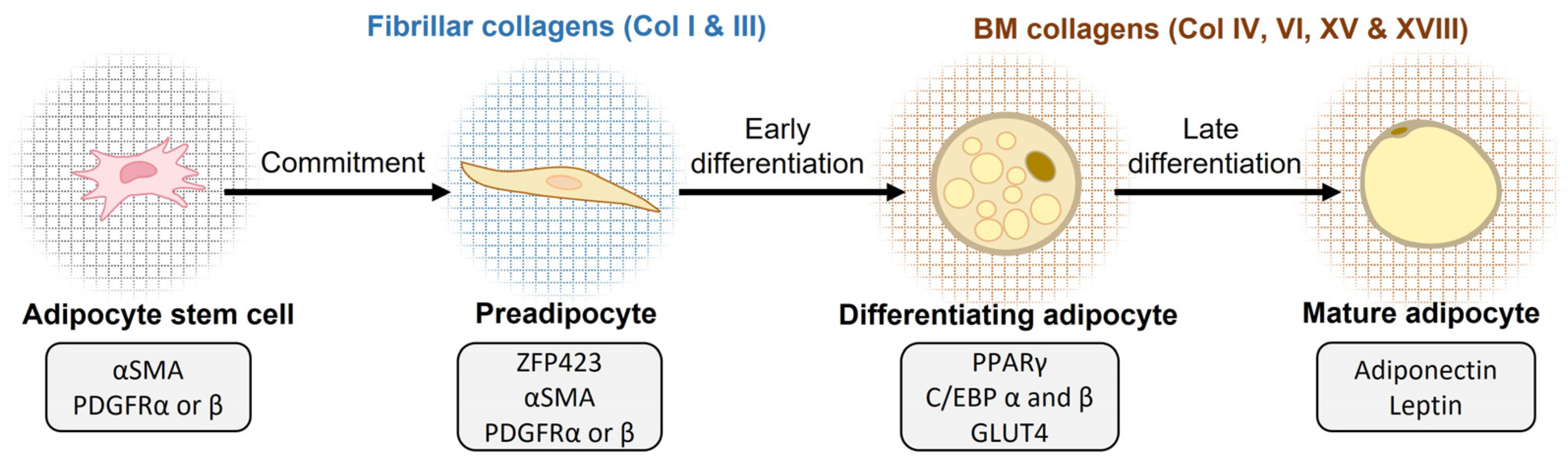
3.1. Collagens in Adipogenesis

Adipogenesis is the process of adipocyte differentiation from fibroblast-like precursor cells to lipid-filled mature adipocytes [22,97,98,99,100]. The first stage in the two-stage process of adipogenesis is the commitment of mesenchymal stem cell-derived adipose progenitor cells to the adipocyte lineage (Figure 2). The commitment step concludes with the formation of a preadipocyte, which still has the outer appearance of a fibroblast and expresses common fibroblast and adipose progenitor cell markers, such as α smooth muscle actin (αSMA) and PDGFRα and PDGFRβ [22]. The expression of Zinc-finger protein 423 (ZFP423) by the preadipocytes promotes adipogenic differentiation by sensitizing the cells to the BMP signal, which facilitates adipogenic progression [101]. Peroxisome proliferator-activated receptor gamma (PPARγ), together with the CCAAT/enhancer-binding protein alpha (C/EBPα), are widely accepted as the master regulators of adipocyte differentiation [22,102,103]. In the second stage, differentiating adipocytes lose their cuboidal shape and change to a morphologically round mature adipocyte shape and accumulate lipid droplets [22]. Adiponectin and leptin hormone secretion indicates that the adipocyte has fully matured. ECM attachments play a major role in regulating the pre- and mature adipocyte shape and size [22,99,102].
Decades ago, an electron microscopic study suggested the need for a three-dimensional collagen fibril-rich environment for proper adipocyte differentiation and maturation [104]. Later, a widely used in vitro model of adipogenesis, the 3T3-L1 murine preadipocytes, confirmed that, during the differentiation process from fibroblasts to adipocytes, the gene expression profiles of collagens shift from fibrillar to BM type collagens (Figure 2). During the undifferentiated fibroblast stage, the 3T3-L1 cells generally express the fibrillar collagen types I, III, and V, whereas collagen types IV and VI are expressed when they differentiate into mature adipocytes [105]. Interestingly, this shift is accompanied by changes in the expression and modification of cytoskeleton components and integrin-attachment modes. Further, the cell morphology changes from the elongated shape of pre-adipocytes to the round one of adipocytes [105] (Figure 1); this is believed to be driven by the change from fibrillar collagen and FN adhesion and signaling through integrin α5β1 to collagen IV and laminin adhesion/signaling through integrin α6β1. In addition to the morphological transformation, the transcription profiles of the cells are also impacted by the different cues from the different ECMs associated with preadipocytes and mature adipocytes [106,107,108]. In summary, the collagenous matrix surrounding the adipocytes is actively modified during the differentiation process (Figure 1) and impacts the regulation of adipogenesis during development as well as in AT expansion [22,56,109].

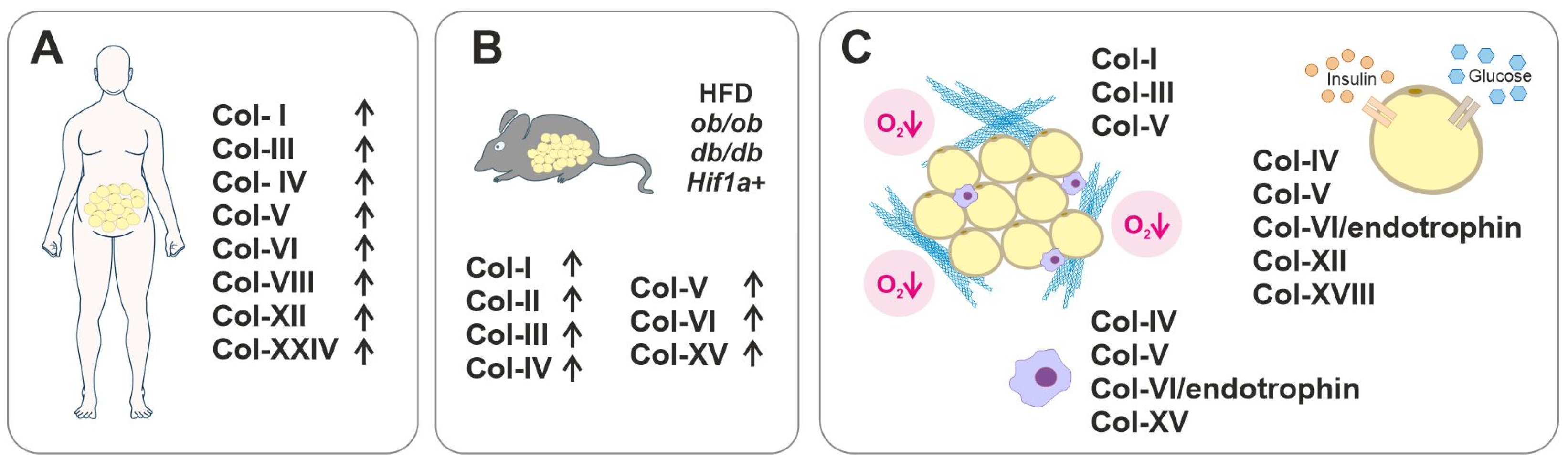
3.2. Collagens in Dysfunctional AT and Metabolic Diseases

Many of the 28 different vertebrate collagen types [60,110] are linked with various metabolic diseases in which AT is affected. In Table 1, we have summarized the current data on the collagens that are expressed in pathological AT and that are suggested to contribute to AT dysfunction and/or metabolic diseases. These collagens and their roles in adipogenesis and pathological AT are discussed in the subsequent paragraphs. To date, and to the best of our knowledge, no detailed data have been reported on the function of collagen types VII, X, XI, XVI, XVII, XIX–XXIII, and XXV–XXVIII in relation to AT and the pathological conditions associated with it; although, transcriptome analyses have revealed changes in the expression of some of these collagens upon adipogenic differentiation in vitro or in obese versus lean individuals, as discussed in Section 3.9.

3.3. Collagen I

Collagen I belongs to the subfamily of fibrillar collagens and is the most common collagen type in vertebrates and an important structural component in multiple tissues. In healthy tissues, collagen I molecules usually occur as heterotrimers which are composed of two α1 polypeptide chains and one α2 chain, encoded by COL1A1 and COL1A2 genes in humans, respectively [110] (Figure 1). Collagen I synthesis is high in AT in general, but it is enriched in SAT compared to VAT in healthy rodents. In contrast, in healthy humans, the expression levels of Col1a1 do not vary significantly between the two depots [53,132]. Its expression is high during the early stages of adipogenesis both in vivo and in the 3T3-L1 in vitro model; however, in rat VAT, the expression decreases when the adipocytes mature and the collagen gene expression profile shifts from fibrillar collagens toward BM-associated collagens (Figure 2). In SAT, collagen I synthesis does not diminish during adipocyte maturation [105,132,133].
Remodeling of the collagen I scaffold is essential for the proper differentiation and functioning of adipocytes. For example, the degradation of collagen I by the membrane-tethered matrix metalloproteinase 1 (MT1-MMP), also known as MMP14, is crucial for the differentiation of WAT (but not BAT) [134]. Without the action of this protease, the preadipocytes are entrapped within a dense fibrillar collagen meshwork that compromises the proper tissue architecture and signaling required for adipocyte differentiation. In the db/db animal model of T2D, the collagen I synthesis rate is notably increased in WAT [34], while in fibrotic SAT and VAT of obese humans, collagen I is typically found as fibrous bundles of various thicknesses alongside collagen III [37]. Macrophage-driven stimuli were shown to induce collagen I as well as other ECM proteins [29]. Further, constitutively active HIF-1α in a transgenic mouse model resulted in an increased expression of the genes of fibrillar collagens and their crosslinking enzymes, as well as increased local inflammation, leading to AT fibrosis and dysfunction [30]. Last, HFDs were shown to further stimulate collagen I production in db/db mice [135]. Differing from T2D and obesity, the serum biomarkers of collagen I were downregulated in patients with T1D with retinopathy. A decreased amount of collagen I was suggested to reduce the vascular integrity in such patients [111].
Although collagen I is often considered to be a primarily structural collagen, certain intriguing functional properties have been observed for it in relation to AT as well. The coating of collagen I has been reported to promote the migration and proliferation of undifferentiated mouse 3T3-L1 preadipocytes in vitro. Collagen I may induce these effects via reactive-oxygen-species generation and the activation of p65-dependent NF-κB signaling [136]. Another explanation for collagen I-induced adipocyte migration is that it occurs due to the activation of the Hippo/YAP pathway, which promotes primary cilia growth, leading to increased 3T3-L1 cell migration [137]. However, the effect that collagen I has on preadipocyte differentiation seems to be detrimental instead. Further, 3T3-L1 preadipocytes cultured on collagen I present an increased YAP expression, which leads to a reduction in the synthesis of adipogenic factors, such as PPARγ and C/EBPα, and the inhibition of adipocyte maturation [138]. Another study showed that interaction between collagen I and aortic carboxypeptidase-like protein, which is a secreted protein that is highly expressed in preadipocytes but downregulated during adipogenesis, leads to a reduced expression of these adipogenic factors, thus providing evidence of ECM-derived cues that influence adipogenic differentiation [139]. Moreover, the collagen I coating was found to repress autophagy in adipocytes through YAP activation and increase their mitochondrial content, causing adipogenesis inhibition and accelerated energy metabolism in 3T3-L1 cells, which was verified by an enhanced glucose uptake, reduced fatty acid release, and increased ATP production [140,141].

3.4. Collagen III

Collagen III is another abundant fibrillar collagen that is found in normal tissues in the same stromal areas as collagen I [110], as well as in the fibrotic ATs of obese humans [37] (Figure 1). However, in all types of mouse fat depots, collagen III synthesis appears to be significantly lower compared to collagen I synthesis [34]. Similar to collagen I, collagen III is also upregulated in mice fed with a HFD, or in mice that experience the constitutive expression of Hif1a [30,135]. In patients with T1D-induced retinopathy, unlike collagen I, elevated collagen III levels were reported [111]. Further, patients with T2D-induced nephropathy present increased levels of type III procollagen aminopeptide in their blood, thus suggesting that this peptide could be a possible biomarker for diabetic nephropathy [113].
Collagen III is downregulated during the differentiation of 3T3-L1, but the depletion of collagen III in 3T3-L1 preadipocytes was shown to prevent adipogenesis, indicating that it serves an important function in this process [133,142]. Collagen III was suggested to convey its effects through its established binding partner, the G protein-coupled receptor 56 (GPR56), as the phenotype of 3T3-L1 cell with CRISPR/Cas9-mediated Col3a1 knockout strongly resembled the phenotype of GPR56 knockout cells. The Col3a1 knockout led to a decrease in the adipogenic markers PPARγ, C/EPBα, and aP2, and reduced cell adhesion and lipid accumulation while maintaining constant canonical Wnt/beta-catenin activity [142], which is known to impair adipogenesis [143]. The knockout of Col3a1 also downregulated the expression of several ECM transcripts during the in vitro differentiation process, including collagens IV and VI [142]. Interestingly, the Col3a1 deficiency also downregulated the expression of FN in undifferentiated 3T3-L1 cells but upregulated its expression in mature adipocytes, suggesting that the functions of these ECM components are interlinked [142].

3.5. Collagen IV

The non-fibrillar collagen IV is a heterotrimer that is composed of various combinations of six different α chains, and these subtypes are often classified into their own category of BM collagens [110] (Figure 1). Collagen IV is the most important structural element of BMs, where it binds laminins, nidogens, and other ECM components, and stabilizes the structure [57]. Adipocytes are encircled by a thin BM, and collagen IV is naturally a prominent ECM component in AT [58]. The synthesis of collagen IV in AT appears to vary between developmental stages and in different adipose depots [107,132].
When 3T3-L1 preadipocytes undergo the adipogenic differentiation process, collagen IV production, or, more accurately, the production of α1(IV) and α2(IV) chains, increases significantly alongside some other basal lamina elements [133,144]. Another study reported that the secretion of collagen IV is increased in hypoxia by as much as 10 times, but the mRNA levels of α1(IV) and α2(IV) are not affected by the low oxygen pressure [145]. Interestingly, bone marrow stromal cells do not undergo adipogenic differentiation when cultured on the native structural form of the collagen IV scaffold but do undergo differentiation in a denatured collagen IV matrix. Adipogenic differentiation with denatured collagen IV was shown to occur through integrin αvβ3 integrin signaling [108]. These findings reiterate the important role of matrix remodeling and the balance of ECM-degrading MMP levels and MMP-inhibiting TIMP levels in adipogenesis and suggest that the inhibition of collagen IV denaturation in AT could serve as a strategy for obesity treatment.
In obesity, collagen IV synthesis is upregulated alongside other BM elements in SAT and is associated with the inflammatory and fibrotic factors TGF-β1 and TGF-β3, as well as with insulin resistance [107]. However, in an in vitro setting, TGF-β1 and TGF-β3 stimulated COL4A1 expression only in endothelial cells isolated from the SAT of patients, and not in isolated adipocytes. After the gastric bypass surgery of severely obese patients and the subsequent weight loss, COL4A1 expression was downregulated in SAT, and this reduction correlated with improved glucose metabolism parameters, such as insulin resistance assessment (HOMA-IR) in the studied patients [107].

3.6. Collagen V

Collagen V is a widely occurring low abundance fibrillar collagen with three different α chains, namely α1(V), α2(V), and α3(V), which form distinct heterotrimeric collagen V molecules. The most common combination which occurs in tissues is composed of two α1(V) chains and one α2(V) chain that assemble into heterotypic collagen fibrils with collagens I and III, thus regulating the fibril geometry [146] (Figure 1). During the differentiation of 3T3-L1 cells and bovine intramuscular preadipocytes, collagen V synthesis rises rapidly at early stages of adipogenesis and, later, the collagen V network undergoes extensive modifications, causing the thickening of fibrils [133,147]. Mice with a Col5a2 knockout do not survive embryonic development [148], but the Ubc-CreERT2; Col5a2fl/fl mice with the tamoxifen-induced postnatal ubiquitous ablation of the α2(V) chain present a drastic reduction in dermal and abdominal AT, with small adipocytes and fibrotic abdominal fat pad seams, highlighting the importance of collagen α2(V) for AT maintenance [149].
Mast cells, which gather and possibly mature in AT during obesity and diabetes progression, excrete inflammatory mast cell protease 6 (MCP-6), which has been shown to promote collagen V, specifically Col5a1, expression [114]. Collagen V is then said to promote AT fibrosis, and it seems to also suppress preadipocyte differentiation [114]. The co-culturing of adipocytes and M2 macrophages also appears to increase collagen V production in the adipocytes, possibly via TGF-β signaling [38], further suggesting crosstalk between immune cells and adipocytes to regulate collagen V levels in AT. In fibrous AT, collagen V bundles gather around blood vessels in significant quantities [37]. Obese individuals have fewer capillaries but more large vessels in their SAT depots than lean individuals, pointing to impaired angiogenesis in the obese individuals [38]. Collagen V inhibits angiogenesis in endothelial cell cultures, suggesting a link between collagen V levels and impaired angiogenesis in AT [38].
The α3(V) chain has more limited tissue distribution and is found in skeletal muscle, pancreas, and WAT in vertebrates and associates into heterotrimers with one α1(V) and one α2(V) chain [150]. In fact, Col5a3 is highly expressed in human adipocytes and mouse WAT [115], and its expression is increased in 3T3-L1 cells upon adipogenic differentiation [115]; however, it is decreased when adipogenic differentiation is compromised [134]. Thus, α3(V) seems to have AT-specific functions beyond its structural roles in collagen fibrils [115,146]. Yet, the normal-chow-fed mice with Col5a3 deletion present only a subtle reduction in SAT and no reduction in abdominal fat. However, feeding them with HFD resulted in a significant decrease in the total body weight of the Col5a3 knockout females, suggesting a sex-specific resistance to diet-induced obesity in the absence of this collagen chain [115]. Col5a3 ablation also leads to a significant decrease in insulin-stimulated Pparg expression in mouse WAT, further indicating a role of α3(V) in adipocytic differentiation [115].
The same study associated the α1(V), α2(V), and α3(V) chains with the regulation of glucose metabolism [115]. The ablation of α3(V) in mice led to impaired insulin sensitivity and hyperglycemia due to the incorrect deployment of glucose transporter GLUT4 receptors in their WAT and muscles, and this finding was replicated in the 3T3-L1 cell line. However, it should be noted that the knockout Col5a3 gene led to impaired glucose-stimulated insulin secretion from pancreatic β cells which can also contribute to the observed glucose imbalance in these mutant mice [115].

3.7. Collagen VI

Collagen VI is a prominent component of the ECM and usually forms heterotrimers composed of α1(VI), α2(VI), and α3(VI) chains. In addition to these, genes for three other α(VI) chains, i.e., α4(VI), α5(VI), and α6(VI), have been annotated; however, at least the COL6A4 gene is most likely not functional [121,151] (Figure 1). Collagen α(VI) chains form a separate subfamily among collagens because they assemble into a distinct network of beaded microfilaments that are located in the interphase between BMs and interstitial ECM. It is widely expressed in tissues such as muscle, bone, cartilage, and tissue, as well as in the nervous system, and serves both biomechanical and biochemical functions that regulate cell survival, differentiation, and proliferation [151].
Collagen VI is an abundant component of AT and is produced by adipocytes [121,151,152]. Collagen VI binds to the collagen IV network in the adipocyte BMs, contributing to the rigidity of the pericellular ECM [56]. In humans, collagen VI genes show fat depot-specific expression profiles and are produced in greater quantities in the SAT than in the VAT, and collagen VI mRNA transcripts are enriched in the stromal vascular fraction containing adipocyte precursors over the mature adipocytes [116]. In mice, collagen VI is the most abundant collagen in mature adipocytes [34]. Collagen VI production increases during the adipogenic differentiation of 3T3-L1 cells and bovine intramuscular preadipocytes, and its fiber network undergoes significant thickening during differentiation (Figure 2), which is similar to the collagen V network modification [133,147].
Collagen VI is overexpressed in fibrotic AT and accumulates in areas of pericellular fibrosis around the adipocytes in humans [37]. Increased COL6A3 expression in obesity restricts fat storage in SAT, which might lead to the lipid accumulation into VAT instead [153]. Moreover, high COL6A3 expression causes reduced oxygenation of AT, contributing to hypoxia and inflammation in the AT [153]. In obesity, COL6A3 expression in SAT is higher in humans with insulin resistance than those who are sensitive to insulin [36,119]. It is worth noting that a high BMI (>28) caused high variability in COL6A3 expression levels [153].
In mice, the Col6a3 knockout resulted in a reduction in epididymal AT mass but had no effect on the SAT or BAT [117], suggesting significant depot-specific functions. The stromal vascular fraction cells isolated from the SAT of Col6a3 knockout mice have an impaired adipogenic capacity in vitro [117]. In the in vitro 3T3-L1 model, the deletion of Col6a3 results in lipid accumulation that is comparable to that of wild type cells, while both Col6a1 and Col6a2 knockouts in these cells lead to impaired lipid accumulation. However, impaired lipolysis, which was observed both for Col6a3 knockout 3T3-L1 cells and Col6a3 knockout mice, may explain the unaltered lipid accumulation in the 3T3-L1 Col6a3 knockout cells [117].
The deletion of Col6a3 in obese ob/ob mice leads to improved outcomes and a metabolic phenotype, including lower AT inflammation, improved lipid clearance, and fewer necrotic adipocyte deaths. Adipocyte cell size was found to be larger in ob/ob and Col6a3−/− crosses than in ob/ob mice expressing Col6a3, which probably improved the metabolic phenotype of these obese mice, as adipocytes were able to grow without restrictions [34]. One study reported that, in obese humans, COL6A3 is upregulated after weight loss and downregulated in obesity, but these fluctuations had no connection to metabolic dysfunction [116]. This is contrary to the results reported by mouse studies and other human studies that support the increased synthesis of collagen α3(VI) chains in obesity [34,119,153].
Animal studies suggest that the α3(VI) chain of collagen VI is the most important α(VI) chain that regulates AT functions. The C-terminal biologically active endotrophin fragment of the α3(VI) chain (Figure 1) has independent roles in AT. During ECM remodeling, endotrophin is cleaved from the parental α3(VI) by BMP-1 protease and does not form a part of the mature collagen VI microfilament network [121]. Endotrophin is associated with a myriad of harmful effects with regard to metabolism, such as increasing the expression of pro-adipogenic genes and causing abnormal increased lipid accumulation and lipolysis [154,155]. It promotes detrimental changes in the AT architecture and leads to a significant increase in serum triglyceride levels during HFD in transgenic mice that overexpress endotrophin specifically in adipocytes [155]. A high endotrophin level has been associated with increased insulin resistance, and, interestingly, blocking the endotrophin function with a specific antibody treatment has been shown to improve metabolic health [155]. Moreover, endotrophin levels were increased in human patients with diabetes [155].
Endotrophin overexpression in adipocytes increases TGF-β signaling and upregulates ECM protein synthesis, including fibrillar collagens, which may account for several of its detrimental effects on metabolism [155]. In another study, endotrophin was also linked to the synthesis of the fibrosis biomarker pro-collagen III, and endotrophin itself has been suggested as a novel biomarker of tissue fibrosis [120,121,156]. Using mice and 3T3-L1 cells, Zhao et al. [154] observed that endotrophin increases the synthesis of various fibrotic proteins in different cell types of AT. For example, in adipocytes, endotrophin induces fibrillar collagen synthesis, whereas, in macrophages, it mainly promotes the expression of the collagen cross-linking enzyme LOX and leads to an increase in AT macrophages and a shift from the M2 phenotype to pro-inflammatory M1 [154,155]. Macrophages appear to be the main mediator through which endotrophin induces AT inflammation, as its pro-inflammatory effects were not observed in adipocytes themselves [154]. In a clinical pilot study where humans with poor glycemic control in T2D were put on a diet and exercise regime, endotrophin levels were high at the start of the study. However, they were significantly lowered due to the change in lifestyle and were correlated with lowered serum glycated HbA1c hemoglobin and urine albumin-creatinine ratio levels, further indicating that α3(VI)/endotrophin expression is associated with metabolic dysfunction [157].

3.8. Multiplexin Collagens XV and XVIII

Besides the major AT collagens (types I, III, IV, and VI), other collagen types are also expressed within AT, and some of them play intriguing structural or functional roles. For example, the structurally homologous non-fibrillar BM-associated collagens XV and XVIII of the multiplexin subclass are implicated in adipogenesis and lipid metabolism [158,159] (Figure 1).
Collagen XV is expressed in many cell types of the connective tissue, including adipocytes and fibroblasts [158]. In AT, the roles of type XV collagen are still not very well understood. Nevertheless, in mice, Col15a1 expression appears to be higher in WAT depots than BAT depots [122]. The adipocyte differentiation process is associated with increased collagen XV synthesis in mice. Collagen XV promotes adipocyte differentiation and inhibits lipolysis, possibly via changes in its DNA methylation and the inhibition of the hormone-responsive cyclic AMP (cAMP)−protein kinase A (PKA) pathway. The cAMP response element binding protein (CREB) suppresses collagen XV expression and, as a result, the collagen XV-dependent promotion of adipocyte differentiation [122]. Moreover, collagen XV levels are significantly increased in obese mice, and this upregulation has been suggested to play a functional role in lipid deposition and adipogenesis [122]. RNA-seq data have demonstrated the involvement of collagen XV in the regulation of abnormal ECM remodeling, which is associated with the induction of adipocyte apoptosis via the collagen XV-activated AMPK pathway [123]. Collagen XV is also involved in the regulation of AT inflammation, as it promotes the polarization of pro-inflammatory M1 macrophages and the upregulation of the endoplasmic reticulum stress-related genes [124].
Type XVIII collagen is a multidomain collagen and heparan sulphate proteoglycan with three alternative forms (short, medium, and long) that differ in terms of domain structure and that have tissue-specific expression patterns and different functional roles [159] (Figure 1). Collagen XVIII production is known to increase during adipogenesis [127,129]. In mice, the effects of collagen XVIII on adipocyte differentiation appear to be largely mediated by the two longest isoforms of collagen XVIII, whose expression is regulated by an alternative internal promoter of the gene, and a specific lack of these isoforms leads to reduced adiposity [126,127]. Mouse embryonic fibroblast isolated from Col18a1−/− mice or mice specifically lacking the two longest isoforms (the Col18a1P2/P2 mice) have an impaired adipogenic capacity, and the stromal vascular fraction of the epididymal AT of these mice contains more adipocyte progenitor cells than wild-type mice [127]. Wnt/β-catenin signaling is an important adipogenic regulator that suppresses adipogenic progression [143]. Interestingly, the longest isoform of collagen XVIII contains a Frizzled-like domain (Figure 1) that has considerable similarity to the Frizzled receptors of the Wnt ligands [159], suggesting that collagen XVIII may regulate adipogenesis through the Wnt/β-catenin pathway. In fact, immunoprecipitation analysis indicates the binding of the Frizzled-containing domain of collagen XVIII with the Wnt10b [127]. An impaired capacity of Col18a1−/− and Col18a1P2/P2 mutants to store lipids in the AT causes ectopic lipid accumulation and dyslipidemia, which leads to increased serum triglyceride levels and higher fat accumulation in the liver compared to wild-type mice [126,127].
Hypertriglyceridemia has also been observed in humans with mutations in the COL18A1 gene [125]. Multiple single-nucleotide polymorphisms (SNP) in COL18A1 are associated with obesity in T2D and with circulating lipid contents [128,129,130]. Moreover, collagen XVIII synthesis in VAT is associated with circulating free fatty acids in obesity, and a genetic linkage analysis shows an association between chromosome 21, where COL18A1 is located, and the familial combined hyperlipidemia-triglyceride trait, as well as increased serum triglycerides in hypertensive pedigrees [127,160]. Knobloch syndrome patients carrying a null mutation of COL18A have increased serum triglyceride levels after fasting and show reduced activity and mass of plasma lipoprotein lipase (LPL), which cleaves fatty acids from circulating lipoproteins [125,161]. The heparan sulphate side chains of collagen XVIII are suggested to carry LPL from the ECM to its receptor on the endothelial cell membrane, and a lack of this collagen may lead to the retention of LPL in the subendothelial matrix, leading to dysregulation in blood lipid profiles and dyslipidemias [125,161].
Furthermore, a type XVIII collagen knockout in mice causes metabolic complications, specifically reduced insulin sensitivity and glucose tolerance, which are most likely caused by the aforementioned lack of adiposity [126]. Interestingly, collagen XVIII deficiency also leads to increased heat production, probably due to the increased thermogenesis in mouse BAT, as well as changes in BAT composition [126]. BAT serves an intriguing function in AT-related lipid regulation. It appears that the increased activation of BAT thermogenesis will positively impact the lipid profile, lowering the risk of atherosclerosis [162,163]. Interestingly, Col18a1−/− mice displayed an improved triglyceride profile in circulation at cold temperatures, likely due to the activated lipid uptake and non-shivering thermogenesis in their BAT [126].

3.9. Other Collagens

Studies on AT-associated collagens have been focused on fibrillar and BM collagens and collagen VI, but a few transcriptome analyses have also revealed interesting expression patterns in AT for other collagen types. For example, the fibril-associated collagen XII and non-fibrillar short-chain collagen VIII were found to be upregulated in the adipocytes of obese individuals. Here, a high expression of COL12A1 is strongly associated with the amount of LDL, the “bad cholesterol,” whereas low COL12A1 is related to improved insulin sensitivity [47]. Collagen VIII is an understudied collagen, but it has been suggested to have functions related to endothelial cells, smooth muscle cells, and myofibroblasts [164], and, thus, probably also in the AT vascularization and fibrosis. Another microarray study revealed the downregulation of COL14A1 when human mesenchymal stem cells were induced to differentiate to mature adipocytes in vitro [165]. This deviates from the findings of studies on 3T3-L1 cells that have reported that collagen XIV, and, specifically, its FN type III domain, triggered adipogenic differentiation in these cells [166]. Finally, collagen XXIV was upregulated in the VAT and skeletal muscles of HFD-fed mice, as well as in the VAT, but not the SAT, of obese diabetic human subjects compared to lean controls, suggesting a pathogenic function of this collagen in T2D and obesity [131].

4. Conclusions and Future Perspectives

AT is a highly active tissue that plays a vital role in metabolism and the general health of the entire body. AT-resident ECM and its major collagen components help guide AT growth by providing a supportive and modifiable scaffold and acting as a restrictive barrier. AT ECM is capable of undergoing a myriad of changes in response to environmental and nutritional cues. Beyond their structural role, AT collagens play significantly functional roles in adipocyte maturation and whole-body metabolism (Figure 2 and Figure 3). The relatively recently discovered ECM component, collagen α3(VI)-derived endotrophin fragment, for example, has emerged as an important regulator of adipogenesis and AT function. High endotrophic levels are significantly associated with many chronic diseases including T2D, vascular, and kidney diseases, and it was proposed to serve as a new prognostic biomarker for such metabolic disorders [153,156]. In addition to the widely studied fibrillar collagens, the BM-associated collagen XVIII appears to majorly contribute to regulating AT development and functions and the related metabolic complications such as dyslipidemia (Figure 3).
Collagen and ECM modulation can be a powerful approach to affect whole AT homeostasis and, thus, research on AT collagens can open interesting avenues into the development of novel anti-T2D and obesity therapies. However, their complex dual role in both the physiology and pathology of AT is a challenge. Further research into the diversity of AT collagens is expected to reveal novel structural and functional roles for collagens. Understanding the developmental and physiological roles of AT collagens versus the pathological effects of fibrotic ECM in AT, and its effects on the insulin sensitivity of adipocytes and other AT cells are central questions to be answered in studies focusing on AT ECM. Furthermore, how much do posttranslational modifications of collagens affect their relevant functions, and do LOX- and transglutaminase-mediated crosslinking alter their impact from positive to negative? How do other ECM components such as fibronectin affect collagenous AT ECM development and functioning? Also, what are the cellular mechanisms that promote the expression and assembly of collagens that are necessary for AT health versus those cues that initiate fibrosis? What is the role of ECM in general, and collagens in particular, in modulating the heterogeneity and plasticity of adipocytes? These and many other questions must be carefully addressed in future AT ECM studies.

Author Contributions

R.H. ideated the manuscript. I.J. and T.P. (Tiina Petäistö) performed the literature search and drafted the manuscript. R.H. made the figures. I.J., T.P. (Tiina Petäistö), E.M.D., M.M., T.P. (Taina Pihlajaniemi), M.T.K. and R.H. revised and updated the manuscript. All authors have read and agreed to the published version of the manuscript.

Funding

T. Petäistö was supported by the Kerttu Saalasti foundation and the University of Oulu Scholarship funds. M.T. Kaartinen was supported by the Canadian Institutes of Health Research (CIHR). E. D. Mirzarazi was supported by Le Fonds de Recherche du Québec—Santé (FRQS). M. Mahmoodi was supported by the Faculty of Dental Medicine and Oral Health Sciences at McGill University.

Institutional Review Board Statement

Not applicable.

Informed Consent Statement

Not applicable.

Data Availability Statement

No new data were created or analyzed in this article. Data sharing is not applicable to this article.

Acknowledgments

The authors thank Audrey Savolainen for proof-reading the text.

Conflicts of Interest

The authors declare no conflict of interest.

References

  1. Zorena, K.; Jachimowicz-Duda, O.; Ślęzak, D.; Robakowska, M.; Mrugacz, M. Adipokines and Obesity. Potential Link to Metabolic Disorders and Chronic Complications. Int. J. Mol. Sci. 2020, 21, 3570. [Google Scholar] [CrossRef] [PubMed]
  2. Zwick, R.K.; Guerrero-Juarez, C.F.; Horsley, V.; Plikus, M.V. Anatomical, Physiological, and Functional Diversity of Adipose Tissue. Cell Metab. 2018, 27, 68–83. [Google Scholar] [CrossRef] [PubMed]
  3. Ibrahim, M.M. Subcutaneous and Visceral Adipose Tissue: Structural and Functional Differences. Obes. Rev. 2010, 11, 11–18. [Google Scholar] [CrossRef] [PubMed]
  4. Giordano, A.; Smorlesi, A.; Frontini, A.; Barbatelli, G.; Cinti, S. White, Brown and Pink Adipocytes: The Extraordinary Plasticity of the Adipose Organ. Eur. J. Endocrinol. 2014, 170, R159–R171. [Google Scholar] [CrossRef] [PubMed]
  5. Wronska, A.; Kmiec, Z. Structural and Biochemical Characteristics of Various White Adipose Tissue Depots. Acta Physiol. 2012, 205, 194–208. [Google Scholar] [CrossRef] [PubMed]
  6. Froy, O.; Garaulet, M. The Circadian Clock in White and Brown Adipose Tissue: Mechanistic, Endocrine, and Clinical Aspects. Endocr. Rev. 2018, 39, 261–273. [Google Scholar] [CrossRef] [PubMed]
  7. Mulya, A.; Kirwan, J.P. Brown and Beige Adipose Tissue. Endocrinol Metab. Clin. N. Am. 2016, 45, 605–621. [Google Scholar] [CrossRef]
  8. Virtanen, K.A.; Lidell, M.E.; Orava, J.; Heglind, M.; Westergren, R.; Niemi, T.; Taittonen, M.; Laine, J.; Savisto, N.-J.; Enerbäck, S.; et al. Functional Brown Adipose Tissue in Healthy Adults. N. Engl. J. Med. 2009, 360, 1518–1525. [Google Scholar] [CrossRef]
  9. Van Marken Lichtenbelt, W.D.; Vanhommerig, J.W.; Smulders, N.M.; Drossaerts, J.M.A.F.L.; Kemerink, G.J.; Bouvy, N.D.; Schrauwen, P.; Teule, G.J.J. Cold-Activated Brown Adipose Tissue in Healthy Men. N. Engl. J. Med. 2009, 360, 1500–1508. [Google Scholar] [CrossRef]
  10. Cypess, A.M.; Lehman, S.; Williams, G.; Tal, I.; Rodman, D.; Goldfine, A.B.; Kuo, F.C.; Palmer, E.L.; Tseng, Y.-H.; Doria, A.; et al. Identification and Importance of Brown Adipose Tissue in Adult Humans. N. Engl. J. Med. 2009, 360, 1509–1517. [Google Scholar] [CrossRef]
  11. Giordano, A.; Frontini, A.; Cinti, S. Convertible Visceral Fat as a Therapeutic Target to Curb Obesity. Nat. Rev. Drug. Discov. 2016, 15, 405–424. [Google Scholar] [CrossRef] [PubMed]
  12. Giordano, A.; Cinti, F.; Canese, R.; Carpinelli, G.; Colleluori, G.; Di Vincenzo, A.; Palombelli, G.; Severi, I.; Moretti, M.; Redaelli, C.; et al. The Adipose Organ Is a Unitary Structure in Mice and Humans. Biomedicines 2022, 10, 2275. [Google Scholar] [CrossRef] [PubMed]
  13. Harb, E.; Kheder, O.; Poopalasingam, G.; Rashid, R.; Srinivasan, A.; Izzi-Engbeaya, C. Brown Adipose Tissue and Regulation of Human Body Weight. Diabetes Metab. Res. Rev. 2023, 39, e3594. [Google Scholar] [CrossRef] [PubMed]
  14. Armani, A.; Feraco, A.; Camajani, E.; Gorini, S.; Lombardo, M.; Caprio, M. Nutraceuticals in Brown Adipose Tissue Activation. Cells 2022, 11, 3996. [Google Scholar] [CrossRef]
  15. Kawai, T.; Autieri, M.V.; Scalia, R. Adipose Tissue Inflammation and Metabolic Dysfunction in Obesity. Am. J. Physiol. Cell Physiol. 2021, 320, C375–C391. [Google Scholar] [CrossRef]
  16. Kolb, R.; Sutterwala, F.S.; Zhang, W. Obesity and Cancer: Inflammation Bridges the Two. Curr. Opin. Pharmacol. 2016, 29, 77–89. [Google Scholar] [CrossRef]
  17. Avgerinos, K.I.; Spyrou, N.; Mantzoros, C.S.; Dalamaga, M. Obesity and Cancer Risk: Emerging Biological Mechanisms and Perspectives. Metabolism 2019, 92, 121–135. [Google Scholar] [CrossRef]
  18. Polyzos, S.A.; Kountouras, J.; Mantzoros, C.S. Obesity and Nonalcoholic Fatty Liver Disease: From Pathophysiology to Therapeutics. Metabolism 2019, 92, 82–97. [Google Scholar] [CrossRef]
  19. Fujii, H.; Kawada, N. The Role of Insulin Resistance and Diabetes in Nonalcoholic Fatty Liver Disease. Int. J. Mol. Sci. 2020, 21, 3863. [Google Scholar] [CrossRef]
  20. Bloksgaard, M.; Lindsey, M.; Martinez-Lemus, L.A. Extracellular Matrix in Cardiovascular Pathophysiology. Am. J. Physiol. Heart Circ. Physiol. 2018, 315, H1687–H1690. [Google Scholar] [CrossRef]
  21. Elagizi, A.; Kachur, S.; Carbone, S.; Lavie, C.J.; Blair, S.N. A Review of Obesity, Physical Activity, and Cardiovascular Disease. Curr. Obes. Rep. 2020, 9, 571–581. [Google Scholar] [CrossRef] [PubMed]
  22. Ghaben, A.L.; Scherer, P.E. Adipogenesis and Metabolic Health. Nat. Rev. Mol. Cell. Biol. 2019, 20, 242–258. [Google Scholar] [CrossRef] [PubMed]
  23. Zatterale, F.; Longo, M.; Naderi, J.; Raciti, G.A.; Desiderio, A.; Miele, C.; Beguinot, F. Chronic Adipose Tissue Inflammation Linking Obesity to Insulin Resistance and Type 2 Diabetes. Front. Physiol. 2020, 10, 1607. [Google Scholar] [CrossRef] [PubMed]
  24. Dilworth, L.; Facey, A.; Omoruyi, F. Diabetes Mellitus and Its Metabolic Complications: The Role of Adipose Tissues. Int. J. Mol. Sci. 2021, 22, 7644. [Google Scholar] [CrossRef]
  25. Crewe, C.; An, Y.A.; Scherer, P.E. The Ominous Triad of Adipose Tissue Dysfunction: Inflammation, Fibrosis, and Impaired Angiogenesis. J. Clin. Investig. 2017, 127, 74–82. [Google Scholar] [CrossRef]
  26. Herold, J.; Kalucka, J. Angiogenesis in Adipose Tissue: The Interplay Between Adipose and Endothelial Cells. Front. Physiol. 2021, 11, 624903. [Google Scholar] [CrossRef]
  27. Lee, M.-J. Transforming Growth Factor Beta Superfamily Regulation of Adipose Tissue Biology in Obesity. Biochim. Biophys. Acta Mol. Basis Dis. 2018, 1864, 1160–1171. [Google Scholar] [CrossRef]
  28. Debari, M.K.; Abbott, R.D. Adipose Tissue Fibrosis: Mechanisms, Models, and Importance. Int. J. Mol. Sci. 2020, 21, 6030. [Google Scholar] [CrossRef]
  29. Henegar, C.; Tordjman, J.; Achard, V.; Lacasa, D.; Cremer, I.; Guerre-Millo, M.; Poitou, C.; Basdevant, A.; Stich, V.; Viguerie, N.; et al. Adipose Tissue Transcriptomic Signature Highlights the Pathological Relevance of Extracellular Matrix in Human Obesity. Genome Biol. 2008, 9, R14. [Google Scholar] [CrossRef]
  30. Halberg, N.; Khan, T.; Trujillo, M.E.; Wernstedt-Asterholm, I.; Attie, A.D.; Sherwani, S.; Wang, Z.V.; Landskroner-Eiger, S.; Dineen, S.; Magalang, U.J.; et al. Hypoxia-Inducible Factor 1α Induces Fibrosis and Insulin Resistance in White Adipose Tissue. Mol. Cell. Biol. 2009, 29, 4467–4483. [Google Scholar] [CrossRef]
  31. Anvari, G.; Bellas, E. Hypoxia Induces Stress Fiber Formation in Adipocytes in the Early Stage of Obesity. Sci. Rep. 2021, 11, 21473. [Google Scholar] [CrossRef] [PubMed]
  32. Pastel, E.; Price, E.; Sjöholm, K.; McCulloch, L.J.; Rittig, N.; Liversedge, N.; Knight, B.; Møller, N.; Svensson, P.-A.; Kos, K. Lysyl Oxidase and Adipose Tissue Dysfunction. Metabolism 2018, 78, 118–127. [Google Scholar] [CrossRef] [PubMed]
  33. Eckert, R.L.; Kaartinen, M.T.; Nurminskaya, M.; Belkin, A.M.; Colak, G.; Johnson, G.V.W.; Mehta, K. Transglutaminase Regulation of Cell Function. Physiol. Rev. 2014, 94, 383–417. [Google Scholar] [CrossRef] [PubMed]
  34. Khan, T.; Muise, E.S.; Iyengar, P.; Wang, Z.V.; Chandalia, M.; Abate, N.; Zhang, B.B.; Bonaldo, P.; Chua, S.; Scherer, P.E. Metabolic Dysregulation and Adipose Tissue Fibrosis: Role of Collagen VI. Mol. Cell. Biol. 2009, 29, 1575–1591. [Google Scholar] [CrossRef] [PubMed]
  35. Lackey, D.E.; Burk, D.H.; Ali, M.R.; Mostaedi, R.; Smith, W.H.; Park, J.; Scherer, P.E.; Seay, S.A.; McCoin, C.S.; Bonaldo, P.; et al. Contributions of Adipose Tissue Architectural and Tensile Properties toward Defining Healthy and Unhealthy Obesity. Am. J. Physiol. Endocrinol. Metab. 2014, 306, E233–E246. [Google Scholar] [CrossRef]
  36. Lawler, H.M.; Underkofler, C.M.; Kern, P.A.; Erickson, C.; Bredbeck, B.; Rasouli, N. Adipose Tissue Hypoxia, Inflammation, and Fibrosis in Obese Insulin-Sensitive and Obese Insulin-Resistant Subjects. J. Clin. Endocrinol. Metab. 2016, 101, 1422–1428. [Google Scholar] [CrossRef]
  37. Divoux, A.; Tordjman, J.; Lacasa, D.; Veyrie, N.; Hugol, D.; Aissat, A.; Basdevant, A.; Guerre-Millo, M.; Poitou, C.; Zucker, J.-D.; et al. Fibrosis in Human Adipose Tissue: Composition, Distribution, and Link with Lipid Metabolism and Fat Mass Loss. Diabetes 2010, 59, 2817–2825. [Google Scholar] [CrossRef]
  38. Spencer, M.; Unal, R.; Zhu, B.; Rasouli, N.; McGehee, R.E.; Peterson, C.A.; Kern, P.A. Adipose Tissue Extracellular Matrix and Vascular Abnormalities in Obesity and Insulin Resistance. J. Clin. Endocrinol. Metab. 2011, 96, E1990–E1998. [Google Scholar] [CrossRef]
  39. Lee, S.G.; Kim, J.S.; Kim, H.-J.; Schlaepfer, D.D.; Kim, I.-S.; Nam, J.-O. Endothelial Angiogenic Activity and Adipose Angiogenesis Is Controlled by Extracellular Matrix Protein TGFBI. Sci. Rep. 2021, 11, 9644. [Google Scholar] [CrossRef]
  40. Sun, K.; Li, X.; Scherer, P.E. Extracellular Matrix (ECM) and Fibrosis in Adipose Tissue: Overview and Perspectives. Compr. Physiol. 2023, 13, 4387–4407. [Google Scholar] [CrossRef]
  41. Jones, J.E.C.; Rabhi, N.; Orofino, J.; Gamini, R.; Perissi, V.; Vernochet, C.; Farmer, S.R. The Adipocyte Acquires a Fibroblast-Like Transcriptional Signature in Response to a High Fat Diet. Sci. Rep. 2020, 10, 2380. [Google Scholar] [CrossRef] [PubMed]
  42. Lin, J.Z.; Rabhi, N.; Farmer, S.R. Myocardin-Related Transcription Factor A Promotes Recruitment of ITGA5+ Profibrotic Progenitors during Obesity-Induced Adipose Tissue Fibrosis. Cell Rep. 2018, 23, 1977–1987. [Google Scholar] [CrossRef]
  43. Iwayama, T.; Steele, C.; Yao, L.; Dozmorov, M.G.; Karamichos, D.; Wren, J.D.; Olson, L.E. PDGFRα Signaling Drives Adipose Tissue Fibrosis by Targeting Progenitor Cell Plasticity. Genes Dev. 2015, 29, 1106–1119. [Google Scholar] [CrossRef] [PubMed]
  44. Marcelin, G.; Ferreira, A.; Liu, Y.; Atlan, M.; Aron-Wisnewsky, J.; Pelloux, V.; Botbol, Y.; Ambrosini, M.; Fradet, M.; Rouault, C.; et al. A PDGFRα-Mediated Switch toward CD9high Adipocyte Progenitors Controls Obesity-Induced Adipose Tissue Fibrosis. Cell Metab. 2017, 25, 673–685. [Google Scholar] [CrossRef]
  45. Shen, H.; Huang, X.; Zhao, Y.; Wu, D.; Xue, K.; Yao, J.; Wang, Y.; Tang, N.; Qiu, Y. The Hippo Pathway Links Adipocyte Plasticity to Adipose Tissue Fibrosis. Nat. Commun. 2022, 13, 6030. [Google Scholar] [CrossRef] [PubMed]
  46. Vigouroux, C.; Caron-Debarle, M.; Le Dour, C.; Magré, J.; Capeau, J. Molecular Mechanisms of Human Lipodystrophies: From Adipocyte Lipid Droplet to Oxidative Stress and Lipotoxicity. Int. J. Biochem. Cell Biol. 2011, 43, 862–876. [Google Scholar] [CrossRef]
  47. Kaartinen, M.T.; Hang, A.; Barry, A.; Arora, M.; Heinonen, S.; Lundbom, J.; Hakkarainen, A.; Lundholm, N.; Rissanen, A.; Kaprio, J.; et al. Matrisome Alterations in Obesity—Adipose Tissue Transcriptome Study on Monozygotic Weight-Discordant Twins. Matrix Biol. 2022, 108, 1–19. [Google Scholar] [CrossRef]
  48. Kaartinen, M.T.; Arora, M.; Heinonen, S.; Rissanen, A.; Kaprio, J.; Pietiläinen, K.H. Transglutaminases and Obesity in Humans: Association of F13A1 to Adipocyte Hypertrophy and Adipose Tissue Immune Response. Int. J. Mol. Sci. 2020, 21, 8289. [Google Scholar] [CrossRef]
  49. Kaartinen, M.T.; Arora, M.; Heinonen, S.; Hang, A.; Barry, A.; Lundbom, J.; Hakkarainen, A.; Lundholm, N.; Rissanen, A.; Kaprio, J.; et al. F13A1 Transglutaminase Expression in Human Adipose Tissue Increases in Acquired Excess Weight and Associates with Inflammatory Status of Adipocytes. Int. J. Obes. 2021, 45, 577–587. [Google Scholar] [CrossRef]
  50. Myneni, V.D.; Melino, G.; Kaartinen, M.T. Transglutaminase 2—A Novel Inhibitor of Adipogenesis. Cell Death Dis. 2015, 6, e1868. [Google Scholar] [CrossRef]
  51. Myneni, V.D.; Mousa, A.; Kaartinen, M.T. Factor XIII-A Transglutaminase Deficient Mice Show Signs of Metabolically Healthy Obesity on High Fat Diet. Sci. Rep. 2016, 6, 35574. [Google Scholar] [CrossRef]
  52. Myneni, V.D.; Hitomi, K.; Kaartinen, M.T. Factor XIII-A Transglutaminase Acts as a Switch between Preadipocyte Proliferation and Differentiation. Blood 2014, 124, 1344–1353. [Google Scholar] [CrossRef]
  53. Soták, M.; Rajan, M.R.; Clark, M.; Biörserud, C.; Wallenius, V.; Hagberg, C.E.; Börgeson, E. Healthy Subcutaneous and Omental Adipose Tissue Is Associated with High Expression of Extracellular Matrix Components. Int. J. Mol. Sci. 2022, 23, 520. [Google Scholar] [CrossRef] [PubMed]
  54. Baker, N.A.; Muir, L.A.; Washabaugh, A.R.; Neeley, C.K.; Chen, S.Y.P.; Flesher, C.G.; Vorwald, J.; Finks, J.F.; Ghaferi, A.A.; Mulholland, M.W.; et al. Diabetes-Specific Regulation of Adipocyte Metabolism by the Adipose Tissue Extracellular Matrix. J. Clin. Endocrinol. Metab. 2017, 102, 1032–1043. [Google Scholar] [CrossRef]
  55. Strieder-Barboza, C.; Baker, N.A.; Flesher, C.G.; Karmakar, M.; Patel, A.; Lumeng, C.N.; O’Rourke, R.W. Depot-Specific Adipocyte-Extracellular Matrix Metabolic Crosstalk in Murine Obesity. Adipocyte 2020, 9, 189–196. [Google Scholar] [CrossRef]
  56. Ruiz-Ojeda, F.J.; Mendez-Gutierrez, A.; Aguilera, C.M.; Plaza-Diaz, J. Extracellular Matrix Remodeling of Adipose Tissue in Obesity and Metabolic Diseases. Int. J. Mol. Sci. 2019, 20, 4888. [Google Scholar] [CrossRef]
  57. Pozzi, A.; Yurchenco, P.D.; Iozzo, R.V. The Nature and Biology of Basement Membranes. Matrix Biol. 2017, 57–58, 1–11. [Google Scholar] [CrossRef] [PubMed]
  58. Mariman, E.C.; Wang, P. Adipocyte Extracellular Matrix Composition, Dynamics and Role in Obesity. Cell. Mol. Life Sci. 2010, 67, 1277–1292. [Google Scholar] [CrossRef]
  59. Lin, D.; Chun, T.-H.; Kang, L. Adipose Extracellular Matrix Remodelling in Obesity and Insulin Resistance. Biochem. Pharmacol. 2016, 119, 8–16. [Google Scholar] [CrossRef] [PubMed]
  60. Karamanos, N.K.; Theocharis, A.D.; Piperigkou, Z.; Manou, D.; Passi, A.; Skandalis, S.S.; Vynios, D.H.; Orian-Rousseau, V.; Ricard-Blum, S.; Schmelzer, C.E.H.; et al. A Guide to the Composition and Functions of the Extracellular Matrix. FEBS J. 2021, 288, 6850–6912. [Google Scholar] [CrossRef]
  61. Frantz, C.; Stewart, K.M.; Weaver, V.M. The Extracellular Matrix at a Glance. J. Cell Sci. 2010, 123, 4195–4200. [Google Scholar] [CrossRef] [PubMed]
  62. Mosher, D.F.; Fogerty, F.J.; Chernousov, M.A.; Barry, E.L.R. Assembly of Fibronectin into Extracellular Matrix. Ann. N. Y. Acad. Sci. 1991, 614, 167–180. [Google Scholar] [CrossRef]
  63. Schwarzbauer, J.E.; DeSimone, D.W. Fibronectins, Their Fibrillogenesis, and In Vivo Functions. Cold Spring Harb. Perspect. Biol. 2011, 3, a005041. [Google Scholar] [CrossRef]
  64. Mezzenga, R.; Mitsi, M. The Molecular Dance of Fibronectin: Conformational Flexibility Leads to Functional Versatility. Biomacromolecules 2019, 20, 55–72. [Google Scholar] [CrossRef] [PubMed]
  65. Fogelgren, B.; Polgár, N.; Szauter, K.M.; Újfaludi, Z.; Laczkó, R.; Fong, K.S.K.; Csiszar, K. Cellular Fibronectin Binds to Lysyl Oxidase with High Affinity and Is Critical for Its Proteolytic Activation. J. Biol. Chem. 2005, 280, 24690–24697. [Google Scholar] [CrossRef] [PubMed]
  66. Saunders, J.T.; Schwarzbauer, J.E. Fibronectin Matrix as a Scaffold for Procollagen Proteinase Binding and Collagen Processing. Mol. Biol. Cell 2019, 30, 2218–2226. [Google Scholar] [CrossRef]
  67. Huang, G.; Zhang, Y.; Kim, B.; Ge, G.; Annis, D.S.; Mosher, D.F.; Greenspan, D.S. Fibronectin Binds and Enhances the Activity of Bone Morphogenetic Protein 1. J. Biol. Chem. 2009, 284, 25879–25888. [Google Scholar] [CrossRef]
  68. Sottile, J.; Hocking, D.C. Fibronectin Polymerization Regulates the Composition and Stability of Extracellular Matrix Fibrils and Cell-Matrix Adhesions. Mol. Biol. Cell 2002, 13, 3546–3559. [Google Scholar] [CrossRef]
  69. Velling, T.; Risteli, J.; Wennerberg, K.; Mosher, D.F.; Johansson, S. Polymerization of Type I and III Collagens Is Dependent On Fibronectin and Enhanced By Integrins A11β1and A2β1. J. Biol. Chem. 2002, 277, 37377–37381. [Google Scholar] [CrossRef]
  70. Kinsey, R.; Williamson, M.R.; Chaudhry, S.; Mellody, K.T.; McGovern, A.; Takahashi, S.; Shuttleworth, C.A.; Kielty, C.M. Fibrillin-1 Microfibril Deposition Is Dependent on Fibronectin Assembly. J. Cell Sci. 2008, 121, 2696–2704. [Google Scholar] [CrossRef]
  71. Sabatier, L.; Chen, D.; Fagotto-Kaufmann, C.; Hubmacher, D.; McKee, M.D.; Annis, D.S.; Mosher, D.F.; Reinhardt, D.P. Fibrillin Assembly Requires Fibronectin. Mol. Biol. Cell 2009, 20, 846–858. [Google Scholar] [CrossRef] [PubMed]
  72. Pankov, R.; Yamada, K.M. Fibronectin at a Glance. J. Cell Sci. 2002, 115, 3861–3863. [Google Scholar] [CrossRef]
  73. To, W.S.; Midwood, K.S. Plasma and Cellular Fibronectin: Distinct and Independent Functions during Tissue Repair. Fibrogenesis Tissue Repair 2011, 4, 21. [Google Scholar] [CrossRef] [PubMed]
  74. Moretti, F.A.; Chauhan, A.K.; Iaconcig, A.; Porro, F.; Baralle, F.E.; Muro, A.F. A Major Fraction of Fibronectin Present in the Extracellular Matrix of Tissues Is Plasma-Derived. J. Biol. Chem. 2007, 282, 28057–28062. [Google Scholar] [CrossRef] [PubMed]
  75. Spiegelman, B.M.; Ginty, C.A. Fibronectin Modulation of Cell Shape and Lipogenic Gene Expression in 3t3-Adipocytes. Cell 1983, 35, 657–666. [Google Scholar] [CrossRef]
  76. Fukai, F.; Iso, T.; Sekiguchi, K.; Miyatake, N.; Tsugita, A.; Katayama, T. An Amino-Terminal Fibronectin Fragment Stimulates the Differentiation of ST-13 Preadipocytes. Biochemistry 1993, 32, 5746–5751. [Google Scholar] [CrossRef]
  77. Kamiya, S.; Kato, R.; Wakabayashi, M.; Tohyama, T.; Enami, I.; Ueki, M.; Yajima, H.; Ishii, T.; Nakamura, H.; Katayama, T.; et al. Fibronectin Peptides Derived from Two Distinct Regions Stimulate Adipocyte Differentiation by Preventing Fibronectin Matrix Assembly. Biochemistry 2002, 41, 3270–3277. [Google Scholar] [CrossRef]
  78. Wang, Y.; Zhao, L.; Smas, C.; Sul, H.S. Pref-1 Interacts with Fibronectin To Inhibit Adipocyte Differentiation. Mol. Cell. Biol. 2010, 30, 3480–3492. [Google Scholar] [CrossRef]
  79. Mosher, D.F. Cross-Linking of Fibronectin to Collagenous Proteins. Mol. Cell. Biochem. 1984, 58, 63–68. [Google Scholar] [CrossRef]
  80. Barry, E.L.; Mosher, D.F. Factor XIIIa-Mediated Cross-Linking of Fibronectin in Fibroblast Cell Layers. Cross-Linking of Cellular and Plasma Fibronectin and of Amino-Terminal Fibronectin Fragments. J. Biol. Chem. 1989, 264, 4179–4185. [Google Scholar] [CrossRef]
  81. Rajak, S.; Hussain, Y.; Singh, K.; Tiwari, S.; Ahmad, B.; Bharti, S.; Prakash, P. Cellular Fibronectin Containing Extra Domain A Causes Insulin Resistance via Toll-like Receptor 4. Sci. Rep. 2020, 10, 9102. [Google Scholar] [CrossRef]
  82. Dejgaard, A.; Andersen, T.; Christoffersen, P.; Clemmensen, I.; Gluud, C. Plasma Fibronectin Concentrations in Morbidly Obese Patients. Scand. J. Clin. Lab. Investig. 1984, 44, 207–210. [Google Scholar] [CrossRef]
  83. Cucuianu, M.; Bodizs, G.; Duncea, I.; Colhon, D. Plasma Fibronectin in Overweight Men and Women: Correlation with Serum Triglyceride Levels and Serum Cholinesterase Activity. Blood Coagul. Fibrinolysis 1996, 7, 779–785. [Google Scholar] [CrossRef] [PubMed]
  84. Aouadi, M.; Tencerova, M.; Vangala, P.; Yawe, J.C.; Nicoloro, S.M.; Amano, S.U.; Cohen, J.L.; Czech, M.P. Gene Silencing in Adipose Tissue Macrophages Regulates Whole-Body Metabolism in Obese Mice. Proc. Natl. Acad. Sci. USA 2013, 110, 8278–8283. [Google Scholar] [CrossRef]
  85. Gómez-Ambrosi, J.; Catalán, V.; Ramírez, B.; Rodríguez, A.; Colina, I.; Silva, C.; Rotellar, F.; Mugueta, C.; Gil, M.J.; Cienfuegos, J.A.; et al. Plasma Osteopontin Levels and Expression in Adipose Tissue Are Increased in Obesity. J. Clin. Endocrinol. Metab. 2007, 92, 3719–3727. [Google Scholar] [CrossRef] [PubMed]
  86. Shirakawa, K.; Yan, X.; Shinmura, K.; Endo, J.; Kataoka, M.; Katsumata, Y.; Yamamoto, T.; Anzai, A.; Isobe, S.; Yoshida, N.; et al. Obesity Accelerates T Cell Senescence in Murine Visceral Adipose Tissue. J. Clin. Investig. 2016, 126, 4626–4639. [Google Scholar] [CrossRef] [PubMed]
  87. Nomiyama, T.; Perez-Tilve, D.; Ogawa, D.; Gizard, F.; Zhao, Y.; Heywood, E.B.; Jones, K.L.; Kawamori, R.; Cassis, L.A.; Tschöp, M.H.; et al. Osteopontin Mediates Obesity-Induced Adipose Tissue Macrophage Infiltration and Insulin Resistance in Mice. J. Clin. Investig. 2007, 117, 2877–2888. [Google Scholar] [CrossRef] [PubMed]
  88. Lancha, A.; Rodríguez, A.; Catalán, V.; Becerril, S.; Sáinz, N.; Ramírez, B.; Burrell, M.A.; Salvador, J.; Frühbeck, G.; Gómez-Ambrosi, J. Osteopontin Deletion Prevents the Development of Obesity and Hepatic Steatosis via Impaired Adipose Tissue Matrix Remodeling and Reduced Inflammation and Fibrosis in Adipose Tissue and Liver in Mice. PLoS ONE 2014, 9, e98398. [Google Scholar] [CrossRef]
  89. Naor, D.; Sionov, R.V.; Ish-Shalom, D. CD44: Structure, Function and Association with the Malignant Process. Adv Cancer Res 1997, 71, 241–319. [Google Scholar] [CrossRef]
  90. Han, C.Y.; Subramanian, S.; Chan, C.K.; Omer, M.; Chiba, T.; Wight, T.N.; Chait, A. Adipocyte-Derived Serum Amyloid A3 and Hyaluronan Play a Role in Monocyte Recruitment and Adhesion. Diabetes 2007, 56, 2260–2273. [Google Scholar] [CrossRef]
  91. Ji, E.; Jung, M.Y.; Park, J.H.; Kim, S.; Seo, C.R.; Park, K.W.; Lee, E.K.; Yeom, C.H.; Lee, S. Inhibition of Adipogenesis in 3T3-L1 Cells and Suppression of Abdominal Fat Accumulation in High-Fat Diet-Feeding C57BL/6J Mice after Downregulation of Hyaluronic Acid. Int. J. Obes. 2014, 38, 1035–1043. [Google Scholar] [CrossRef] [PubMed]
  92. Zhu, Y.; Kruglikov, I.L.; Akgul, Y.; Scherer, P.E. Hyaluronan in Adipogenesis, Adipose Tissue Physiology and Systemic Metabolism. Matrix Biol. 2019, 78–79, 284–291. [Google Scholar] [CrossRef] [PubMed]
  93. Wilson, N.; Steadman, R.; Muller, I.; Draman, M.; Rees, D.A.; Taylor, P.; Dayan, C.M.; Ludgate, M.; Zhang, L. Role of Hyaluronan in Human Adipogenesis: Evidence from in-Vitro and in-Vivo Studies. Int. J. Mol. Sci. 2019, 20, 2675. [Google Scholar] [CrossRef]
  94. Kang, L.; Lantier, L.; Kennedy, A.; Bonner, J.S.; Mayes, W.H.; Bracy, D.P.; Bookbinder, L.H.; Hasty, A.H.; Thompson, C.B.; Wasserman, D.H. Hyaluronan Accumulates with High-Fat Feeding and Contributes to Insulin Resistance. Diabetes 2013, 62, 1888–1896. [Google Scholar] [CrossRef]
  95. Li, Y.; Tong, X.; Rumala, C.; Clemons, K.; Wang, S. Thrombospondin1 Deficiency Reduces Obesity-Associated Inflammation and Improves Insulin Sensitivity in a Diet-Induced Obese Mouse Model. PLoS ONE 2011, 6, e26656. [Google Scholar] [CrossRef] [PubMed]
  96. Varma, V.; Yao-Borengasser, A.; Bodles, A.M.; Rasouli, N.; Phanavanh, B.; Nolen, G.T.; Kern, E.M.; Nagarajan, R.; Spencer, H.J.; Lee, M.-J.; et al. Thrombospondin-1 Is an Adipokine Associated with Obesity, Adipose Inflammation, and Insulin Resistance. Diabetes 2008, 57, 432–439. [Google Scholar] [CrossRef]
  97. García-Bernal, D.; García-Arranz, M.; Yáñez, R.M.; Hervás-Salcedo, R.; Cortés, A.; Fernández-García, M.; Hernando-Rodríguez, M.; Quintana-Bustamante, Ó.; Bueren, J.A.; García-Olmo, D.; et al. The Current Status of Mesenchymal Stromal Cells: Controversies, Unresolved Issues and Some Promising Solutions to Improve Their Therapeutic Efficacy. Front. Cell Dev. Biol. 2021, 9, 650664. [Google Scholar] [CrossRef]
  98. Hepler, C.; Vishvanath, L.; Gupta, R.K. Sorting out Adipocyte Precursors and Their Role in Physiology and Disease. Genes Dev. 2017, 31, 127–140. [Google Scholar] [CrossRef]
  99. Kuri-Harcuch, W.; Velez-delValle, C.; Vazquez-Sandoval, A.; Hernández-Mosqueira, C.; Fernandez-Sanchez, V. A Cellular Perspective of Adipogenesis Transcriptional Regulation. J. Cell. Physiol. 2019, 234, 1111–1129. [Google Scholar] [CrossRef]
  100. Abuhattum, S.; Gefen, A.; Weihs, D. Ratio of Total Traction Force to Projected Cell Area Is Preserved in Differentiating Adipocytes. Integr. Biol. 2015, 7, 1212–1217. [Google Scholar] [CrossRef]
  101. Gupta, R.K.; Arany, Z.; Seale, P.; Mepani, R.J.; Ye, L.; Conroe, H.M.; Roby, Y.A.; Kulaga, H.; Reed, R.R.; Spiegelman, B.M. Transcriptional Control of Preadipocyte Determination by Zfp423. Nature 2010, 464, 619–623. [Google Scholar] [CrossRef] [PubMed]
  102. Lefterova, M.I.; Haakonsson, A.K.; Lazar, M.A.; Mandrup, S. PPARγ and the Global Map of Adipogenesis and Beyond. Trends Endocrinol. Metab. 2014, 25, 293–302. [Google Scholar] [CrossRef]
  103. Rosen, E.D.; Sarraf, P.; Troy, A.E.; Bradwin, G.; Moore, K.; Milstone, D.S.; Spiegelman, B.M.; Mortensen, R.M. PPARγ Is Required for the Differentiation of Adipose Tissue In Vivo and In Vitro. Mol. Cell 1999, 4, 611–617. [Google Scholar] [CrossRef]
  104. Napolitano, L. The Differentiation of White Adipose Cells. J. Cell Biol. 1963, 18, 663–679. [Google Scholar] [CrossRef] [PubMed]
  105. Mor-Yossef Moldovan, L.; Lustig, M.; Naftaly, A.; Mardamshina, M.; Geiger, T.; Gefen, A.; Benayahu, D. Cell Shape Alteration during Adipogenesis Is Associated with Coordinated Matrix Cues. J. Cell. Physiol. 2019, 234, 3850–3863. [Google Scholar] [CrossRef]
  106. Huang, G.; Greenspan, D.S. ECM Roles in the Function of Metabolic Tissues. Trends Endocrinol. Metab. 2012, 23, 16–22. [Google Scholar] [CrossRef] [PubMed]
  107. Reggio, S.; Rouault, C.; Poitou, C.; Bichet, J.-C.; Prifti, E.; Bouillot, J.-L.; Rizkalla, S.; Lacasa, D.; Tordjman, J.; Clément, K. Increased Basement Membrane Components in Adipose Tissue During Obesity: Links With TGFβ and Metabolic Phenotypes. J. Clin. Endocrinol. Metab. 2016, 101, 2578–2587. [Google Scholar] [CrossRef]
  108. Mauney, J.; Volloch, V. Human Bone Marrow-Derived Stromal Cells Show Highly Efficient Stress-Resistant Adipogenesis on Denatured Collagen IV Matrix but Not on Its Native Counterpart: Implications for Obesity. Matrix Biol. 2010, 29, 9–14. [Google Scholar] [CrossRef]
  109. Johnston, E.K.; Abbott, R.D. Adipose Tissue Development Relies on Coordinated Extracellular Matrix Remodeling, Angiogenesis, and Adipogenesis. Biomedicines 2022, 10, 2227. [Google Scholar] [CrossRef]
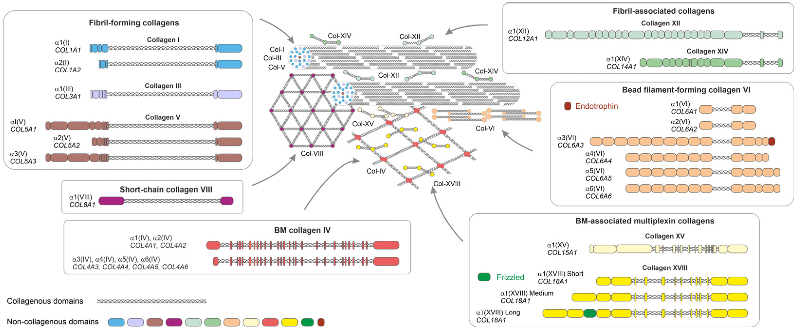
  110. Ricard-Blum, S. The Collagen Family. Cold Spring Harb. Perspect. Biol. 2011, 3, a004978. [Google Scholar] [CrossRef]
  111. Arkkila, P.E.T.; Rönnemaa, T.; Koskinen, P.J.; Kantola, I.M.; Seppänen, E.; Viikari, J.S.A. Biochemical Markers of Type III and I Collagen: Association with Retinopathy and Neuropathy in Type 1 Diabetic Subjects. Diabet. Med. 2001, 18, 816–821. [Google Scholar] [CrossRef]
  112. Gupta, R.K.; Mepani, R.J.; Kleiner, S.; Lo, J.C.; Khandekar, M.J.; Cohen, P.; Frontini, A.; Bhowmick, D.C.; Ye, L.; Cinti, S.; et al. Zfp423 Expression Identifies Committed Preadipocytes and Localizes to Adipose Endothelial and Perivascular Cells. Cell Metab. 2012, 15, 230–239. [Google Scholar] [CrossRef] [PubMed]
  113. Ishimura, E.; Nishizawa, Y.; Shoji, S.; Morii, H. Serum Type III, IV Collagens and TIMP in Patients with Type II Diabetes Mellitus. Life Sci. 1996, 58, 1331–1337. [Google Scholar] [CrossRef] [PubMed]
  114. Hirai, S.; Ohyane, C.; Kim, Y.-I.; Lin, S.; Goto, T.; Takahashi, N.; Kim, C.-S.; Kang, J.; Yu, R.; Kawada, T. Involvement of Mast Cells in Adipose Tissue Fibrosis. Am. J. Physiol. Endocrinol. Metab. 2014, 306, E247–E255. [Google Scholar] [CrossRef]
  115. Huang, G.; Ge, G.; Wang, D.; Gopalakrishnan, B.; Butz, D.H.; Colman, R.J.; Nagy, A.; Greenspan, D.S. A3(V) Collagen Is Critical for Glucose Homeostasis in Mice Due to Effects in Pancreatic Islets and Peripheral Tissues. J. Clin. Investig. 2011, 121, 769–783. [Google Scholar] [CrossRef] [PubMed]
  116. McCulloch, L.J.; Rawling, T.J.; Sjöholm, K.; Franck, N.; Dankel, S.N.; Price, E.J.; Knight, B.; Liversedge, N.H.; Mellgren, G.; Nystrom, F.; et al. COL6A3 Is Regulated by Leptin in Human Adipose Tissue and Reduced in Obesity. Endocrinology 2015, 156, 134–146. [Google Scholar] [CrossRef]
  117. Oh, J.; Kim, C.S.; Kim, M.; Jo, W.; Sung, Y.H.; Park, J. Type VI Collagen and Its Cleavage Product, Endotrophin, Cooperatively Regulate the Adipogenic and Lipolytic Capacity of Adipocytes. Metabolism 2021, 114, 154430. [Google Scholar] [CrossRef]
  118. Spencer, M.; Yao-Borengasser, A.; Unal, R.; Rasouli, N.; Gurley, C.M.; Zhu, B.; Peterson, C.A.; Kern, P.A. Adipose Tissue Macrophages in Insulin-Resistant Subjects Are Associated with Collagen VI and Fibrosis and Demonstrate Alternative Activation. Am. J. Physiol. Endocrinol. Metab. 2010, 299, E1016–E1027. [Google Scholar] [CrossRef]
  119. Dankel, S.N.; Svärd, J.; Matthä, S.; Claussnitzer, M.; Klöting, N.; Glunk, V.; Fandalyuk, Z.; Grytten, E.; Solsvik, M.H.; Nielsen, H.J.; et al. COL6A3 Expression in Adipocytes Associates with Insulin Resistance and Depends on PPARγ and Adipocyte Size. Obesity 2014, 22, 1807–1813. [Google Scholar] [CrossRef]
  120. Williams, L.M.; McCann, F.E.; Cabrita, M.A.; Layton, T.; Cribbs, A.; Knezevic, B.; Fang, H.; Knight, J.; Zhang, M.; Fischer, R.; et al. Identifying Collagen VI as a Target of Fibrotic Diseases Regulated by CREBBP/EP300. Proc. Natl. Acad. Sci. USA 2020, 117, 20753–20763. [Google Scholar] [CrossRef]
  121. Williams, L.; Layton, T.; Yang, N.; Feldmann, M.; Nanchahal, J. Collagen VI as a Driver and Disease Biomarker in Human Fibrosis. FEBS J. 2022, 289, 3603–3629. [Google Scholar] [CrossRef] [PubMed]
  122. Liu, G.; Li, M.; Xu, Y.; Wu, S.; Saeed, M.; Sun, C. ColXV Promotes Adipocyte Differentiation via Inhibiting DNA Methylation and CAMP/PKA Pathway in Mice. Oncotarget 2017, 8, 60135–60148. [Google Scholar] [CrossRef] [PubMed]
  123. Xia, T.; Shen, Z.; Cai, J.; Pan, M.; Sun, C. ColXV Aggravates Adipocyte Apoptosis by Facilitating Abnormal Extracellular Matrix Remodeling in Mice. Int. J. Mol. Sci. 2020, 21, 959. [Google Scholar] [CrossRef] [PubMed]
  124. Li, C.; Liu, Y.; Li, Y.; Tai, R.; Sun, Z.; Wu, Q.; Liu, Y.; Sun, C. Collagen XV Promotes ER Stress-Induced Inflammation through Activating Integrin Β1/FAK Signaling Pathway and M1 Macrophage Polarization in Adipose Tissue. Int. J. Mol. Sci. 2021, 22, 9997. [Google Scholar] [CrossRef]
  125. Bishop, J.R.; Passos-Bueno, M.R.; Fong, L.; Stanford, K.I.; Gonzales, J.C.; Yeh, E.; Young, S.G.; Bensadoun, A.; Witztum, J.L.; Esko, J.D.; et al. Deletion of the Basement Membrane Heparan Sulfate Proteoglycan Type XVIII Collagen Causes Hypertriglyceridemia in Mice and Humans. PLoS ONE 2010, 5, e13919. [Google Scholar] [CrossRef] [PubMed]
  126. Petäistö, T.; Vicente, D.; Mäkelä, K.A.; Finnilä, M.A.; Miinalainen, I.; Koivunen, J.; Izzi, V.; Aikio, M.; Karppinen, S.; Devarajan, R.; et al. Lack of Collagen XVIII Leads to Lipodystrophy and Perturbs Hepatic Glucose and Lipid Homeostasis. J. Physiol. 2020, 598, 3373–3393. [Google Scholar] [CrossRef]
  127. Aikio, M.; Elamaa, H.; Vicente, D.; Izzi, V.; Kaur, I.; Seppinen, L.; Speedy, H.E.; Kaminska, D.; Kuusisto, S.; Sormunen, R.; et al. Specific Collagen XVIII Isoforms Promote Adipose Tissue Accrual via Mechanisms Determining Adipocyte Number and Affect Fat Deposition. Proc. Natl. Acad. Sci. USA 2014, 111, 3043. [Google Scholar] [CrossRef]
  128. Peloso, G.M.; Auer, P.L.; Bis, J.C.; Voorman, A.; Morrison, A.C.; Stitziel, N.O.; Brody, J.A.; Khetarpal, S.A.; Crosby, J.R.; Fornage, M.; et al. Association of Low-Frequency and Rare Coding-Sequence Variants with Blood Lipids and Coronary Heart Disease in 56,000 Whites and Blacks. Am. J. Hum. Genet. 2014, 94, 223–232. [Google Scholar] [CrossRef]
  129. Errera, F.I.; Canani, L.H.; Yeh, E.; Kague, E.; Armelin-Correa, L.M.; Suzuki, O.T.; Tschiedel, B.; Silva, M.E.; Sertie, A.L.; Passos-Bueno, M.R. COL18A1 Is Highly Expressed during Human Adipocyte Differentiation and the SNP c.1136C T in Its “Frizzled” Motif Is Associated with Obesity in Diabetes Type 2 Patients. An Acad. Bras. Cienc. 2008, 80, 167–177. [Google Scholar] [CrossRef]
  130. Kaur, I.; Ruskamo, S.; Koivunen, J.; Heljasvaara, R.; Lackman, J.J.; Izzi, V.; Petaja-Repo, U.E.; Kursula, P.; Pihlajaniemi, T. The N-Terminal Domain of Unknown Function (DUF959) in Collagen XVIII Is Intrinsically Disordered and Highly O-Glycosylated. Biochem. J. 2018, 475, 3577–3593. [Google Scholar] [CrossRef]
  131. Weng, X.; Lin, D.; Huang, J.T.J.; Stimson, R.H.; Wasserman, D.H.; Kang, L. Collagen 24 α1 Is Increased in Insulin-Resistant Skeletal Muscle and Adipose Tissue. Int. J. Mol. Sci. 2020, 21, 5738. [Google Scholar] [CrossRef] [PubMed]
  132. Mori, S.; Kiuchi, S.; Ouchi, A.; Hase, T.; Murase, T. Characteristic Expression of Extracellular Matrix in Subcutaneous Adipose Tissue Development and Adipogenesis; Comparison with Visceral Adipose Tissue. Int. J. Biol. Sci. 2014, 10, 825–833. [Google Scholar] [CrossRef]
  133. Ojima, K.; Oe, M.; Nakajima, I.; Muroya, S.; Nishimura, T. Dynamics of Protein Secretion during Adipocyte Differentiation. FEBS Open Bio 2016, 6, 816–826. [Google Scholar] [CrossRef]
  134. Chun, T.H.; Hotary, K.B.; Sabeh, F.; Saltiel, A.R.; Allen, E.D.; Weiss, S.J. A Pericellular Collagenase Directs the 3-Dimensional Development of White Adipose Tissue. Cell 2006, 125, 577–591. [Google Scholar] [CrossRef] [PubMed]
  135. Huber, J.; Löffler, M.; Bilban, M.; Reimers, M.; Kadl, A.; Todoric, J.; Zeyda, M.; Geyeregger, R.; Schreiner, M.; Weichhart, T.; et al. Prevention of High-Fat Diet-Induced Adipose Tissue Remodeling in Obese Diabetic Mice by n-3 Polyunsaturated Fatty Acids. Int. J. Obes. 2007, 31, 1004–1013. [Google Scholar] [CrossRef]
  136. Liu, X.; Xu, Q.; Liu, W.; Yao, G.; Zhao, Y.; Xu, F.; Hayashi, T.; Fujisaki, H.; Hattori, S.; Tashiro, S.; et al. Enhanced Migration of Murine Fibroblast-like 3T3-L1 Preadipocytes on Type I Collagen-Coated Dish Is Reversed by Silibinin Treatment. Mol. Cell. Biochem. 2018, 441, 35–62. [Google Scholar] [CrossRef] [PubMed]
  137. Xu, Q.; Liu, X.; Liu, W.; Hayashi, T.; Yamato, M.; Fujisaki, H.; Hattori, S.; Tashiro, S.; Onodera, S.; Ikejima, T. Type I Collagen-Induced YAP Nuclear Expression Promotes Primary Cilia Growth and Contributes to Cell Migration in Confluent Mouse Embryo Fibroblast 3T3-L1 Cells. Mol. Cell. Biochem. 2019, 450, 87–96. [Google Scholar] [CrossRef]
  138. Liu, X.; Long, X.; Gao, Y.; Liu, W.; Hayashi, T.; Mizuno, K.; Hattori, S.; Fujisaki, H.; Ogura, T.; Onodera, S.; et al. Type I Collagen Inhibits Adipogenic Differentiation via YAP Activation in Vitro. J. Cell. Physiol. 2020, 235, 1821–1837. [Google Scholar] [CrossRef]
  139. Gusinjac, A.; Gagnon, A.; Sorisky, A. Effect of Collagen I and Aortic Carboxypeptidase-like Protein on 3T3-L1 Adipocyte Differentiation. Metabolism 2011, 60, 782–788. [Google Scholar] [CrossRef]
  140. Clemente-Postigo, M.; Tinahones, A.; El Bekay, R.; Malagón, M.M.; Tinahones, F.J. The Role of Autophagy in White Adipose Tissue Function: Implications for Metabolic Health. Metabolites 2020, 10, 179. [Google Scholar] [CrossRef] [PubMed]
  141. Gao, Y.; Ma, K.; Kang, Y.; Liu, W.; Liu, X.; Long, X.; Hayashi, T.; Hattori, S.; Mizuno, K.; Fujisaki, H.; et al. Type I Collagen Reduces Lipid Accumulation during Adipogenesis of Preadipocytes 3T3-L1 via the YAP-MTOR-Autophagy Axis. Biochim. Biophys. Acta Mol. Cell Biol. Lipids 2022, 1867, 159181. [Google Scholar] [CrossRef]
  142. Al Hasan, M.; Martin, P.E.; Shu, X.; Patterson, S.; Bartholomew, C. Type III Collagen Is Required for Adipogenesis and Actin Stress Fibre Formation in 3T3-L1 Preadipocytes. Biomolecules 2021, 11, 156. [Google Scholar] [CrossRef] [PubMed]
  143. de Winter, T.J.J.; Nusse, R. Running Against the Wnt: How Wnt/β-Catenin Suppresses Adipogenesis. Front. Cell Dev. Biol. 2021, 9, 627429. [Google Scholar] [CrossRef] [PubMed]
  144. Aratani, Y.; Kitagawa, Y. Enhanced Synthesis and Secretion of Type IV Collagen and Entactin during Adipose Conversion of 3T3-L1 Cells and Production of Unorthodox Laminin Complex. J. Biol. Chem. 1988, 263, 16163–16169. [Google Scholar] [CrossRef]
  145. Tajima, R.; Kawaguchi, N.; Horino, Y.; Takahashi, Y.; Toriyama, K.; Inou, K.; Torii, S.; Kitagawa, Y. Hypoxic Enhancement of Type IV Collagen Secretion Accelerates Adipose Conversion of 3T3-L1 Fibroblasts. Biochim. Biophys. Acta Mol. Cell Res. 2001, 1540, 179–187. [Google Scholar] [CrossRef] [PubMed]
  146. Mak, K.M.; Png, C.Y.M.; Lee, D.J. Type V Collagen in Health, Disease, and Fibrosis. Anat. Rec. 2016, 299, 613–629. [Google Scholar] [CrossRef]
  147. Nakajima, I.; Muroya, S.; Tanabe, R.-I.; Chikuni, K. Extracellular Matrix Development during Differentiation into Adipocytes with a Unique Increase in Type V and VI Collagen. Biol. Cell 2002, 94, 197–203. [Google Scholar] [CrossRef]
  148. Wenstrup, R.J.; Florer, J.B.; Brunskill, E.W.; Bell, S.M.; Chervoneva, I.; Birk, D.E. Type V Collagen Controls the Initiation of Collagen Fibril Assembly. J. Biol. Chem. 2004, 279, 53331–53337. [Google Scholar] [CrossRef]
  149. Park, A.C.; Phan, N.; Massoudi, D.; Liu, Z.; Kernien, J.F.; Adams, S.M.; Davidson, J.M.; Birk, D.E.; Liu, B.; Greenspan, D.S. Deficits in Col5a2 Expression Result in Novel Skin and Adipose Abnormalities and Predisposition to Aortic Aneurysms and Dissections. Am. J. Pathol. 2017, 187, 2300–2311. [Google Scholar] [CrossRef]
  150. Imamura, Y.; Scott, I.C.; Greenspan, D.S. The Pro-A3(V) Collagen Chain: Complete Primary Structure, Expression Domains in Adult and Developing Tissues, and Comparison to the Structures and Expression Domains of the Other Types V and XI Procollagen Chains. J. Biol. Chem. 2000, 275, 8749–8759. [Google Scholar] [CrossRef]
  151. Cescon, M.; Gattazzo, F.; Chen, P.; Bonaldo, P. Collagen VI at a Glance. J. Cell Sci. 2015, 128, 3525–3531. [Google Scholar] [CrossRef]
  152. Divoux, A.; Clément, K. Architecture and the Extracellular Matrix: The Still Unappreciated Components of the Adipose Tissue. Obes. Rev. 2011, 12, e494–e503. [Google Scholar] [CrossRef]
  153. Pasarica, M.; Gowronska-Kozak, B.; Burk, D.; Remedios, I.; Hymel, D.; Gimble, J.; Ravussin, E.; Bray, G.A.; Smith, S.R. Adipose Tissue Collagen VI in Obesity. J. Clin. Endocrinol. Metab. 2009, 94, 5155–5162. [Google Scholar] [CrossRef]
  154. Zhao, Y.; Gu, X.; Zhang, N.; Kolonin, M.G.; An, Z.; Sun, K. Divergent Functions of Endotrophin on Different Cell Populations in Adipose Tissue. Am. J. Physiol. Endocrinol. Metab. 2016, 311, E952–E963. [Google Scholar] [CrossRef]
  155. Sun, K.; Park, J.; Gupta, O.T.; Holland, W.L.; Auerbach, P.; Zhang, N.; Marangoni, R.G.; Nicoloro, S.M.; Czech, M.P.; Varga, J.; et al. Endotrophin Triggers Adipose Tissue Fibrosis and Metabolic Dysfunction. Nat. Commun. 2014, 5, 3485. [Google Scholar] [CrossRef]
  156. Staunstrup, L.M.; Bager, C.L.; Frederiksen, P.; Helge, J.W.; Brunak, S.; Christiansen, C.; Karsdal, M. Endotrophin Is Associated with Chronic Multimorbidity and All-Cause Mortality in a Cohort of Elderly Women. EBioMedicine 2021, 68, 103391. [Google Scholar] [CrossRef] [PubMed]
  157. Yoldemir, S.A.; Arman, Y.; Akarsu, M.; Altun, O.; Ozcan, M.; Tukek, T. Correlation of Glycemic Regulation and Endotrophin in Patients with Type 2 Diabetes; Pilot Study. Diabetol. Metab. Syndr. 2021, 13, 9. [Google Scholar] [CrossRef] [PubMed]
  158. Bretaud, S.; Guillon, E.; Karppinen, S.-M.; Pihlajaniemi, T.; Ruggiero, F. Collagen XV, a Multifaceted Multiplexin Present across Tissues and Species. Matrix Biol. Plus 2020, 6–7, 100023. [Google Scholar] [CrossRef]
  159. Heljasvaara, R.; Aikio, M.; Ruotsalainen, H.; Pihlajaniemi, T. Collagen XVIII in Tissue Homeostasis and Dysregulation—Lessons Learned from Model Organisms and Human Patients. Matrix Biol. 2017, 57–58, 55–75. [Google Scholar] [CrossRef] [PubMed]
  160. North, K.E.; Miller, M.B.; Coon, H.; Martin, L.J.; Peacock, J.M.; Arnett, D.; Zhang, B.; Province, M.; Oberman, A.; Blangero, J.; et al. Evidence for a Gene Influencing Fasting LDL Cholesterol and Triglyceride Levels on Chromosome 21q. Atherosclerosis 2005, 179, 119–125. [Google Scholar] [CrossRef] [PubMed]
  161. Gordts, P.L.S.M.; Esko, J.D. The Heparan Sulfate Proteoglycan Grip on Hyperlipidemia and Atherosclerosis. Matrix Biol. 2018, 71–72, 262–282. [Google Scholar] [CrossRef] [PubMed]
  162. Bartelt, A.; John, C.; Schaltenberg, N.; Berbée, J.F.P.; Worthmann, A.; Cherradi, M.L.; Schlein, C.; Piepenburg, J.; Boon, M.R.; Rinninger, F.; et al. Thermogenic Adipocytes Promote HDL Turnover and Reverse Cholesterol Transport. Nat. Commun. 2017, 8, 15010. [Google Scholar] [CrossRef]
  163. Berbée, J.F.P.; Boon, M.R.; Khedoe, P.P.S.J.; Bartelt, A.; Schlein, C.; Worthmann, A.; Kooijman, S.; Hoeke, G.; Mol, I.M.; John, C.; et al. Brown Fat Activation Reduces Hypercholesterolaemia and Protects from Atherosclerosis Development. Nat. Commun. 2015, 6, 6356. [Google Scholar] [CrossRef]
  164. Shuttleworth, C.A. Type VIII Collagen. Int. J. Biochem. Cell Biol. 1997, 29, 1145–1148. [Google Scholar] [CrossRef]
  165. Ullah, M.; Sittinger, M.; Ringe, J. Extracellular Matrix of Adipogenically Differentiated Mesenchymal Stem Cells Reveals a Network of Collagen Filaments, Mostly Interwoven by Hexagonal Structural Units. Matrix Biol. 2013, 32, 452–465. [Google Scholar] [CrossRef] [PubMed]
  166. Ruehl, M.; Erben, U.; Schuppan, D.; Wagner, C.; Zeller, A.; Freise, C.; Al-Hasani, H.; Loesekann, M.; Notter, M.; Wittig, B.M.; et al. The Elongated First Fibronectin Type III Domain of Collagen XIV Is an Inducer of Quiescence and Differentiation in Fibroblasts and Preadipocytes. J. Biol. Chem. 2005, 280, 38537–38543. [Google Scholar] [CrossRef] [PubMed]
Figure 1. Structure and supramolecular assembly of the collagen types reported to contribute to AT physiology and pathology. Domain organizations of collagen α chains of different subfamilies are depicted in boxes. Collagens I, III, and V form collagen fibrils. Collagens XII and XIV bind to the fibrils and regulate their organization. Collagen XVIII binds to collagen IV network in the BM. Collagen XV resides at the interphase of the BM and collagen fibrils. Collagen IV forms beaded filaments at the fibrillar matrix-BM interphase. Collagen VIII forms hexagonal lattices at the BM. Triple helix, collagenous domains. Non-collagenous domains of different collagens are shown with different colours. Endotrophin domain of collagen VI and frizzled domain of collagen XVIII are indicated. The sizes of collagens and their assemblies are not presented at their correct scale. Post-translational modifications such as glycosaminoglycan chains in collagens XII, XIV, XV, and XVIII are not illustrated.
Biomedicines 11 01412 g001
Figure 2. Key collagens associated with white adipocyte differentiation. During the adipogenic differentiation of adipocyte stem cells to mature adipocytes, the expression of collagens shifts from the fibrillar collagens I and III to the BM-associated collagens IV, VI, XV, and XVIII. This view is largely based on the in vitro model of murine 3T3-L1 preadipocytes, as discussed in the main text. The gray boxes present some common cell markers expressed at different stages of adipocyte differentiation. Abbreviations: αSMA—α smooth muscle actin; BM—basement membrane; C/EBP—CCAAT/enhancer-binding proteins; Col—collagen; GLUT4—glucose transporter 4; PDGFR—platelet-derived growth factor receptor; PPARγ—peroxisome proliferation-activated receptor γ; ZFP423—Zinc-finger protein 423.
Biomedicines 11 01412 g002
Figure 3. Summary of collagens in AT dysfunction. (A) Upregulated collagens (arrows) in the WAT of obese humans. (B) Upregulated collagens (arrows) in the WAT of mice fed with a high-fat diet (HFD), or in obese ob/ob or diabetic db/db mouse models, or in the Hif1a+ mice with constitutively active HIF-1α. (C) Left: Key collagens forming large collagen bundles (blue) in hypoxic (pink) and fibrotic AT. Collagens which are reported to be associated with insulin resistance (right upper corner), or with AT inflammation (bottom, a macrophage depicted).
Biomedicines 11 01412 g003
Table 1. Expression of selected collagens in dysregulated human and mouse adipose tissue (AT).
CollagenPathological ConditionExpression/ManifestationReferences
IFibrotic AT
T1D
Increased expression in obese AT compared to lean subjects
Decreased level of crosslinked telopeptide in the serum of T1D patients with retinopathy
[34,37,111,112]
IIT2DIncreased expression in epididymal AT in diabetic (db/db) mice[34]
IIIFibrotic AT
T1D
T2D
Increased expression in obese AT compared to lean subjects and in patients with T1D with retinopathy
Increased levels of procollagen aminopeptide in patients with T2D and progressing diabetic nephropathy
[37,111,113]
IVT2DIncreased Col4a1 and Col4a2 expression in the WAT of diabetic mice
Downregulation of COL4A1 in SAT after gastric bypass and improvement of HOMA-IR
[34,107]
VFibrotic AT
Impaired glucose metabolism
Insulin resistance
Increased expression in the WAT of diabetic mice
Increased expression in obesity; accumulation in fibrotic areas, especially around large blood vessels.
Fibrotic promotion causes insulin resistance
Lack of Col5a3 leads to impaired glucose metabolism
[34,38,114,115]
VI Fibrotic AT
Altered glucose metabolism
Increased expression in obese patients associates with pericellular fibrosis
Biomarker in AT fibrosis
Conflicting results in glucose metabolism; insulin resistance vs. improved glucose metabolism
Increased expression in obese/diabetic mice while downregulated in obese humans
[34,37,116,117,118,119,120,121]
VIIIObesityIn the twins study, an increased expression of COL8A2 was found in the heavier twin[47]
XIIInsulin resistanceIn the twins study, COL12A1 expression positively associated with LDL cholesterol, and low expression associated with increased insulin sensitivity[47]
XVObesityIncreased expression in AT in HFD-induced obesity in mice
Regulates adipocyte apoptosis and inflammation in AT
[122,123,124]
XVIIIVisceral obesity in
T2D
Dyslipidemia
Lipodystrophy
Specific SNPs associate with obesity in patients with T2D (c.1136C > T) and with abnormal circulating lipid content (c.331G > A, p.Gly111Arg)
Patients with Knobloch syndrome due to COL18A1 null mutation have fasting hypertriglyceridemia
Lack of Col18a1 in mice causes lipodystrophy, T2D, and increased serum triglyceride levels
Expression of long isoforms of collagen XVIII in visceral fat positively correlates with free fatty acid levels in the plasma
[125,126,127,128,129,130]
XXIVT2DIncreased expression in insulin-resistant obese VAT[131]
Abbreviations: AT—adipose tissue; HFD—high-fat diet; HOMA-IR—homeostatic model assessment for insulin resistance; LDL—low-density lipoprotein; SAT—subcutaneous adipose tissue; T1D/T2D—type 1/2 diabetes; WAT—white AT.
Disclaimer/Publisher’s Note: The statements, opinions and data contained in all publications are solely those of the individual author(s) and contributor(s) and not of MDPI and/or the editor(s). MDPI and/or the editor(s) disclaim responsibility for any injury to people or property resulting from any ideas, methods, instructions or products referred to in the content.

