Review

Epidural and Intrathecal Drug Delivery in Rats and Mice for Experimental Research: Fundamental Concepts, Techniques, Precaution, and Application

1
Gachon Pain Center and Department of Physiology, Gachon University College of Medicine, Incheon 21999, Republic of Korea
2
Department of Anesthesiology and Pain Medicine, Gachon University, Gil Medical Center, Incheon 21565, Republic of Korea
*
Authors to whom correspondence should be addressed.
These authors contributed equally to this work.
Biomedicines 2023, 11(5), 1413; https://doi.org/10.3390/biomedicines11051413
Submission received: 15 April 2023 / Revised: 4 May 2023 / Accepted: 7 May 2023 / Published: 10 May 2023
(This article belongs to the Special Issue Neuropathic Pain: From Mechanisms to Therapeutic Approaches)

Abstract

:
Epidural and intrathecal routes are the most effective drug administration methods for pain management in clinical and experimental medicine to achieve quick results, reduce required drug dosages, and overcome the adverse effects associated with the oral and parenteral routes. Beyond pain management with analgesics, the intrathecal route is more widely used for stem cell therapy, gene therapy, insulin delivery, protein therapy, and drug therapy with agonist, antagonist, or antibiotic drugs in experimental medicine. However, clear information regarding intrathecal and epidural drug delivery in rats and mice is lacking, despite differences from human medicine in terms of anatomical space and proximity to the route of entry. In this study, we discussed and compared the anatomical locations of the epidural and intrathecal spaces, cerebrospinal fluid volume, dorsal root ganglion, techniques and challenges of epidural and intrathecal injections, dosage and volume of drugs, needle and catheter sizes, and the purpose and applications of these two routes in different disease models in rats and mice. We also described intrathecal injection in relation to the dorsal root ganglion. The accumulated information about the epidural and intrathecal delivery routes could contribute to better safety, quality, and reliability in experimental research.

1. Introduction

Delivering a therapeutic agent to the central nervous system (CNS) is challenging because of the blood–brain barrier and blood–spinal cord barrier of the CNS, which consist of endothelial cells possessing tight junctions and efflux pumps [1]. Thus, the administration of drugs via parenteral routes ultimately results in low concentrations of drugs in the CNS because of the blood–brain and blood–spinal cord barriers. To overcome these par-enteral delivery challenges, intraspinal drug delivery systems are one of the most common approaches used. The epidural [2,3,4] and intrathecal routes [5,6,7] are the most used intraspinal drug delivery routes in rats and mice. The target of each drug delivery route is the cerebrospinal fluid (CSF). With an intrathecal injection, a drug is delivered directly into the CSF within the intrathecal space of the spinal column [1]. Intrathecally administered drugs are restricted within the CSF circulating in the spinal column and brain ventricles. However, drugs delivered to the epidural space cross the dura membrane to reach the CSF. Thus, epidurally administered drugs can also reach systemic circulation. These two routes are compared in Table 1.
Intraspinal drug delivery through an epidural or intrathecal injection is comparatively more challenging in small experimental animals such as rats and mice than in large animals and humans because of the small size of the rat and mouse spinal column, spinal canal, epidural space, and intrathecal or subarachnoid space, and the small volume of CSF. Substantial experimental research and a review of the literature in human medicine have been reported [1,8,9,10]. Numerous research studies have used the epidural [4,11,12] and intrathecal routes [13,14,15] in mice and rats; however, to the best of our knowledge, no review of the literature has discussed the delivery process, challenges, and application of these routes in rats and mice.
Furthermore, the intraspinal intrathecal space [16,17] and intracerebroventricular route through the brain parenchyma into the lateral ventricles of the brain have been used to directly access the CSF [1]. However, the intraspinal/intrathecal route is broadly used because it is less invasive and is suitable for chronic use with surgical catheterization [13,18] and for direct single-dose injections via needle puncturing [6,17,19].
The objectives of this comparative study were to discuss the anatomical locations of the epidural and intrathecal spaces, CSF volume, techniques and challenges of epidural and intrathecal injection, dosage and volume of drugs, and the purpose and applications of these two routes in different disease models in rats and mice. We further describe the sites of injection, needle and catheter types and size, injection volume, concentration, and purpose and indications of epidural and intrathecal injections in experimental research in mice and rats, as well as how intrathecal injection can be effective for therapeutic agents targeting the dorsal root ganglion (DRG).

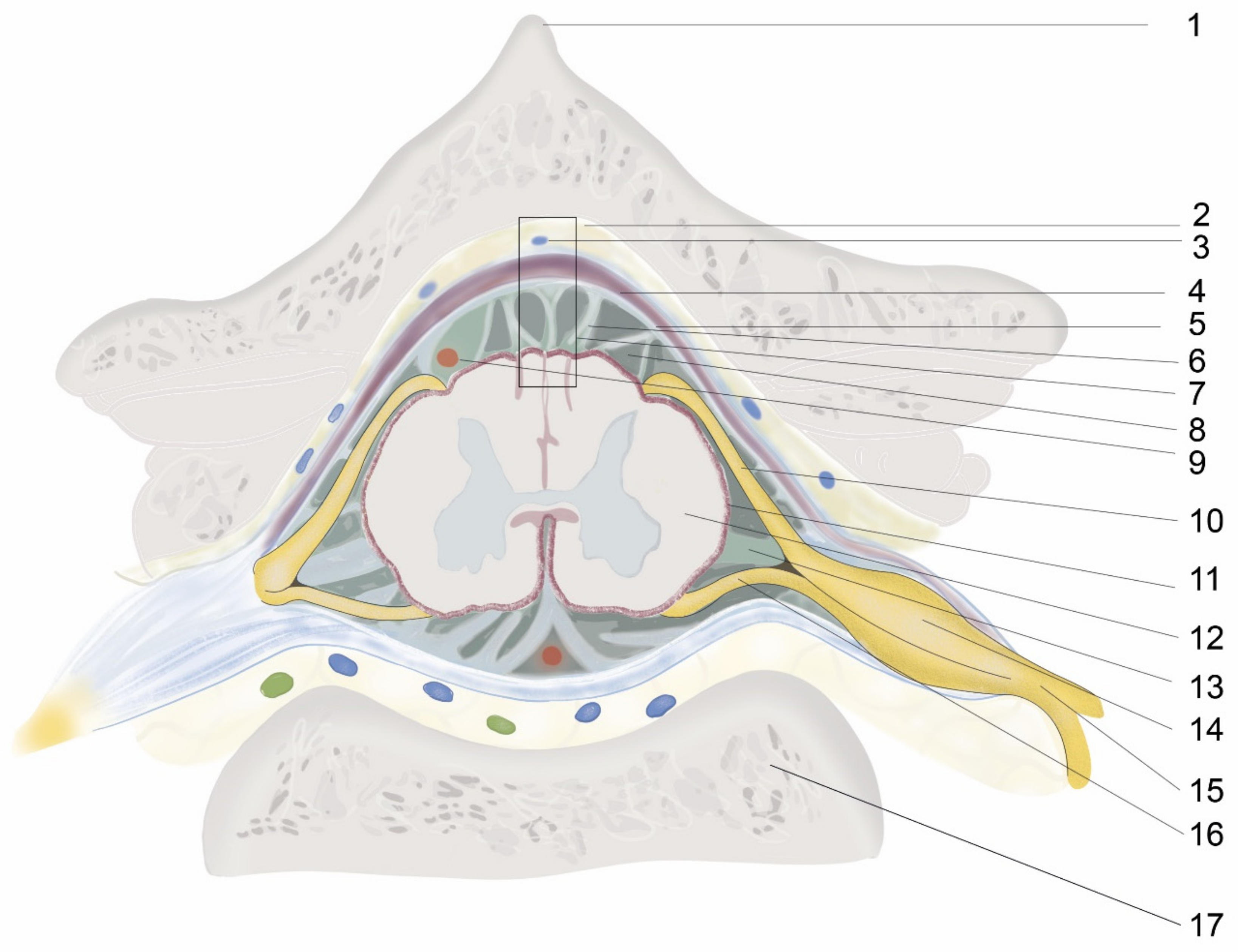
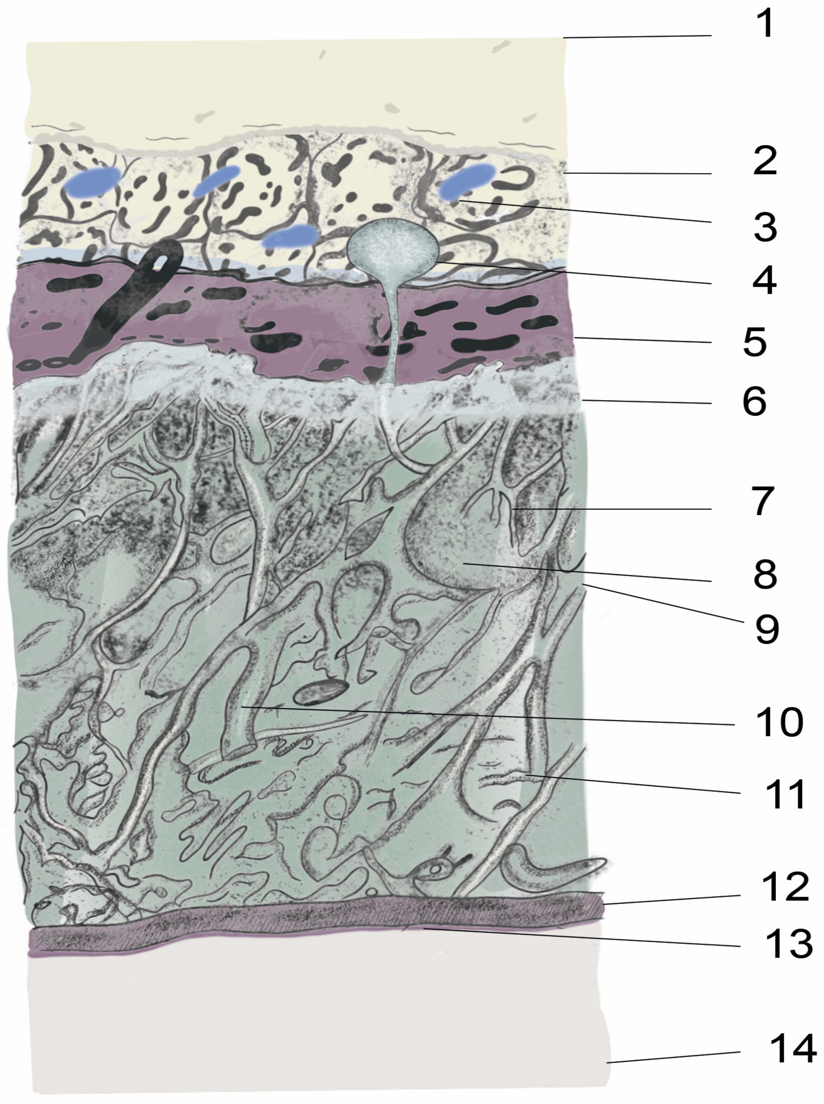
2. Anatomy and Physiology of the Spinal Meninges

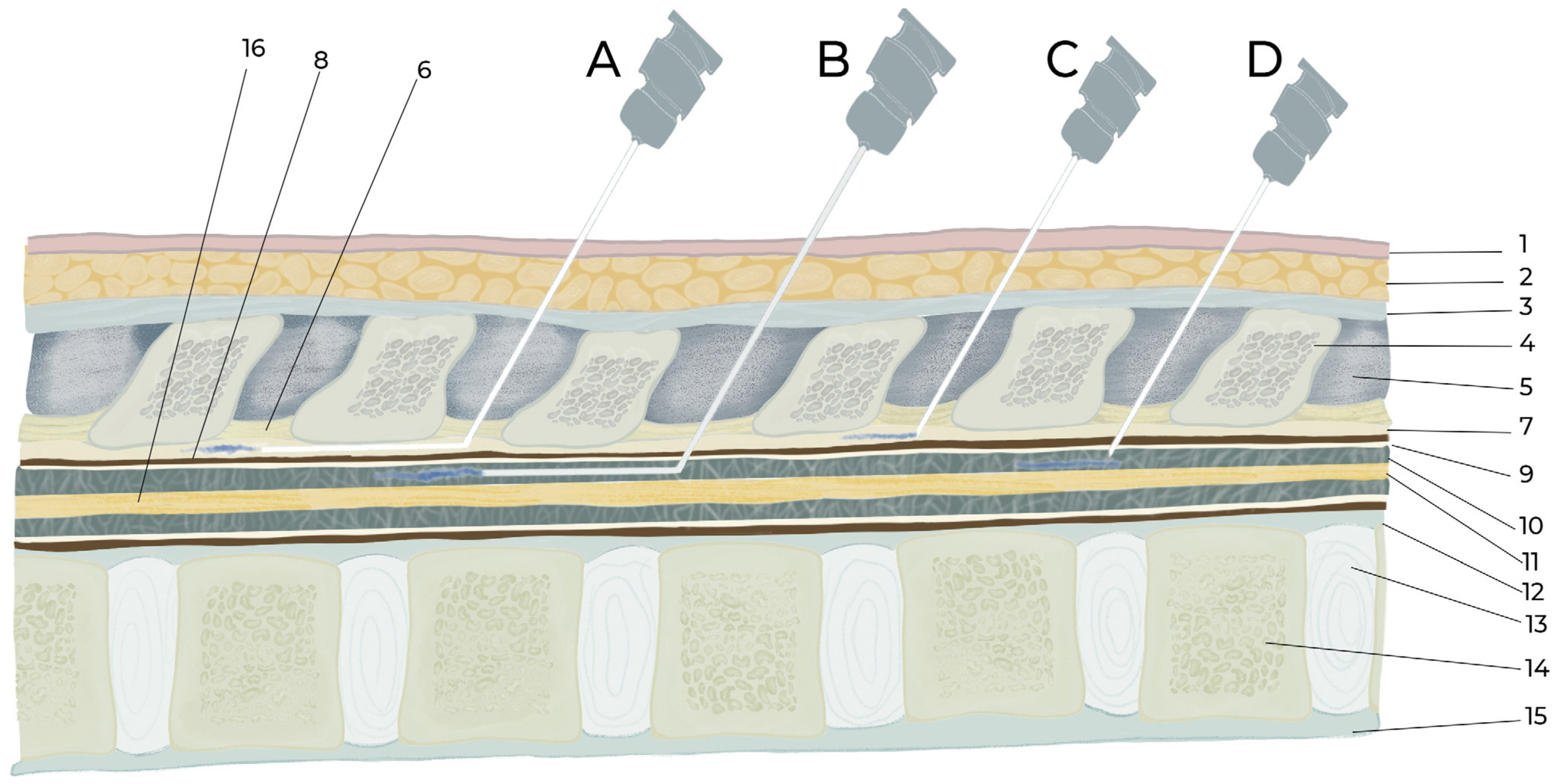
2.1. Organs, Tissues from Skin to Spinal Cord

The intrathecal or epidural administration of a therapeutic agent requires an in-depth knowledge of the structures from the skin to the spinal cord and the vertebral column anatomy, as well as considerable technical skill. For intraspinal drug delivery, the most important structure is the meninges, whose function is to hold and maintain the CSF surrounding the brain and spinal cord, thereby protecting and regulating their function. The meninges are the outer coverings of the brain and spinal cord, called the cranial meninges and spinal meninges, respectively, which separate the brain and spinal cord from the adjacent bony structures of the skull and vertebral column, respectively. The spinal and cranial meninges are continuous with each other in that the unique structure of both consists of the same three meningeal layers (from superficial to deep): the dura mater, arachnoid mater, and pia mater (Figure 1 and Figure 2). In addition, the upper parts of the spinal canal in mice, rats, and other tetrapods are formed by the bony part, vertebral arch (inside a single vertebra), and ligament flavum (i.e., intervertebral space; Figure 3). The ligament flavum is important for intraspinal drug delivery, especially for epidural and intrathecal injections. The structures from the superficial to deep regions are the skin, subcutaneous fat, muscle, supraspinous ligament, interspinous ligament, ligament flavum, epidural space, spinal meninges (dura mater, arachnoid mater, and pia mater), and spinal cord (Figure 3).

2.2. Epidural Space

The term “epidural” is composed of the prefix “epi-”, which means “up” or “above”, and “dural”, which refers to the dura mater layer of the meninges. Thus, an epidural is the upper part of the dura mater. The epidural space is the space between the outer layer of the meninges (i.e., dura mater) and the inner periosteum or ligament flavum of the vertebral column [20]. Mice, rats, and other tetrapods have two adjacent upper parts of the epidural space: the first is the bony part (i.e., the vertebral arch (inside a single vertebra)), and the second is the connective tissue part (i.e., ligament flavum (intervertebral space)). It is filled with loose connective tissue, epidural fats, veins, arteries, internal vertebral venous plexuses, spinal nerve roots, and lymphatics [21] (Figure 1, Figure 2 and Figure 3).

2.3. Dura mater

The dura mater is the outermost of the three layers of the meninges and lies between the epidural space and the arachnoid mater. It consists of irregularly oriented collagen fiber bundles that accommodate blood vessels and through which nerve fibers and nerve fascicles pass [22].

2.4. Subdural Space

The term “subdural” is composed of the prefix “sub-”, which means below”, and “dural”, which refers to the dura mater layer of the meninges. Thus, the subdural space is the lower part of the dura mater or the space between the inner layer of the dura mater and the upper surface of the arachnoid mater. This very thin layer contains a thin film of fluid. The application of drugs to the subdural space in human medicine has been reported [23], but this application in experimental medicine in mice and rats is difficult owing to the layer’s negligible thickness. However, this space is important for experimental cranial subdural hemorrhage [24].

2.5. Arachnoid Mater

The origin of the word “arachnoid” is the Greek word “Arachne”, meaning “spider”, because of the web-like collagenous fibroblast structure of the arachnoid membranes, trabeculae, and septae that cross the subarachnoid space from the arachnoid mater to the pia mater [25]. The arachnoid mater underlying the dura mater comprises the outermost arachnoid barrier cell layer, which includes tightly packed cells, numerous tight junctions, and no extracellular collagen but an innermost collagenous portion. Owing to its numerous tight junctions, this layer is a physiological and morphological barrier between the subarachnoid CSF and blood circulation in the dura [26]. This layer has two parts: the basal arachnoid mater and the interweaving arachnoid trabeculae. The arachnoid trabeculae are rich in mitochondria and originate from the inner surface of the arachnoid mater to the pia mater, thereby contributing to the subarachnoid space, which contains CSF [26]. In addition, the arachnoid matter protrudes into or through the dura mater into the epidural space. This protrusion is defined as arachnoid villi or granulations (Figure 2), and it produces CSF. The vascular and lymphatic interface area has a role in CSF drainage [27]. Most arachnoid villi originate near the nerve root area and provide expansions, such as the lumbar cistern, in the lower lumbar spinal cord [28].

2.6. Subarachnoid Space/Intrathecal Space

As with the word “subdural”, the term “subarachnoid” is composed of the prefix “sub-”, meaning “below”, and “arachnoid”, referring to the arachnoid mater layer of the meninges. Thus, the subarachnoid space is the space between the arachnoid and pia mater, and it is more commonly called the “intrathecal space.” The prefix “intra-” means “inside”, and “thecal”, which originated from the Greek word theke, means “a hard outer covering” and refers to the arachnoid mater. The term “intrathecal” literally means “in-side the arachnoid mater”. This space contains CSF, numerous arachnoid trabeculae, col-lagen fibrils, and major blood vessels (i.e., veins and arteries) that cross the CNS to the epidural space. The subarachnoid spaces of the cranium and vertebral column are connected to each other and form a single closed route for CSF circulation [27,28]. This space is mostly used for intraspinal drug delivery in mice and rats via intrathecal injection and catheterization.

2.7. Pia Mater

The pia mater is the innermost layer of the meninges that directly envelops the brain and spinal cord. The pia mater consists of flattened thin fibroblast tissues, collagen micro-fibrils, and capillaries containing a vascular plexus for the spinal cord tissue. This layer is separated from the neural tissue of the spinal cord by loose collagen fibers and the glia limitans. The single-cell layer thickness and fenestration of the basal lamina in many areas of the pia expose the glia limitans directly to the subarachnoid space. Pial and arachnoid cells also envelop the nerves, collagen bundles, and blood vessels that are within or cross the subarachnoid space and those around arteries that penetrate the CNS [29]. The spinal pia mater also has ligamentous lateral projections, called denticulate ligaments, to both sides of the spinal cord. These emerge through the arachnoid and attach to the spinal dura mater. Each pair of denticulate ligaments is located between pairs of spinal nerves, and they fix and hold the spinal cord in place [30].

2.8. Cerebrospinal Fluid Production, Volume, and Circulation

2.8.1. CSF Production and Volume

Clear knowledge about CSF production, circulation, absorption, and total volume is necessary before intraspinal drug delivery because an extra volume of any drug may change the volume and pressure of the CSF [27; 31]. CSF is a colorless fluid that is secreted by the choroid plexus into the ventricular cavities (80%) and by other structures, such as the brain parenchyma (20%) [31]. Compared to blood plasma, CSF contains a higher concentration of sodium, chloride, and magnesium ions and a lower concentration of glucose; proteins; amino acids; uric acid; and calcium, phosphate, and potassium ions [32]. The rate of CSF production and the total CSF volume vary across species (Table 2), but CSF circulates similarly inside the ventricular system and the cranial and spinal subarachnoid spaces. CSF lubricates, protects, and serves as a natural cushion for the brain and spinal cord, constantly providing ions, nutrients, hormones, and neurotransmitters and removing metabolic waste products.

2.8.2. CSF Circulation

In brief, after CSF is produced by the choroid plexus of the lateral ventricles and the parenchyma of the brain, it moves to the third ventricle and then to the fourth ventricle. From the fourth ventricle, CSF enters the subarachnoid space, basal cisterns, and the cisterna magna. In the cisterna magna, CSF flows along three routes: anteriorly to the pre-medullary prepontine cisterns and cerebellopontine cisterns, superiorly to the hemispheric subarachnoid space, and inferiorly to the spinal subarachnoid space [25,31,33]. It circulates through the single, closed cranial, and spinal subarachnoid space. Finally, the CSF is absorbed into the dural venous sinuses by the cranial and spinal subarachnoid villi and granulations via diffusion, and osmotic pressure plays a role in the filtration process. The circulation of CSF is believed to occur by several actions, such as continuous production and absorption of CSF by ciliary action applied by the ventricular ependyma for different pressure gradients of the arachnoid villi. CSF circulation is also influenced by brain movement, the cardiac cycle, and arterial blood flow [25,34].

3. Dorsal Root Ganglion (DRG)

In general, there are 31 pairs of DRGs in mice and rats on both sides, including 8 cervical, 13 thoracic, 6 lumbar, and 4 sacral pairs [35]. DRG refers to the dorsal root of the spinal nerve, which emerges through the intervertebral neural foramen and expands to form a ganglion, called the DRG. The DRG is close to the spinal cord, located just before the dorsal and ventral spinal roots unify to form a single spinal nerve. The DRG is composed of sensory neurons, satellite glia, endothelial cells, and macrophages [36]. It is highly vascularized and contains capillary tissue supplied by the branches of the segmental arteries. In particular, the DRG has high blood vessel permeability for unique endothelial cells, allowing the biodistribution and absorption of systemic drugs, which are limited in other parts of the nervous system [36]. This characteristic of the DRG has clinical and preclinical significance for systemic coverage. However, the systemic approach is not typical because it exposes multiple tissues often related to serious adverse effects. Intra-DRG injection is widely used in humans and large animals, but it is challenging to administer in rats and mice because of their small size. The intrathecal injection is frequently used by targeting the DRG, although controversy exists regarding whether the DRG has direct contact with the CSF. An interesting study in human cadavers demonstrated that an intra-DRG injection of green ink frequently reached the spinal cord [37], indicating the DRG’s connection with the CSF and spinal cord. Intra-DRG injection of toluidine blue also consistently stains the ipsilateral CSF and spinal cord in rats [38]. Chang et al. [36] also found that an intraspinal nerve injection stained the ipsilateral spinal cord, thereby indicating that nerve tissue can convey drugs even if it has no direct contact with the CSF. Furthermore, Chang et al. [38] also demonstrated that an intrathecal injection stained the DRG and the spinal nerve. Another study [39] reconfirmed that intrathecal injection of methylene blue in rats also stained the DRG and spinal nerves. CSF and subarachnoid space reaching half of the DRG length has also been demonstrated in rats [39] and in some length of the DRG in humans [40]. Therefore, the intrathecal route can be a good choice for targeting the DRG in rats and mice.

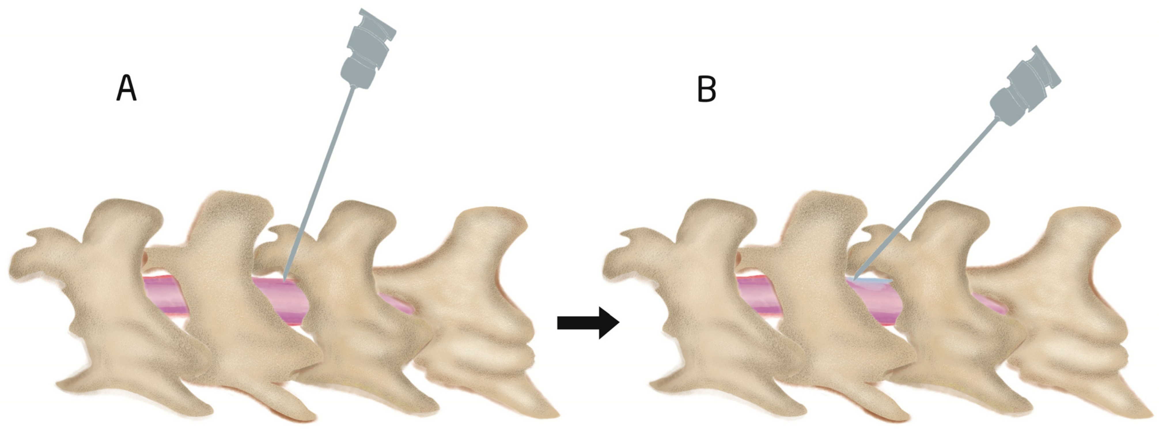
4. Epidural and Intrathecal Injection Procedures

Epidural and intrathecal injections can be performed via acute needle puncture or catheterization. Acute needle puncture is usually used for single drug administration but can also be used for multiple doses [5], and intervals of 24 h, 2 days, and 7 days have been reported (Table 3). Acute needle puncture can be performed in anesthetized [41] or unanesthetized animals [5,42]; however, intrathecal injections in unanesthetized animals require greater skills. The intervertebral space of the spinal column is commonly used, but the lumbar region [41,43], especially L5–L6, is most commonly used for single acute injection [16,44]. However, multiple injections were performed through a single location, e.g., L5-L6 [5,6], L4-L5 [45], and top of the foramen [46] (Table 3). Proper needle insertion to the appropriate location and an accurate volume and concentration of the injectate, based on the species, are critical for the success and the recovery of animals; these are briefly discussed in Table 3, Table 4, Table 5 and Table 6. For an effective IT injection via acute puncture, only one try is required; however, if the first effort fails, the needle should be removed, and a second attempt may be made. If the second effort fails, a different intervertebral space should be chosen [15]. In addition, shaving and aseptic preparation of the skin, needles, and administering agents are also important for success.
The exact positioning of a needle to the epidural space is difficult noninvasively and requires high skill because the distance between the epidural and intrathecal spaces is very small in rats and mice. Therefore, the applications of direct epidural injections using needle puncture in mice and rats are limited because the needle can easily penetrate the dura mater and enter the intrathecal space. Therefore, intrathecal injection by acute needle puncture [5,6,17], intrathecal catheterization [11,13,14], and epidural catheterization [2,3,4] but not epidural injection by needle puncture is frequently used in mice and rats.

4.1. Procedure of Intrathecal Injection by Acute Needle Puncturing

After administering anesthesia (e.g., volatile isoflurane), the thoracolumbar region is shaved. Needles are inserted through the intervertebral space where bony parts are absent, which is covered by the ligamentum flavum [20]. The angle of needle insertion varies but has been reported at over 20° [42,47,48,49], 45° [50,51], at 45° shown in videos but reported to be 70–80° in the discussion section [16], and at 70–80° shown in videos [17] in mice; an angle of 15–30° has been reported in rats [51]. Therefore, it is difficult to conclude what the optimal injection angle is (Figure 3 and Figure 4). However, the authors of the current review also insert needles at angles of 70–80° and then reduce the angle to 30–45° during drug injection to easily spread out the injected drug from a narrow needle and to prevent CSF leakage during needle withdrawal (Figure 4). During insertion, when the tip of the needle reaches the bottom of the spinal canal (upper part of the vertebral body), the needle should not be pushed further as it may penetrate the intervertebral disc into the abdominal cavity [52] (Figure 1 and Figure 3). The depth of needle insertion has been reported to be approximately 0.30 cm in rats after skin and muscle incision and 0.30–0.40 cm without skin and muscle incision in mice [50].
The iliac crest and the L5–L6 intervertebral space are located by detection of the supraspinous process (Figure 1) of these two vertebrae (Figure 5). The pelvic girdle is then softly held by one hand to fix the dorsoventral position, and, on the other hand, a sterile needle is inserted at the appropriate angle to penetrate the ligamentum flavum and dura mater to reach the arachnoid space. However, some researchers use 1–3 cm longitudinal skin incisions over the spinous process of the desired intervertebral space in rats for better confirmation of the location during a single acute intrathecal needle puncture [51,53,54]. The drug is then delivered slowly to allow for spreading. The delivery time varies (Table 3) and should be selected considering the characteristics of the injected solution and the species. In addition, after drug delivery, care should be taken to wait 15 s to 1 min before withdrawing the syringe (Table 3); otherwise, drugs may be pulled back into the syringe. Appropriate intrathecal positioning can typically be confirmed via the tail-flick test, but this response does not occur every time. Dura mater puncture can also be confirmed by other characteristics, such as the formation of an “S” shape by the tail, by hind paw retraction, and occasionally by backflow of the CSF [14,15,20]. After injecting a drug, temporary motor paralysis also occurs, which is a sign of successful drug administration.
The injection volume is usually 5–10 μL in mice and 10–50 μL in rats, but different volumes have also been used (Table 3, Table 4, Table 5 and Table 6). An intrathecal injection of 30–50% of the volume of the total CSF volume is defined as a larger volume [27]. After the thoracolumbar intrathecal administration of a larger volume, it is immediately transferred to the cere-bro-cervical region. Thirty-three and forty-two percent of the total CSF volume in a thoraco-lumbar intrathecal administration was reported as tolerable in humans and non-human primates, respectively [27]. However, the limits of safe injection volumes have not been characterized in mice and rats, and further experimental studies are needed. The total volume of CSF in mice and rats is 35–40 μL and 150 μL, respectively [1]. The needle size for intrathecal injection by needle puncture in rats and mice varies between articles, but a 30-G needle is commonly used for mice [5,7], and 25-G is used for rats [15,17] (Table 3 and Table 4).
Furthermore, the dead space is sometimes ignored, but it has a significant impact on the accurate dosing of a drug because a very small total drug volume is injected via these routes. Dead space is the internal volume of the catheter or needle through which a drug passes from the syringe to the targeted region (e.g., epidural or intrathecal space). If the dead space is 10 µL and the injected volume is 10 µL, then the entire amount of the drug will remain in the catheter. To solve this problem, the dead space can first be filled with the injected agent, and the targeted volume should then be withdrawn in the syringe and then injected. Another method is by flushing with an equal volume of saline to replace the dead space occupied by the drugs [3,14,18]. However, care should be taken to prevent air bubbles when changing the syringe, which may cause adverse effects such as the alteration of subarachnoid pressure and injury to the nerves or meninges [55].

4.2. Epidural and Intrathecal Catheterization

Epidural and intrathecal catheterization are convenient and less stressful for animals when applying multiple doses for long-term medication. Intrathecal catheterization is administered in the atlanto-occipital and thoracolumbar regions [18,43], but epidural catheterization is only administered in the thoracolumbar region (Table 4 and Table 5). Catheters are inserted into the epidural and intrathecal space either through the intraspinal space by penetrating/cutting the ligament flavum [20,43] or by laminectomy (e.g., dorsal laminectomy, lateral laminectomy) [20]. In addition, atlanto-occipital catheterization is conducted by penetrating the anterior atlanto-occipital membrane that joins the upper cranial border of the anterior arch of the atlas (C1) to the anterior inferior surface of the foramen magnum [13,18]. The catheter is usually extended from L4 to L6 until the lumbar enlargement region [11,56] because the spinal nerves of these regions have pain and motor function-related clinical significance for supplying the hind legs. However, in a spinal cord injury model, the target is to reach the injured area (e.g., from the atlanto-occipital ar-ea to the T10–T11 injured area) [57]. The length of the spinal column of rats and mice should be known before catheterization, and the length from the atlanto-occipital space to the L6 vertebra is approximately 11 cm in rats and 4.2 cm in mice (Figure 5). One study [58] showed that the length was consistently 11 cm in rats. Therefore, in atlanto-occipital catheterization, a catheter should be extended 8–11 cm in rats and 3–4 cm in mice (Table 4 and Table 5). Therefore, care should be taken to avoid dura mater puncture in epidural catheterization and to avoid spinal cord injury in intrathecal catheterization. Usually, the length of catheter insertion for thoraco-lumbar catheterization in mice and rats is 1–2 cm but varies, with a maximum reported length of 4 cm [59]. The other end of the catheter is tunneled subcutaneously, and the opening is at the skin of the neck region, and the opening of the catheter is sealed or joined to a pump. Dura mater puncture can be confirmed based on behavioral responses such as those that occur with intrathecal injection. If animals exhibit any neurological deficits arising from the surgical procedure, they should be excluded from further experiments [11]. Additionally, 3 days after surgery, 10 μL of 2% lidocaine is often added to observe temporary motor paralysis and sensory loss for 20–40 min. If this does not occur, the animal should be excluded [14,18,49,49]. For epidural catheterization, animals should be excluded if CSF is aspirated or behaviors associated with dura mater puncture are observed [2] because epidural catheters should remain in the epidural space and above the dura mater where CSF is absent. Invasive epidural and intrathecal catheterization can be performed by direct observation of the dura mater by placing the catheter above the dura mater for epidural catheterization and by puncturing the dura and maintaining the catheter below the dura mater. Dead space should also be cautiously considered because of long catheters. PE-10 tubing is commonly used for intrathecal and epidural catheterization in rats and mice (Table 4 and Table 5). There are no studies showing how long catheters can be maintained after implantation. However, accurate placement of catheters was confirmed after 45 days in mice [60] and 9 months in rats [58]. Nonetheless, after experiments, the proper placement of catheters must be confirmed [49,60,61]. The injection site, injectate volume, concentration, frequency, duration, and purpose of epidural and intrathecal injections by catheterization are discussed in Table 3, Table 4, Table 5 and Table 6.
Table 3. Intrathecal injection by acute needle puncture in mice and rats.
SpeciesInjection SiteVolumeSyringe SizeTime of InjectionTime of Syringe
Withdrawal
Number of
Injections
Reference
C57BL/6 miceL4–510 μL30-G needle to 50-μL Hamilton3 μL/minThe needle was removed 1 min after completion and was kept in Trendelenburg position 5 min moreSingle[17]
C57BL/6 miceL5–610 μL30-G 0.5-in needle--Three injections, 24-h intervals[7]
Kunming miceL5–L610 μL Delivered for more than 30 sSyringe maintained for an additional 15 s to ensure diffusion before removalSingle[6]
C57BL/6J miceL5–65 μL30-G in 10-μL Hamilton- Three injections at two-day intervals[5]
FVB/NJ miceL5–L68 μL27-G needle
25-μL Hamilton syringe
1 μL/4 s1 min after finishing deliverySingle[16]
MiceL5–L65 μL30 G1 μL/6 s15 sSingle[47]
C57BL/6 micetop of the foramen magnum20 μL25-G, 1-mL syringeSlowlyAfter 2 minThree injections at 7 days intervals[46]
CDI miceL5–610 μL30-G needle--Single[62]
Mice 20 μL30-G 1/2 in 50 μL HamiltonInjections
were delivered as a bolus within 5 s
Single[63]
SD ratL2–30.2 mL or 2 mL1-mL syringe1-mL syringeAfter injection, rats placed upside-down at a 45° angle for 15 minSingle[54]
SD ratL5–630 μL31 G -Single[44]
Wistar ratsL4–515 μL26 G3 μL/min-Two injections, 24-h intervals[45]
Wistar ratsL6–S10.02 mL/kg, average of 0.05 mL per rat25 G1 mL/min, average: 3 s/injection1 mL/min, average: 3 s /injectionSingle[15]
Wistar ratsL3–4 or L4–5 25 G1 min Single[53]
Table 4. Intrathecal injection by catheterization in rat and in mouse.
SpeciesSite of InsertionCatheter Size and Total LengthInserted LengthDead Space and Filling AgentInjected VolumeReference
Lumbar
SD ratL4–5PE-10 (0.6 mm diameter)1–2 cm20 µL, saline10 μL[11]
SD ratsL4–5PE-10 tube, 12 cm2 cm--[43]
SD ratsL5–6PE-10 (0.6 mm diameter), 10 cm4 cm-10 μL[59]
SD ratsL5–6PE-10 (0.6 mm diameter), 15 cm3 cm4.5 µL, saline (7 µL)10 μL[14]
SD ratL2 laminectomy, tip located between L3 and L5SUBL-14L3–L510 µL25 or 50 μL[64]
RatsT13–L1PE-5 catheters (outside diameter: 0.36 mm)L2–L56 µL, PBS20 µL[60]
Atlanto-occipital
SD ratAtlanto-occipitalALZET
catheter (PU-10 28G
8 cm caudally to reach lumbar enlargement10 μL, sterile saline20 μL[18]
SD ratAtlanto-occipitalPE-108.5 cm caudally to reach lumbar enlargement10 μL, sterile saline10 μL[65]
MiceAtlanto-occipital-ALZET IT mice
catheter
-O’Buckley IT catheter
2.5 cm [13]
Table 5. Epidural catheterization in rat and in mouse.
SpeciesSite of InsertionCatheter Size and Total LengthInserted LengthDead Space and Filling AgentInjected VolumeReference
SD ratL4–5PE-10 (0.6 mm diameter)1–2 cm20 µL, saline10 μL[11]
SD ratT13–L1PE-10~3.0 cm until L5–6100 µL of hyaluronic acid, 0.9% saline100 µL of hyaluronic acid, 0.9% saline[2]
SD artT13–L1PE-10 catheter~3.0 cm until L5–610 µL of sa160 µL[3]
MiceT11–T12PU-10catheter1 cm-50 µL[4]

5. Uses and Application of Epidural and Intrathecal Injection

The epidural route is widely used for the induction of anesthesia in large animals and humans [66]. However, this route is widely used for analgesic purposes and not for anesthesia. In rats and mice, the intraperitoneal, intravenous, and intrarespiratory routes (i.e., for volatile anesthetics) are used to induce anesthesia. These routes are most commonly used for pain management with analgesics [65]. However, beyond pain management, the intrathecal route is widely used for stem cell therapy [45,46,62,67,68], gene therapy [7,17,38,68], delivery of immune cells [63], sedation [41], protein therapy, insulin delivery [69], mineral delivery [70], chemical delivery [2,70,71,72], and drug therapy using agonists [43,72,73], antagonists [43,64], antibiotics [70,74], and antiparasitic drugs [75] (Table 6). Intraspinal injection used in pain models can be divided into cancer pain models [68], including chemotherapy, induced pain models [5,75], and non-cancer pain models, such as models of arthritis [24], rheumatoid arthritis [72], diabetes-induced neuropathic pain [69,73], chronic pancreatitis-induced pain [19], spinal injury-induced pain [57,60], post-herpetic neuralgia [43], foraminal stenosis-induced pain [2,3], chronic DRG compression-induced pain [45], spared nerve injury [76], intrathecal capsaicin-induced spontaneous pain [71], chronic post-ischemia neuropathic pain [77], spinal nerve ligation-induced pain [11,44], and the acetic acid-induced writhing test [70] (Table 6). Furthermore, the intraspinal route is also used for the evaluation of safety and analgesic effects in normal healthy animals [12,65]. Beyond pain management, the intrathecal route is also used for drug administration for the amelioration of spinal injury-induced spasticity [60] and the induction of itching and scratching in behavioral models [78] and a pruritis model [42] (Table 6).
Table 6. Different uses and applications of epidural and intrathecal injections.
SpeciesMethod of Drugs AdministrationDisease ModelTypes of Agents InjectedPurpose of InjectionConcentrationInjected Volume and VehicleReference
SD ratITcResiniferatoxin-induced postherpetic neuralgia-Amiloride, a potent ASIC3 inhibitor
-7,8-DHF, TrkB agonist, 3 mg/kg
-To evaluate involvement of ASIC3 and TrkB signaling in pain in dorsal root ganglia100-μg amiloride daily for 7 days
-3-mg/kg, TrkB agonist for 7 days
10 μL[43]
SD ratITcSpinal nerve ligation-induced pain modelPhosphodiesterase 4B-specific siRNA-To reduce neuroinflammation2 μg [11]
SD ratITinjChronic pancreatis modelCognate receptor C–X–C chemokine receptor type 4 (CXCR4) inhibitor-to reduce pancreatic
pain
5 μg/10 μL daily for one week10 μL[19]
SD ratITcFreund’s complete adjuvant-induced rheumatoid arthritisCrocin-To reduce rheumatoid arthritis-induced pain100 mg/kg20 μL[72]
SD ratITcBone cancer pain modelGenetically engineered human bone marrow stem cells-To reduce bone cancer pain6 × 106
cells
10 μL[68]
SD ratITinjNeuropathic painAdipose tissue-derived stem cells (ASCs)-To relieve neuropathic pain1 × 106 cells30 μL DMEM[44]
SD ratEDcForaminal stenosis-induced painHyaluronic acid (HA)-To relieve neuropathic pain100 µL of HA100 µL of HA[2]
SD ratEDcHealthy ratsGabapentin-To evaluate safety and toxicity30 mg300 μL[12]
SD ratEDcLumbar foraminal stenosis-induced painPolydeoxyribonucleotide-To evaluate analgesic effect0.1 mg/kg160 µL[3]
Wistar RatITcSpinal cord ischemiahuman umbilical cord blood stem cellsTo improve spinal cord function1 × 104
HUCBSCs
10 μL[67]
SD ratITcSpinal cord injury modelEmbryonic Stem Cell-Derived Spinal
GABAergic Neural Precursor Cells
To reduce central neuropathic pain and motor function1 × 106 cells-[57]
Wistar ratITinjChronic DRG compression-induced pain modelBone marrow stromal cell-To reduce neuropathic
Pain
1 × 10⁶ cells15 μL[45]
CD1 miceITinjCCI-induced neuropathic pain modelBone marrow stromal cell-To reduce neuropathic pain1 or 2.5 × 10⁵ cells10 μL[62]
RatITcSpinal cord injury-induced spasticity-Potassium-chloride cotransporter KCC2
- BDNF
-To evaluate the involvement of KCC2 and BDNF in spasticity20 μg
10 μg
20 μL[60]
RatITcPhasic andincisional painGentamycin, Streptomycin, Neomycin-To evaluate Antinociceptive potency of aminoglycoside antibiotics5 μg, 15 μg, 15 μg, respectively10 μL[74]
RatITcDiabetes-induced neuropathic painInsulin-To evaluate Antinociceptive potency of insulin0.2 U10 μL[69]
Rats, MiceITcDiabetes-induced neuropathic painSirtuin 1 agonist, SRT1720-To reduce neuropathic
pain
0.8 μg in rats, 1.4 μg in mice10 μL[73]
RatITcSpared nerve injury (SNI)TMEM16A, U0126 inhibitors-To find out the mechanisms of neuropathic pain10 μg, 10 μg10 μL[76]
MiceITinjChemotherapy (Paclitaxel)-induced neuropathic pain-Artesunate-To reduce chemotherapy-induced neuropathic pain100 μg5 μL[75]
Mice Neuropathic PainDecursin-To reduce pain [5]
MiceITinjSpontaneous painCapsaicinTo induce spontaneous pain0.5 µg in 10 μL [71]
MiceITinjPAR-2 activator trypsin-induced scratching behavior-gastrin-releasing peptide (GRP)
-Opioids
-To reduce scratching behavior1 nmol/5 μL5 μL[78]
MiceITinjMorphine-induced pruritisMorphine-To induce scratching behavior0.3 nmol5 μL[42]
MiceITinjChronic post-ischemia neuropathic pain modelHuman mesenchymal stem cells-To reduce pain behavior2 × 105 cells5 μL[77]
MiceITinjCollagen-induced arthritisERK1/2 inhibitor (U0126), Tramadol, and NMDAR antagonist D-2-amino-5-phosphonovaleric acid-To reduce pain behavior1.6 µg, 250 µg, 0.5 µg, respectively5 μL[24]
MiceITcAcetic acid-induced writhing testNeomycin,
gentamicin
-To evaluate antinociceptive effects0.5–20.0 µg, 5–40 µg, respectively10 μL[70]
ITc, intrathecal catheterization; ITInj, intrathecal injection; EDc epidural catheterization, TMEM16A, transmembrane protein 16A.

6. Limitation

This review described the anatomy and physiology of spinal meninges, CSF, and DRG, focusing on epidural and intrathecal drug delivery. However, we did not include comparative studies of rats/mice with humans. The spine [79,80], meninges [81], and DRG [82] of humans and rodents have similar structures and components. Similarly, as compared to humans, rodents have a lower relative amount of CSF and slower rates of CSF circulation, but they have similar systems for production and circulation [1,31,33,81]. Therefore, we provided a general discussion of the spinal meninges, CSF, and DRG to provide clear information for researchers planning to deliver drugs epidurally and intraspinally.

7. Conclusions

The success of an experimental study depends on the administration of therapeutic agents, and intraspinal drug administration is challenging in mice and rats. Researchers should have adequate knowledge, and studies should be planned and conducted appropriately. Researchers or experimental administrators must consider many factors, and they should have clear knowledge of the anatomy and physiology of the spinal column.
In this review, we described the anatomy of the spinal column; the layers and contents of the spinal meninges; and the location, production, volume, and circulation of CSF in the hopes of drawing careful consideration and attention, contributing to experimental success, and thus minimizing adverse effects in animals. However, further animal research is needed to evaluate the ranges of the epidural, subdural, and intrathecal space thickness in mice and rats based on morphological studies or diagnostic imaging (e.g., micro-computed tomography or ultrasonography). The length of the spinal column, especially from the antero-occipital space to C7, T13, L5, and L6 in mice and rats, should be studied.
This comparative description of injection sites, needle and catheter types and sizes, injection volumes, concentrations, and the purpose and different indications of epidural and intrathecal injections for experimental research in mice and rats will enhance the knowledge in the neuroscience research fields and improve the outcomes of experiments requiring intrathecally and epidurally injected drugs for treating pain, stem cell treatments, and other uses. We concluded that the intrathecal injection route is effective for targeting the DRG in rats and mice. The injection volume is generally larger in epidural injections than in intrathecal injections, possibly because of the consideration of CSF volumes and their pressure. Epidural injection volumes range from 10–50 μL in mice and 10–300 μL in rats. Intrathecal injection volumes range from 5–10 μL in mice and 10–50 μL in rats. Furthermore, the detection of the maximal safe intrathecal and epidural injection volumes in mice and rats is still required. It also has to be made clear whether or not using a different place for each injection while administering multiple doses by acute intrathecal injections would be less painful and harmful than using a single location. The accumulation of information regarding the delivery of drugs through the epidural and intrathecal routes may allow better safety, quality, and reliability in experimental research.

Author Contributions

M.M.R. and J.Y.L. searched and collected the information, M.M.R. contributed in writing original draft of the manuscript; J.Y.L., Y.H.K. and C.-K.P. reviewed, edited and made grammar corrections. M.M.R., Y.H.K. and C.-K.P. conceptualized the manuscript. All authors have read and agreed to the published version of the manuscript.

Funding

This work was supported by grants from the National Research Foundation (NRF) of Korea NRF-2022M3E5E8081191 to C.-K.P. and NRF-2022R1C1C1008226 to Y.H.K.).

Institutional Review Board Statement

Not applicable.

Informed Consent Statement

Not applicable.

Data Availability Statement

Not applicable.

Conflicts of Interest

The authors have no conflict of interest to declare.

References

  1. Fowler, M.J.; Cotter, J.D.; Knight, B.E.; Sevick-Muraca, E.M.; Sandberg, D.I.; Sirianni, R.W. Intrathecal drug delivery in the era of nanomedicine. Adv. Drug Deliv. Rev. 2020, 165–166, 77–95. [Google Scholar] [CrossRef] [PubMed]
  2. Nahm, F.S.; Lee, P.B.; Choe, G.Y.; Lim, Y.J.; Kim, Y.C. Therapeutic effect of epidural hyaluronic acid in a rat model of foraminal stenosis. J. Pain Res. 2017, 10, 241–248. [Google Scholar] [CrossRef] [PubMed]
  3. Lee, H.J.; Ju, J.; Choi, E.; Nahm, F.S.; Choe, G.Y.; Lee, P.B. Effect of epidural polydeoxyribonucleotide in a rat model of lumbar foraminal stenosis. Korean J. Pain 2021, 34, 394–404. [Google Scholar] [CrossRef] [PubMed]
  4. Xu, Z.; Agbigbe, O.; Nigro, N.; Yakobi, G.; Shapiro, J.; Ginosar, Y. Use of high-resolution thermography as a validation measure to confirm epidural anesthesia in mice: A cross-over study. Int. J. Obstet. Anesth. 2021, 46, 102981. [Google Scholar] [CrossRef] [PubMed]
  5. Son, D.B.; Choi, W.; Kim, M.; Go, E.J.; Jeong, D.; Park, C.K.; Kim, Y.H.; Lee, H.; Suh, J.W. Decursin Alleviates Mechanical Allodynia in a Paclitaxel-Induced Neuropathic Pain Mouse Model. Cells 2021, 10, 547. [Google Scholar] [CrossRef]
  6. Qian, Y.; Wang, Q.; Jiao, J.; Wang, G.; Gu, Z.; Huang, D.; Wang, Z. Intrathecal injection of dexmedetomidine ameliorates chronic neuropathic pain via the modulation of MPK3/ERK1/2 in a mouse model of chronic neuropathic pain. Neurol. Res. 2019, 41, 1059–1068. [Google Scholar] [CrossRef] [PubMed]
  7. Njoo, C.; Heinl, C.; Kuner, R. In vivo SiRNA transfection and gene knockdown in spinal cord via rapid noninvasive lumbar intrathecal injections in mice. J. Vis. Exp. 2014, 85, e51229. [Google Scholar] [CrossRef]
  8. Bottros, M.M.; Christo, P.J. Current perspectives on intrathecal drug delivery. J. Pain Res. 2014, 7, 615–626. [Google Scholar] [CrossRef]
  9. Bhaskar, A.K. Interventional management of cancer pain. Curr. Opin. Support. Palliat. Care 2012, 6, 1–9. [Google Scholar] [CrossRef]
  10. House, L.M.; Barrette, K.; Mattie, R.; McCormick, Z.L. Cervical Epidural Steroid Injection: Techniques and Evidence. Phys. Med. Rehabil. Clin. N. Am. 2018, 29, 1–17. [Google Scholar] [CrossRef]
  11. Ji, Q.; Di, Y.; He, X.; Liu, Q.; Liu, J.; Li, W.; Zhang, L. Intrathecal injection of phosphodiesterase 4B-specific siRNA attenuates neuropathic pain in rats with L5 spinal nerve ligation. Mol. Med. Rep. 2016, 13, 1914–1922. [Google Scholar] [CrossRef] [PubMed]
  12. Choi, S.S.; Kim, Y.C.; Lim, Y.J.; Lee, C.J.; Lee, P.B.; Lee, S.C.; Sim, W.S.; Choi, Y.L. The neurological safety of epidural gabapentin in rats: A light microscopic examination. Anesth. Analg. 2005, 101, 1422–1426. [Google Scholar] [CrossRef] [PubMed]
  13. Oladosu, F.A.; Ciszek, B.P.; O’Buckley, S.C.; Nackley, A.G. Novel intrathecal and subcutaneous catheter delivery systems in the mouse. J. Neurosci. Methods 2016, 264, 119–128. [Google Scholar] [CrossRef] [PubMed]
  14. Storkson, R.V.; Kjorsvik, A.; Tjolsen, A.; Hole, K. Lumbar catheterization of the spinal subarachnoid space in the rat. J. Neurosci. Methods 1996, 65, 167–172. [Google Scholar] [CrossRef]
  15. Thomas, A.A.; Detilleux, J.; Sandersen, C.F.; Flecknell, P.A. Minimally invasive technique for intrathecal administration of morphine in rats: Practicality and antinociceptive properties. Lab Anim. 2017, 51, 479–489. [Google Scholar] [CrossRef]
  16. Li, D.; Li, Y.; Tian, Y.; Xu, Z.; Guo, Y. Direct Intrathecal Injection of Recombinant Adeno-associated Viruses in Adult Mice. J. Vis. Exp. 2019, 144, e58565. [Google Scholar] [CrossRef]
  17. Bey, K.; Ciron, C.; Dubreil, L.; Deniaud, J.; Ledevin, M.; Cristini, J.; Blouin, V.; Aubourg, P.; Colle, M.A. Efficient CNS targeting in adult mice by intrathecal infusion of single-stranded AAV9-GFP for gene therapy of neurological disorders. Gene Ther. 2017, 24, 325–332. [Google Scholar] [CrossRef]
  18. Kaya, C.; Atalay, Y.O.; Meydan, B.C.; Ustun, Y.B.; Koksal, E.; Caliskan, S. Evaluation of the neurotoxic effects of intrathecal administration of (S)-(+)-Ketoprofen on rat spinal cords: Randomized controlled experimental study. Braz. J. Anesthesiol. 2019, 69, 403–412. [Google Scholar] [CrossRef]
  19. Zhu, H.Y.; Liu, X.; Miao, X.; Li, D.; Wang, S.; Xu, G.Y. Up-regulation of CXCR4 expression contributes to persistent abdominal pain in rats with chronic pancreatitis. Mol. Pain 2017, 13, 1744806917697979. [Google Scholar] [CrossRef]
  20. Chen, L.; Jiang, M.; Pei, L. Comparison of three methods of drug delivery in the rat lumbar spinal subarachnoid space. Anat. Rec. 2012, 295, 1212–1220. [Google Scholar] [CrossRef]
  21. Macpherson, D.; Quondamatteo, F.; Broom, M. Update on applied epidural anatomy. BJA Educ. 2022, 22, 182–189. [Google Scholar] [CrossRef] [PubMed]
  22. Reina, M.A.; de Leon-Casasola, O.A.; Lopez, A.; De Andres, J.; Martin, S.; Mora, M. An in vitro study of dural lesions produced by 25-gauge Quincke and Whitacre needles evaluated by scanning electron microscopy. Reg. Anesth. Pain Med. 2000, 25, 393–402. [Google Scholar] [CrossRef] [PubMed]
  23. Lee, B.H.; Braehler, M. Use of test dose allows early detection of subdural local anesthetic injection with lumbar plexus block. J. Clin. Anesth. 2017, 37, 111–113. [Google Scholar] [CrossRef] [PubMed]
  24. Xu, Y.; Zhang, K.; Miao, J.; Zhao, P.; Lv, M.; Li, J.; Fu, X.; Luo, X.; Zhu, P. The spinal NR2BR/ERK2 pathway as a target for the central sensitization of collagen-induced arthritis pain. PLoS ONE 2018, 13, e0201021. [Google Scholar] [CrossRef]
  25. Adeeb, N.; Deep, A.; Griessenauer, C.J.; Mortazavi, M.M.; Watanabe, K.; Loukas, M.; Tubbs, R.S.; Cohen-Gadol, A.A. The intracranial arachnoid mater: A comprehensive review of its history, anatomy, imaging, and pathology. Child’s Nerv. Syst. 2013, 29, 17–33. [Google Scholar] [CrossRef]
  26. Vandenabeele, F.; Creemers, J.; Lambrichts, I. Ultrastructure of the human spinal arachnoid mater and dura mater. J. Anat. 1996, 189, 417–430. [Google Scholar]
  27. Belov, V.; Appleton, J.; Levin, S.; Giffenig, P.; Durcanova, B.; Papisov, M. Large-Volume Intrathecal Administrations: Impact on CSF Pressure and Safety Implications. Front. Neurosci. 2021, 15, 604197. [Google Scholar] [CrossRef]
  28. Ghannam, J.Y.; Al Kharazi, K.A. Neuroanatomy, Cranial Meninges; StatPearls: Treasure Island, FL, USA, 2022. [Google Scholar]
  29. Miller, A.D.; Subramanian, A.; Viljoen, H.J. Theoretically proposed optimal frequency for ultrasound induced cartilage restoration. Theor. Biol. Med. Model. 2017, 14, 21. [Google Scholar] [CrossRef]
  30. Ceylan, D.; Tatarli, N.; Abdullaev, T.; Seker, A.; Yildiz, S.D.; Keles, E.; Konya, D.; Bayri, Y.; Kilic, T.; Cavdar, S. The denticulate ligament: Anatomical properties, functional and clinical significance. Acta Neurochir. 2012, 154, 1229–1234. [Google Scholar] [CrossRef]
  31. Brinker, T.; Stopa, E.; Morrison, J.; Klinge, P. A new look at cerebrospinal fluid circulation. Fluids Barriers CNS 2014, 11, 10. [Google Scholar] [CrossRef]
  32. Wakamatsu, K.; Chiba, Y.; Murakami, R.; Miyai, Y.; Matsumoto, K.; Kamada, M.; Nonaka, W.; Uemura, N.; Yanase, K.; Ueno, M. Metabolites and Biomarker Compounds of Neurodegenerative Diseases in Cerebrospinal Fluid. Metabolites 2022, 12, 343. [Google Scholar] [CrossRef] [PubMed]
  33. Li, J.C.; Pei, M.C.; Bo, B.S.; Zhao, X.X.; Cang, J.; Fang, F.; Liang, Z.F. Whole-brain mapping of mouse CSF flow via HEAP-METRIC phase-contrast MRI. Magn. Reason. Med. 2022, 87, 2851–2861. [Google Scholar] [CrossRef] [PubMed]
  34. Murtha, L.A.; Yang, Q.; Parsons, M.W.; Levi, C.R.; Beard, D.J.; Spratt, N.J.; McLeod, D.D. Cerebrospinal fluid is drained primarily via the spinal canal and olfactory route in young and aged spontaneously hypertensive rats. Fluids Barriers CNS 2014, 11, 12. [Google Scholar] [CrossRef] [PubMed]
  35. Sleigh, J.N.; Weir, G.A.; Schiavo, G. A simple, step-by-step dissection protocol for the rapid isolation of mouse dorsal root ganglia. BMC Res. Notes 2016, 9, 82. [Google Scholar] [CrossRef]
  36. Berta, T.; Qadri, Y.; Tan, P.H.; Ji, R.R. Targeting dorsal root ganglia and primary sensory neurons for the treatment of chronic pain. Expert Opin. Ther. Targets 2017, 21, 695–703. [Google Scholar] [CrossRef]
  37. Hogan, Q.H. Labat lecture: The primary sensory neuron: Where it is, what it does, and why it matters. Reg. Anesth. Pain Med. 2010, 35, 306–311. [Google Scholar] [CrossRef]
  38. Chang, M.F.; Hsieh, J.H.; Chiang, H.; Kan, H.W.; Huang, C.M.; Chellis, L.; Lin, B.S.; Miaw, S.C.; Pan, C.L.; Chao, C.C.; et al. Effective gene expression in the rat dorsal root ganglia with a non-viral vector delivered via spinal nerve injection. Sci. Rep. 2016, 6, 35612. [Google Scholar] [CrossRef]
  39. Joukal, M.; Klusakova, I.; Dubovy, P. Direct communication of the spinal subarachnoid space with the rat dorsal root ganglia. Ann. Anat. 2016, 205, 9–15. [Google Scholar] [CrossRef]
  40. Sakka, L.; Coll, G.; Chazal, J. Anatomy and physiology of cerebrospinal fluid. Eur. Ann. Otorhinolaryngol. Head Neck Dis. 2011, 128, 309–316. [Google Scholar] [CrossRef]
  41. Li, X.; Zhang, W. Influence of intrathecal injection with dexmedetomidine on the behavioral ability and analgesic effects on rats with neuropathic pain and expression of protein kinase C in the spinal dorsal horn. Exp. Ther. Med. 2018, 16, 3835–3840. [Google Scholar] [CrossRef]
  42. Ye, Y.S.; Pan, A.Z.; Zhen, Y.; Kang, M.R.; Zhang, B.; Yi, W.M. Antipruritic effects of electroacupuncture on morphine-induced pruritus model mice through the TLR2/4-MyD88-NF-kappaB pathway. Neuroreport 2019, 30, 331–337. [Google Scholar] [CrossRef]
  43. Wei, X.; Wang, L.; Hua, J.; Jin, X.H.; Ji, F.; Peng, K.; Zhou, B.; Yang, J.; Meng, X.W. Inhibiting BDNF/TrkB.T1 receptor improves resiniferatoxin-induced postherpetic neuralgia through decreasing ASIC3 signaling in dorsal root ganglia. J. Neuroinflamm. 2021, 18, 96. [Google Scholar] [CrossRef] [PubMed]
  44. Jwa, H.S.; Kim, Y.H.; Lee, J.; Back, S.K.; Park, C.K. Adipose Tissue-Derived Stem Cells Alleviate Cold Allodynia in a Rat Spinal Nerve Ligation Model of Neuropathic Pain. Stem Cells Int. 2020, 2020, 8845262. [Google Scholar] [CrossRef] [PubMed]
  45. Teng, Y.; Zhang, Y.; Yue, S.; Chen, H.; Qu, Y.; Wei, H.; Jia, X. Intrathecal injection of bone marrow stromal cells attenuates neuropathic pain via inhibition of P2X4R in spinal cord microglia. J. Neuroinflamm. 2019, 16, 271. [Google Scholar] [CrossRef] [PubMed]
  46. Harris, V.K.; Yan, Q.J.; Vyshkina, T.; Sahabi, S.; Liu, X.; Sadiq, S.A. Clinical and pathological effects of intrathecal injection of mesenchymal stem cell-derived neural progenitors in an experimental model of multiple sclerosis. J. Neurol. Sci. 2012, 313, 167–177. [Google Scholar] [CrossRef] [PubMed]
  47. Shao, H.; Xue, Q.; Zhang, F.; Luo, Y.; Zhu, H.; Zhang, X.; Zhang, H.; Ding, W.; Yu, B. Spinal SIRT1 activation attenuates neuropathic pain in mice. PLoS ONE 2014, 9, e100938. [Google Scholar] [CrossRef]
  48. Crowley, S.T.; Fukushima, Y.; Uchida, S.; Kataoka, K.; Itaka, K. Enhancement of Motor Function Recovery after Spinal Cord Injury in Mice by Delivery of Brain-Derived Neurotrophic Factor mRNA. Mol. Ther. Nucleic Acids 2019, 17, 465–476. [Google Scholar] [CrossRef]
  49. Hou, Y.; Wang, L.; Gao, J.; Jin, X.; Ji, F.; Yang, J. A modified procedure for lumbar intrathecal catheterization in rats. Neurol. Res. 2016, 38, 725–732. [Google Scholar] [CrossRef]
  50. Chen, Y.; Mazur, C.; Luo, Y.; Sun, L.; Zhang, M.; McCampbell, A.; Tomassy, G.S. Intrathecal Delivery of Antisense Oligonucleotides in the Rat Central Nervous System. J. Vis. Exp. 2019, 152, e60274. [Google Scholar] [CrossRef]
  51. Wang, H.C.; Cheng, K.I.; Chen, P.R.; Tseng, K.Y.; Kwan, A.L.; Chang, L.L. Glycine receptors expression in rat spinal cord and dorsal root ganglion in prostaglandin E2 intrathecal injection models. BMC Neurosci. 2018, 19, 72. [Google Scholar] [CrossRef]
  52. Mazur, C.; Fitzsimmons, B.; Kamme, F.; Nichols, B.; Powers, B.; Wancewicz, E. Development of a simple, rapid, and robust intrathecal catheterization method in the rat. J. Neurosci. Methods 2017, 280, 36–46. [Google Scholar] [CrossRef] [PubMed]
  53. Quintana, E.; Judas; Kallenbach, K. Intrathecal injection of human umbilical cord blood stem cells attenuates spinal cord ischaemic compromise in rats CONFERENCE DISCUSSION. Interact. Cardiovasc. Thorac. Surg. 2014, 18, 762. [Google Scholar]
  54. Kim, H.; Na, D.L.; Lee, N.K.; Kim, A.R.; Lee, S.; Jang, H. Intrathecal Injection in A Rat Model: A Potential Route to Deliver Human Wharton’s Jelly-Derived Mesenchymal Stem Cells into the Brain. Int. J. Mol. Sci. 2020, 21, 1272. [Google Scholar] [CrossRef] [PubMed]
  55. Hubler, M.; Litz, R.J.; von Kummer, R.; Albrecht, D.M. Intrathecal air following spinal anaesthesia. Anaesthesia 2002, 57, 307–308. [Google Scholar] [CrossRef]
  56. Kawamata, T.; Omote, K.; Kawamata, M.; Iwasaki, H.; Namiki, A. Antinociceptive interaction of intrathecal alpha2-adrenergic agonists, tizanidine and clonidine, with lidocaine in rats. Anesthesiology 1997, 87, 436–448. [Google Scholar] [CrossRef]
  57. Hwang, I.; Hahm, S.C.; Choi, K.A.; Park, S.H.; Jeong, H.; Yea, J.H.; Kim, J.; Hong, S. Intrathecal Transplantation of Embryonic Stem Cell-Derived Spinal GABAergic Neural Precursor Cells Attenuates Neuropathic Pain in a Spinal Cord Injury Rat Model. Cell Transpl. 2016, 25, 593–607. [Google Scholar] [CrossRef]
  58. Ouchi, K.; Sekine, J.; Koga, Y.; Nakao, S.; Sugiyama, K. Establishment of an animal model of sedation using epidural anesthesia that uses the tail-flick test for evaluating local anesthetic effects in rats. Exp. Anim. 2013, 62, 137–144. [Google Scholar] [CrossRef]
  59. Hou, J.; Xia, Z.; Xiao, X.; Wan, X.; Zhao, B. Neurotoxicity of intrathecal injections of dexmedetomidine into the rat spinal dorsal horn. Neural Regen. Res. 2012, 7, 1765–1770. [Google Scholar]
  60. Boulenguez, P.; Liabeuf, S.; Bos, R.; Bras, H.; Jean-Xavier, C.; Brocard, C.; Stil, A.; Darbon, P.; Cattaert, D.; Delpire, E.; et al. Down-regulation of the potassium-chloride cotransporter KCC2 contributes to spasticity after spinal cord injury. Nat. Med. 2010, 16, 302–307. [Google Scholar] [CrossRef]
  61. Jasmin, L.; Ohara, P.T. Long-term intrathecal catheterization in the rat. J. Neurosci. Methods 2001, 110, 81–89. [Google Scholar] [CrossRef]
  62. Chen, G.; Park, C.K.; Xie, R.G.; Ji, R.R. Intrathecal bone marrow stromal cells inhibit neuropathic pain via TGF-beta secretion. J. Clin. Investig. 2015, 125, 3226–3240. [Google Scholar] [CrossRef] [PubMed]
  63. Taiwo, O.B.; Kovacs, K.J.; Larson, A.A. Chronic daily intrathecal injections of a large volume of fluid increase mast cells in the thalamus of mice. Brain Res. 2005, 1056, 76–84. [Google Scholar] [CrossRef] [PubMed]
  64. Estrada, J.A.; Ducrocq, G.P.; Kim, J.S.; Kaufman, M.P. Intrathecal injection of brilliant blue G, a P2X7 antagonist, attenuates the exercise pressor reflex in rats. Am. J. Physiol. Regul. Integr. Comp. Physiol. 2020, 319, R223–R232. [Google Scholar] [CrossRef] [PubMed]
  65. Yoon, M.H.; Park, K.D.; Lee, H.G.; Kim, W.M.; An, T.H.; Kim, Y.O.; Huang, L.J.; Hua, C.J. Additive antinociception between intrathecal sildenafil and morphine in the rat formalin test. J. Korean Med. Sci. 2008, 23, 1033–1038. [Google Scholar] [CrossRef]
  66. Schaffer, D.P.H.; de Araujo, N.; Otero, A.R.; Dorea Neto, F.A.; Barbosa, V.F.; Martins Filho, E.F.; Oria, A.P. Cardiorespiratory effects of epidural anesthesia using lidocaine with morphine or dexmedetomidine in capuchin monkeys (Sapajus sp.) undergoing bilateral tubal ligation surgery, anesthetized with isoflurane. J. Med. Primatol. 2017, 46, 311–319. [Google Scholar] [CrossRef]
  67. Judas, G.I.; Ferreira, S.G.; Simas, R.; Sannomiya, P.; Benicio, A.; da Silva, L.F.; Moreira, L.F. Intrathecal injection of human umbilical cord blood stem cells attenuates spinal cord ischaemic compromise in rats. Interact. Cardiovasc. Thorac. Surg. 2014, 18, 757–762. [Google Scholar] [CrossRef]
  68. Sun, Y.; Tian, Y.; Li, H.; Zhang, D.; Sun, Q. Antinociceptive Effect of Intrathecal Injection of Genetically Engineered Human Bone Marrow Stem Cells Expressing the Human Proenkephalin Gene in a Rat Model of Bone Cancer Pain. Pain Res. Manag. 2017, 2017, 7346103. [Google Scholar] [CrossRef]
  69. Kou, Z.Z.; Li, C.Y.; Hu, J.C.; Yin, J.B.; Zhang, D.L.; Liao, Y.H.; Wu, Z.Y.; Ding, T.; Qu, J.; Li, H.; et al. Alterations in the neural circuits from peripheral afferents to the spinal cord: Possible implications for diabetic polyneuropathy in streptozotocin-induced type 1 diabetic rats. Front. Neural Circuits 2014, 8, 6. [Google Scholar] [CrossRef]
  70. Dogrul, A.; Yesilyurt, O. Effects of intrathecally administered aminoglycoside antibiotics, calcium-channel blockers, nickel and calcium on acetic acid-induced writhing test in mice. Gen. Pharmacol. 1998, 30, 613–616. [Google Scholar] [CrossRef]
  71. Han, Q.; Kim, Y.H.; Wang, X.; Liu, D.; Zhang, Z.J.; Bey, A.L.; Lay, M.; Chang, W.; Berta, T.; Zhang, Y.; et al. SHANK3 Deficiency Impairs Heat Hyperalgesia and TRPV1 Signaling in Primary Sensory Neurons. Neuron 2016, 92, 1279–1293. [Google Scholar] [CrossRef]
  72. Wang, J.F.; Xu, H.J.; He, Z.L.; Yin, Q.; Cheng, W. Crocin Alleviates Pain Hyperalgesia in AIA Rats by Inhibiting the Spinal Wnt5a/beta-Catenin Signaling Pathway and Glial Activation. Neural Plast. 2020, 2020, 4297483. [Google Scholar] [CrossRef] [PubMed]
  73. Zhang, Z.; Ding, X.; Zhou, Z.; Qiu, Z.; Shi, N.; Zhou, S.; Du, L.; Zhu, X.; Wu, Y.; Yin, X.; et al. Sirtuin 1 alleviates diabetic neuropathic pain by regulating synaptic plasticity of spinal dorsal horn neurons. Pain 2019, 160, 1082–1092. [Google Scholar] [CrossRef] [PubMed]
  74. Prado, W.A.; Machado Filho, E.B. Antinociceptive potency of aminoglycoside antibiotics and magnesium chloride: A comparative study on models of phasic and incisional pain in rats. Braz. J. Med. Biol. Res. 2002, 35, 395–403. [Google Scholar] [CrossRef] [PubMed]
  75. Li, Y.; Kang, J.; Xu, Y.; Li, N.; Jiao, Y.; Wang, C.; Wang, C.; Wang, G.; Yu, Y.; Yuan, J.; et al. Artesunate Alleviates Paclitaxel-Induced Neuropathic Pain in Mice by Decreasing Metabotropic Glutamate Receptor 5 Activity and Neuroinflammation in Primary Sensory Neurons. Front. Mol. Neurosci. 2022, 15, 902572. [Google Scholar] [CrossRef] [PubMed]
  76. Chen, Q.; Kong, L.; Xu, Z.; Cao, N.; Tang, X.; Gao, R.; Zhang, J.; Deng, S.; Tan, C.; Zhang, M.; et al. The Role of TMEM16A/ERK/NK-1 Signaling in Dorsal Root Ganglia Neurons in the Development of Neuropathic Pain Induced by Spared Nerve Injury (SNI). Mol. Neurobiol. 2021, 58, 5772–5789. [Google Scholar] [CrossRef]
  77. Yoo, S.H.; Lee, S.H.; Lee, S.; Park, J.H.; Lee, S.; Jin, H.; Park, H.J. The effect of human mesenchymal stem cell injection on pain behavior in chronic post-ischemia pain mice. Korean J. Pain 2020, 33, 23–29. [Google Scholar] [CrossRef]
  78. Maciel, I.S.; Azevedo, V.M.; Pereira, T.C.; Bogo, M.R.; Souza, A.H.; Gomez, M.V.; Campos, M.M. The spinal inhibition of N-type voltage-gated calcium channels selectively prevents scratching behavior in mice. Neuroscience 2014, 277, 794–805. [Google Scholar] [CrossRef]
  79. Jaumard, N.V.; Leung, J.; Gokhale, A.J.; Guarino, B.B.; Welch, W.C.; Winkelstein, B.A. Relevant Anatomic and Morphological Measurements of the Rat Spine. Spine 2015, 40, E1084–E1092. [Google Scholar] [CrossRef]
  80. Toossi, A.; Bergin, B.; Marefatallah, M.; Parhizi, B.; Tyreman, N.; Everaert, D.G.; Rezaei, S.; Seres, P.; Gatenby, J.C.; Perlmutter, S.I.; et al. Comparative neuroanatomy of the lumbosacral spinal cord of the rat, cat, pig, monkey, and human. Sci. Rep. 2021, 11, 1955. [Google Scholar] [CrossRef]
  81. Weller, R.O.; Sharp, M.M.; Christodoulides, M.; Carare, R.O.; Mollgard, K. The meninges as barriers and facilitators for the movement of fluid, cells and pathogens related to the rodent and human CNS. Acta Neuropathol. 2018, 135, 363–385. [Google Scholar] [CrossRef]
  82. Schwaid, A.G.; Krasowka-Zoladek, A.; Chi, A.; Cornella-Taracido, I. Comparison of the Rat and Human Dorsal Root Ganglion Proteome. Sci. Rep. 2018, 8, 13469. [Google Scholar] [CrossRef] [PubMed]
Figure 1. Illustration of cross-sectional anatomy of the spinal colum. 1. Spinous process, 2. Epidural space, 3. Intervertebral venous plexus, 4. Duramater, 5. Arachnoid mater, 6. Arachnoid trabeculae, 7. Figure 2, 8. Subarachnoid space and CSF, 9. Blood vessels, 10. Dorsal nerve root, 11. Pia mater, 12. Spinal cord, 13. Ligamentum denticulatum, 14. Dorsal nerve root ganglion, 15. Dorsal nerve root, 16. Ventral nerve root, 17. Vertebral body.
Biomedicines 11 01413 g001
Figure 2. Illustration of anatomy of the spinal meninges and adjacent parts. 1. Ligament flavum, 2. Epidural space, 3. Intervertebral venous plexus, epidural fat 4. Arachnoid villi, 5. Duramater, 6. Arachnoid mater, 7. Arachnoid trabeculae, 8. CSF, 9. Subarachnoid space, 10. Major blood vessel, 11. Collagen fibrils, 12. Pia mater, 13. Glia limitans, 14. Spinal cord.
Biomedicines 11 01413 g002
Figure 3. Illustration of intraspinal epidural and intrathecal drug delivery by catheterization and injection. A. Epidural catheterization, B. Intrathecal catheterization, C. Epidural injection, D. Intrathecal injection. 1. Skin, 2. Subcutaneous fat and muscle, 3. Supraspinal ligament, 4. Supraspinal process of vertebra, 5. Interspinous ligament, 6. Ligament flavum, 7. Epidural space, 8. Duramater, 9. Arachnoid mater, 10. Subarachnoid space, 11. Spinal cord, 12. Posterior longitudinal ligament, 13. Inter vertebral disc, 14. Vertebral body, 15. Anterior longitudinal ligament, 16. Spinal cord.
Biomedicines 11 01413 g003
Figure 4. Illustration of intraspinal intrathecal injection site and angle. First insertion of needle at 70–80° angle (A) and then reduction until 30–45° (B) during drug injection to spread the injected drug easily and to prevent CSF leak out during withdrawing of needle.
Biomedicines 11 01413 g004
Figure 5. External location of antero-occipital space, C7, T13, L5, L6 spinous process, and iliac crest in 8 weeks old male Sprague Dawley rat and 8 weeks old male C57bl/6 mice. The average distance from antero-occipital space to L6 spinous process is 11 cm in rats and 4.2 cm in mice.
Biomedicines 11 01413 g005
Table 1. Comparison of epidural and intrathecal space and drug delivery.
EpiduralIntrathecal
ContentThe epidural space contains fat, the dural sac, spinal nerves, blood vessels, and connective tissue.The subarachnoid space consists of the cerebrospinal fluid (CSF), major blood vessels, and cisterns.
Site of drug deliveryEpidural space.Intrathecal space, subarachnoid space, or superficial potential space of spinal cord.
Confirmation of proper injection
-
No dura mater layer puncture movement of tail, legs
-
No CSF found in needle.
-
Dura mater layer puncture movement of tail, legs present
-
CSF may be found in needle.
Blood–brain barrierMostly avoids absorptive problems made by the blood–brain barrier.Avoids absorptive problems made by the blood–brain barrier.
Onset of actionAfter epidural drug administration, drug diffuses through the dura mater into the CSF, which is a significant barrier that needs to be passed, and thus, onset of action is slow.As the drug is delivered directly into the CSF, the onset of action is fast and instantaneous.
Involvement of systemic circulationDespite of diffusion of drug through dura mater, some portions of drug also reachss systemic circulation through epidural blood vessels.There is no involvement of systemic circulation, only restricted within the CSF, circulating in the spinal canal and the brain ventricles.
DoseUsually 10× that of intrathecal dose, depending on drugs.Usually 10× lower than epidural dose, depending on drugs.
Occurrence of side effectsComparatively higher occurrence of side effects due to systemic involvement.Comparatively lower occurrence of side effects as there is no systemic involvement.
Pain reliefMore suitable for short term.Better for long term.
UsesAnalgesia, anesthesia.Analgesia, anesthesia, spasticity, chemotherapy, stem cell therapy, antibiotic, protein therapy, etc.
Table 2. CSF volume, production, and turnover of experimental animals and humans [1].
SpeciesVolume (μL)Production (μL/min)Turnover (Times/24 h)
Mus musculus35–400.32–0.3512–14
Rattus norvegicus1501.7–2.89–12
Homo sapiens100,000–200,000350–3703–5
Disclaimer/Publisher’s Note: The statements, opinions and data contained in all publications are solely those of the individual author(s) and contributor(s) and not of MDPI and/or the editor(s). MDPI and/or the editor(s) disclaim responsibility for any injury to people or property resulting from any ideas, methods, instructions or products referred to in the content.

