

Effects of Virtual Reality Cognitive Training on Neuroplasticity: A Quasi-Randomized Clinical Trial in Patients with Stroke

 ,
,  ,
,
Abstract
:
1. Introduction
2. Materials and Methods
2.1. Study Setting and Participants
2.2. Procedures
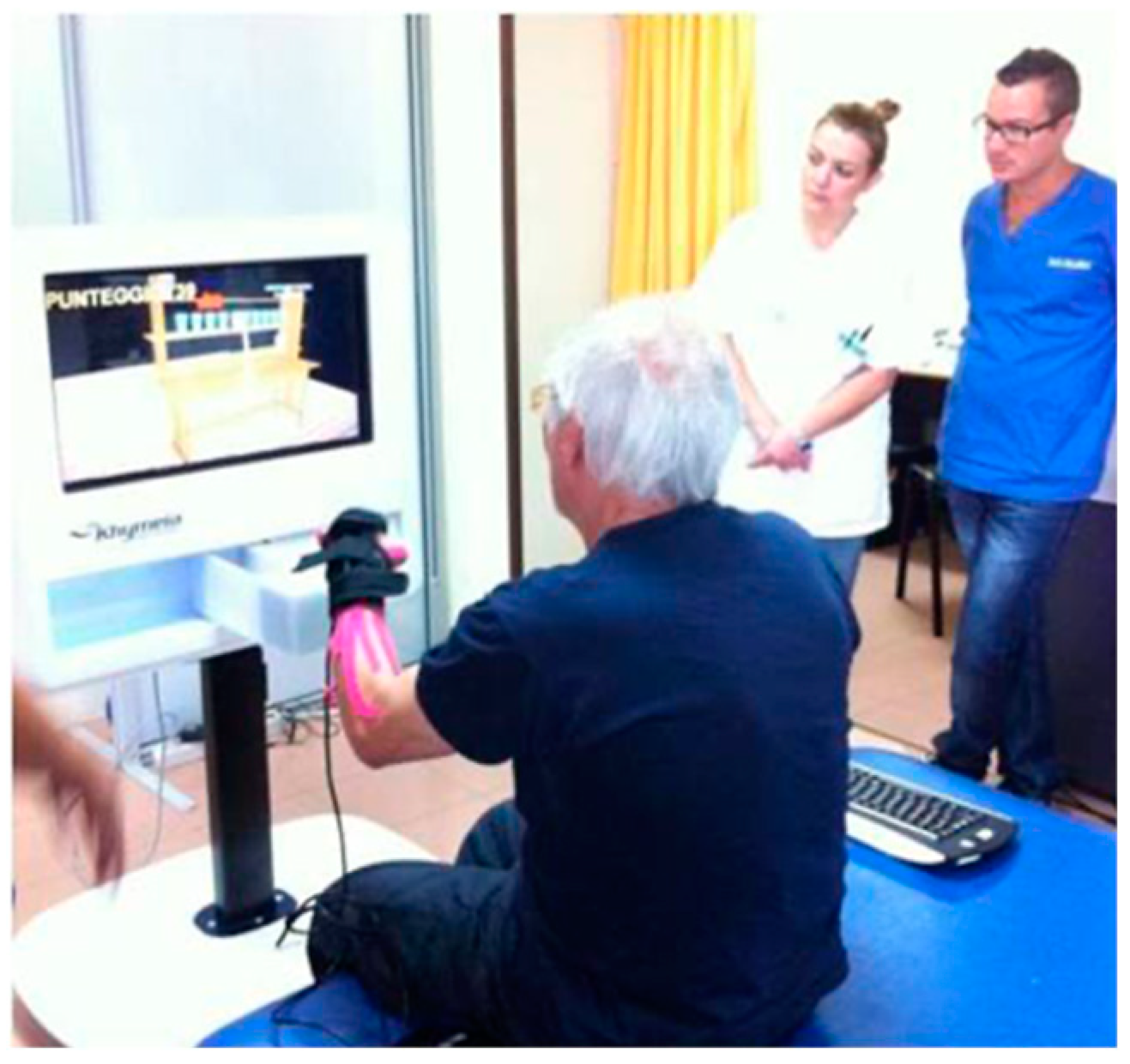
2.3. Virtual Cognitive Task Using VRRS
2.4. Standard Cognitive Training
2.5. Statistical Analysis
3. Results
4. Discussion
5. Limitations of the Study and Future Perspectives
6. Conclusions
Author Contributions
Funding
Institutional Review Board Statement
Informed Consent Statement
Data Availability Statement
Conflicts of Interest
References
- Chohan, S.A.; Venkatesh, P.K.; How, C.H. Long-term complications of stroke and secondary prevention: An overview for primary care physicians. Singapore Med. J. 2019, 60, 616–620. [Google Scholar] [CrossRef] [PubMed]
- Katan, M.; Luft, A. Global Burden of Stroke. Sem. Neurol. 2018, 38, 208–211. [Google Scholar] [CrossRef]
- Kim, K.; Kim, Y.M.; Kim, E.K. Correlation between the Activities of Daily Living of Stroke Patients in a Community Setting and Their Quality of Life. J. Phys. Ther. Sci. 2014, 26, 417–419. [Google Scholar] [CrossRef] [PubMed]
- Lekoubou, A.; Nguyen, C.; Kwon, M.; Nyalundja, A.D.; Agrawal, A. Post-stroke Everything. Curr. Neurol. Neurosci. Rep. 2023, 23, 785–800. [Google Scholar] [CrossRef] [PubMed]
- Béjot, Y.; Daubail, B.; Giroud, M. Epidemiology of stroke and transient ischemic attacks: Current knowledge and perspectives. Revue Neurol. 2016, 172, 59–68. [Google Scholar] [CrossRef]
- Runde, D. Calculated decisions: NIH Stroke Scale/Score (NIHSS). Emerg. Med. Pract. 2021, 23 (Suppl. 6), CD3–CD5. [Google Scholar]
- Alijanpour, S.; Mostafazdeh-Bora, M.; Ahmadi Ahangar, A. Different Stroke Scales; Which Scale or Scales Should Be Used? Caspian J. Int. Med. 2021, 4, 1–21. [Google Scholar]
- Sánchez-Herrera-Baeza, P.; Cano-de-la-Cuerda, R.; Serrada-Tejeda, S.; Fernández-Vázquez, D.; Navarro-López, V.; González-Alted, C.; Miangolarra-Page, J.C. Influence of Age, Gender and Education Level on Executive Functions and Functioning in People with Stroke. Biomedicines 2023, 11, 1603. [Google Scholar] [CrossRef]
- Lai, S.M.; Duncan, P.W. Stroke recovery profile and the Modified Rankin assessment. Neuroepidemiology 2001, 20, 26–30. [Google Scholar] [CrossRef]
- Rowland, T.J.; Cooke, D.M.; Gustafsson, L.A. Role of occupational therapy after stroke. Ann. Indian Acad. Neurol. 2008, 11 (Suppl. 1), S99–S107. [Google Scholar] [CrossRef]
- Gibson, E.; Koh, C.L.; Eames, S.; Bennett, S.; Scott, A.M.; Hoffmann, T.C. Occupational therapy for cognitive impairment in stroke patients. Cochrane Database Syst. Rev. 2022, 3, CD006430. [Google Scholar] [PubMed]
- O’Donoghue, M.; Boland, P.; Leahy, S.; Galvin, R.; McManus, J.; Lisiecka, D.; Hayes, S. Exploring the perspectives of key stakeholders on the design and delivery of a cognitive rehabilitation intervention for people post-stroke. PLoS ONE 2022, 17, 0269961. [Google Scholar]
- Torregrossa, W.; Torrisi, M.; De Luca, R.; Casella, C.; Rifici, C.; Bonanno, M.; Calabrò, R.S. Neuropsychological Assessment in Patients with Traumatic Brain Injury: A Comprehensive Review with Clinical Recommendations. Biomedicines 2023, 11, 1991. [Google Scholar] [CrossRef]
- Parisi, A.; Bellinzona, F.; Di Lernia, D.; Repetto, C.; De Gaspari, S.; Brizzi, G.; Riva, G.; Tuena, C. Efficacy of Multisensory Technology in Post-Stroke Cognitive Rehabilitation: A Systematic Review. J. Clin. Med. 2022, 11, 6324. [Google Scholar] [CrossRef]
- Malik, A.N.; Tariq, H.; Afridi, A.; Rathore, F.A. Technological advancements in stroke rehabilitation. JPMA. J. Pak. Med. Assoc. 2022, 72, 1672–1674. [Google Scholar] [PubMed]
- Hatem, S.M.; Saussez, G.; Della Faille, M.; Prist, V.; Zhang, X.; Dispa, D.; Bleyenheuft, Y. Rehabilitation of Motor Function after Stroke: A Multiple Systematic Review Focused on Techniques to Stimulate Upper Extremity Recovery. Front. Hum. Neurosci. 2016, 10, 442. [Google Scholar] [CrossRef] [PubMed]
- Laver, K.E.; Adey-Wakeling, Z.; Crotty, M.; Lannin, N.A.; George, S.; Sherrington, C. Telerehabilitation services for stroke. Cochrane Database Syst. 2020, 1, CD010255. [Google Scholar] [CrossRef]
- Kleim, J.A.; Jones, T.A. Principles of experience-dependent neural plasticity: Implications for rehabilitation after brain damage. J. Speech Hear. Res. 2008, 51, S225–S239. [Google Scholar] [CrossRef]
- Russo, M.; De Luca, R.; Naro, A.; Sciarrone, F.; Aragona, B.; Silvestri, G.; Manuli, A.; Bramanti, A.; Casella, C.; Bramanti, P.; et al. Does body shadow improve the efficacy of virtual reality-based training with BTS NIRVANA? A pilot study. Medicine 2017, 96, 8096. [Google Scholar] [CrossRef]
- Maggio, M.G.; Latella, D.; Maresca, G.; Sciarrone, F.; Manuli, A.; Naro, A.; De Luca, R.; Calabrò, R.S. Virtual Reality and Cognitive Rehabilitation in People with Stroke: An Overview. J. Neurosc. Nurs. 2019, 51, 101–105. [Google Scholar] [CrossRef]
- Proffitt, R.; Lange, B. Considerations in the efficacy and effectiveness of virtual reality interventions for stroke rehabilitation: Moving the field forward. Phys. Ther. 2015, 95, 441–448. [Google Scholar] [CrossRef]
- Mancuso, M.; Varalta, V.; Sardella, L.; Capitani, D.; Zoccolotti, P.; Antonucci, G. Italian OCS Group. Italian normative data for a stroke specific cognitive screening tool: The Oxford Cognitive Screen. (OCS). Neurol. Sci. 2016, 37, 1713–1721. [Google Scholar] [CrossRef] [PubMed]
- Laver, K.E.; Lange, B.; George, S.; Deutsch, J.E.; Saposnik, G.; Crotty, M. Virtual reality for stroke rehabilitation. Cochrane Database Syst. Rev. 2017, 11, CD008349. [Google Scholar] [CrossRef]
- Calabrò, R.S.; Cerasa, A.; Ciancarelli, I.; Pignolo, L.; Tonin, P.; Iosa, M.; Morone, G. The Arrival of the Metaverse in Neurorehabilitation: Fact, Fake or Vision? Biomedicines 2022, 10, 2602. [Google Scholar] [CrossRef]
- Maggio, M.G.; Naro, A.; Manuli, A.; Maresca, G.; Balletta, T.; Latella, D.; De Luca, R.; Calabrò, R.S. Effects of Robotic Neurorehabilitation on Body Representation in Individuals with Stroke: A Preliminary Study Focusing on an EEG-Based Approach. Brain Topogr. 2021, 34, 348–362. [Google Scholar] [CrossRef] [PubMed]
- Steinisch, M.; Tana, M.G.; Comani, S. A post-stroke rehabilitation system integrating robotics, VR and high-resolution EEG imaging. IEEE Trans. Neural Syst. Rehab. Eng. 2013, 21, 849–859. [Google Scholar] [CrossRef] [PubMed]
- Juhász, C.; Kamondi, A.; Szirmai, I. Spectral EEG analysis following hemispheric stroke: Evidences of transhemispheric diaschisis. Acta Neurol. Scand. 1997, 96, 397–400. [Google Scholar] [CrossRef] [PubMed]
- Mingyu, L.; Jue, W.; Nan, Y.; Qin, Y. Development of EEG biofeedback system based on virtual reality environment. IEEE Eng. Med. Biol. Soc. 2005, 3, 5362–5364. [Google Scholar]
- Pyasik, M.; Scandola, M.; Moro, V. Electrophysiological correlates of action monitoring in brain-damaged patients: A systematic review. Neuropsychologia 2022, 174, 108333. [Google Scholar] [CrossRef]
- Mishra, S.; Kumar, A.; Padmanabhan, P.; Gulyás, B. Neurophysiological Correlates of Cognition as Revealed by Virtual Reality: Delving the Brain with a Synergistic Approach. Brain Sci. 2021, 11, 51. [Google Scholar] [CrossRef]
- Arcuri, F.; Porcaro, C.; Ciancarelli, I.; Tonin, P.; Cerasa, A. Electrophysiological Correlates of Virtual-Reality Applications in the Rehabilitation Setting: New Perspectives for Stroke Patients. Electronics 2021, 10, 836. [Google Scholar] [CrossRef]
- Gangemi, A.; Colombo, B.; Fabio, R.A. Effects of short- and long-term neurostimulation (tDCS) on Alzheimer’s disease patients: Two randomized studies. Aging Clin. Exp. Res. 2021, 33, 383–390. [Google Scholar] [CrossRef]
- Lin, C.T.; Chung, I.F.; Ko, L.W.; Chen, Y.C.; Liang, S.F.; Duann, J.R. EEG-based assessment of driver cognitive responses in a dynamic virtual-reality driving environment. IEEE. Trans. Biomed. Eng. 2007, 54, 1349–1352. [Google Scholar]
- Beppi, C.; Ribeiro Violante, I.; Scott, G.; Sandrone, S. EEG, MEG and neuromodulatory approaches to explore cognition: Current status and future directions. Brain Cogn. 2021, 148, 105677. [Google Scholar] [CrossRef] [PubMed]
- Delorme, A.; Makeig, S. EEGLAB: An open-source toolbox for analysis of single-trial EEG dynamics including independent component analysis. J. Neurosci. Methods 2004, 134, 9–21. [Google Scholar] [CrossRef] [PubMed]
- Kamarajan, C.; Porjesz, B. Advances in electrophysiological research. Alcohol. Res. Curr. Rev. 2015, 37, 53–87. [Google Scholar]
- Welch, P.D. The use of Fast Fourier Transform for the estimation of power spectra: A method based on time averaging over short, modified periodograms. IEEE Trans. Audio Electroacoust. 1967, AU-15, 70–73. [Google Scholar] [CrossRef]
- Georgiev, D.D.; Georgieva, I.; Gong, Z.; Nanjappan, V.; Georgiev, G.V. Virtual Reality for Neurorehabilitation and Cognitive Enhancement. Brain Sci. 2021, 11, 221. [Google Scholar] [CrossRef]
- SPSS Statistics-IBM Data Science Community. Available online: https://www.ibm.com/products/spss-statistics (accessed on 30 June 2021).
- Souza, R.H.C.E.; Naves, E.L.M. Attention Detection in Virtual Environments Using EEG Signals: A Scoping Review. Front. Physiol. 2021, 12, 727840. [Google Scholar] [CrossRef]
- Calabrò, R.S.; Naro, A.; Russo, M.; Leo, A.; De Luca, R.; Balletta, T.; Buda, A.; La Rosa, G.; Bramanti, A.; Bramanti, P. The role of virtual reality in improving motor performance as revealed by EEG: A randomized clinical trial. J. Neuroeng. Rehab 2017, 14, 53. [Google Scholar] [CrossRef]
- De Luca, R.; Bonanno, M.; Rifici, C.; Pollicino, P.; Caminiti, A.; Morone, G.; Calabrò, R.S. Does Non-Immersive Virtual Reality Improve Attention Processes in Severe Traumatic Brain Injury? Encouraging Data from a Pilot Study. Brain Sci. 2022, 12, 1211. [Google Scholar] [CrossRef]
- Leonardi, S.; Maggio, M.G.; Russo, M.; Bramanti, A.; Arcadi, F.A.; Naro, A.; Calabrò, R.S.; De Luca, R. Cognitive recovery in people with relapsing/remitting multiple sclerosis: A randomized clinical trial on virtual reality-based neurorehabilitation. Clin. Neurol. Neurosurg. 2021, 208, 106828. [Google Scholar] [CrossRef] [PubMed]
- De Luca, R.; Bonanno, M.; Marra, A.; Rifici, C.; Pollicino, P.; Caminiti, A.; Castorina, M.V.; Santamato, A.; Quartarone, A.; Calabrò, R.S. Can Virtual Reality Cognitive Rehabilitation Improve Executive Functioning and Coping Strategies in Traumatic Brain Injury? A Pilot Study. Brain Sci. 2023, 13, 578. [Google Scholar] [CrossRef]
- Naro, A.; Calabrò, R.S. What Do We Know about The Use of Virtual Reality in the Rehabilitation Field? A Brief Overview. Electronics 2021, 10, 1042. [Google Scholar] [CrossRef]
- Zak, M.; Wasik, M.; Sikorski, T.; Aleksandrowicz, K.; Miszczuk, R.; Courteix, D.; Dutheil, F.; Januszko-Szakiel, A.; Brola, W. Rehabilitation in Older Adults Affected by Immobility Syndrome, Aided by Virtual Reality Technology: A Narrative Review. J. Clin. Med. 2023, 12, 5675. [Google Scholar] [CrossRef] [PubMed]
- Tompkins, C.A.; Lehman, M.T.; Wyatt, A.D.; Schulz, R. Functional outcome assessment of adults with right hemisphere brain damage. In Seminars Speech Lang; Thieme Medical Publishers, Inc.: New York, NY, USA, 1998; Volume 19, pp. 303–321. [Google Scholar]
- Tompkins, C.A. Rehabilitation for cognitive-communication disorders in right hemisphere brain damage. Arch. Phys. Med. Rehab. 2012, 93 (Suppl. 1), S61–S69. [Google Scholar] [CrossRef] [PubMed]
- Sturm, W.; Willmes, K. On the functional neuroanatomy of intrinsic and phasic alertness. NeuroImage 2001, 14, S76–S84. [Google Scholar] [CrossRef]
- Ferber, S.; Ruppel, J.; Danckert, J. Visual working memory deficits following right brain damage. Brain Cogn. 2020, 142, 105566. [Google Scholar] [CrossRef]
- Audet, T.; Mercier, L.; Collard, S.; Rochette, A.; Hebert, R. Attention deficits: Is there a right hemisphere specialization for simple reaction time, sustained attention and phasic alertness? Brain Cogn. 2000, 43, 17–21. [Google Scholar] [PubMed]
- Gillespie, D.C.; Bowen, A.; Chung, C.S.; Cockburn, J.; Knapp, P.; Pollock, A. Rehabilitation for post-stroke cognitive impairment: An overview of recommendations arising from systematic reviews of current evidence. Clin. Rehabil. 2015, 29, 120–128. [Google Scholar] [CrossRef]
- Cicerone, K.D.; Langenbahn, D.M.; Braden, C.; Malec, J.F.; Kalmar, K.; Fraas, M.; Felicetti, T.; Laatsch, L.; Harley, J.P.; Bergquist, T.; et al. Evidence-based cognitive rehabilitation: Updated review of the literature from 2003 through 2008. Arch. Phys. Med. Rehabil. 2011, 92, 519–530. [Google Scholar] [CrossRef]
- Spaccavento, S.; Marinelli, C.V.; Nardulli, R.; Macchitella, L.; Bivona, U.; Piccardi, L.; Zoccolotti, P.; Angelelli, P. Attention Deficits in Stroke Patients: The Role of Lesion Characteristics, Time from Stroke, and Concomitant Neuropsychological Deficits. Behav. Neurol. 2019, 2019, 7835710. [Google Scholar] [CrossRef] [PubMed]
- Nieto-Escamez, F.; Cortés-Pérez, I.; Obrero-Gaitán, E.; Fusco, A. Virtual Reality Applications in Neurorehabilitation: Current Panorama and Challenges. Brain Sci. 2023, 13, 819. [Google Scholar] [CrossRef]
- Feitosa, J.A.; Casseb, R.F.; Camargo, A.; Brandao, A.F.; Li, L.M.; Castellano, G. Graph analysis of cortical reorganization after virtual reality-based rehabilitation following stroke: A pilot randomized study. Front. Neurol. 2023, 14, 1241639. [Google Scholar] [CrossRef] [PubMed]
- Alouani, A.T.; Elfouly, T. Traumatic Brain Injury (TBI) Detection: Past, Present, and Future. Biomedicines 2022, 10, 2472. [Google Scholar] [CrossRef]
- Smith, E.E.; Reznik, S.J.; Stewart, J.L.; Allen, J.J. Assessing and conceptualizing frontal EEG asymmetry: An updated primer on recording, processing, analyzing, and interpreting frontal alpha asymmetry. Int. J. Psychophysiol. 2017, 111, 98–114. [Google Scholar] [CrossRef] [PubMed]
- Johnson, D.A.; Wilson, G.S. Telemetry for Biosensor Systems. In Electrochem. Methods for Neuroscience; Michael, A.C., Ed.; CRC Press/Taylor & Francis: Boca Raton, FL, USA, 2007. [Google Scholar]
- Lee, M.H.; Kwon, O.Y.; Kim, Y.J.; Kim, H.K.; Lee, Y.E.; Williamson, J.; Fazli, S.; Lee, S.W. EEG dataset and OpenBMI toolbox for three BCI paradigms: An investigation into BCI illiteracy. GigaScience 2019, 8, giz002. [Google Scholar] [CrossRef] [PubMed]
- Klimesch, W. An algorithm for the EEG frequency architecture of consciousness and brain body coupling. Front. Hum. Neurosci. 2013, 7, 766. [Google Scholar] [CrossRef]
- Park, W.; Kwon, G.H.; Kim, Y.H.; Lee, J.H.; Kim, L. EEG response varies with lesion location in patients with chronic stroke. J. Neuroeng. Rehab. 2016, 13, 21. [Google Scholar] [CrossRef]
- Engel, A.K.; Fries, P.; Singer, W. Dynamic predictions: Oscillations and synchrony in top-down processing. Nature Rev. Neurosci. 2001, 2, 704–716. [Google Scholar] [CrossRef]
- Torregrossa, W.; Raciti, L.; Rifici, C.; Rizzo, G.; Raciti, G.; Casella, C.; Naro, A.; Calabrò, R.S. Behavioral and Psychiatric Symptoms in Patients with Severe Traumatic Brain Injury: A Comprehensive Overview. Biomedicines 2023, 11, 1449. [Google Scholar] [CrossRef] [PubMed]
- Maresca, G.; Maggio, M.G.; Latella, D.; Cannavò, A.; De Cola, M.C.; Portaro, S.; Stagnitti, M.C.; Silvestri, G.; Torrisi, M.; Bramanti, A.; et al. Toward Improving Poststroke Aphasia: A Pilot Study on the Growing Use of Telerehabilitation for the Continuity of Care. J. Stroke Cerebrovasc. Dis. 2019, 28, 104303. [Google Scholar] [CrossRef] [PubMed]
- Calabrò, R.S.; Bonanno, M.; Torregrossa, W.; Cacciante, L.; Celesti, A.; Rifici, C.; Tonin, P.; De Luca, R.; Quartarone, A. Benefits of Telerehabilitation for Patients with Severe Acquired Brain Injury: Promising Results From a Multicenter Randomized Controlled Trial Using Nonimmersive Virtual Reality. J. Med. Int. Res. 2023, 25, e45458. [Google Scholar] [CrossRef] [PubMed]
- Cha, K.; Wang, J.; Li, Y.; Shen, L.; Chen, Z.; Long, J. A novel upper-limb tracking system in a virtual environment for stroke rehabilitation. J. Neuroeng. Rehab. 2021, 18, 166. [Google Scholar] [CrossRef] [PubMed]
- Dey, A.; Chatburn, A.; Billinghurst, M. Exploration of an EEG-Based Cognitively Adaptive Training System in Virtual Reality. In Proceedings of the IEEE Conference on Virtual Reality and 3D User Interfaces (VR), Osaka, Japan, 23–27 March 2019; pp. 220–226. [Google Scholar]
- Magosso, E.; De Crescenzio, F.; Ricci, G.; Piastra, S.; Ursino, M. EEG Alpha Power Is Modulated by Attentional Changes during Cognitive Tasks and Virtual Reality Immersion. Comput. Intell. Neurosci. 2019, 2019, 7051079. [Google Scholar] [CrossRef] [PubMed]
- Fabio, R.A.; Gangemi, A.; Capri, T.; Budden, S.; Falzone, A. Neurophysiological and cognitive effects of transcranial direct current stimulation in three girls with Rett Syndrome with chronic language impairments. Res. Dev. Disabil. 2018, 76, 76–87. [Google Scholar] [CrossRef]


| Subject | Age (Years) | Gender | Education (Years) | Barthel Index | Rankin Scale | Time Elapsed Since the Event |
|---|---|---|---|---|---|---|
| Experimental Group | ||||||
| 1 | 57 | M | 8 | 15 | 5 | 6 |
| 2 | 61 | M | 8 | 5 | 5 | 8 |
| 3 | 69 | M | 5 | 40 | 4 | 7 |
| 4 | 65 | M | 13 | 15 | 4 | 6 |
| 5 | 63 | F | 13 | 10 | 4 | 12 |
| 6 | 57 | F | 8 | 10 | 4 | 6 |
| 7 | 57 | F | 13 | 35 | 4 | 8 |
| 8 | 39 | M | 8 | 70 | 3 | 8 |
| 9 | 59 | M | 8 | 10 | 5 | 7 |
| 10 | 56 | F | 8 | 40 | 4 | 6 |
| 11 | 60 | F | 8 | 65 | 3 | 7 |
| 12 | 43 | M | 13 | 10 | 5 | 9 |
| 13 | 67 | M | 13 | 5 | 5 | 9 |
| 14 | 66 | M | 13 | 10 | 5 | 12 |
| 15 | 53 | M | 13 | 25 | 4 | 6 |
| Control Group | Age (Years) | Gender | Education (Years) | Barthel Index | Rankin Scale | Time Elapsed Since the Event |
| 1 | 38 | M | 8 | 10 | 5 | 6 |
| 2 | 73 | M | 13 | 5 | 5 | 6 |
| 3 | 59 | M | 8 | 35 | 4 | 7 |
| 4 | 73 | M | 5 | 20 | 4 | 12 |
| 5 | 68 | M | 5 | 15 | 4 | 8 |
| 6 | 69 | F | 8 | 15 | 4 | 8 |
| 7 | 65 | F | 8 | 30 | 4 | 6 |
| 8 | 64 | M | 13 | 70 | 3 | 6 |
| 9 | 55 | M | 5 | 40 | 5 | 6 |
| 10 | 54 | F | 8 | 40 | 4 | 7 |
| 11 | 48 | M | 13 | 55 | 4 | 9 |
| 12 | 55 | F | 10 | 5 | 5 | 6 |
| 13 | 50 | M | 13 | 20 | 4 | 7 |
| 14 | 45 | F | 8 | 15 | 4 | 7 |
| 15 | 44 | F | 8 | 10 | 4 | 8 |
| Domain | Sub-Domain | VRRS Task | Standard Activities |
|---|---|---|---|
| -Attention Processes | Selective | To administer the scanning exercise, the user must locate the target symbols in a grid and select the matching virtual symbols. To select and immediately recall feedback (audio and video) similar to various elements (colors, musical strings, geometric or abstract forms, animals, numbers) observed in the virtual environment, the patient touches the virtual target element within a specific time. This action causes a visual change with a specific audio feedback (positive reinforcement), using VVRS—interaction between the cognitive therapist and the patient. Otherwise, the element disappears (negative reinforcement). | To administer the attention exercise, the user must locate the target symbols while facing a paper-and-pencil grid and select the matching real symbols. To select and immediately recall feedback (audio and video) resembling various elements (colors, musical strings, geometric or abstract forms, animals, numbers) observed in the real environment, the patient touches the target element within a specific time, using a timer and the interaction between the cognitive therapist and the patient. |
| Sustained | To stimulate sustained attention processes, the patient observes from 3 to 5 target stimuli for a variable and progressive time (10–15 min), with an attentional focus on the virtual tasks administered. | To stimulate sustained attention processes, the patient observes from 3 to 5 target stimuli for a variable and progressive time (10–15 min), with an attentional focus on the real activities administered. | |
| Memory Abilities | Verbal | To work on recognition and remembrance in virtual tasks involving verbal material, reminiscence and validation therapy, mnemonic techniques, and strategic skills. | To work on recognition and remembrance in traditional tasks with paper-and-pencil verbal material, reminiscence and validation therapy, mnemonic techniques and strategic skills, face to face with a therapist, without a virtual tool. |
| Visuo-Spatial | To work on recognition and remembrance virtual tasks with not verbal/visuo-spatial tasks (pictures; image; number; colors…) mnemonic techniques and strategic skills. | To work on recognition and remembrance using paper-and-pencil tasks without verbal/visuo-spatial tasks (pictures, images, numbers, colors), employing conventional mnemonic techniques and strategic skills, face to face with a therapist, without the use of virtual tools. |
| Socio-Demographic and Clinical Variables | Experimental Group | Control Group | Statistic | Pairwise Comparisons |
|---|---|---|---|---|
| Sex (male/female) a | M = 10 | M = 10 | 0.00 | (p = 1) |
| F = 5 | F = 5 | |||
| Age (years) b | 58.13 (8.33) | 57.33 | 0.24 | p = 0.82 |
| Education level (years) b | 10.13 (2.87) | 8.96 (2.92) | 1.19 | p = 0.24 |
| Barthel index (0–100) b | 24.33 (21.20) | 25.66 (19.07) | 0.18 | p = 0.85 |
| Rankin Scale score (0–6) b | 4.26 (0.70) | 4.20 (0.56) | 0.28 | p = 0.77 |
| Years from ischemic stroke b | 7.8 (2.00) | 7.26 (1.62) | 0.80 | p = 0.43 |
| Pre-Test | p | Post-Test | p | |||
|---|---|---|---|---|---|---|
| Right Hemisphere (Hz) | Experimental | Control | Experimental | Control | ||
| Theta band | (M = 17.80; SD = 2.24) | (M = 18.30; SD = 1.73) | 0.23 | (M = 18.10; SD = 2.24) | (M = 18.02; SD = 1.76) | 0.16 |
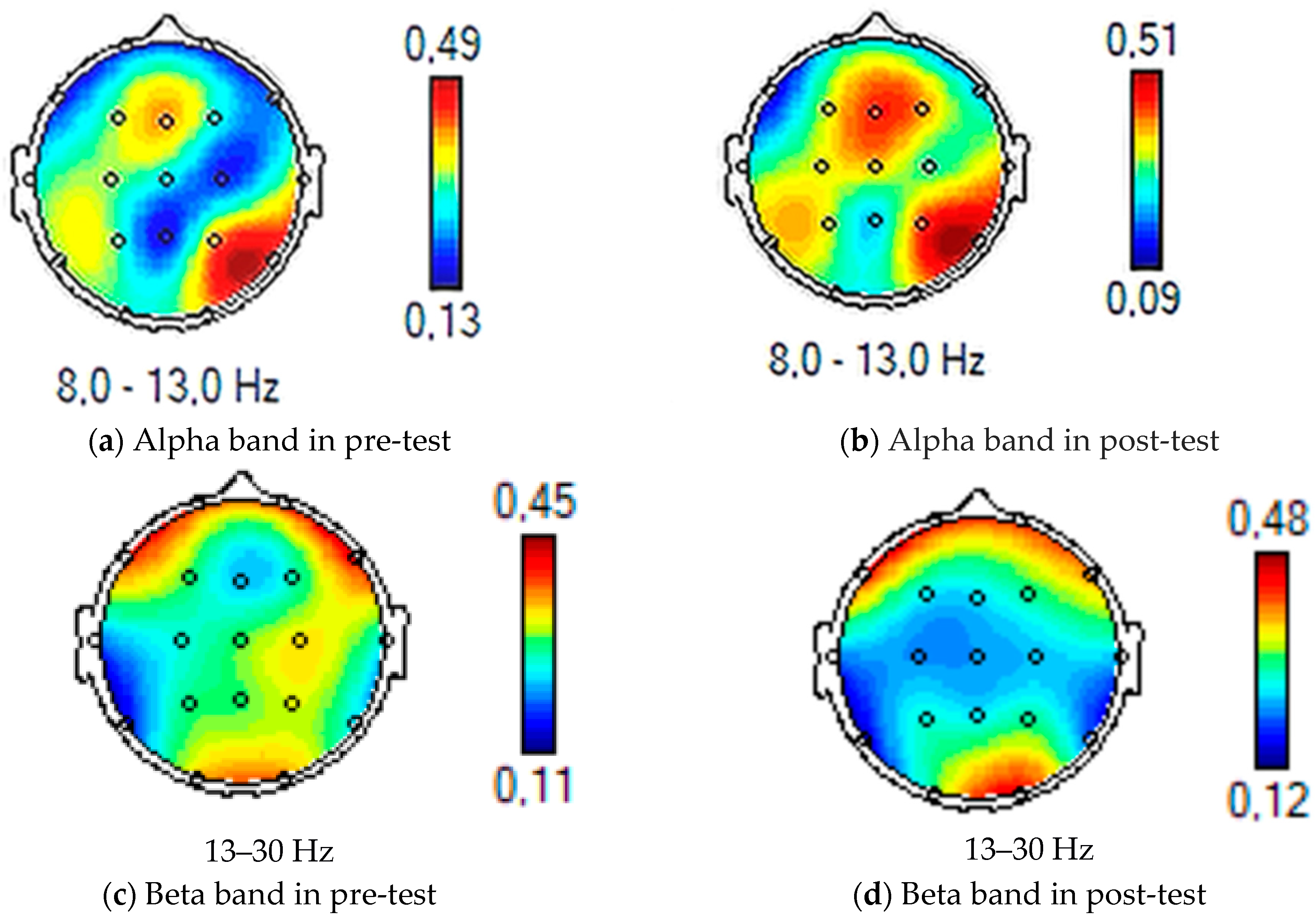
| Alpha band | (M = 21.33; SD = 0.97) | (M = 21.41; SD = 1.02) | 0.31 | (M = 30.23; SD = 2.99) | (M = 21.8; SD = 1.02) | 0.01 |
| Beta band | (M = 23.13; SD = 2.74) | (M = 23.27; SD = 2.89) | 0.37 | (M = 28.27 SD = 2.37) | (M = 23.27; SD = 2.43) | 0.01 |
| Left Hemisphere Theta band | (M = 17.40; SD = 2.74) | (M = 18.03; SD = 1.77) | 0.22 | (M = 18.25; SD = 2.24) | (M = 18.72; SD = 1.76) | 0.14 |
| Alpha band | (M = 22.43; SD = 1.67) | (M = 21.32; SD = 1.32) | 0.36 | (M = 30.23; SD = 2.99) | (M = 21.8; SD = 1.02) | 0.01 |
| Beta band | (M = 23.53; SD = 3.15) | (M = 23.40; SD = 2.47) | 0.49 | (M = 26.97 SD = 3.81) | (M = 23.13; SD = 2.90) | 0.05 |
Disclaimer/Publisher’s Note: The statements, opinions and data contained in all publications are solely those of the individual author(s) and contributor(s) and not of MDPI and/or the editor(s). MDPI and/or the editor(s) disclaim responsibility for any injury to people or property resulting from any ideas, methods, instructions or products referred to in the content. |
© 2023 by the authors. Licensee MDPI, Basel, Switzerland. This article is an open access article distributed under the terms and conditions of the Creative Commons Attribution (CC BY) license (https://creativecommons.org/licenses/by/4.0/).
Share and Cite
Gangemi, A.; De Luca, R.; Fabio, R.A.; Lauria, P.; Rifici, C.; Pollicino, P.; Marra, A.; Olivo, A.; Quartarone, A.; Calabrò, R.S. Effects of Virtual Reality Cognitive Training on Neuroplasticity: A Quasi-Randomized Clinical Trial in Patients with Stroke. Biomedicines 2023, 11, 3225. https://doi.org/10.3390/biomedicines11123225
Gangemi A, De Luca R, Fabio RA, Lauria P, Rifici C, Pollicino P, Marra A, Olivo A, Quartarone A, Calabrò RS. Effects of Virtual Reality Cognitive Training on Neuroplasticity: A Quasi-Randomized Clinical Trial in Patients with Stroke. Biomedicines. 2023; 11(12):3225. https://doi.org/10.3390/biomedicines11123225
Chicago/Turabian StyleGangemi, Antonio, Rosaria De Luca, Rosa Angela Fabio, Paola Lauria, Carmela Rifici, Patrizia Pollicino, Angela Marra, Antonella Olivo, Angelo Quartarone, and Rocco Salvatore Calabrò. 2023. "Effects of Virtual Reality Cognitive Training on Neuroplasticity: A Quasi-Randomized Clinical Trial in Patients with Stroke" Biomedicines 11, no. 12: 3225. https://doi.org/10.3390/biomedicines11123225