
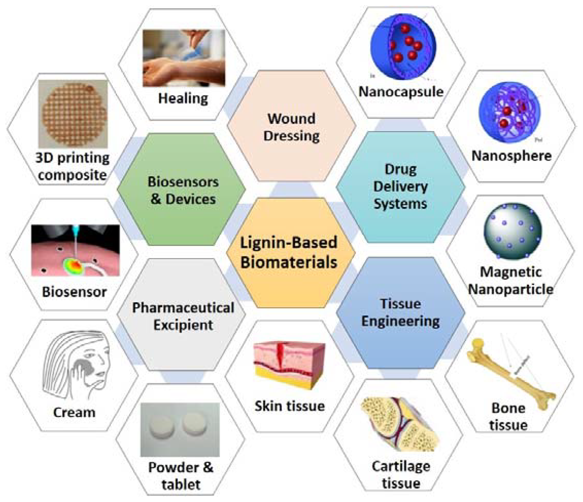
Lignin-Based Porous Biomaterials for Medical and Pharmaceutical Applications


Abstract
:1. Introduction
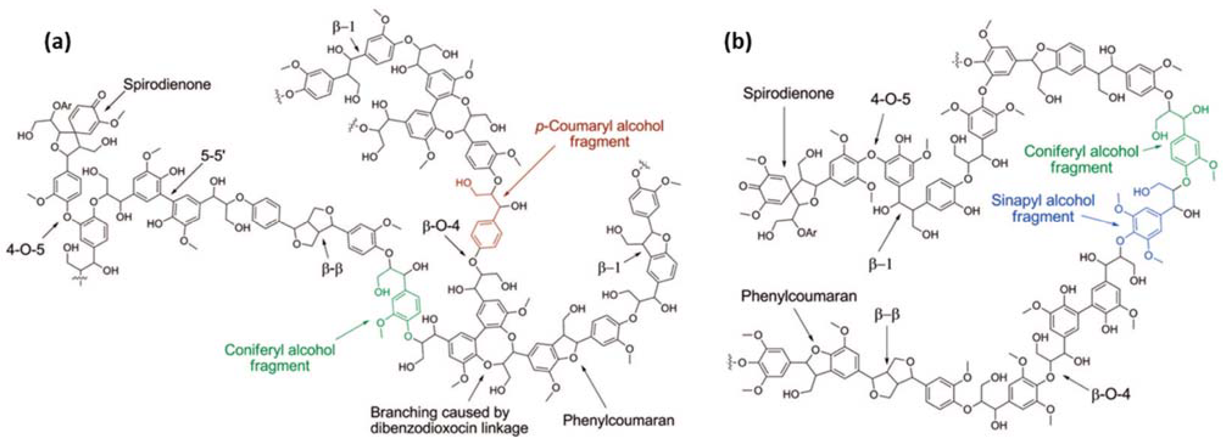
2. Lignin-Based Porous Hydrogels
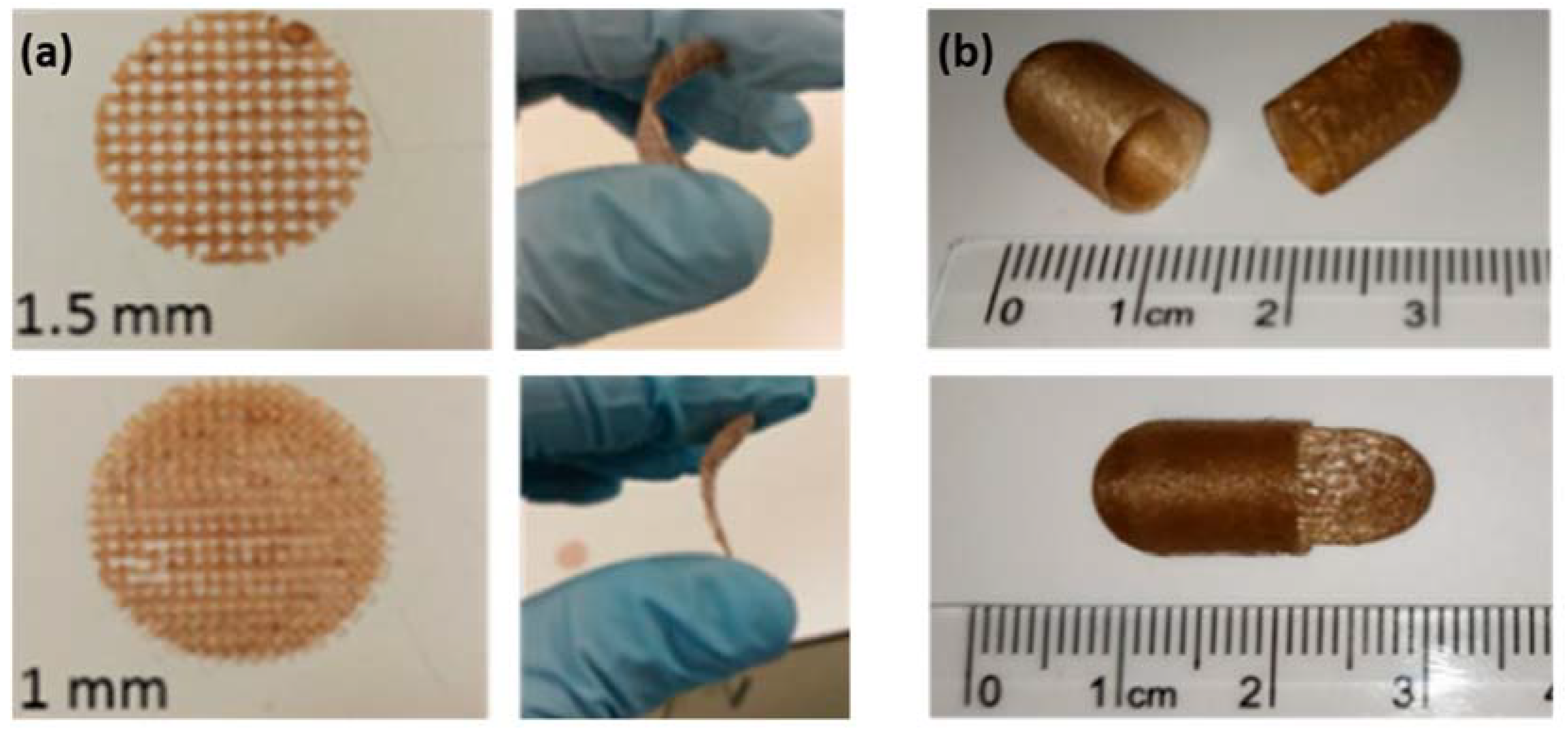
2.1. Porous Structures of Lignin-Based Hydrogels
2.2. Current Status of Lignin-Based Hydrogels for Medical Applications

| Hydrogel | Methodology | Advanced Properties | Application | Ref. |
|---|---|---|---|---|
| lignin-agarose-SF- ZnCr2O4 | cross-linking reaction between lignin and agarose by ECH, and addition of SF and ZnCr2O4 | cellular ingrowth & vascularization in designed scaffolds | wound healing, tissue engineering | [22] |
| carrageenan-lignin-AgNPs-MgCl2 | one-pot synthesis of AgNPs using lignin as a reducing and capping agent in the carrageenan matrix cross-linked with divalent cations | wound healing speed | wound dressing | [32] |
| chitosan-alkali lignin | combining lignin with an aqueous-acidic solution of chitosan | forming electrostatic cross-links between the chitosan chains | wound healing, tissue engineering | [34] |
| lignin-chitosan-PVA | mixing an aqueous acidicsolution of chitosan with solutions of lignin and PVA | mechanical strength, protein adsorption, wound environmental regulation ability | wound dressing | [33] |
| lignin-GAN; lignin-PEG | esterification reaction with microwave radiation | welling, sustaining drug delivery, adhesion reduction | drug delivery (curcumin) | [30] |
| xanthan-lignin | mixing lignin with xanthan using ECH as crosslinking agent | the amount of drug loading | drug delivery (bisoprolol fumarate) | [31] |
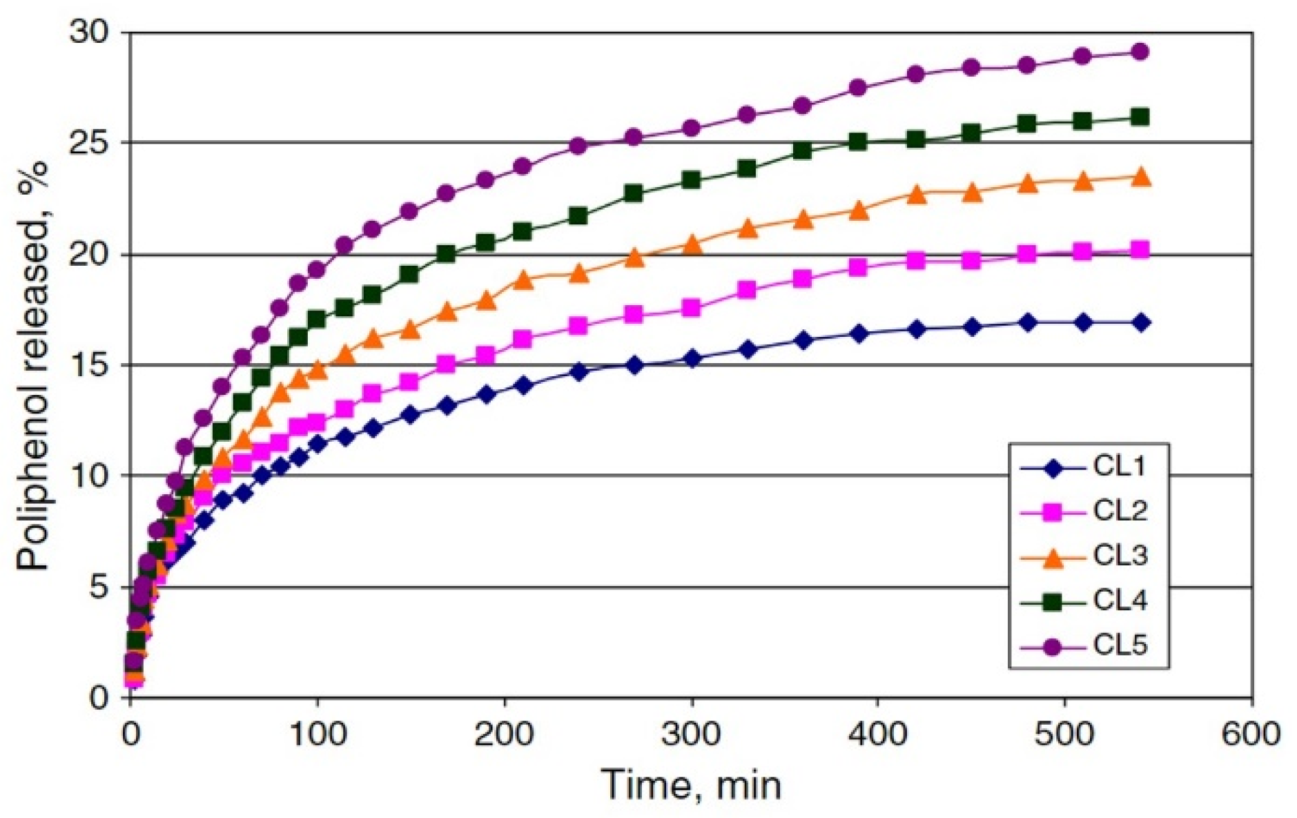
| cellulose-lignin | mixing cellulose alkaline solution with lignin, followed by the crosslinking with ECH | the swelling capacity and drug release | drug delivery (polyphenols) | [24] |
3. Lignin-Based Hollow Nanoparticles
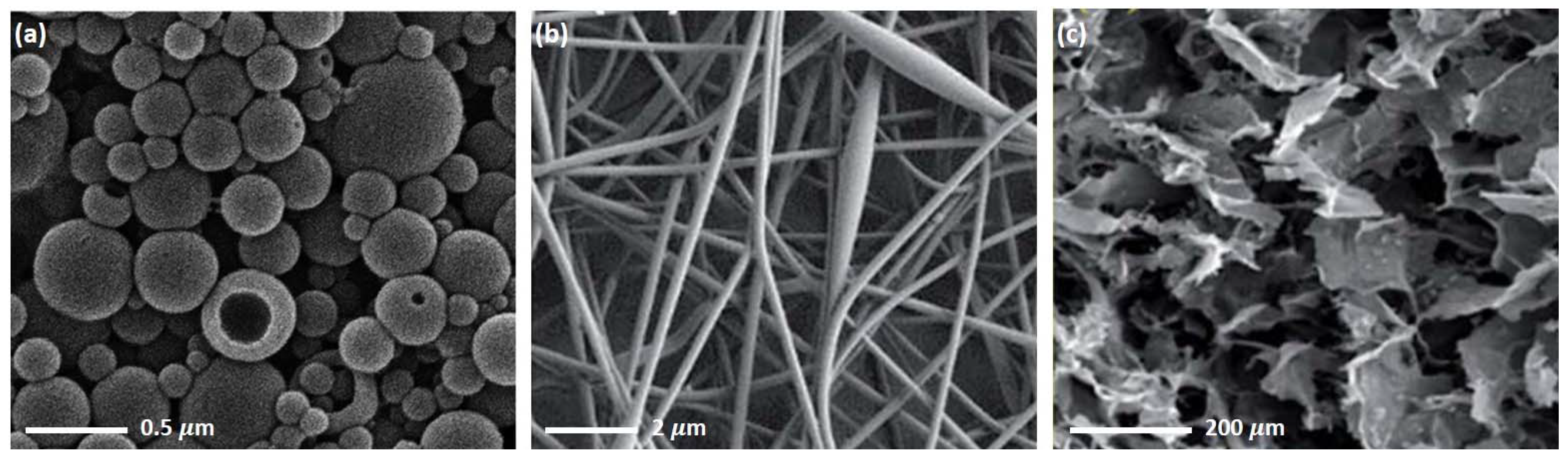
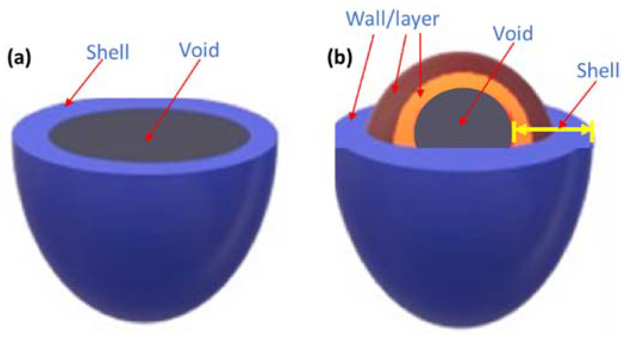
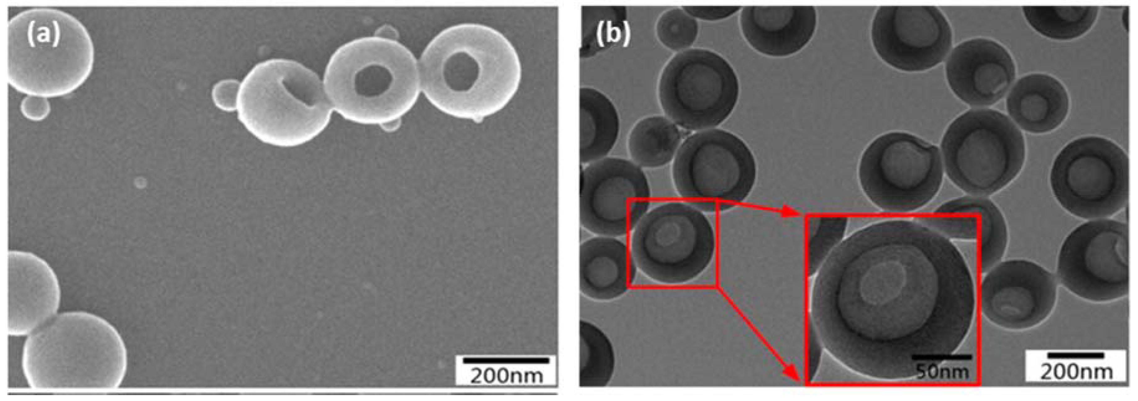
3.1. Hollow Structure of the LHNPs
3.2. Current Status of Lignin Nanoparticles for Pharmaceutical and Medical Applications
4. Lignin-Based Porous Biocomposites
5. Promising Fabrication Technologies of Lignin-Based Medical Materials
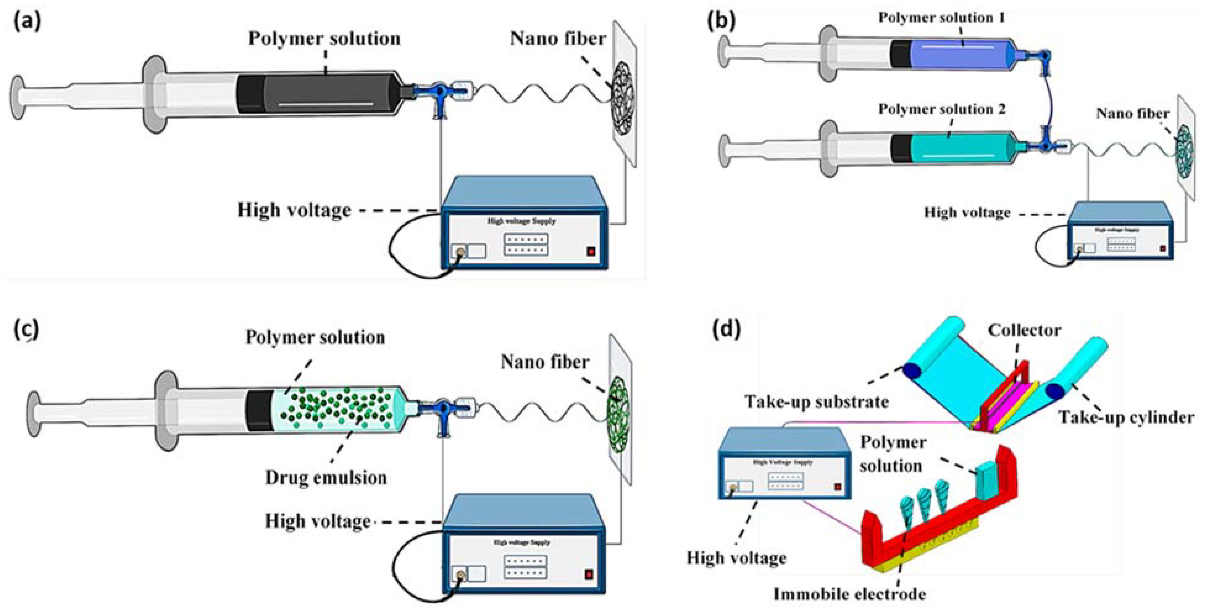
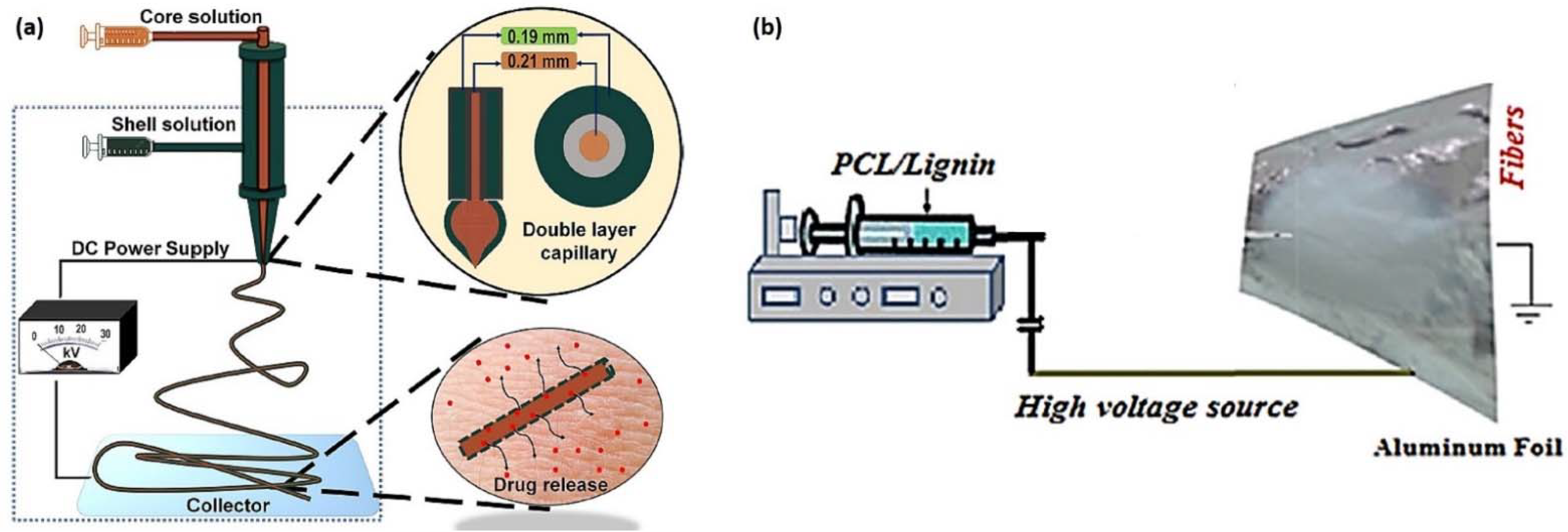
5.1. Electrospinning
5.2. Three-Dimensional Printing
6. Conclusions
Author Contributions
Funding
Conflicts of Interest
References
- Rico-García, D.; Ruiz-Rubio, L.; Pérez-Alvarez, L.; Hernández-Olmos, S.L.; Guerrero-Ramírez, G.L.; Vilas-Vilela, J.L. Lignin-Based Hydrogels: Synthesis and Applications. Polymers 2020, 12, 81. [Google Scholar] [CrossRef] [PubMed] [Green Version]
- Zakzeski, J.; Bruijnincx, P.C.A.; Jongerius, A.L.; Weckhuysen, B.M. The Catalytic Valorization of Lignin for the Production of Renewable Chemicals. Chem. Rev. 2010, 110, 3552–3599. [Google Scholar] [CrossRef] [PubMed]
- Zhou, Y.; Han, Y.; Li, G.; Yang, S.; Xiong, F.; Chu, F. Preparation of Targeted Lignin–Based Hollow Nanoparticles for the Delivery of Doxorubicin. Nanomaterials 2019, 9, 188. [Google Scholar] [CrossRef] [Green Version]
- Zhou, Y.; Han, Y.; Li, G.; Yang, S.; Chu, F. Lignin-Based Hollow Nanoparticles for Controlled Drug Delivery: Grafting Preparation Using β-Cyclodextrin/Enzymatic-Hydrolysis Lignin. Nanomaterials 2019, 9, 997. [Google Scholar] [CrossRef] [PubMed] [Green Version]
- Abudula, T.; Gauthaman, K.; Mostafavi, A.; Alshahrie, A.; Salah, N.; Morganti, P.; Chianese, A.; Tamayol, A.; Memic, A. Sustainable Drug Release from Polycaprolactone Coated Chitin-Lignin Gel Fibrous Scaffolds. Sci. Rep. 2020, 10, 20428. [Google Scholar] [CrossRef]
- Yiamsawas, D.; Beckers, S.J.; Lu, H.; Landfester, K.; Wurm, F.R. Morphology-Controlled Synthesis of Lignin Nanocarriers for Drug Delivery and Carbon Materials. ACS Biomater. Sci. Eng. 2017, 3, 2375–2383. [Google Scholar] [CrossRef]
- Figueiredo, P.; Lintinen, K.; Kiriazis, A.; Hynninen, V.; Liu, Z.; Bauleth-Ramos, T.; Rahikkala, A.; Correia, A.; Kohout, T.; Sarmento, B.; et al. In Vitro Evaluation of Biodegradable Lignin-Based Nanoparticles for Drug Delivery and Enhanced Antiproliferation Effect in Cancer Cells. Biomaterials 2017, 121, 97–108. [Google Scholar] [CrossRef]
- Chen, N.; Dempere, L.A.; Tong, Z. Synthesis of PH-Responsive Lignin-Based Nanocapsules for Controlled Release of Hydrophobic Molecules. ACS Sustain. Chem. Eng. 2016, 4, 5204–5211. [Google Scholar] [CrossRef]
- Frangville, C.; Rutkevičius, M.; Richter, A.P.; Velev, O.D.; Stoyanov, S.D.; Paunov, V.N. Fabrication of Environmentally Biodegradable Lignin Nanoparticles. Chemphyschem 2012, 13, 4235–4243. [Google Scholar] [CrossRef]
- Alqahtani, M.S.; Alqahtani, A.; Al-Thabit, A.; Roni, M.; Syed, R. Novel Lignin Nanoparticles for Oral Drug Delivery. J. Mater. Chem. B 2019, 7, 4461–4473. [Google Scholar] [CrossRef]
- Christoforidis, J.B.; Chang, S.; Jiang, A.; Wang, J.; Cebulla, C.M. Intravitreal Devices for the Treatment of Vitreous Inflammation. Mediat. Inflamm. 2012, 2012, e126463. [Google Scholar] [CrossRef] [PubMed] [Green Version]
- Gunay, M.S.; Ozer, A.Y.; Chalon, S. Drug Delivery Systems for Imaging and Therapy of Parkinson’s Disease. Curr. Neuropharmacol. 2016, 14, 376–391. [Google Scholar] [CrossRef] [PubMed]
- Kalirajan, C.; Dukle, A.; Nathanael, A.J.; Oh, T.-H.; Manivasagam, G. A Critical Review on Polymeric Biomaterials for Biomedical Applications. Polymers 2021, 13, 3015. [Google Scholar] [CrossRef] [PubMed]
- Khan, S.; Siddique, R.; Huanfei, D.; Shereen, M.A.; Nabi, G.; Bai, Q.; Manan, S.; Xue, M.; Ullah, M.W.; Bowen, H. Perspective Applications and Associated Challenges of Using Nanocellulose in Treating Bone-Related Diseases. Front. Bioeng. Biotechnol. 2021, 9, 350. [Google Scholar] [CrossRef]
- Bayer, I.S. A Review of Sustained Drug Release Studies from Nanofiber Hydrogels. Biomedicines 2021, 9, 1612. [Google Scholar] [CrossRef]
- Domínguez-Robles, J.; Martin, N.K.; Fong, M.L.; Stewart, S.A.; Irwin, N.J.; Rial-Hermida, M.I.; Donnelly, R.F.; Larrañeta, E. Antioxidant PLA Composites Containing Lignin for 3D Printing Applications: A Potential Material for Healthcare Applications. Pharmaceutics 2019, 11, 165. [Google Scholar] [CrossRef] [Green Version]
- Shi, K.; Tan, D.K.; Nokhodchi, A.; Maniruzzaman, M. Drop-On-Powder 3D Printing of Tablets with an Anti-Cancer Drug, 5-Fluorouracil. Pharmaceutics 2019, 11, 150. [Google Scholar] [CrossRef] [Green Version]
- Tang, W.; Bhushan, B. Adhesion, Friction and Wear Characterization of Skin and Skin Cream Using Atomic Force Microscope. Colloids Surf. B: Biointerfaces 2010, 76, 1–15. [Google Scholar] [CrossRef]
- Jin, C.; Song, W.; Liu, T.; Xin, J.; Hiscox, W.; Zhang, J.; Liu, G.; Kong, Z. Temperature and PH Responsive Hydrogels Using Methacrylated Lignosulfonate Cross-Linker: Synthesis, Characterization, and Properties. ACS Sustain. Chem. Eng. 2017, 6, 1763–1771. [Google Scholar] [CrossRef]
- Capecchi, E.; Piccinino, D.; Bizzarri, B.M.; Pelosi, C.; Colantonio, C.; Calabro, G.; Saladino, R. Enzyme-Lignin Nanocapsules Are Sustainable Catalysts and Vehicles for the Preparation of Unique Polyvalent Bioinks. Biomacromolecules 2019, 20, 1975–1988. [Google Scholar] [CrossRef]
- Ago, M.; Borghei, M.; Haataja, J.; Rojas, O. Mesoporous Carbon Soft-Templated from Lignin Nanofiber Networks: Microphase Separation Boosts Supercapacitance in Conductive Electrodes. RSC Adv. 2016, 6, 85802–85810. [Google Scholar] [CrossRef] [Green Version]
- Eivazzadeh-Keihan, R.; Aliabadi, H.A.M.; Radinekiyan, F.; Sobhani, M.; Khalili, F.; Maleki, A.; Madanchi, H.; Mahdavi, M.; Shalan, A.E. Investigation of the Biological Activity, Mechanical Properties and Wound Healing Application of a Novel Scaffold Based on Lignin–Agarose Hydrogel and Silk Fibroin Embedded Zinc Chromite Nanoparticles. RSC Adv. 2021, 11, 17914–17923. [Google Scholar] [CrossRef]
- Domínguez-Robles, J.; Cárcamo-Martínez, Á.; Stewart, S.A.; Donnelly, R.F.; Larrañeta, E.; Borrega, M. Lignin for Pharmaceutical and Biomedical Applications—Could This Become a Reality? Sustain. Chem. Pharm. 2020, 18, 100320. [Google Scholar] [CrossRef]
- Ciolacu, D.; Oprea, A.M.; Anghel, N.; Cazacu, G.; Cazacu, M. New Cellulose–Lignin Hydrogels and Their Application in Controlled Release of Polyphenols. Mater. Sci. Eng. C 2012, 32, 452–463. [Google Scholar] [CrossRef]
- Lin, C.-C.; Metters, A.T. Hydrogels in Controlled Release Formulations: Network Design and Mathematical Modeling. Adv. Drug Deliv. Rev. 2006, 58, 1379–1408. [Google Scholar] [CrossRef]
- Shen, X.; Shamshina, J.L.; Berton, P.; Gurau, G.; Rogers, R.D. Hydrogels Based on Cellulose and Chitin: Fabrication, Properties, and Applications. Green Chem. 2015, 18, 53–75. [Google Scholar] [CrossRef] [Green Version]
- Khan, J.; Rudrapal, M.; Bhat, E.A.; Ali, A.; Alaidarous, M.; Alshehri, B.; Banwas, S.; Ismail, R.; Egbuna, C. Perspective Insights to Bio-Nanomaterials for the Treatment of Neurological Disorders. Front. Bioeng. Biotechnol. 2021, 9, 912. [Google Scholar] [CrossRef]
- Thakur, V.K.; Thakur, M.K. Recent Advances in Green Hydrogels from Lignin: A Review. Int. J. Biol. Macromol. 2015, 72, 834–847. [Google Scholar] [CrossRef]
- Wróblewska-Krepsztul, J.; Rydzkowski, T.; Michalska-Pożoga, I.; Thakur, V.K. Biopolymers for Biomedical and Pharmaceutical Applications: Recent Advances and Overview of Alginate Electrospinning. Nanomaterials 2019, 9, E404. [Google Scholar] [CrossRef] [Green Version]
- Larrañeta, E.; Imízcoz, M.; Toh, J.X.; Irwin, N.J.; Ripolin, A.; Perminova, A.; Domínguez-Robles, J.; Rodríguez, A.; Donnelly, R.F. Synthesis and Characterization of Lignin Hydrogels for Potential Applications as Drug Eluting Antimicrobial Coatings for Medical Materials. ACS Sustain. Chem. Eng. 2018, 6, 9037–9046. [Google Scholar] [CrossRef]
- Raschip, I.; Panainte, A.; Daniela, P.; Profire, L.; Vasile, C. In Vitro Testing of Xanthan/Lignin Hydrogels as Carriers for Controlled Delivery of Bisoprolol Fumarate. Rev. Med. Chir. Soc. Med. Nat. Iasi 2015, 119, 1189–1194. [Google Scholar] [PubMed]
- Jaiswal, L.; Shankar, S.; Rhim, J.-W.; Hahm, D.-H. Lignin-Mediated Green Synthesis of AgNPs in Carrageenan Matrix for Wound Dressing Applications. Int. J. Biol. Macromol. 2020, 159, 859–869. [Google Scholar] [CrossRef] [PubMed]
- Zhang, Y.; Jiang, M.; Zhang, Y.; Cao, Q.; Wang, X.; Han, Y.; Sun, G.; Li, Y.; Zhou, J. Novel Lignin–Chitosan–PVA Composite Hydrogel for Wound Dressing. Mater. Sci. Eng. C 2019, 104, 110002. [Google Scholar] [CrossRef]
- Ravishankar, K.; Venkatesan, M.; Desingh, R.P.; Mahalingam, A.; Sadhasivam, B.; Subramaniyam, R.; Dhamodharan, R. Biocompatible Hydrogels of Chitosan-Alkali Lignin for Potential Wound Healing Applications. Mater. Sci. Eng. C 2019, 102, 447–457. [Google Scholar] [CrossRef]
- Tian, J.; Zhang, F.; Han, Y.; Zhao, X.; Chen, C.; Zhang, C.; Jia, G. Template-Directed Synthesis, Properties, and Dual-Modal Bioapplications of Multifunctional GdPO4 Hierarchical Hollow Spheres. Appl. Surf. Sci. 2019, 475, 264–272. [Google Scholar] [CrossRef]
- Stanisz, M.; Klapiszewski, Ł.; Jesionowski, T. Recent Advances in the Fabrication and Application of Biopolymer-Based Micro- and Nanostructures: A Comprehensive Review. Chem. Eng. J. 2020, 397, 125409. [Google Scholar] [CrossRef]
- Lu, Q.; Zhu, M.; Zu, Y.; Liu, W.; Yang, L.; Zhang, Y.; Zhao, X.; Zhang, X.; Zhang, X.; Li, W. Comparative Antioxidant Activity of Nanoscale Lignin Prepared by a Supercritical Antisolvent (SAS) Process with Non-Nanoscale Lignin. Food Chem. 2012, 135, 63–67. [Google Scholar] [CrossRef]
- Zhang, X.; Yang, M.; Yuan, Q.; Cheng, G. Controlled Preparation of Corncob Lignin Nanoparticles and Their Size-Dependent Antioxidant Properties: Toward High Value Utilization of Lignin. ACS Sustain. Chem. Eng. 2019, 7, 17166–17174. [Google Scholar] [CrossRef]
- Ten, E.; Ling, C.; Wang, Y.; Srivastava, A.; Dempere, L.A.; Vermerris, W. Lignin Nanotubes As Vehicles for Gene Delivery into Human Cells. Biomacromolecules 2014, 15, 327–338. [Google Scholar] [CrossRef]
- Dai, L.; Liu, R.; Hu, L.-Q.; Zou, Z.-F.; Si, C.-L. Lignin Nanoparticle as a Novel Green Carrier for the Efficient Delivery of Resveratrol. ACS Sustain. Chem. Eng. 2017, 5, 8241–8249. [Google Scholar] [CrossRef]
- Jaganathan, G.; Manivannan, K.; Lakshmanan, S.; Sithique, M.A. Fabrication and Characterization of Artocarpus Heterophyllus Waste Derived Lignin Added Chitosan Biocomposites for Wound Dressing Application. Sustain. Chem. Pharm. 2018, 10, 27–32. [Google Scholar] [CrossRef]
- Barros, A.; Quraishi, S.; Martins, M.; Gurikov, P.; Subrahmanyam, R.; Smirnova, I.; Duarte, A.R.C.; Reis, R.L. Hybrid Alginate-Based Cryogels for Life Science Applications. Chem. Ing. Tech. 2016, 88, 1770–1778. [Google Scholar] [CrossRef]
- Memic, A.; Abudula, T. Antioxidant, Antibacterial, Injectable Lignin-Gelatin Composite Cryogels for Wound Healing and Tissue Engineering. U.S. Patent US10881760B1, 5 January 2021. [Google Scholar]
- Quraishi, S.; Martins, M.; Barros, A.A.; Gurikov, P.; Raman, S.P.; Smirnova, I.; Duarte, A.R.C.; Reis, R.L. Novel Non-Cytotoxic Alginate–Lignin Hybrid Aerogels as Scaffolds for Tissue Engineering. J. Supercrit. Fluids 2015, 105, 1–8. [Google Scholar] [CrossRef]
- Danti, S.; Trombi, L.; Fusco, A.; Azimi, B.; Lazzeri, A.; Morganti, P.; Coltelli, M.-B.; Donnarumma, G. Chitin Nanofibrils and Nanolignin as Functional Agents in Skin Regeneration. Int. J. Mol. Sci. 2019, 20, 2669. [Google Scholar] [CrossRef] [PubMed] [Green Version]
- Erakovic, S.; Jankovic, A.; Tsui, G.C.P.; Tang, C.-Y.; Miskovic-Stankovic, V.; Stevanovic, T. Novel Bioactive Antimicrobial Lignin Containing Coatings on Titanium Obtained by Electrophoretic Deposition. Int. J. Mol. Sci. 2014, 15, 12294–12322. [Google Scholar] [CrossRef] [Green Version]
- Eraković, S.; Janković, A.; Veljović, D.; Palcevskis, E.; Mitrić, M.; Stevanović, T.; Janaćković, D.; Mišković-Stanković, V. Corrosion Stability and Bioactivity in Simulated Body Fluid of Silver/Hydroxyapatite and Silver/Hydroxyapatite/Lignin Coatings on Titanium Obtained by Electrophoretic Deposition. J. Phys. Chem. B 2013, 117, 1633–1643. [Google Scholar] [CrossRef] [PubMed]
- Erakovic, S.; Veljovic, D.; Diouf, P.N.; Stevanovic, T.; Mitric, M.; Milonjic, S.; Miskovic-Stankovic, V.B. Electrophoretic Deposition of Biocomposite Lignin/Hydroxyapatite Coatings on Titanium. Int. J. Chem. React. Eng. 2009, 7, 5391. [Google Scholar] [CrossRef]
- Eraković, S.; Janković, A.; Matić, I.Z.; Juranić, Z.D.; Vukašinović-Sekulić, M.; Stevanović, T.; Mišković-Stanković, V. Investigation of Silver Impact on Hydroxyapatite/Lignin Coatings Electrodeposited on Titanium. Mater. Chem. Phys. 2013, 142, 521–530. [Google Scholar] [CrossRef]
- Erakovic, S.; Veljovic, D.; Diouf, P.; Stevanovic, T.; Mitrić, M.; Janaćković, D.; Matić, I.; Juranic, Z.; Mišković-Stanković, V. The Effect of Lignin on the Structure and Characteristics of Composite Coatings Electrodeposited on Titanium. Prog. Org. Coat. 2012, 75, 275–283. [Google Scholar] [CrossRef]
- Wang, K.; Loo, L.S.; Goh, K.L. A Facile Method for Processing Lignin Reinforced Chitosan Biopolymer Microfibres: Optimising the Fibre Mechanical Properties through Lignin Type and Concentration. Mater. Res. Express 2016, 3, 035301. [Google Scholar] [CrossRef]
- Abudula, T.; Gzara, L.; Simonetti, G.; Alshahrie, A.; Salah, N.; Morganti, P.; Chianese, A.; Fallahi, A.; Tamayol, A.; Bencherif, S.A.; et al. The Effect of Poly (Glycerol Sebacate) Incorporation within Hybrid Chitin–Lignin Sol–Gel Nanofibrous Scaffolds. Materials 2018, 11, 451. [Google Scholar] [CrossRef] [PubMed] [Green Version]
- Salami, M.A.; Kaveian, F.; Rafienia, M.; Saber-Samandari, S.; Khandan, A.; Naeimi, M. Electrospun Polycaprolactone/Lignin-Based Nanocomposite as a Novel Tissue Scaffold for Biomedical Applications. J. Med. Signals Sens. 2017, 7, 228–238. [Google Scholar] [PubMed]
- Saudi, A.; Amini, S.; Amirpour, N.; Kazemi, M.; Zargar Kharazi, A.; Salehi, H.; Rafienia, M. Promoting Neural Cell Proliferation and Differentiation by Incorporating Lignin into Electrospun Poly(Vinyl Alcohol) and Poly(Glycerol Sebacate) Fibers. Mater. Sci. Eng. C 2019, 104, 110005. [Google Scholar] [CrossRef] [PubMed]
- Zhang, N. Preparation of Nanofibre Material Based on Electrospinning Technology and Its Application in Rehabilitation of Lower Limb Joint Motion. Int. J. Nanotechnol. 2020, 17, 325–338. [Google Scholar] [CrossRef]
- Lee, E.-S.; Kim, Y.-O.; Ha, Y.-M.; Lim, D.; Hwang, J.Y.; Kim, J.; Park, M.; Cho, J.W.; Jung, Y.C. Antimicrobial Properties of Lignin-Decorated Thin Multi-Walled Carbon Nanotubes in Poly(Vinyl Alcohol) Nanocomposites. Eur. Polym. J. 2018, 105, 79–84. [Google Scholar] [CrossRef]
- Saha, D.; Payzant, E.A.; Kumbhar, A.S.; Naskar, A.K. Sustainable Mesoporous Carbons as Storage and Controlled-Delivery Media for Functional Molecules. ACS Appl. Mater. Interfaces 2013, 5, 5868–5874. [Google Scholar] [CrossRef]
- Zuber, S.H.; Hashikin, N.A.A.; Mohd Yusof, M.F.; Hashim, R. Lignin and Soy Flour as Adhesive Materials in the Fabrication of Rhizophora Spp. Particleboard for Medical Physics Applications. J. Adhes. 2020, 98, 429–448. [Google Scholar] [CrossRef]
- Pishnamazi, M.; Hafizi, H.; Shirazian, S.; Culebras, M.; Walker, G.M.; Collins, M.N. Design of Controlled Release System for Paracetamol Based on Modified Lignin. Polymers 2019, 11, 1059. [Google Scholar] [CrossRef] [PubMed] [Green Version]
- Pishnamazi, M.; Ismail, H.Y.; Shirazian, S.; Iqbal, J.; Walker, G.M.; Collins, M.N. Application of Lignin in Controlled Release: Development of Predictive Model Based on Artificial Neural Network for API Release. Cellulose 2019, 26, 6165–6178. [Google Scholar] [CrossRef]
- Gil-Chávez, J.; Padhi, S.S.P.; Leopold, C.S.; Smirnova, I. Application of Aquasolv Lignin in Ibuprofen-Loaded Pharmaceutical Formulations Obtained via Direct Compression and Wet Granulation. Int. J. Biol. Macromol. 2021, 174, 229–239. [Google Scholar] [CrossRef]
- Li, Z.; Zhang, J.; Qin, L.; Ge, Y. Enhancing Antioxidant Performance of Lignin by Enzymatic Treatment with Laccase. ACS Sustain. Chem. Eng. 2018, 6, 2591–2595. [Google Scholar] [CrossRef]
- Luís, Â.; Domingues, F.; Duarte, A.P. Bioactive Compounds, RP-HPLC Analysis of Phenolics, and Antioxidant Activity of Some Portuguese Shrub Species Extracts. Nat. Prod. Commun. 2011, 6, 1863–1872. [Google Scholar] [CrossRef] [PubMed] [Green Version]
- Yang, X.; Wang, J.; Guo, H.; Liu, L.; Xu, W.; Duan, G. Structural Design toward Functional Materials by Electrospinning: A Review. e-Polymers 2020, 20, 682–712. [Google Scholar] [CrossRef]
- Alven, S.; Buyana, B.; Feketshane, Z.; Aderibigbe, B.A. Electrospun Nanofibers/Nanofibrous Scaffolds Loaded with Silver Nanoparticles as Effective Antibacterial Wound Dressing Materials. Pharmaceutics 2021, 13, 964. [Google Scholar] [CrossRef]
- Zhong, W. Chapter 3—Nanofibres for Medical Textiles. In Advances in Smart Medical Textiles; van Langenhove, L., Ed.; Woodhead Publishing Series in Textiles; Woodhead Publishing: Oxford, UK, 2016; pp. 57–70. ISBN 978-1-78242-379-9. [Google Scholar]
- Parham, S.; Kharazi, A.Z.; Bakhsheshi-Rad, H.R.; Ghayour, H.; Ismail, A.F.; Nur, H.; Berto, F. Electrospun Nano-Fibers for Biomedical and Tissue Engineering Applications: A Comprehensive Review. Materials 2020, 13, 2153. [Google Scholar] [CrossRef]
- Chabaud, G. 3D and 4D Printing of High Performance Continuous Synthetic and Natural Fibre Composites for Structural and Morphing Applications. Ph.D. Thesis, Université de Bretagne Sud, Lorient, France, 2020. [Google Scholar]
- Xu, W.; Wang, X.; Sandler, N.; Willför, S.; Xu, C. Three-Dimensional Printing of Wood-Derived Biopolymers: A Review Focused on Biomedical Applications. ACS Sustain. Chem. Eng. 2018, 6, 5663–5680. [Google Scholar] [CrossRef]
- Liu, J.; Sun, L.; Xu, W.; Wang, Q.; Yu, S.; Sun, J. Current Advances and Future Perspectives of 3D Printing Natural-Derived Biopolymers. Carbohydr. Polym. 2019, 207, 297–316. [Google Scholar] [CrossRef]
- Veeman, D.; Sai, M.S.; Sureshkumar, P.; Jagadeesha, T.; Natrayan, L.; Ravichandran, M.; Mammo, W.D. Additive Manufacturing of Biopolymers for Tissue Engineering and Regenerative Medicine: An Overview, Potential Applications, Advancements, and Trends. Int. J. Polym. Sci. 2021, 2021, e4907027. [Google Scholar] [CrossRef]
- Yang, J.; An, X.; Liu, L.; Tang, S.; Cao, H.; Xu, Q.; Liu, H. Cellulose, Hemicellulose, Lignin, and Their Derivatives as Multi-Components of Bio-Based Feedstocks for 3D Printing. Carbohydr. Polym. 2020, 250, 116881. [Google Scholar] [CrossRef]
- Fan, D.; Staufer, U.; Accardo, A. Engineered 3D Polymer and Hydrogel Microenvironments for Cell Culture Applications. Bioengineering 2019, 6, 113. [Google Scholar] [CrossRef] [Green Version]
- Feng, X.; Yang, Z.; Chmely, S.; Wang, Q.; Wang, S.; Xie, Y. Lignin-Coated Cellulose Nanocrystal Filled Methacrylate Composites Prepared via 3D Stereolithography Printing: Mechanical Reinforcement and Thermal Stabilization. Carbohydr. Polym. 2017, 169, 272–281. [Google Scholar] [CrossRef] [PubMed]
- Ramiah, P.; du Toit, L.C.; Choonara, Y.E.; Kondiah, P.P.D.; Pillay, V. Hydrogel-Based Bioinks for 3D Bioprinting in Tissue Regeneration. Front. Mater. 2020, 7, 76. [Google Scholar] [CrossRef]


| Lignin | Methodology | Advanced LNPs | Drug | Ref. |
|---|---|---|---|---|
| enzymatic hydrolysis lignin | LNPs covered by Fe3O4 nanoparticles and grafted with folic acid; lignin was dissolved in THF and water for self-assembly | folic-magnetic-functionalized LHNPs | DOX | [3] |
| enzymatic-hydrolysis lignin | grafting β-CD onto lignin; modified lignin prepared LHNPs via self-assembly to encapsulate HCPT. | LHNPs and β-CD-LHNPs | antitumor drug HCPT | [4] |
| kraft lignin | methacrylation of lignin, mini-emulsion with solvent evaporation | lignin nanocarriers | hexadecane | [6] |
| kraft lignin (softwood) | solvent exchange by dissolving lignin in tetrahydrofuran (THF) and adding water into the system via dialysis | LNPs, magnetic Fe3O4-LNPs | SFN, BZL, CAP | [7] |
| Sodium lignosulfonate | grafting lignin with allyl groups by etherification, further dispersed in an oil-in-water miniemulsion system by ultrasonication, then form nanocapsules via a thiolene radical reaction | lignin-based pH-responsive nanocapsules | coumarin-6 | [8] |
| low-sulfonated lignin | anti-solvent preparation: lignin was dissolved in ethylene glycol and precipitated by HCl | LNPs | rhodamine 6G | [9] |
| organosolv lignin | using a modified phase separation | LNPs | curcumin | [10] |
| alkali lignin (sorghum, loblolly pine, poplar, sugar cane bagasse) | synthesized with an alumina membrane template | lignin nanotubes | DNA | [39] |
| alkaline lignin | diluting the stock solution with organic solvent, then self-assembly | magnetic Fe3O4-LNPs | resveratrol | [40] |
| Lignin | Lignin-Based Biocomposites | Technique | Application | Improved Properties | Ref. |
|---|---|---|---|---|---|
| lignin (artocarpus heterophyllus waste) | lignin-chitosan biocomposites | freezing technique | wound healing & dressing | mechanical stability | [41] |
| enzyme hydrolysis lignin (wheat straw) | lignin-alginate cryogels porous scaffolds | freezing technique | tissue engineering, regenerative medicine | porous structure | [42] |
| lignin nanoparticles | lignin-gelatin cryogels | freezing technique | wound healing, tissue engineering | mechanical properties, antibacterial | [43] |
| alginate-lignin (wheat straw) | wet and dry alginate-lignin aerogels | using CO2 induced gelation | tissue engineering, regenerative medicine | reduce hydrophility of alginate | [44] |
| bio-lignin | chitin nanofibrils and nanolignin complexes loaded with GA | spray drier | skin regeneration | cytocmpatible, anti-inflammatory | [45] |
| organosolv lignin (North American hardwoods) | HAP-lignin and Ag-HAP-lignin coatings on titanium | modified chemical precipitation, and EPD | bone and hard tissue implant/repairing coatings | Surface porosity, antibacterial | [46,47,48,49,50] |
| alkali lignin | lignin-chitosan microfibres | wet-spinning | tissue repair & regeneration | mechnical properties | [51] |
| bio-lignin | PLC-chitin-lignin gel fibrous scaffolds | coaxial electrospinning | drug release, wound dressing | controlled drug release, antibacterial | [5] |
| bio-lignin | chitin-lignin sol-gel nanofibrous scaffolds | electrospinning | wound care products | mechanical properties, antibacteria | [52] |
| lignin (Mn: 3000; Aldrich) | PCL-lignin nano scaffold | electrospinning | tissue engineering | biological response of the cells | [53] |
| lignin | nanofibrous PVA-PGS-lignin scaffolds | aligned electrospinning | nerve tissue engineering | neural cell proliferation, differentiation | [54] |
| lignin | lignin-PLA, PLLA-lignin nanofibrous membranes | electrostatic spinning | cartilage tissue engineering | mechanical properties | [55] |
| lignin (Sigma Aldrich, Korea) | lignin-t-MWNT-PVA nanofibers | electrospinning | wound healing | antimicrobial properties | [56] |
| kraft lignin (softwood) | lignin-PLA nanocomposite | 3D printing | wound dressing | mechanical & surface properties, antioxidant | [16] |
Publisher’s Note: MDPI stays neutral with regard to jurisdictional claims in published maps and institutional affiliations. |
© 2022 by the authors. Licensee MDPI, Basel, Switzerland. This article is an open access article distributed under the terms and conditions of the Creative Commons Attribution (CC BY) license (https://creativecommons.org/licenses/by/4.0/).
Share and Cite
Nan, N.; Hu, W.; Wang, J. Lignin-Based Porous Biomaterials for Medical and Pharmaceutical Applications. Biomedicines 2022, 10, 747. https://doi.org/10.3390/biomedicines10040747
Nan N, Hu W, Wang J. Lignin-Based Porous Biomaterials for Medical and Pharmaceutical Applications. Biomedicines. 2022; 10(4):747. https://doi.org/10.3390/biomedicines10040747
Chicago/Turabian StyleNan, Nan, Wanhe Hu, and Jingxin Wang. 2022. "Lignin-Based Porous Biomaterials for Medical and Pharmaceutical Applications" Biomedicines 10, no. 4: 747. https://doi.org/10.3390/biomedicines10040747