Article

Expression Profiles of CDKN2A, MDM2, E2F2 and LTF Genes in Oral Squamous Cell Carcinoma

by
Karolina Gołąbek
1,*,
Grzegorz Rączka
2,
Jadwiga Gaździcka
1,
Katarzyna Miśkiewicz-Orczyk
3,
Natalia Zięba
3,
Łukasz Krakowczyk
4,
Maciej Misiołek
3 and
Joanna Katarzyna Strzelczyk
1
1
Department of Medical and Molecular Biology, Faculty of Medical Sciences in Zabrze, Medical University of Silesia in Katowice, 19 Jordana Str., 41-808 Zabrze, Poland
2
Department of Forest Management Planning, Poznań University of Life Sciences, 71 C Wojska Polskiego Str., 60-625 Poznan, Poland
3
Department of Otorhinolaryngology and Oncological Laryngology, Faculty of Medical Sciences in Zabrze, Medical University of Silesia in Katowice, 10 C Skłodowskiej Str., 41-800 Zabrze, Poland
4
Clinic of Oncological and Reconstructive Surgery, Maria Sklodowska-Curie National Research Institute of Oncology, 15 Wybrzeże Armii Krajowej Str., 44-102 Gliwice, Poland
*
Author to whom correspondence should be addressed.
Biomedicines 2022, 10(12), 3011; https://doi.org/10.3390/biomedicines10123011
Submission received: 1 November 2022 / Revised: 17 November 2022 / Accepted: 20 November 2022 / Published: 23 November 2022
(This article belongs to the Special Issue State-of-the-Art Cancer Biology and Therapeutics in Poland)

Abstract

:
Background: Oral squamous cell carcinoma (OSCC) is one of the most commonly detected neoplasms worldwide. Not all mechanisms associated with cell cycle disturbances are known in OSCC. Examples of genes involved in the control of the cell cycle are CDKN2A, MDM2, E2F2 and LTF. The aim of this study was to examine the possible association between CDKN2A, MDM2, E2F2 and LTF mRNA expression and influence on clinical variables. Methods: The study group consisted of 88 Polish patients. The gene expression levels were assessed by quantitative reverse transcription PCR. Results: We found no statistically significant differences in the expression level of CDKN2A, MDM2, E2F2 and LTF genes in tumour samples compared to margin samples. No association was found between the gene expression levels and clinical parameters, except E2F2. The patients with G2 tumours had a significantly higher gene expression level of E2F2 than patients with low-grade G1 tumours. Conclusions: We have not demonstrated that a change in expression profiles of genes has a significant impact on the pathogenesis of OSCC. It may also be useful to conduct further studies on the use of E2F2 expression profile changes as a factor to describe the invasiveness and dynamics of OSCC development.

1. Introduction

Oral squamous cell carcinoma (OSCC) is one of the most commonly detected neoplasms worldwide. This type of cancer accounted for 2% of all discovered cancers in 2020. In the same year, there were 177,757 deaths [1]. OSCC develops as a result of the individual’s genetic predisposition, deregulation of many genes (such as oncogenes, tumour suppressor genes, DNA repair genes), loss of genome integrity and external factors such as viruses, tobacco smoking and alcohol consumption [2,3]. It seems useful to carry out molecular studies that could deepen our understanding of cell cycle regulation disorders, which could lead to carcinogenesis in OSCC. Examples of genes involved in the control of the cell cycle are CDKN2A (cyclin dependent kinase inhibitor 2A), MDM2 (murine double minute 2), E2F2 (E2F transcription factor 2) and LTF (lactotransferrin). Given the function of these genes, they are crucial for the course of this process. CDKN2A plays an important role in cell cycle regulation and the codes for the two proteins p14-ARF and p16-INK4A. p14-ARF interacts with MDM2 and as a result leads to the ubiquitination of p53 [4,5], while p16-INK4A blocks and phosphorylates serine and threonine residues of retinoblastoma (Rb) protein and negatively regulates the cell cycle by obstructing cells in the late G1 phase [5]. MDM2 is oncogene while encoding E3 ubiquitin ligase [6] and plays a role as a negative factor of many protein such as p53, pRb, P21 or p14 [7]. E2F2 belongs to the E2F family of transcription factors and it can bind to a Rb protein. It is known that the RB/E2F signalling pathway is associated with an important proliferation control at the G1/S checkpoint [8]. LTF encode lactotransferrin. This glycoprotein arrested the G1 to S phase transition and it regulates the expression and function of proteins such as Akt, p21, p19, p27, Cdk2, cyclin E, Cdk4, and cyclin D1 [9,10].
The aim of this study was to examine the possible association between CDKN2A, MDM2, E2F2 and LTF mRNA expression in the OSCC tumour and margin samples and influence on clinical variables in the Caucasian Polish population.

2. Materials and Methods

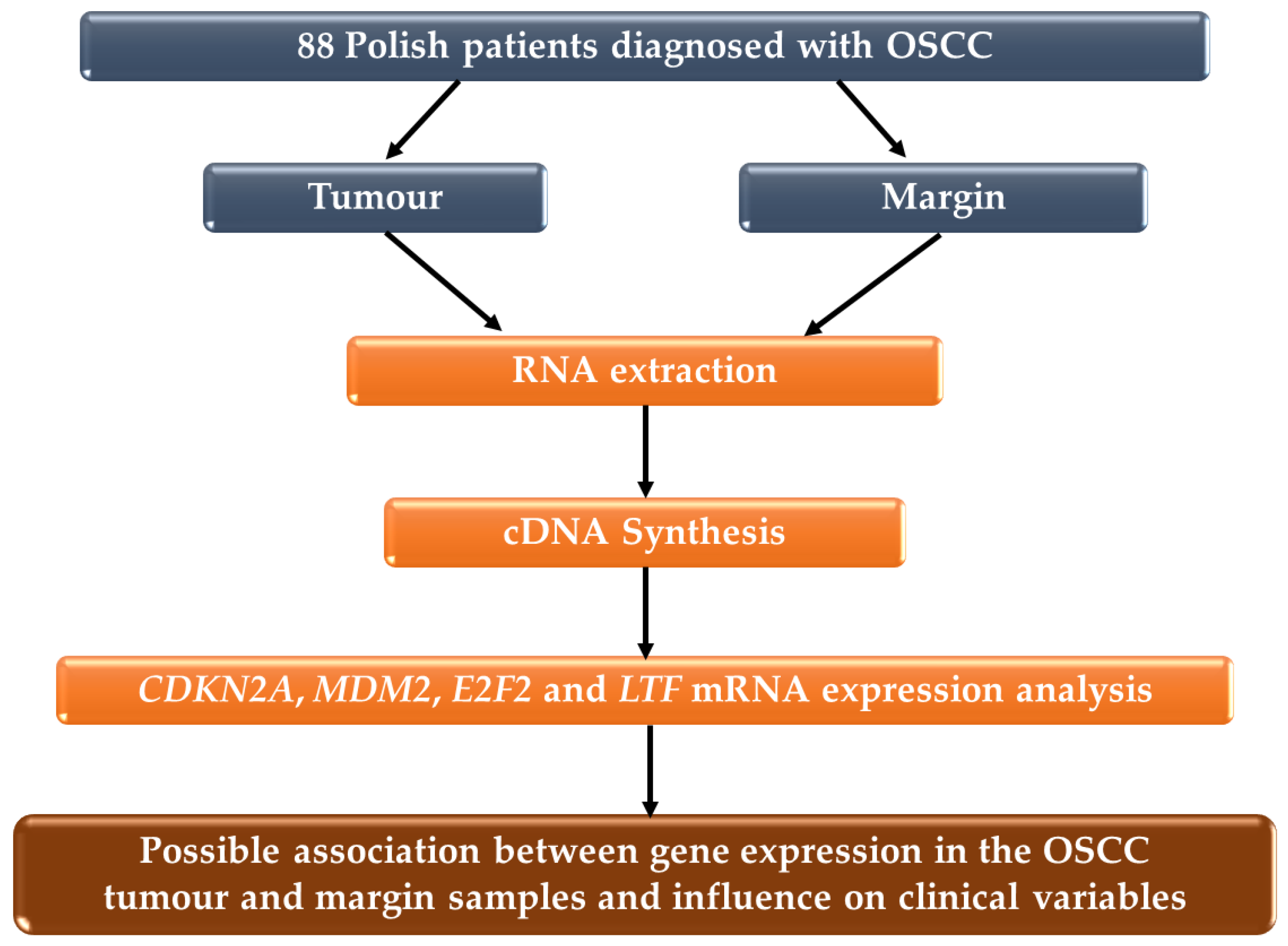
2.1. Patient and Samples

The study group consisted of 88 Polish patients diagnosed with OSCC. The tumour and matching margin specimens were collected following surgical resection at the Department of Otorhinolaryngology and Oncological Laryngology, Faculty of Medical Sciences in Zabrze, Medical University of Silesia in Katowice, Poland and the Maria Sklodowska-Curie National Research Institute of Oncology (formerly known as the Maria Sklodowska-Curie Memorial Cancer Centre and Institute of Oncology), Gliwice, Poland. Tumour staging was based on the American Joint Committee on Cancer (AJCC, version 2007) [11,12] and WHO Classification of Head and Neck Tumours [13]. Margins were verified as being free of cancer by pathologists. All samples after resection were immediately submerged in stabilization solution, RNAlater® (Sigma-Aldrich, Saint Louis, MO, USA), then frozen at −80 °C until RNA extraction. The main inclusion criteria for the OSCC group included a diagnosis of primary tumours and no history of preoperative radio- or chemotherapy. The study was approved by the Bioethics Committee of the Medical University of Silesia (approval no. KNW/022/KB1/49/16 and no. KNW/002/KB1/49/II/16/17) and the Institutional Review Board on Medical Ethics of the Maria Sklodowska-Curie Memorial Cancer Centre and Institute of Oncology in Gliwice (approval no. KB/493-15/08 and no. KB/430-47/13). The study protocol diagram is shown in Figure 1.

2.2. RNA Extraction

The tumour and margin tissue samples were homogenized with FastPrep®-24 homogenizer (MP Biomedicals, Solon, CA, USA) with ceramic beads Lysing Matrix D (MP Biomedicals, Solon, CA, USA). The RNA was isolated using the RNA isolation kit (BioVendor, Brno, Czech Republic) to the standard instruction. The qualitative and quantitative analysis of all isolated RNA was performed by spectrophotometry in Biochrom WPA Biowave DNA UV/Vis Spectrophotometer (Biochrom, Cambridge, UK).

2.3. Complementary DNA (cDNA) Synthesis

Total RNA (5 ng) was reverse-transcribed into cDNA using High Capacity cDNA Reverse Transcription Kit with RNase Inhibitor (Applied Biosystems, Foster City, CA, USA) according to manufacturers’ protocol. The reaction was performed in 20 μL volume containing: 2 μL of 10x Buffer RT; 0.8 μL of 25x dNTP mix (100 mM); 2 μL of 10x RT Random Primers; 1 μL of MultiScribe™ Reverse Transcriptase; 1 μL of RNase inhibitor; 3.2 μL of nuclease free H2O and 10 μL of previously isolated RNA. The reaction was carried out in Mastercycler personal (Eppendorf, Hamburg, Germany) with the following thermal profile: 25 °C for 10 min, 37 °C for 120 min, 85 °C for 5 min and 4 °C–∞.

2.4. Gene Expression Analysis

The relative genes expression (RQ) analysis was performed by Real Time PCR (qPCR) using TaqManTM Gene Expression Assays, QuantStudio 5 RealTime PCR System and Analysis Software v1.5.1 (Applied Biosystems, Foster City, CA, USA). The glyceraldehyde-3-phosphate dehydrogenase gene (GAPDH) was used as an endogenous control. The comparative threshold cycle (Ct) method 2−∆∆Ct was used to determine the RQ. Seven surgical margin samples were used as a calibrator. The qPCR was performed in a volume of 20 µL using 1 µL of cDNA, 10 µL of TaqManTM Fast Advanced Master Mix (Applied Biosystems, Foster City, CA, USA), 1 µL of TaqManTM Gene Expression Assays (Assay ID: Hs00923894_m1 for CDKN2A, Assay ID: Hs01066930_m1 for MDM2, Assay ID: Hs00914334_m1 for E2F2, Assay ID: Hs00918090_m1 for LTF and Assay ID: Hs03929097_g1 for GAPH), and 8 µL of nuclease free H2O (EURx, Gdańsk, Poland). Thermal cycle for all analyzed genes was: 95 °C for 20 s, followed by 40 cycles of 95 °C for 1 s and 60 °C for 20 s.

2.5. Statistical Analysis
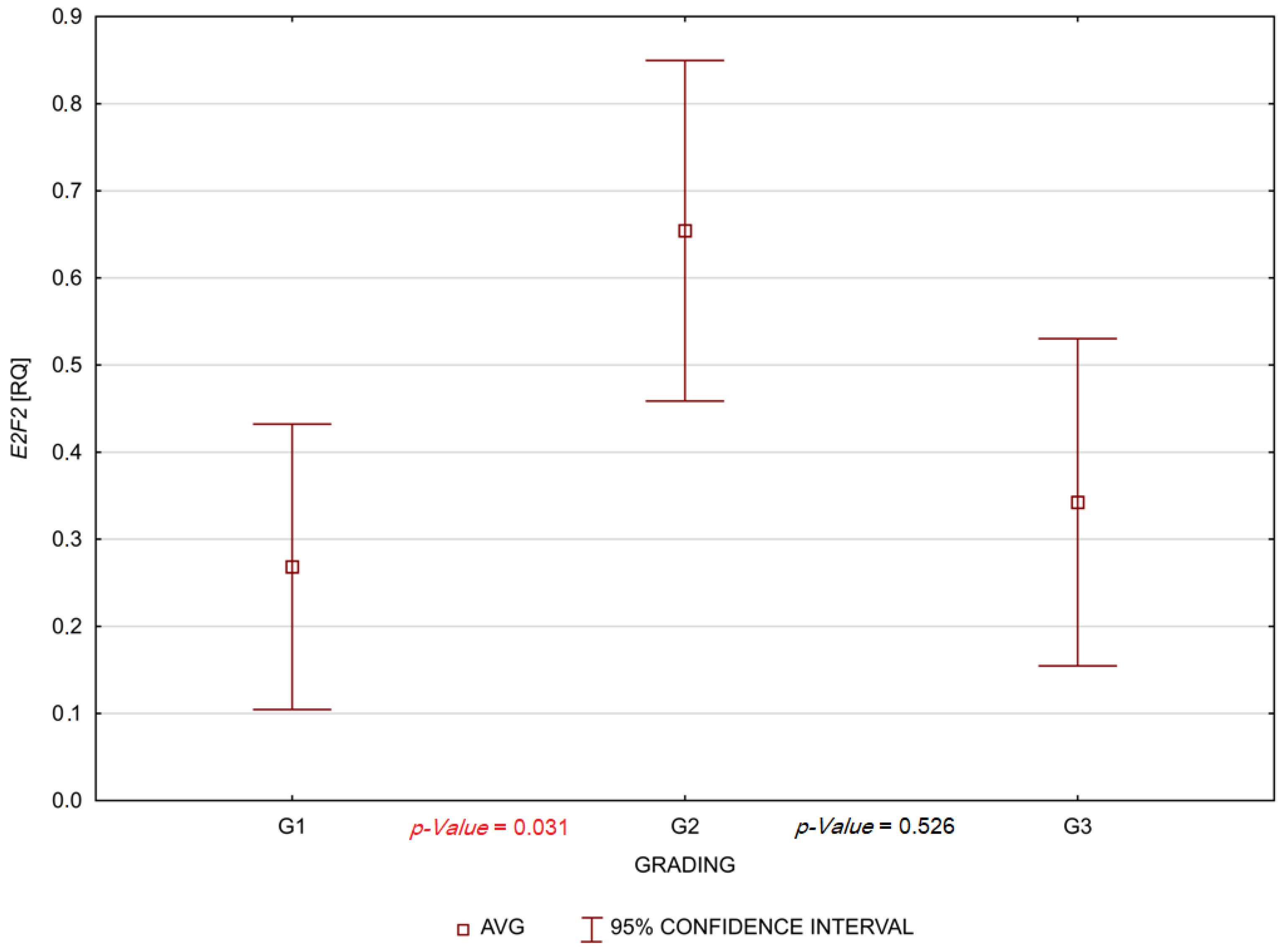
The distribution of variables was evaluated by the Shapiro-Wilk’s test. The significance between genes expression in the tumour and margin were tested using Spearman’s rank correlation analysis. The Mann–Whitney U test and Kruskal-Wallis test were used for comparison gender, age, smoking, alcohol consumption and genes’ expression. The Kruskal-Wallis test with post hoc tests based on mean-ranks were used to determine the effect of the expression of genes in the tumour and margin on clinical parameters (TNM, G). p-Value < 0.05 were considered as statistically significant. The statistical software STATISTICA version 13 (TIBCO Software Inc., Palo Alto, CA, USA) was used to perform all analyses.

3. Results

Clinical parameters of the OSCC group are shown in Table 1. In one patient, the histological gradient (G) was not available. The average age was 56.4 years (range: 18–75 years). There were 60 (68.2%) men and 28 (31.8%) women; 69 (78.4%) patients were smokers; 65 (73.9%) reported alcohol consumption; 51 (57.9%) were both smokers and alcohol users.
We found no statistically significant differences in the expression level of CDKN2A, MDM2, E2F2 and LTF genes in tumour samples compared to margin samples. The relative levels of the expression (RQ values) of CDKN2A, MDM2, E2F2 and LTF genes are shown in Table 2.
We also assessed the correlation of the expression levels of selected genes with age, gender, smoking, alcohol consumption and clinical characteristics of the patients. We demonstrated several statistically significant relationships. The expression of the MDM2 gene was negative correlated with age only in men (p-Value = 0.03) in tumour samples. A statistically significant negative correlation between patient age and MDM2 gene expression in tumour was found only in non-smokers (p-Value = 0.021). We also showed statistically significant positive correlation between age and E2F2 expression in the non-drinker in margin (p-Value = 0.013). Statistically significant positive correlations occurred in non-drinkers and smokers, referring to the expression of the CDKN2A gene (p-Value = 0.016) and the E2F2 gene also in the margin (p-Value = 0.001).
No association was found between the gene expression levels, age, gender, smoking, alcohol consumption and clinical parameters (TNM and G) in tumour and margin samples, except E2F2. The patients with G2 (moderately differentiated) tumours had a significantly higher gene expression level of E2F2 than patients with low-grade G1 (well-differentiated) tumours (p-Value = 0.031) in tumour samples. These results are shown in Figure 2.

4. Discussion

One of the most common malignant tumours of the oral cavity is OSCC [3]. Not all the molecular mechanisms that lead to this type of cancer have yet been elucidated. Therefore, it seems useful to carry out analyses to investigate changes in gene expression, including those related to the regulation of the cell cycle. In this manuscript, we performed an analysis of CDKN2A, MDM2, E2F2 and LTF expressions in the pathogenesis of OSCC.
We found no statistically significant differences in the expression level CDKN2A, MDM2, E2F2 and LTF genes in tumour samples compared to margin samples. Interestingly, we demonstrated a statistically significant association with the expression of CDKN2A in margin tissue in non-drinkers and smokers. CDKN2A expression may be higher in human papillomavirus (HPV) infections [14]. Bearing in mind the above results, it seems useful to investigate the status of HPV infection in our samples. However, the change in the expression of p16 (a protein encoded by CDKN2A) could be not only outcome virus infection but also DNA damage and gene mutation [15]. Tobacco smoke contains mutagenic substances [16], which could be related to our changes in CDKN2A expression in smokers. The association between CDKN2A expression and HPV infection in oral and cervical cancer demonstrated that HPV acts via the oncoprotein E7, which can bind the Rb protein, resulting in the inhibition of the formation of the Rb complex with E2F family of transcription factors (for example E2F2). As a result, free E2F2 in absence of the complex with Rb can increase the expression of CDKN2A [17,18,19]. Some studies have demonstrated that changes in the expression, promoter methylation or copy number deletion of CDKN2A were relatively common in the development and prognosis of OSCC [14,20,21,22,23,24,25]. There are reports that researchers have observed that decreased CDKN2A expression was associated with OSCC [26,27]. There are also studies demonstrating the overexpression of CDKN2A in OSCC [28,29]. The Wang and others’ [30] analysis demonstrated that the expression of CDKN2A was upregulated in OSCC tissues compared with normal tissues in Oncomine database (p-Value < 0.01). Furthermore, mRNA expression of the CDKN2A in HNSCC (head and neck squamous cell carcinoma) was upregulated compared with normal tissues in the GEPIA (Gene Expression Profiling Interactive Analysis) database (p-Value < 0.01). On the other hand, in the GEPIA database, a high expression of CDKN2A was associated with better survival in HNSCC patients (p-Value < 0.05) [30]. The different results of the studies can be explained by differences in the pathogenesis of a subset of OSCCs [14].
The next gene we analyzed in our work was the oncogene MDM2. The expression of MDM2 gene was negative correlated with age only in men (p-Value = 0.03) in tumour samples. A statistically significant negative correlation between patient age and MDM2 gene expression in tumour was found only in non-smokers (p-Value = 0.021). The lowest expression of MDM2 protein was observed in larynx carcinoma specimens. It is worth adding that the expression of MDM2 by activation of p53 may be caused by DNA damage associated with exposure to tobacco and/or alcohol carcinogens [31]. MDM2 overexpression can be observed in many cancers such as lung cancer, breast cancer, liver cancer, esophagogastric cancer and colorectal cancer. Overexpression of MDM2 is the most common change, especially in patients with lung cancer [6]. Higher expression of this protein has also been observed in HNSCC [32,33]. Change in MDM2 expression is also associated with a poorer prognosis in the squamous cell carcinoma of the tonsillar region [34]. Millon and other [31] noted the overexpressed MDM2 mRNA and a significant decrease in MDM expression in an advanced T stage in tumour samples with HNSCC. The gene expression level was not related with either lymph node involvement or differentiation status of the tumour.
It is known that E2F2 play a significant role in promoting the cell cycle [8]. Some studies demonstrate increased E2F2 gene expression in cancers such as ovarian cancer, non-small cell lung cancer, hepatocellular carcinoma and breast cancer [35,36,37,38]. Our study demonstrated a statistically significant positive correlation between age and E2F2 expression in the non-drinker in margin (p-Value = 0.013). Statistically significant positive correlations occurred in non-drinkers and smokers, referring to the expression of E2F2 gene also in the margin (p-Value = 0.001). It is also worth emphasizing that patients with G2 (moderately differentiated) tumours had a significantly higher gene expression level of E2F2 than patients with low-grade G1 (well-differentiated) tumours. Perhaps these results are related to the fact that E2F2 can act as both a promoter of cell division and a suppressor. Moreover, the E2F2 expression may be associated with the CDKN2A expression. Another study analyzed E2F2 mRNA expression levels in the human brain and central nervous system cancers types via Oncomine and GEPIA databases. Researchers have demonstrated the overexpression of E2F2 in glioblastoma multiforme and low-grade glioma tissues [39]. Li and others [40] documented that E2F2 variants are predictive biomarkers for recurrence risk in patients with OPSCC (oropharyngeal squamous cell carcinoma) [40]. On the other hand, E2F2 can also have a function as a suppressor in colon cancer. This mechanism is related to the fact that E2F2 inhibited the proliferation of cancer cells by inhibiting the expression of survivin and regulating the expression of molecules such as cyclin A2, proto-oncogene C-Myc, minichromosome maintenance complex component 4 and cyclin dependent kinase 2 [41]. In addition, the other study demonstrated a low level of E2F2 expression in clear cell renal cell carcinoma cancer tissues [42]. It seems that molecules such as small non-coding RNAs—miRNA (microRNA) and long non-coding RNA (lncRNA) could play important roles in the regulation of E2F expression. Some authors were suggested that miRNAs (for example microRNA-let-7a, miR-125b, miR-146b-3p, miR-638, miR-31, miR-218, miR-454-3p and miRNA-936) by targeting E2F2, could be influenced by cancer progression, overall survival of patients and response to radiochemotherapy. These studies focused on types of cancer such as breast cancer, ovarian cancer, colon cancer, gastric cancer, prostate cancer, hepatocellular carcinoma, non-small cell lung cancer cell, glioma and laryngeal cancer [37,38,41,43,44,45,46,47,48]. It is also known that circ_RPPH1/miR-146b-3p/E2F2 axis can promote the progression of breast cancer [48]. Moreover, circCUL2 suppresses retinoblastoma cells by regulating miR-214-5p/E2F2 axis [49].
It is also known that LTF could be used for cancer prognosis [50]. Chiu and others [51] demonstrated that primary tumours of clear cell renal cell carcinoma in women characterized by the downregulation of LTF had higher pathologic stages [51]. Another study demonstrated that LTF expression was negatively correlated with metastases and T-stage in nasopharyngeal carcinoma (NPC) samples. LTF-mRNA expression in NPC tissues was significantly lower than in healthy tissue [52]. In our study, we found no statistically significant differences in the expression of LTF genes in tumour samples compared to margin samples and with clinical features of the OSCC. Some studies demonstrate that hypermethylation in promoter regions was the factor that may be involved in expression regulation. Zhang and others [53] demonstrated that the low or absent LTF expression in OSCC tissues and TCA8113 cells may be a result of hypermethylation in the promoter regions of LTF, which exerted negative effects on the malignance of OSSC cells [53]. The LTF promoter hypermethylation was also reported in prostate cancer [54]. Another factor determined that LTF expression could be miRNA (mikroRNA), for example miR-214. Deng and others [55] found that the overexpression of miR-214 reduced LTF mRNA levels and protein levels in NPC [55].
In summary, to the best of our knowledge, our study is the first that aimed to determine the association between MDM2, E2F2, and LTF mRNA expression in oral squamous cell carcinoma tumour and margin samples. This is given the idea of Slaughter et al. [56,57] of the initiation of field cancerization being associated with various types of molecular damage, e.g., altered gene expression. A molecular approach to the matching margin can contribute to cancer prevention and control. Our result should be validated on larger and different cohorts to better comprehend the role of CDKN2A, MDM2, E2F2, and LTF genes in OSCC. In addition, in vitro/pre-clinical tests with cell lines, and two-dimensional (2D) and three-dimensional (3D) models are required, which could provide valuable information for a better understanding of the oral carcinogenesis process such as invasion and metastasis [58].

5. Conclusions

In summary, we have not demonstrated that a change in expression profiles of CDKN2A, MDM2, E2F2 and LTF genes in tumour and margin tissues has a significant impact on the pathogenesis of OSCC. Therefore, further molecular analysis of other genes involved in the regulation of the cell cycle is required. It may also be useful to conduct further studies on the use of E2F2 expression profile changes as a factor to describe the invasiveness and dynamics of the OSCC development.

Author Contributions

K.G., research concept and design; K.M.-O., N.Z. and Ł.K., collection of tumour samples; K.G. and J.G., RNA extraction; K.G., cDNA synthesis and gene expression analyses; K.G. and G.R., data analysis and interpretation; K.G., writing of the article; M.M. and J.K.S., critical revision of the article. All authors have read and agreed to the published version of the manuscript.

Funding

This research was funded from the grant by the Medical University of Silesia (KNW-1-014/N/9/O).

Institutional Review Board Statement

The study was conducted according to the guidelines of the Declaration of Helsinki, and approved by the Bioethics Committee of the Medical University of Silesia (Katowice, Poland; approval no. KNW/022/KB1/49/16 and no. KNW/002/KB1/49/II/16/17) and the Institutional Review Board on the Medical Ethics of the Maria Sklodowska-Curie Memorial Cancer Centre and Institute of Oncology in Gliwice (Poland; approval no. KB/493-15/08 and no. KB/430-47/13).

Informed Consent Statement

Informed consent was obtained from all subjects involved in the study.

Data Availability Statement

The data used to support the findings of this study are available from the corresponding author upon request.

Conflicts of Interest

The authors declare no conflict of interest. The funders had no role in the design of the study, in the collection, analyses, or interpretation of data, in the writing of the manuscript, or in the decision to publish the results.

References

  1. Sung, H.; Ferlay, J.; Siegel, R.L.; Laversanne, M.; Soerjomataram, I.; Jemal, A.; Bray, F. Global Cancer Statistics 2020: GLOBOCAN Estimates of Incidence and Mortality Worldwide for 36 Cancers in 185 Countries. CA Cancer J. Clin. 2021, 71, 2096–2249. [Google Scholar] [CrossRef] [PubMed]
  2. Gollin, S.M. Cytogenetic alterations and their molecular genetic correlates in head and neck squamous cell carcinoma: A next generation window to the biology of disease. Genes Chromosom. Cancer 2014, 53, 972–990. [Google Scholar] [CrossRef] [PubMed]
  3. Cho, U.; Sung, Y.E.; Kim, M.S.; Lee, Y.S. Prognostic Role of Systemic Inflammatory Markers in Patients Undergoing Surgical Resection for Oral Squamous Cell Carcinoma. Biomedicines 2022, 10, 1268. [Google Scholar] [CrossRef]
  4. Gallagher, S.; Kefford, R.F.; Rizos, H. Enforced expression of p14ARF induces p53-dependent cell cycle arrest but not apoptosis. Cell Cycle 2005, 4, 465–472. [Google Scholar] [CrossRef] [PubMed]
  5. Ausoni, S.; Boscolo-Rizzo, P.; Singh, B.; Da Mosto, M.C.; Spinato, G.; Tirelli, G.; Spinato, R.; Azzarello, G. Targeting cellular and molecular drivers of head and neck squamous cell carcinoma: Current options and emerging perspectives. Cancer Metastasis Rev. 2016, 35, 413–426. [Google Scholar] [CrossRef] [PubMed] [Green Version]
  6. Hou, H.; Sun, D.; Zhang, X. The role of MDM2 amplification and overexpression in therapeutic resistance of malignant tumors. Cancer Cell Int. 2019, 19, 216. [Google Scholar] [CrossRef] [Green Version]
  7. Rayburn, E.; Zhang, R.; He, J.; Wang, H. MDM2 and human malig-nancies: Expression, clinical pathology, prognostic markers, and implications for chemotherapy. Curr. Cancer Drug Targets 2005, 5, 27–41. [Google Scholar] [CrossRef] [Green Version]
  8. Li, L.; Wang, S.; Zhang, Y.; Pan, J. The E2F transcription factor 2: What do we know? BioSci. Trends 2021, 15, 83–92. [Google Scholar] [CrossRef]
  9. Cutone, A.; Rosa, L.; Ianiro, G.; Lepanto, M.S.; Bonaccorsi di Patti, M.C.; Valenti, P.; Musci, G. Lactoferrin’s Anti-Cancer Properties: Safety, Selectivity, and Wide Range of Action. Biomolecules 2020, 15, 456. [Google Scholar] [CrossRef] [Green Version]
  10. Bukowska-Ośko, I.; Sulejczak, D.; Kaczyńska, K.; Kleczkowska, P.; Kramkowski, K.; Popiel, M.; Wietrak, E.; Kowalczyk, P. Lactoferrin as a Human Genome “Guardian”—An Overall Point of View. Int. J. Mol. Sci. 2022, 23, 5248. [Google Scholar] [CrossRef]
  11. Brunner, M.; Ng, B.C.; Veness, M.J.; Clark, J.R. Comparison of the AJCC N staging system in mucosal and cutaneous squamous head and neck cancer. Laryngoscope 2014, 124, 1598–1602. [Google Scholar] [CrossRef] [PubMed]
  12. Rodrigues, P.C.; Miguel, M.C.; Bagordakis, E.; Fonseca, F.P.; de Aquino, S.N.; Santos-Silva, A.R.; Lopes, M.A.; Graner, E.; Salo, T.; Kowalski, L.P.; et al. Clinicopathological prognostic factors of oral tongue squamous cell carcinoma: A retrospective study of 202 cases. Int. J. Oral. Maxillofac. Surg. 2014, 43, 795–801. [Google Scholar] [CrossRef] [PubMed]
  13. El-Naggar, A.K.; Chan, J.K.C.; Grandis, J.R.; Takata, T.; Slootweg, P.J. WHO Classification of Head and Neck Tumours International Agency for Research on Cancer (IARC), 4th ed.; IARC Publications: Lyon, France, 2017. [Google Scholar]
  14. Allameh, A.; Moazeni-Roodi, A.; Harirchi, I.; Ravanshad, M.; Motiee-Langroudi, M.; Garajei, A.; Hamidavi, A.; Mesbah-Namin, S.A. Promoter DNA Methylation and mRNA Expression Level of p16 Gene in Oral Squamous Cell Carcinoma: Correlation with Clinicopathological Characteristics. Pathol. Oncol. Res. 2019, 25, 1535–1543. [Google Scholar] [CrossRef]
  15. Biesaga, B.; Smolarczyk, R.; Mucha-Małecka, A.; Czapla, J.; Ryś, J.; Małecki, K. Prognostic Significance of STING Immunoexpression in Relation to HPV16 Infection in Patients with Squamous Cell Carcinomas of Oral Cavity and Oropharynx. Biomedicines 2022, 10, 2538. [Google Scholar] [CrossRef] [PubMed]
  16. Omolaoye, T.S.; El Shahawy, O.; Skosana, B.T.; Boillat, T.; Loney, T.; du Plessis, S.S. The mutagenic effect of tobacco smoke on male fertility. Env. Sci. Pollut. Res. 2022, 29, 62055–62066. [Google Scholar] [CrossRef]
  17. König, F.; Krekeler, G.; Hönig, J.F.; Cordon-Cardo, C.; Fischer, G.; Korabiowska, M. Relation between human papillomavirus positivity and p16 expression in head and neck carcinomas--a tissue microarray study. Anticancer Res. 2007, 27, 283–288. [Google Scholar] [PubMed]
  18. Li, J.; Poi, M.J.; Tsai, M.D. Regulatory mechanisms of tumor suppressor p16(INK4A) and their relevance to cancer. Biochemistry 2011, 50, 5566–5582. [Google Scholar] [CrossRef] [PubMed] [Green Version]
  19. El-Naggar, A.K.; Westra, W.H. p16 expression as a surrogate marker for HPV-related oropharyngeal carcinoma: A guide for interpretative relevance and consistency. Head Neck 2012, 34, 459–461. [Google Scholar] [CrossRef]
  20. Wang, D.; Grecula, J.C.; Gahbauer, R.A.; Schuller, D.E.; Jatana, K.R.; Biancamano, J.D.; Lang, J.C. p16 gene alterations in locally advanced squamous cell carcinoma of the head and neck. Oncol. Rep. 2006, 15, 661–665. [Google Scholar] [CrossRef] [Green Version]
  21. Su, P.F.; Huang, W.L.; Wu, H.T.; Wu, C.H.; Liu, T.Y.; Kao, S.Y. p16(INK4A) promoter hypermethylation is associated with invasiveness and prognosis of oral squamous cell carcinoma in an age-dependent manner. Oral Oncol. 2010, 46, 734–739. [Google Scholar] [CrossRef]
  22. Cabanillas, R.; Astudillo, A.; Valle, M.; de la Rosa, J.; Álvarez, R.; Durán, N.S.; Cadiñanos, J. Novel germline CDKN2A mutation associated with head and neck squamous cell carcinomas and melanomas. Head Neck 2013, 35, E80–E84. [Google Scholar] [CrossRef] [PubMed]
  23. Padhi, S.S.; Roy, S.; Kar, M.; Saha, A.; Roy, S.; Adhya, A.; Baisakh, M.; Banerjee, B. Role of CDKN2A/p16 expression in the prognostication of oral squamous cell carcinoma. Oral Oncol. 2017, 73, 27–35. [Google Scholar] [CrossRef] [PubMed]
  24. Yalniz, Z.; Demokan, S.; Karabulut, B.; Ulusan, M.; Suoglu, Y.; Dalay, N. Copy number profiling of tumor suppressor genes in head and neck cancer. Head Neck 2017, 39, 341–346. [Google Scholar] [CrossRef]
  25. Jie, W.; Bai, J.; Yan, J.; Chi, Y.; Li, B.B. Multi-Site Tumour Sampling Improves the Detection of Intra-Tumour Heterogeneity in Oral and Oropharyngeal Squamous Cell Carcinoma. Front. Med. 2021, 8, 670305. [Google Scholar] [CrossRef]
  26. Bhatia, V.; Goel, M.M.; Makker, A.; Tewari, S.; Yadu, A.; Shilpi, P.; Kumar, S.; Agarwal, S.P.; Goel, S.K. Promoter region hypermethylation and mRNA expression of MGMT and p16 genes in tissue and blood samples of human premalignant oral lesions and oral squamous cell carcinoma. Biomed Res. Int. 2014, 2014, 248419. [Google Scholar] [CrossRef] [PubMed] [Green Version]
  27. Pérez-Sayáns, M.; Suárez-Peñaranda, J.M.; Padín-Iruegas, M.E.; Gayoso-Diz, P.; Reis-De Almeida, M.; Barros-Angueira, F.; Gándara-Vila, P.; Blanco-Carrión, A.; García-García, A. The Loss of p16 Expression Worsens the Prognosis of OSCC. Appl. Immunohistochem. Mol. Morphol. 2015, 23, 724–732. [Google Scholar] [CrossRef]
  28. Salehinejad, J.; Sharifi, N.; Amirchaghmaghi, M.; Ghazi, N.; Shakeri, M.T.; Ghazi, A. Immunohistochemical expression of p16 protein in oral squamous cell carcinoma and lichen planus. Ann. Diagn. Pathol. 2014, 18, 210–213. [Google Scholar] [CrossRef]
  29. Jitani, A.K.; Raphael, V.; Mishra, J.; Shunyu, N.B.; Khonglah, Y.; Medhi, J. Analysis of Human Papilloma Virus 16/18 DNA and its Correlation with p16 Expression in Oral Cavity Squamous Cell Carcinoma in North-Eastern India: A Chromogenic in-situ Hybridization Based Study. J. Clin. Diagn. Res. 2015, 9, EC04–EC07. [Google Scholar] [CrossRef]
  30. Wang, S.; Li, T.; Liu, H.; Wei, W.; Yang, Y.; Wang, C.; Li, B.; Han, Z.; Feng, Z. A Combined Prediction Model for Lymph Node Metastasis Based on a Molecular Panel and Clinicopathological Factors in Oral Squamous Cell Carcinoma. Front. Oncol. 2021, 11, 660615. [Google Scholar] [CrossRef]
  31. Millon, R.; Muller, D.; Schultz, I.; Salvi, R.; Ghnassia, J.P.; Frebourg, T.; Wasylyk, B.; Abecassis, J. Loss of MDM2 expression in human head and neck squamous cell carcinomas and clinical significance. Oral Oncol. 2001, 37, 620–631. [Google Scholar] [CrossRef]
  32. Carroll, P.E.; Okuda, M.; Horn, H.F.; Biddinger, P.; Stambrook, P.J.; Gleich, L.L.; Li, Y.Q.; Tarapore, P.; Fukasawa, K. Centrosome hyperamplification in human cancer: Chromosome instability induced by p53 mutation and/or Mdm2 overexpression. Oncogene 1999, 18, 1935–1944. [Google Scholar] [CrossRef] [PubMed] [Green Version]
  33. Valentin-Vega, Y.A.; Barboza, J.A.; Chau, G.P.; El-Naggar, A.K.; Lozano, G. High levels of the p53 inhibitor MDM4 in head and neck squamous carcinomas. Hum. Pathol. 2007, 38, 1553–1562. [Google Scholar] [CrossRef] [PubMed] [Green Version]
  34. Friesland, S.; Kanter-Lewensohn, L.; Tell, R.; Munck-Wikland, E.; Lewensohn, R.; Nilsson, A. Expression of Ku86 confers favorable outcome of tonsillar carcinoma treated with radiotherapy. Head Neck 2003, 25, 313–321. [Google Scholar] [CrossRef]
  35. Dong, Y.; Zou, J.; Su, S.; Huang, H.; Deng, Y.; Wang, B.; Li, W. MicroRNA-218 and microRNA-520a inhibit cell proliferation by downregulating E2F2 in hepatocellular carcinoma. Mol. Med. Rep. 2015, 12, 1016–1022. [Google Scholar] [CrossRef] [PubMed] [Green Version]
  36. Xie, L.; Li, T.; Yang, L.H. E2F2 induces MCM4, CCNE2 and WHSC1 upregulation in ovarian cancer and predicts poor overall survival. Eur. Rev. Med. Pharm. Sci. 2017, 21, 2150–2156. [Google Scholar]
  37. Zhou, X.; Tao, H. Overexpression of microRNA-936 suppresses non-small cell lung cancer cell proliferation and invasion via targeting E2F2. Exp. Ther. Med. 2018, 16, 2696–2702. [Google Scholar] [CrossRef] [Green Version]
  38. Lin, Q.Y.; Wang, J.Q.; Wu, L.L.; Zheng, W.E.; Chen, P.R. miR-638 represses the stem cell characteristics of breast cancer cells by targeting E2F2. Breast Cancer 2020, 27, 147–158. [Google Scholar] [CrossRef]
  39. Liao, P.; Han, S.; Qu, H. Expression, Prognosis, and Immune Infiltrates Analyses of E2Fs in Human Brain and CNS Cancer. Biomed Res. Int. 2020, 2020, 6281635. [Google Scholar] [CrossRef]
  40. Li, Y.; Sturgis, E.M.; Zhu, L.; Cao, X.; Wei, Q.; Zhang, H.; Li, G. E2F transcription factor 2 variants as predictive biomarkers for recurrence risk in patients with squamous cell carcinoma of the oropharynx. Mol. Carcinog. 2017, 56, 1335–1343. [Google Scholar] [CrossRef]
  41. Li, T.; Luo, W.; Liu, K.; Lv, X.; Xi, T. miR-31 promotes proliferation of colon cancer cells by targeting E2F2. Biotechnol. Lett. 2015, 37, 523–532. [Google Scholar] [CrossRef]
  42. Gao, Y.; Ma, X.; Yao, Y.; Li, H.; Fan, Y.; Zhang, Y.; Zhao, C.; Wang, L.; Ma, M.; Lei, Z.; et al. miR-155 regulates the proliferation and invasion of clear cell renal cell carcinoma cells by targeting E2F2. Oncotarget 2016, 7, 20324–20337. [Google Scholar] [CrossRef] [PubMed]
  43. Zhang, Y.; Han, D.; Wei, W.; Cao, W.; Zhang, R.; Dong, Q.; Zhang, J.; Wang, Y.; Liu, N. MiR-218 Inhibited Growth and Metabolism of Human Glioblastoma Cells by Directly Targeting E2F2. Cell. Mol. Neurobiol. 2015, 35, 1165–1173. [Google Scholar] [CrossRef]
  44. Wang, H.; Zhang, X.; Liu, Y.; Ni, Z.; Lin, Y.; Duan, Z.; Shi, Y.; Wang, G.; Li, F. Downregulated miR-31 level associates with poor prognosis of gastric cancer and its restoration suppresses tumor cell malignant phenotypes by inhibiting E2F2. Oncotarget 2016, 7, 36577–36589. [Google Scholar] [CrossRef] [Green Version]
  45. Fang, Z.Q.; Li, M.C.; Zhang, Y.Q.; Liu, X.G. MiR-490-5p inhibits the metastasis of hepatocellular carcinoma by down-regulating E2F2 and ECT2. J. Cell Biochem. 2018, 119, 8317–8324. [Google Scholar] [CrossRef] [PubMed]
  46. Cui, X.; Xiao, D.; Cui, Y.; Wang, X. Exosomes-Derived Long Non-Coding RNA HOTAIR Reduces Laryngeal Cancer Radiosensitivity by Regulating microRNA-454-3p/E2F2 Axis. Onco Targets Ther. 2019, 12, 10827–10839. [Google Scholar] [CrossRef] [PubMed] [Green Version]
  47. Miyamoto, M.; Sawada, K.; Nakamura, K.; Yoshimura, A.; Ishida, K.; Kobayashi, M.; Shimizu, A.; Yamamoto, M.; Kodama, M.; Hashimoto, K.; et al. Paclitaxel exposure downregulates miR-522 expression and its downregulation induces paclitaxel resistance in ovarian cancer cells. Sci. Rep. 2020, 10, 16755. [Google Scholar] [CrossRef] [PubMed]
  48. Feng, H.; Sun, S.Z.; Cheng, F.; Zhang, N.Q. Mediation of circ_RPPH1 on miR-146b-3p/E2F2 pathway to hinder the growth and metastasis of breast carcinoma cells. Aging 2021, 13, 20552–20568. [Google Scholar] [CrossRef]
  49. Zhang, H.; Qiu, X.; Song, Z.; Lan, L.; Ren, X.; Ye, B. CircCUL2 suppresses retinoblastoma cells by regulating miR-214-5p/E2F2 Axis. Anticancer Drugs 2022, 33, e218–e227. [Google Scholar] [CrossRef]
  50. Shaheduzzaman, S.; Vishwanath, A.; Furusato, B.; Cullen, J.; Chen, Y.; Bañez, L.; Nau, M.; Ravindranath, L.; Kim, K.H.; Mohammed, A.; et al. Silencing of Lactotransferrin expression by methylation in prostate cancer progression. Cancer Biol. Ther. 2007, 6, 1088–1095. [Google Scholar] [CrossRef] [Green Version]
  51. Chiu, I.J.; Hsu, Y.H.; Chang, J.S.; Yang, J.C.; Chiu, H.W.; Lin, Y.F. Lactotransferrin Downregulation Drives the Metastatic Progression in Clear Cell Renal Cell Carcinoma. Cancers 2020, 12, 847. [Google Scholar] [CrossRef] [Green Version]
  52. Zhang, W.; Fan, S.; Zou, G.; Shi, L.; Zeng, Z.; Ma, J.; Zhou, Y.; Li, X.; Zhang, X.; Li, X.; et al. Lactotransferrin could be a novel independent molecular prognosticator of nasopharyngeal carcinoma. Tumor Biol. 2015, 36, 675–683. [Google Scholar] [CrossRef] [PubMed]
  53. Zhang, J.; Ling, T.; Wu, H.; Wang, K. Re-expression of Lactotransferrin, a candidate tumor suppressor inactivated by promoter hypermethylation, impairs the malignance of oral squamous cell carcinoma cells. J. Oral. Pathol. Med. 2015, 44, 578–584. [Google Scholar] [CrossRef] [PubMed]
  54. Porter, C.M.; Haffner, M.C.; Kulac, I.; Maynard, J.P.; Baena-Del Valle, J.A.; Isaacs, W.B.; Yegnasubramanian, S.; De Marzo, A.M.; Sfanos, K.S. Lactoferrin CpG Island Hypermethylation and Decoupling of mRNA and Protein Expression in the Early Stages of Prostate Carcinogenesis. Am. J. Pathol. 2019, 189, 2311–2322. [Google Scholar] [CrossRef]
  55. Deng, M.; Zhang, W.; Tang, H.; Ye, Q.; Liao, Q.; Zhou, Y.; Wu, M.; Xiong, W.; Zheng, Y.; Guo, X.; et al. Lactotransferrin acts as a tumor suppressor in nasopharyngeal carcinoma by repressing AKT through multiple mechanisms. Oncogene 2013, 32, 4273–4283. [Google Scholar] [CrossRef] [Green Version]
  56. Slaughter, D.P.; Southwick, H.W.; Smejkal, W. Field cancerization in oral stratified squamous epithelium; clinical implications of multicentric origin. Cancer 1953, 6, 963–968. [Google Scholar] [CrossRef] [PubMed]
  57. Braakhuis, B.J.; Tabor, M.P.; Kummer, J.A.; Leemans, C.R.; Brakenhoff, R.H. A genetic explanation of Slaughter’s concept of field cancerization: Evidence and clinical implications. Cancer Res. 2003, 63, 1727–1730. [Google Scholar] [PubMed]
  58. Hoque Apu, E.; Akram, S.U.; Rissanen, J.; Wan, H.; Salo, T. Desmoglein 3—Influence on oral carcinoma cell migration and invasion. Exp. Cell Res. 2018, 370, 353–364. [Google Scholar] [CrossRef]
Figure 1. Study protocol diagram.
Biomedicines 10 03011 g001
Figure 2. Relative quantification (RQ) values of E2F2 in the group of patients with OSCC according to G1, G2 and G3 in tumour samples.
Biomedicines 10 03011 g002
Table 1. Clinical parameters of patients with OSCC.
Clinical ParametersPatients, n (%)
Histological grading
G1 (Well differentiated)14 (16)
G2 (Moderately differentiated)60 (69)
G3 (Poorly differentiated)13 (15)
T classification
T18 (9)
T224 (27.3)
T323 (26.2)
T433 (37.5)
Nodal status
N040 (45, 5)
N125 (28, 4)
N220 (22, 7)
N33 (3, 4)
Table 2. Relative quantification (RQ) values for CDKN2A, MDM2, E2F2 and LTF genes in tumour vs. margin in patients with OSCC (Spearman’s rank correlation coefficients r, p-Value).
GeneMean RQ ± SDrp-Value
TumourMargin
CDKN2A2.08 ± 4.990.45 ± 0.99−0.0310.782
MDM20.7 ± 0.80.68 ± 0.390.1880.088
E2F20.54 ± 0.650.5 ± 0.440.2080.061
LTF0.04 ± 0.130.13 ± 0.290.0970.378
Publisher’s Note: MDPI stays neutral with regard to jurisdictional claims in published maps and institutional affiliations.

