
Major Hepatectomy En Bloc with Cava Vein Resection for Locally Invasive Caudate Lobe Hepatocarcinoma

Abstract
:1. Introduction
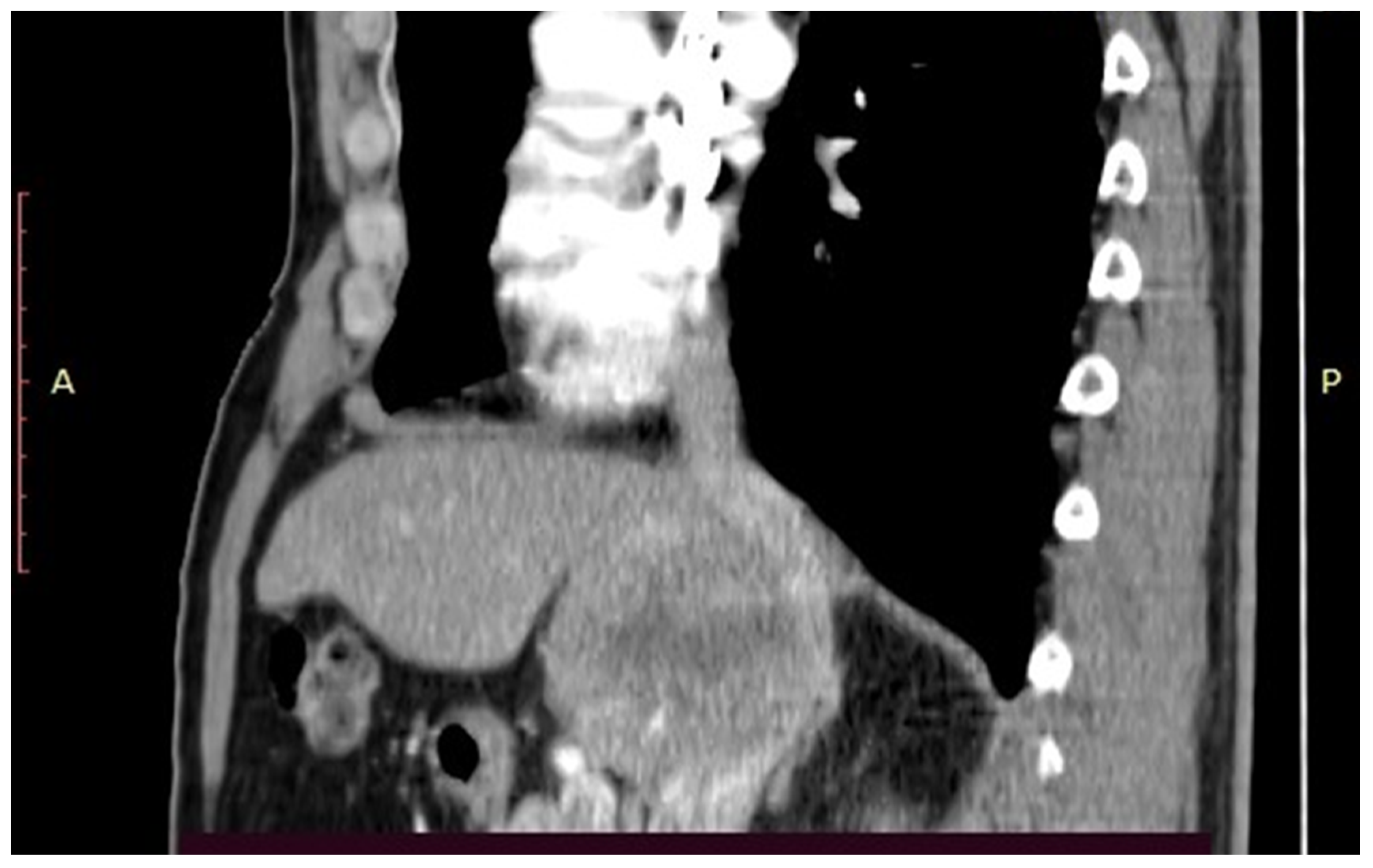
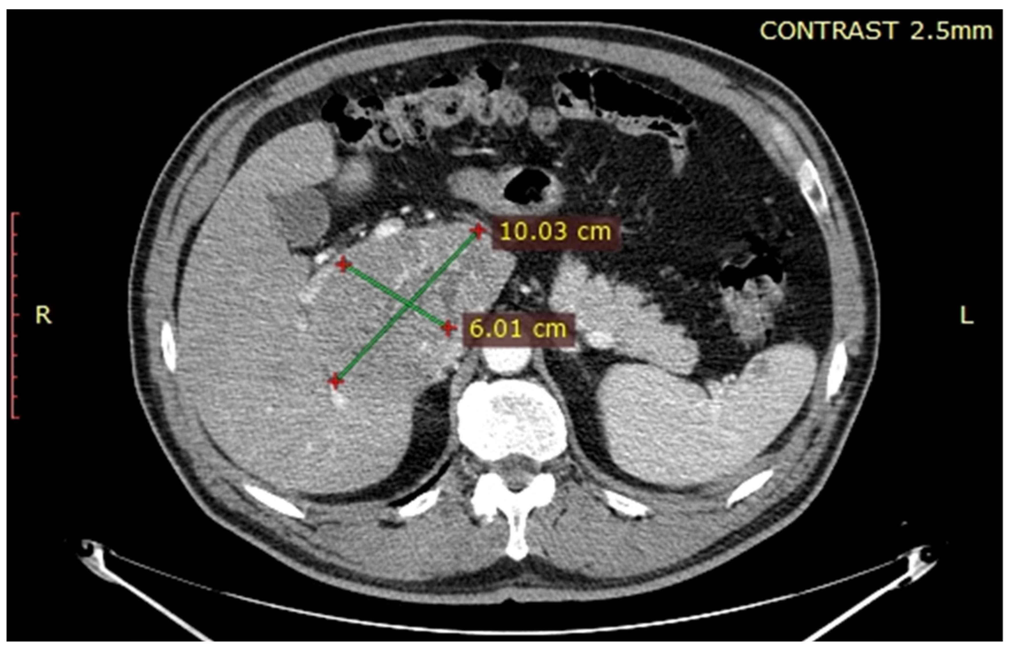
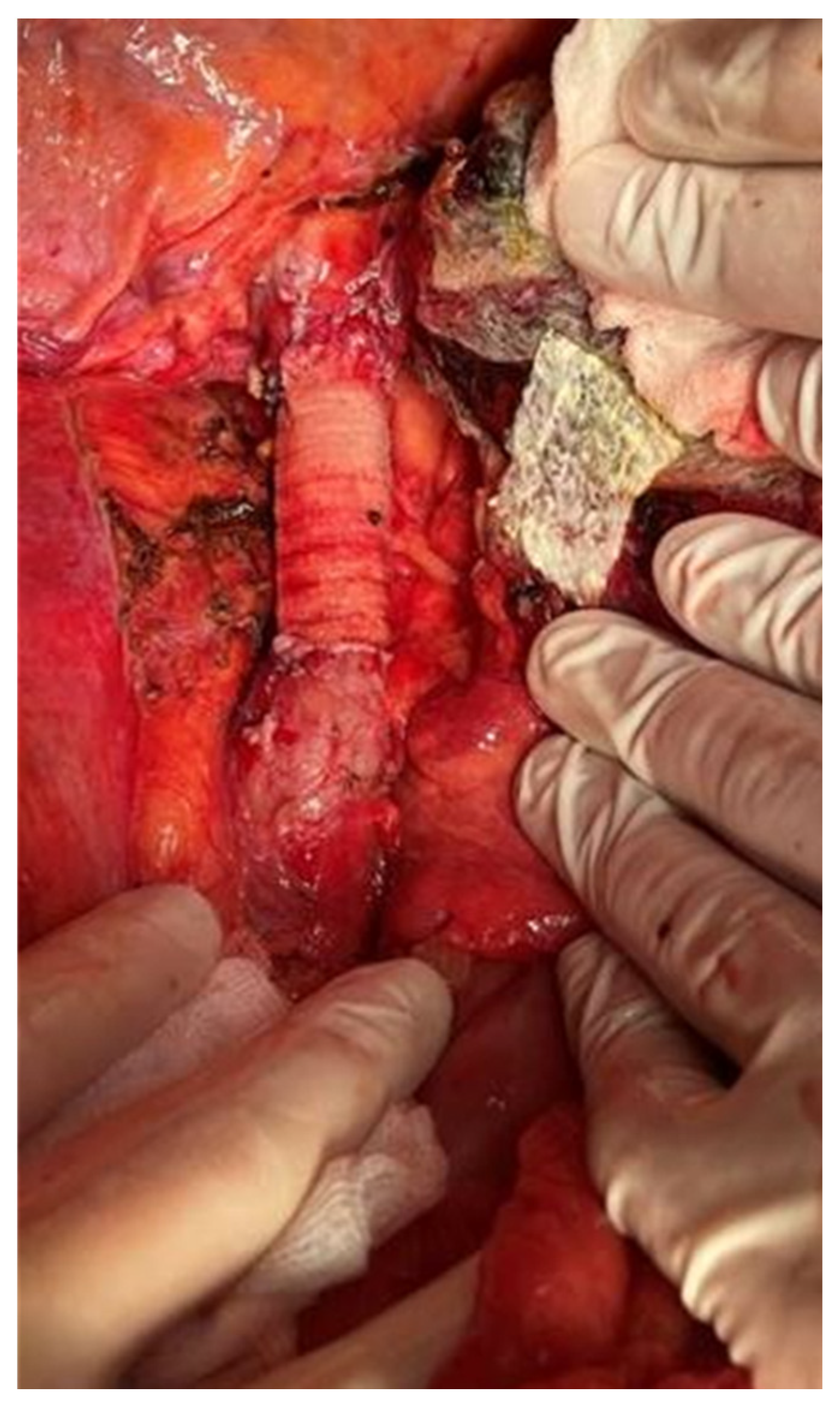
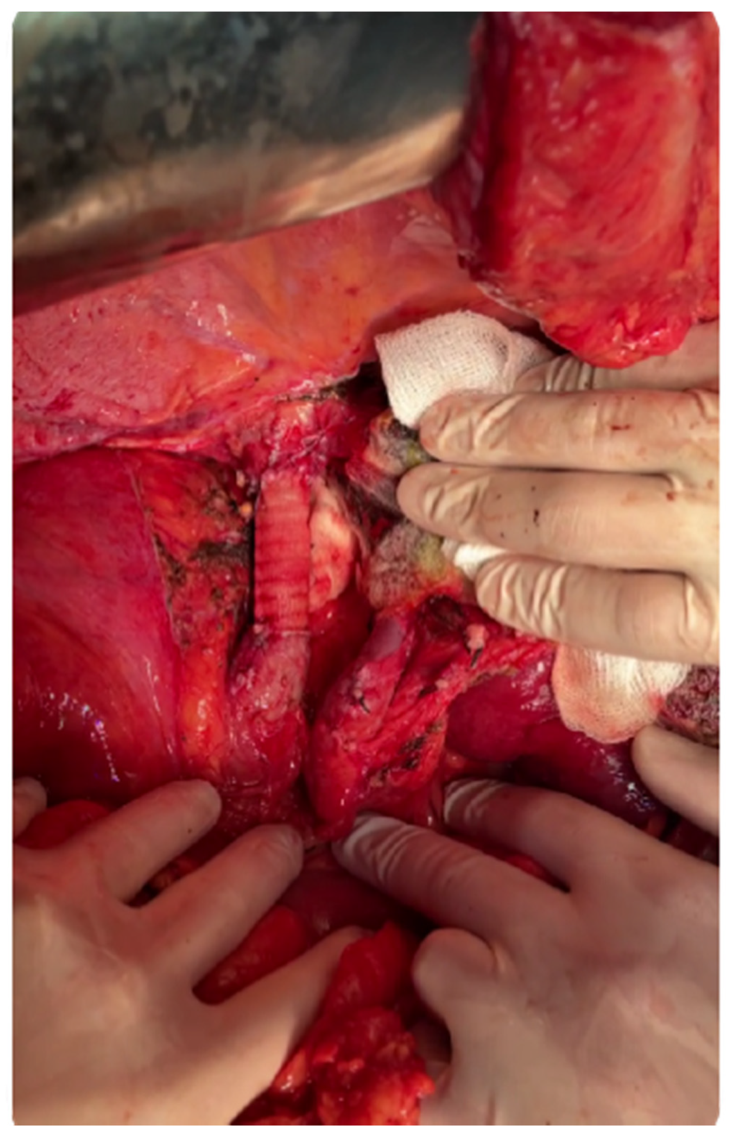
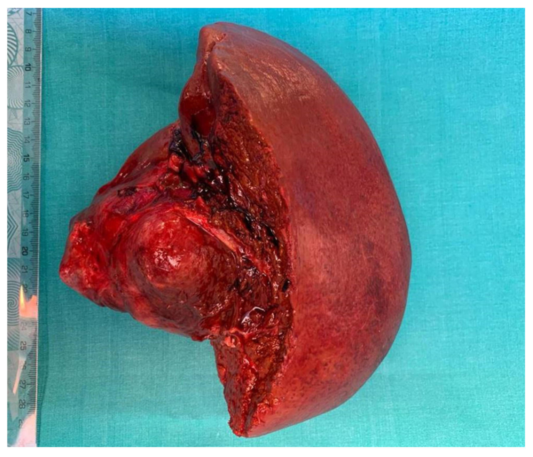
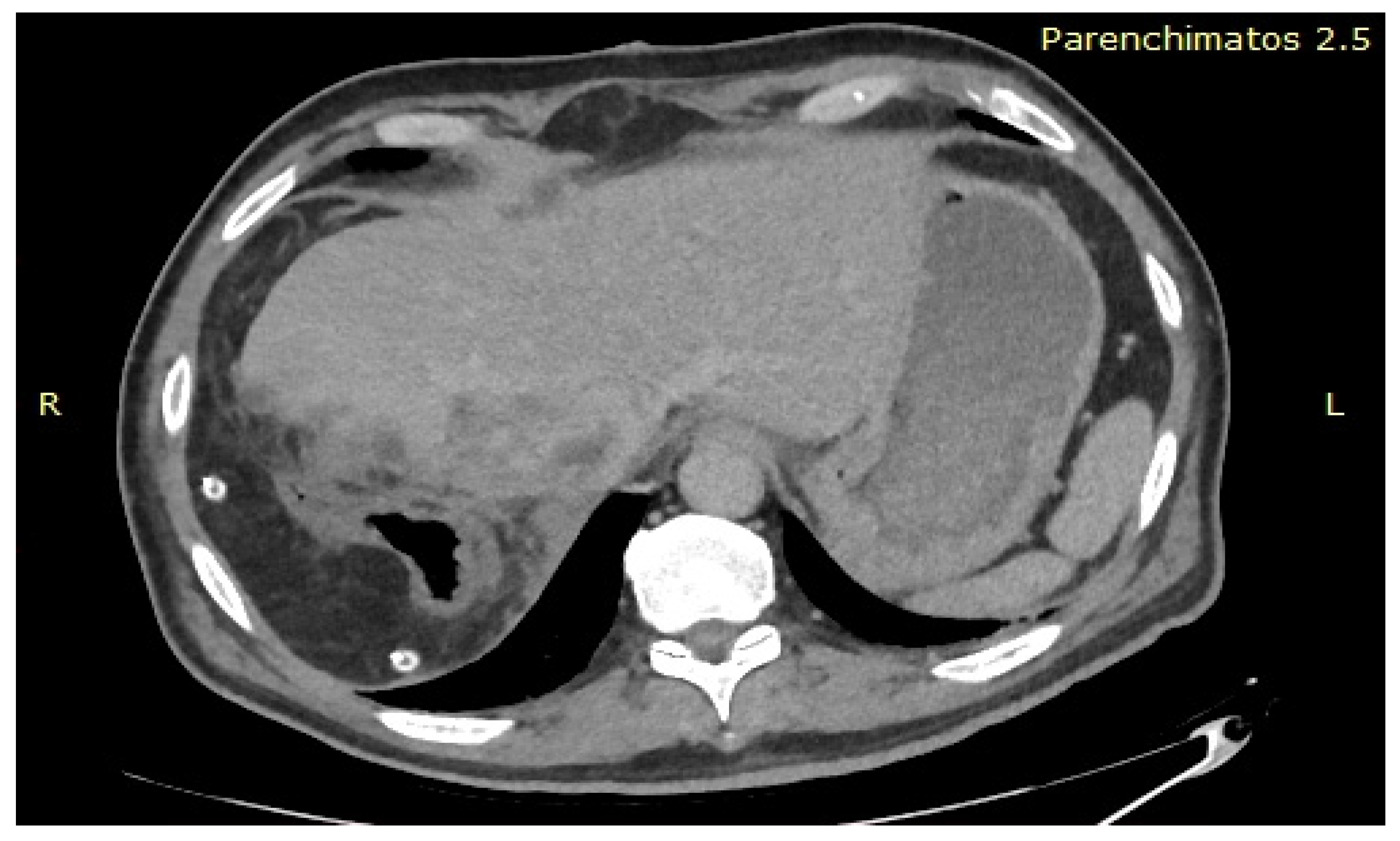
2. Case Report
3. Discussion
4. Conclusions
Author Contributions
Funding
Institutional Review Board Statement
Informed Consent Statement
Data Availability Statement
Conflicts of Interest
References
- Cwinn, M.; Molinari, M. Regional variations in treatment modalities for hepatocellular carcinoma in Canada. HPB 2016, 18, 196–197. [Google Scholar] [CrossRef] [Green Version]
- Seong, J. Challenge and hope in radiotherapy of hepatocellular carcinoma. Yonsei Med. J. 2009, 50, 601–612. [Google Scholar] [CrossRef] [Green Version]
- Petrick, J.L.; Florio, A.A.; Znaor, A.; Ruggieri, D.; Laversanne, M.; Alvarez, C.S.; Ferlay, J.; Valery, P.C.; Bray, F.; McGlynn, K.A. International trends in hepatocellular carcinoma incidence. Int. J. Cancer 2020, 147, 317–330. [Google Scholar] [CrossRef]
- Thomas, M.B.; Chadha, R.; Glover, K.; Wang, X.; Morris, J.; Brown, T.; Rashid, A.; Dancey, J.; Abruzzese, J.L. Phase 2 study of erlotinib in patients with unresectable hepatocellular carcinoma. Cancer 2007, 110, 1059–1067. [Google Scholar] [CrossRef] [PubMed]
- Bekaii-Saab, T.; Markowitz, J.; Prescott, N.; Sadee, W.; Heerema, N.; Wei, L.; Dai, Z.; Papp, A.; Campbell, A.; Culler, K.; et al. A multi-institutional phase II study of the efficacy and tolerability of lapatinib in patients with advanced hepatocellular carcinomas. Clin. Cancer Res. 2009, 15, 5895–5901. [Google Scholar] [CrossRef] [PubMed] [Green Version]
- Yanming, Z.; Lupeng, W.; Dong, X.; Tao, W.; Xiaoying, S. A pooled analysis of combined liver and inferior vena cava resection for hepatic malignancy. HPB 2017, 19, 768–774. [Google Scholar] [CrossRef] [Green Version]
- Kumada, K.; Shimahara, Y.; Fukui, K.; Itoh, K.; Morikawa, S.; Ozawa, K. Extended right hepatic lobectomy: Combined resection of inferior vena cava and its reconstruction by EPTFE graft (Gore-Tex). Case report. Acta Chir. Scand. 1988, 154, 481–483. [Google Scholar] [PubMed]
- Madariaga, J.R.; Fung, J.; Gutierrez, J.; Bueno, J.; Iwatsuki, S. Liver resection combined with excision of vena cava. J. Am. Coll. Surg. 2000, 191, 244–250. [Google Scholar] [CrossRef]
- Bacalbasa, N.; Brezean, I.; Anghel, C.; Barbu, I.; Pautov, M.; Balescu, I.; Brasoveanu, V. Management of a Fulminant Upper Gastrointestinal Bleeding Exteriorized Through Hemobilia Due to Arteriobiliary Fistula Between the Common Bile Duct and a Right Hepatic Artery Aneurysm—A Case Report. In Vivo 2017, 31, 983–989. [Google Scholar] [CrossRef] [Green Version]
- Brasoveanu, V.; Anghel, C.; Barbu, I.; Pautov, M.; Ionescu, M.I.; Motthor, M.; Balescu, I.; Dima, S.; Bacalbasa, N. Pancreatoduodenectomy En Bloc with Portal and Superior Mesenteric Artery Resection—A Case Report and Literature Review. Anticancer Res. 2015, 35, 1613–1618. [Google Scholar]
- Bacalbasa, N.; Balescu, I.; Tanase, A.; Pautov, M.; Brezean, I.; Vilcu, M.; Brasoveanu, V. Spleno-pancreatectomy En Bloc with Parcelar Gastrectomy for Splenic Artery Aneurysm—A Case Report and Literature Review. In Vivo 2018, 32, 915–919. [Google Scholar] [CrossRef]
- Bacalbasa, N.; Balescu, I.; Tanase, A.; Brezean, I.; Vilcu, M.; Brasoveanu, V. Successful Resection of a Non-functional Paraganglioma with Celiac Trunk Invasion Followed by Common Hepatic Artery Reimplantation—A Case Report and Literature Review. In Vivo 2018, 32, 911–914. [Google Scholar] [CrossRef]
- Brasoveanu, V.; Ionescu, M.I.; Grigorie, R.; Mihaila, M.; Bacalbasa, N.; Dumitru, R.; Herlea, V.; Iorgescu, A.; Tomescu, D.; Popescu, I. Living Donor Liver Transplantation for Unresectable Liver Adenomatosis Associated with Congenital Absence of Portal Vein: A Case Report and Literature Review. Am. J. Case Rep. 2015, 16, 637–644. [Google Scholar] [CrossRef] [Green Version]
- Ruiz, C.B.; Kalbaugh, C.A.; Browder, S.E.; McGinigle, K.L.; Kibbe, M.R.; Farber, M.A.; Crowner, J.R.; Marston, W.A.; Pascarella, L. Operative strategies for inferior vena cava repair in oncologic surgery. J. Vasc. Surg. Venous Lymphat. Disord. 2019, 8, 396–404. [Google Scholar] [CrossRef]
- Quinones-Baldrich, W.; Farley, S. Techniques for inferior vena cava resection and reconstruction for retroperitoneal tumor excision. J. Vasc. Surg. Venous Lymphat. Disord. 2013, 1, 84–89. [Google Scholar] [CrossRef]
- Hardwigsen, J.; Baque, P.; Crespy, B.; Moutardier, V.; Delpero, J.R.; Le Treut, Y.P. Resection of the inferior vena cava for neoplasms with or without prosthetic replacement: A 14-patient series. Ann. Surg. 2001, 233, 242–249. [Google Scholar] [CrossRef]
- Daylami, R.; Amiri, A.; Goldsmith, B.; Troppmann, C.; Schneider, P.D.; Khatri, V.P. Inferior vena cava leiomyosarcoma: Is reconstruction necessary after resection? J. Am. Coll. Surg. 2010, 210, 185–190. [Google Scholar] [CrossRef] [PubMed]
- Sarkar, R.; Eilber, F.R.; Gelabert, H.A.; Quinones-Baldrich, W.J. Prosthetic replacement of the inferior vena cava for malignancy. J. Vasc. Surg. 1998, 28, 75–81. [Google Scholar] [CrossRef] [Green Version]
- Bower, T.C.; Nagorney, D.M.; Cherry, K.J.; Toomey, B.J.; Hallett, J.W.; Panneton, J.M.; Gloviczki, P. Replacement of the inferior vena cava for malignancy: An update. J. Vasc. Surg. 2000, 31, 270–281. [Google Scholar] [CrossRef] [Green Version]
- Stauffer, J.A.; Fakhre, G.P.; Dougherty, M.K.; Nakhleh, R.E.; Maples, W.J.; Nguyen, J.H. Pancreatic and multiorgan resection with inferior vena cava reconstruction for retroperitoneal leiomyosarcoma. World J. Surg. Oncol. 2009, 7, 3. [Google Scholar] [CrossRef] [Green Version]
- Wörns, M.A.; Bosslet, T.; Victor, A.; Koch, S.; Hoppe-Lotichius, M.; Heise, M.; Hansen, T.; Pitton, M.B.; Niederle, I.M.; Schuchmann, M.; et al. Prognostic factors and outcomes of patients with hepatocellular carcinoma in non-cirrhotic liver. Scand. J. Gastroenterol. 2012, 47, 718–728. [Google Scholar] [CrossRef] [PubMed]


Publisher’s Note: MDPI stays neutral with regard to jurisdictional claims in published maps and institutional affiliations. |
© 2021 by the authors. Licensee MDPI, Basel, Switzerland. This article is an open access article distributed under the terms and conditions of the Creative Commons Attribution (CC BY) license (https://creativecommons.org/licenses/by/4.0/).
Share and Cite
Bacalbasa, N.; Balescu, I.; Ichim, F.; Barbu, I.; Ristea, A.; Lazea, R.; Danciuc, I.; Popa, I.; Magdoiu, O.; Smira, G.; et al. Major Hepatectomy En Bloc with Cava Vein Resection for Locally Invasive Caudate Lobe Hepatocarcinoma. Healthcare 2021, 9, 1396. https://doi.org/10.3390/healthcare9101396
Bacalbasa N, Balescu I, Ichim F, Barbu I, Ristea A, Lazea R, Danciuc I, Popa I, Magdoiu O, Smira G, et al. Major Hepatectomy En Bloc with Cava Vein Resection for Locally Invasive Caudate Lobe Hepatocarcinoma. Healthcare. 2021; 9(10):1396. https://doi.org/10.3390/healthcare9101396
Chicago/Turabian StyleBacalbasa, Nicolae, Irina Balescu, Florin Ichim, Ion Barbu, Alexandru Ristea, Razvan Lazea, Ioana Danciuc, Ioana Popa, Ovidiu Magdoiu, Gabriela Smira, and et al. 2021. "Major Hepatectomy En Bloc with Cava Vein Resection for Locally Invasive Caudate Lobe Hepatocarcinoma" Healthcare 9, no. 10: 1396. https://doi.org/10.3390/healthcare9101396