
Ectopic Intrauterine Device Revealed by Ureteral Colic in a 37-Week Pregnant Woman: Case Report


Abstract
:1. Introduction
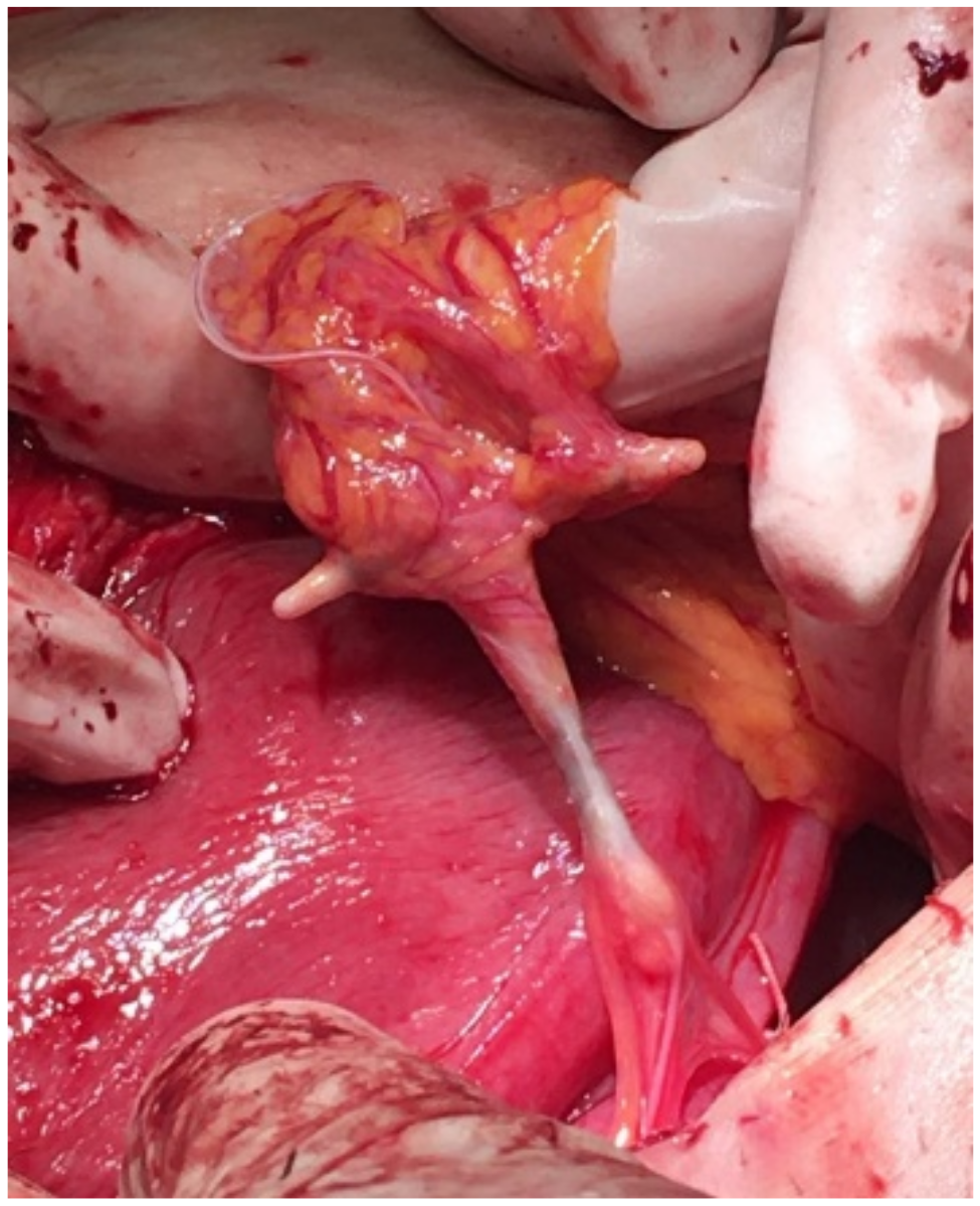
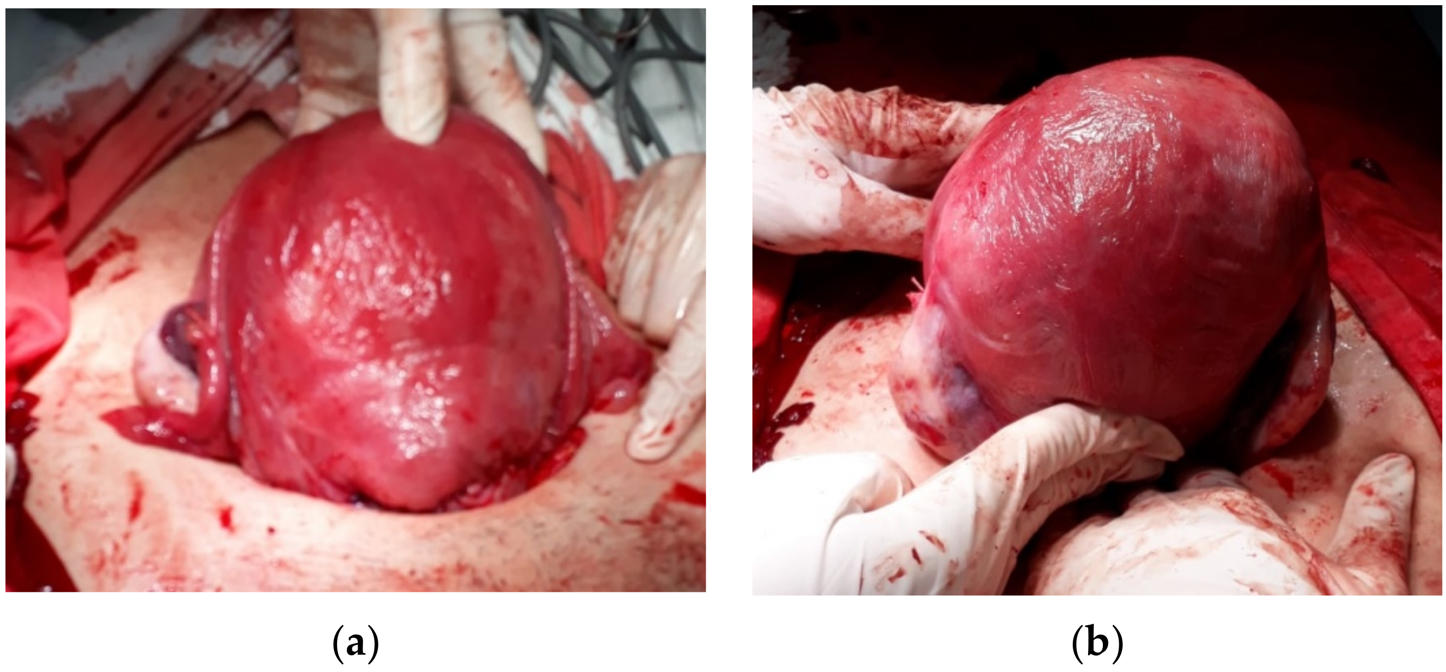
2. Case Report
3. Discussion
4. Conclusions
Author Contributions
Funding
Institutional Review Board Statement
Informed Consent Statement
Data Availability Statement
Conflicts of Interest
References
- Kaislasuo, J.; Suhonen, S.; Gissler, M.; Lähteenmäki, P.; Heikinheimo, O. Uterine perforation caused by intrauterine devices: Clinical course and treatment. Hum. Reprod. 2013, 28, 1546–1551. [Google Scholar] [CrossRef] [PubMed]
- Population Reference Bureau: Family Planning Data Sheet 2019. Available online: https://www.prb.org/wp-content/uploads/2019/09/fp-data-sheet-2019.pdf (accessed on 11 March 2022).
- Sarver, J.; Cregan, M.; Cain, D. Fractured copper intrauterine device (IUD) retained in the uterine wall leading to hysterectomy: A case report. Case Rep. Womens Health 2021, 29, e00287. [Google Scholar] [CrossRef] [PubMed]
- Boortz, H.E.; Margolis, D.J.; Ragavendra, N.; Patel, M.K.; Kadell, B.M. Migration of intrauterine devices: Radiologic findings and implications for patient care. Radiographics 2012, 32, 335–352. [Google Scholar] [CrossRef] [PubMed] [Green Version]
- Ortiz, M.E.; Croxatto, H.B. Copper-T intrauterine device and levonorgestrel intrauterine system: Biological bases of their mechanism of action. Contraception 2007, 75 (Suppl. 6), S16–S30. [Google Scholar] [CrossRef]
- Horvath, S.; Schreiber, C.A.; Sonalkar, S. Contraception. In Endotext [Internet]; Feingold, K.R., Anawalt, B., Boyce, A., Chrousos, G., de Herder, W., Dhatariya, K., Dungan, K., Hershman, J.M., Hofland, J., Kalra, S., et al., Eds.; MDText.com, Inc.: South Dartmouth, MA, USA, 2000. Available online: https://www.ncbi.nlm.nih.gov/books/NBK279148/ (accessed on 29 May 2022).
- Bahamondes, L.; Fernandes, A.; Monteiro, I.; Bahamondes, M.V. Long-acting reversible contraceptive (LARCs) methods. Best Pract. Res. Clin. Obstet. Gynaecol. 2020, 66, 28–40. [Google Scholar] [CrossRef]
- Barnett, C.; Moehner, S.; Do Minh, T.; Heinemann, K. Perforation risk and intra-uterine devices: Results of the EURAS-IUD 5-year extension study. Eur. J. Contracept. Reprod. Health Care 2017, 22, 424–428. [Google Scholar] [CrossRef]
- Zolnierczyk, P.; Cendrowski, K.; Sawicki, W. Intrauterine contraceptive device embedded in the omentum—Case report. Int. J. Womens Health 2015, 7, 945–948. [Google Scholar] [CrossRef] [Green Version]
- Benaguida, H.; Kiram, H.; Telmoudi, E.C.; Ouafidi, B.; Benhessou, M.; Ennachit, M.; Elkarroumi, M. Intraperitoneal migration of an intrauterine device (IUD): A case report. Ann. Med. Surg. 2021, 68, 102547. [Google Scholar] [CrossRef]
- Tarafdari, A.; Malek, M.; Pahlevan Falahy, E.; Hadizadeh, A. IUD perforation and embedment within omentum: A rare and perplexing incidence. Clin. Case Rep. 2022, 10, e05732. [Google Scholar] [CrossRef]
- Wakrim, S.; Lahlou, L. Spontaneously expelled IUD and missing fragments in the uterine cavity. Radiol. Case Rep. 2020, 15, 1654–1656. [Google Scholar] [CrossRef]
- Kondo, W.; Tessmann Zomer, M.; Erzinger, F.L. Uterine arteriovenous fistula after perforation during the placement of an intrauterine device-Minimally invasive treatment using uterine artery embolization. Clin. Exp. Obstet. Gynecol. 2016, 43, 602–605. [Google Scholar] [CrossRef] [PubMed]
- Jatlaoui, T.C.; Riley, H.E.M.; Curtis, K.M. The safety of intrauterine devices among young women: A systematic review. Contraception 2017, 95, 17–39. [Google Scholar] [CrossRef] [PubMed]
- Sparic, R.; Dotlic, J.; Mirkovic, L.; Stamenkovic, J.; Kotlica, B.K.; Nejkovic, L.; Babovic, I.; Malvasi, A.; Tinelli, A. Asymptomatic isthmico-cervical uterine perforation with IUD—Our experience and literature review. Clin. Exp. Obstet. Gynecol. 2016, 43, 896–898. [Google Scholar] [CrossRef] [PubMed]
- Neumann, D.A.; Graversen, J.A.; Pugh, S.K. Intrauterine device embedded in omentum of postpartum patient with a markedly retroverted uterus: A case report. J. Med. Case Rep. 2017, 11, 299. [Google Scholar] [CrossRef] [PubMed] [Green Version]
- Xiong, B.J.; Tao, G.J.; Jiang, D. Bladder-embedded ectopic intrauterine device with calculus. Open Med. 2020, 15, 501–507. [Google Scholar] [CrossRef]
- Akad, M.; Tardif, D.; Fawzy, A.; Socolov, R.V. Management of an Intrauterine Device Migration Resulting in a Pregnancy—Clinical Case. Maedica 2020, 15, 549–551. [Google Scholar] [CrossRef]
- McLaughlin, D.I.; Bevins, W.; Karas, B.K.; Sonnenberg, L. IUD appendicitis during pregnancy. West. J. Med. 1988, 149, 601–602. [Google Scholar]
- Peleg, D.; Latta, R. Removal of an intraabdominal levonorgestrel-releasing intrauterine device during pregnancy. Am. J. Obstet. Gynecol. 2013, 208, e4–e5. [Google Scholar] [CrossRef]
- Gao, T.; Lacue, A.; Brandi, K. Intra-abdominal IUD Requiring Bowel Resection in Pregnancy. J. Minim. Invasive Gynecol. 2021, 28, 1812–1813. [Google Scholar] [CrossRef]
- Amsriza, F.R.; Fakhriani, R. Far-migration of an intrauterine device in the intrathoracic cavity: A rare case report. Clin. Case Rep. 2021, 9, e04127. [Google Scholar] [CrossRef]
- Ziani, I.; Boualaoui, I.; Ibrahimi, A.; El Sayegh, H.; Benslimane, L.; Nouini, Y. Retroperitoneal Ectopic Location of an Intrauterine Device Revealed by Renal Colic: An Exceptional Case. Case Rep. Urol. 2020, 2020, 8893750. [Google Scholar] [CrossRef] [PubMed]


Publisher’s Note: MDPI stays neutral with regard to jurisdictional claims in published maps and institutional affiliations. |
© 2022 by the authors. Licensee MDPI, Basel, Switzerland. This article is an open access article distributed under the terms and conditions of the Creative Commons Attribution (CC BY) license (https://creativecommons.org/licenses/by/4.0/).
Share and Cite
Matei, A.; Dimitriu, M.C.T.; Pacu, I.; Ionescu, C. Ectopic Intrauterine Device Revealed by Ureteral Colic in a 37-Week Pregnant Woman: Case Report. Healthcare 2022, 10, 1060. https://doi.org/10.3390/healthcare10061060
Matei A, Dimitriu MCT, Pacu I, Ionescu C. Ectopic Intrauterine Device Revealed by Ureteral Colic in a 37-Week Pregnant Woman: Case Report. Healthcare. 2022; 10(6):1060. https://doi.org/10.3390/healthcare10061060
Chicago/Turabian StyleMatei, Alexandra, Mihai Cornel Traian Dimitriu, Irina Pacu, and Crîngu Ionescu. 2022. "Ectopic Intrauterine Device Revealed by Ureteral Colic in a 37-Week Pregnant Woman: Case Report" Healthcare 10, no. 6: 1060. https://doi.org/10.3390/healthcare10061060