
Breathing, (S)Training and the Pelvic Floor—A Basic Concept

 , , , and
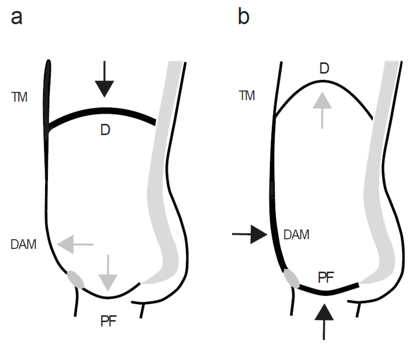
Abstract
:1. Introduction
2. Physiological Basics
2.1. Reappraisal of Physiology and Function of the Muscles Surrounding the Abdominal Cavity
2.2. Involvement of Abdominal and Pelvic Floor Muscles during Quiet Breathing
2.3. Switching from Quiet to Forceful Breathing in Situations with Increased Mental and Physical Strain
3. Straining the Pelvic Floor during Training—The Good and the Bad about Exercises and Their Relationship to Breathing
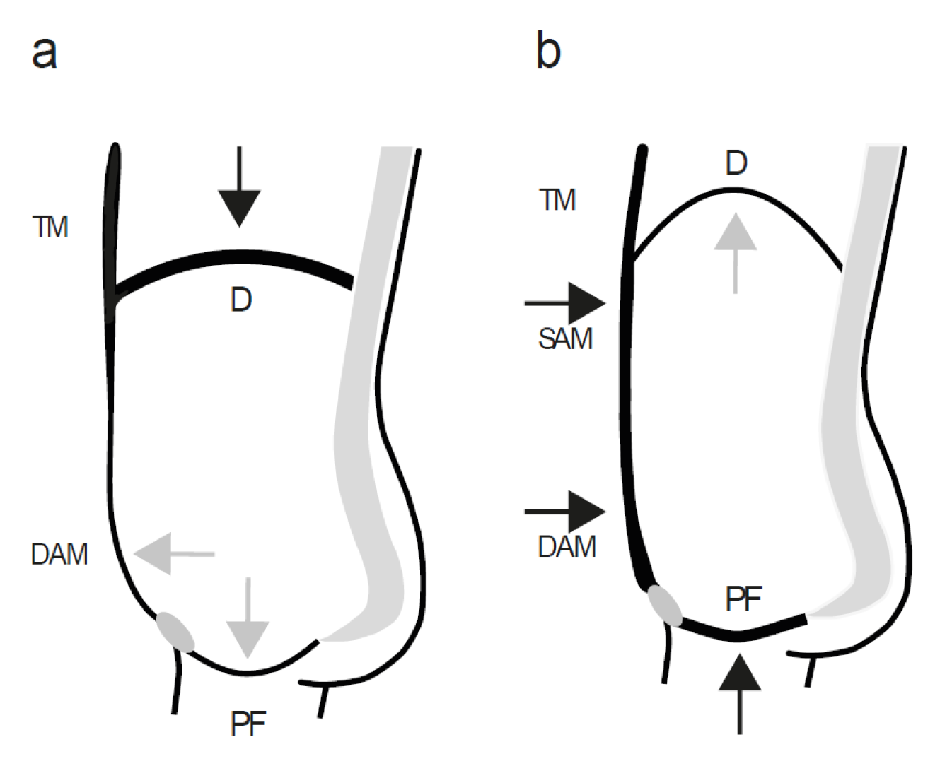
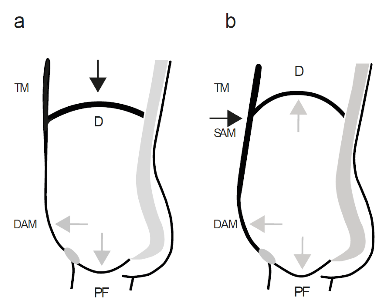
4. Effects of Straining Manoeuvres on the Pelvic Floor Visualized by Dynamic Magnetic Resonance Imaging
5. Discussion
6. Conclusions
Supplementary Materials
Author Contributions
Funding
Institutional Review Board Statement
Informed Consent Statement
Acknowledgments
Conflicts of Interest
References
- Nygaard, I.E.; Shaw, J.M. Physical activity and the pelvic floor. Am. J. Obstet. Gynecol. 2016, 214, 164–171. [Google Scholar] [CrossRef] [PubMed] [Green Version]
- Bø, K.; Nygaard, I.E. Is Physical Activity Good or Bad for the Female Pelvic Floor? A Narrative Review. Sports Med. 2020, 50, 471–484. [Google Scholar] [CrossRef] [PubMed] [Green Version]
- Tim, S.; Mazur-Bialy, A.I. The Most Common Functional Disorders and Factors Affecting Female Pelvic Floor. Life 2021, 11, 1397. [Google Scholar] [CrossRef] [PubMed]
- de Mattos Lourenco, T.R.; Matsuoka, P.K.; Baracat, E.C.; Haddad, J.M. Urinary incontinence in female athletes: A systematic review. Int. Urogynecol. J. 2018, 29, 1757–1763. [Google Scholar] [CrossRef]
- Pires, T.; Pires, P.; Moreira, H.; Viana, R. Prevalence of Urinary Incontinence in High-Impact Sport Athletes: A Systematic Review and Meta-Analysis. J. Hum. Kinet. 2020, 73, 279–288. [Google Scholar] [CrossRef]
- Casey, E.K.; Temme, K. Pelvic floor muscle function and urinary incontinence in the female athlete. Phys. Sportsmed. 2017, 45, 399–407. [Google Scholar] [CrossRef]
- Park, H.; Han, D. The effect of the correlation between the contraction of the pelvic floor muscles and diaphragmatic motion during breathing. J. Phys. Ther. Sci. 2015, 27, 2113–2115. [Google Scholar] [CrossRef] [Green Version]
- Hodges, P.W.; Sapsford, R.; Pengel, L.H. Postural and respiratory functions of the pelvic floor muscles. Neurourol. Urodyn. 2007, 26, 362–371. [Google Scholar] [CrossRef]
- Messelink, B.; Benson, T.; Berghmans, B.; Bø, K.; Corcos, J.; Fowler, C.; Laycock, J.; Huat-Chye Lim, P.; van Lunsen, R.; Lycklama á Nijeholt, G.; et al. Standardization of terminology of pelvic floor muscle function and dysfunction: Report from the pelvic floor clinical assessment group of the International Continence Society. Neurourol. Urodyn. 2005, 24, 374–380. [Google Scholar] [CrossRef]
- Sapsford, R. Rehabilitation of pelvic floor muscles utilizing trunk stabilization. Man. Ther. 2004, 9, 3–12. [Google Scholar] [CrossRef]
- Neumann, P.; Gill, V. Pelvic floor and abdominal muscle interaction: EMG activity and intra-abdominal pressure. Int. Urogynecol. J. Pelvic. Floor Dysfunct. 2002, 13, 125–132. [Google Scholar] [CrossRef] [PubMed]
- Hodges, P.W.; Gandevia, S.C. Changes in intra-abdominal pressure during postural and respiratory activation of the human diaphragm. J. Appl. Physiol. 2000, 89, 967–976. [Google Scholar] [CrossRef] [PubMed]
- Talasz, H.; Kremser, C.; Kofler, M.; Kalchschmid, E.; Lechleitner, M.; Rudisch, A. Phase-locked parallel movement of diaphragm and pelvic floor during breathing and coughing-a dynamic MRI investigation in healthy females. Int. Urogynecol. J. 2011, 22, 61–68. [Google Scholar] [CrossRef] [PubMed]
- Bordoni, B.; Zanier, E. Anatomic connections of the diaphragm: Influence of respiration on the body system. J. Multidiscip. Healthc. 2013, 6, 281–291. [Google Scholar] [CrossRef] [PubMed] [Green Version]
- Borley, N.R. Abdomen and Pelvis. In Gray’s Anatomy, 39th ed.; Standring, S., Ed.; Elsevier: London, UK, 2005; pp. 1041–1047. [Google Scholar]
- Aljuraifani, R.; Stafford, R.E.; Hall, L.M.; van den Hoorn, W.; Hodges, P.W. Task-specific differences in respiration-related activation of deep and superficial pelvic floor muscles. J. Appl. Physiol. 2019, 126, 1343–1351. [Google Scholar] [CrossRef]
- Betschart, C.; Kim, J.; Miller, J.M.; Ashton-Miller, J.A.; DeLancey, J.O. Comparison of muscle fiber directions between different levator ani muscle subdivisions: In vivo MRI measurements in women. Int. Urogynecol. J. 2014, 25, 1263–1268. [Google Scholar] [CrossRef] [Green Version]
- Vodusek, D.B. Neural Control of the Pelvic Floor Muscles. In Pelvic Floor Re-Education, 2nd ed.; Baessler, K., Schuessler, B., Moore, K.L., Norton, P.A., Stanton, S.L., Eds.; Springer: London, UK, 2009; pp. 22–35. [Google Scholar]
- Borley, N.R. True Pelvis, Pelvic Floor and Perineum. In Gray’s Anatomy, 39th ed.; Standring, S., Ed.; Elsevier: London, UK, 2005; pp. 1083–1098. [Google Scholar]
- Sherwood, L. Skeletal muscle metabolism and fiber types. In Human Physiology, 8th ed.; Sherwood, L., Ed.; Brooke/Cole: Belmont, CA, USA, 2013; pp. 277–285. [Google Scholar]
- Rowell, L.B. Pulmonary Structure and Function. In Exercise Physiology, 6th ed.; McArdle, W.D., Katch, F.I., Katch, V.L., Eds.; Lippincott Williams & Wilkins: Philadelphia, PA, USA, 2007; pp. 260–269. [Google Scholar]
- Caix, M.; Outrequin, G.; Descottes, B.; Kalfon, M.; Pouget, X. The muscles of the abdominal wall: A new functional approach with anatomoclinical deductions. Anat. Clin. 1984, 6, 101–108. [Google Scholar] [CrossRef]
- Polla, B.; D’Antona, G.; Bottinelli, R.; Reggiani, C. Respiratory muscle fibres: Specialisation and plasticity. Thorax 2004, 59, 808–817. [Google Scholar] [CrossRef] [Green Version]
- Gosling, J.A.; Dixon, J.S.; Critchley, H.O.; Thompson, S.A. A comparative study of the human external sphincter and periurethral levator ani muscles. Br. J. Urol. 1981, 53, 35–41. [Google Scholar] [CrossRef]
- Wigley, C. Functional Anatomy of the Musculoskeletal System. In Gray’s Anatomy, 39th ed.; Standring, S., Ed.; Elsevier: London, UK, 2005; pp. 110–113. [Google Scholar]
- Hennemann, E.; Somjen, G.; Carpenter, D.O. Functional significance of cell size in spinal motoneurons. J. Neurophysiol. 1965, 28, 560–580. [Google Scholar] [CrossRef]
- Lumb, A.B. Control of Breathing. In Nunn’s Applied Respiratory Physiology, 8th ed.; Springer: Berlin/Heidelberg, Germany, 2017; pp. 51–72. [Google Scholar]
- Laycock, J. Concept of Neuromuscular Rehabilitation and Pelvic Floor Muscle Training. In Pelvic Floor Re-Education, 2nd ed.; Baessler, K., Schuessler, B., Moore, K.L., Norton, P.A., Stanton, S.L., Eds.; Springer: London, UK, 2009; pp. 179–180. [Google Scholar]
- Devreese, A.; Staes, F.; Janssens, L.; Penninckx, F.; Vereecken, R.; De Weerdt, W. Incontinent women have altered pelvic floor muscle contraction patterns. J. Urol. 2007, 178, 558–562. [Google Scholar] [CrossRef] [PubMed]
- Sherwood, L. Respiratory Mechanics. In Human Physiology, 8th ed.; Sherwood, L., Ed.; Brooke/Cole: Belmont, CA, USA, 2013; pp. 461–472. [Google Scholar]
- Sherwood, L. Control of Respiration. In Human Physiology, 8th ed.; Sherwood, L., Ed.; Brooke/Cole: Belmont, CA, USA, 2013; pp. 491–499. [Google Scholar]
- Ashton-Miller, J.A.; DeLancey, J.O. Functional anatomy of the female pelvic floor. Ann. N. Y. Acad. Sci. 2007, 1101, 266–296. [Google Scholar] [CrossRef]
- Nayak, B.; Rodenbaugh, D.W. Modeling the anatomy and function of the pelvic diaphragm and perineal body using a “string model”. Adv. Physiol. Educ. 2008, 32, 169–170. [Google Scholar] [CrossRef]
- Talasz, H.; Kofler, M.; Kalchschmid, E.; Pretterklieber, M.; Lechleitner, M. Breathing with the pelvic floor? Correlation of pelvic floor muscle function and expiratory flows in healthy young nulliparous women. Int. Urogynecol. J. 2010, 21, 475–481. [Google Scholar] [CrossRef]
- Gatzoulis, M.A. Diaphragm. In Gray’s Anatomy, 39th ed.; Standring, S., Ed.; Elsevier: London, UK, 2005; pp. 1007–1012. [Google Scholar]
- Kolar, P.; Neuwirth, J.; Sanda, J.; Suchanek, V.; Svata, Z.; Volejnik, J.; Pivec, M. Analysis of diaphragm movement during tidal breathing and during its activation while breath holding using MRI synchronized with spirometry. Physiol. Res. 2009, 58, 383–392. [Google Scholar] [CrossRef]
- Abe, T.; Kusuhara, N.; Yoshimura, N.; Tomita, T.; Easton, P.A. Differential respiratory activity of four abdominal muscles in humans. J. Appl. Physiol. 1996, 80, 1379–1389. [Google Scholar] [CrossRef]
- Fiammetti, R. Respiration Totale Animation. 2013. Available online: https://www.youtube.com/watch?v=JexU_-7PYCI (accessed on 10 April 2022).
- Park, H.; Hwang, B.; Kim, Y. The impact of the pelvic floor muscles on dynamic ventilation maneuvers. J. Phys. Ther. Sci. 2015, 27, 3155–3157. [Google Scholar] [CrossRef] [PubMed] [Green Version]
- Emerich Gordon, K.; Reed, O. The Role of the Pelvic Floor in Respiration: A Multidisciplinary Literature Review. J. Voice 2020, 34, 243–249. [Google Scholar] [CrossRef]
- Blazek, D.; Stastny, P.; Maszczyk, A.; Krawczyk, M.; Matykiewicz, P.; Petr, M. Systematic review of intra-abdominal and intrathoracic pressures initiated by the Valsalva manoeuvre during high-intensity resistance exercises. Biol. Sport 2019, 36, 373–386. [Google Scholar] [CrossRef]
- Macdonald, I.; Rubin, J.S.; Blake, E.; Hirani, S.; Epstein, R. An investigation of abdominal muscle recruitment for sustained phonation in 25 healthy singers. J. Voice 2012, 26, e9–e16. [Google Scholar] [CrossRef]
- Talasz, H.; Kremser, C.; Kofler, M.; Kalchschmid, E.; Lechleitner, M.; Rudisch, A. Proof of concept: Differential effects of Valsalva and straining maneuvers on the pelvic floor. Eur. J. Obstet. Gynecol. Reprod. Biol. 2012, 164, 227–233. [Google Scholar] [CrossRef] [PubMed]
- Tayashiki, K.; Takai, Y.; Maeo, S.; Kanehisa, H. Intra-abdominal Pressure and Trunk Muscular Activities during Abdominal Bracing and Hollowing. Int. J. Sports Med. 2016, 37, 134–143. [Google Scholar] [CrossRef] [PubMed]
- Hodges, P.W.; Eriksson, A.E.; Shirley, D.; Gandevia, S.C. Intra-abdominal pressure increases stiffness of the lumbar spine. J. Biomech. 2005, 38, 1873–1880. [Google Scholar] [CrossRef] [PubMed]
- Giagio, S.; Innocenti, T.; Pillastrini, P.; Gava, G.; Salvioli, S. What is known from the existing literature about the available interventions for pelvic floor dysfunction among female athletes? A scoping review. Neurourol. Urodyn. 2022, 41, 573–584. [Google Scholar] [CrossRef]
- Bo, K.; Frawley, H.C.; Haylen, B.T.; Abramov, Y.; Almeida, F.G.; Berghmans, B.; Bortolini, M.; Dumoulin, C.; Gomes, M.; McClurg, D.; et al. An International Urogynecological Association (IUGA)/International Continence Society (ICS) joint report on the terminology for the conservative and nonpharmacological management of female pelvic floor dysfunction. Int. Urogynecol. J. 2017, 28, 191–213. [Google Scholar] [CrossRef] [Green Version]
- García-Jaén, M.; Cortell-Tormo, J.M.; Hernández-Sánchez, S.; Tortosa-Martínez, J. Influence of Abdominal Hollowing Maneuver on the Core Musculature Activation during the Prone Plank Exercise. Int. J. Environ. Res. Public Health 2020, 17, 7410. [Google Scholar] [CrossRef]
- Martín-Rodríguez, S.; Bø, K. Is abdominal hypopressive technique effective in the prevention and treatment of pelvic floor dysfunction? Marketing or evidence from high-quality clinical trials? Br. J. Sports Med. 2019, 53, 135–136. [Google Scholar] [CrossRef]
- Talasz, H.; Kofler, M.; Lechleitner, M. Misconception of the Valsalva maneuver. Int. Urogynecol. J. 2011, 22, 1197–1198. [Google Scholar] [CrossRef]
- Hagins, M.; Lamberg, E.M. Individuals with low back pain breathe differently than healthy individuals during a lifting task. J. Orthop. Sports Phys. Ther. 2011, 41, 141–148. [Google Scholar] [CrossRef] [Green Version]
- Hagins, M.; Lamberg, E.M. Natural breath control during lifting tasks: Effect of load. Eur. J. Appl. Physiol. 2006, 96, 453–458. [Google Scholar] [CrossRef]
- HajGhanbari, B.; Yamabayashi, C.; Buna, T.R.; Coelho, J.D.; Freedman, K.D.; Morton, T.A.; Palmer, S.A.; Toy, M.A.; Walsh, C.; Sheel, A.W.; et al. Effects of respiratory muscle training on performance in athletes: A systematic review with meta-analyses. J. Strength Cond. Res. 2013, 27, 1643–1663. [Google Scholar] [CrossRef] [PubMed]
- Shi, Z.H.; Jonkman, A.; de Vries, H.; Jansen, D.; Ottenheijm, C.; Girbes, A.; Spoelstra-de Man, A.; Zhou, J.-X.; Brochard, L.; Heunks, L. Expiratory muscle dysfunction in critically ill patients: Towards improved understanding. Intensive Care Med. 2019, 45, 1061–1071. [Google Scholar] [CrossRef] [Green Version]
- Abrams, P.; Artibani, W.; Cardozo, L.; Dmochowski, R.; van Kerrebroeck, P.; Sand, P. Reviewing the ICS 2002 terminology report: The ongoing debate. Neurourol. Urodyn. 2009, 28, 287. [Google Scholar] [CrossRef] [PubMed]
- Eliasson, K.; Larsson, T.; Mattsson, E. Prevalence of stress incontinence in nulliparous elite trampolinists. Scand. J. Med. Sci. Sports 2002, 12, 106–110. [Google Scholar] [CrossRef] [PubMed]
- Frawley, H.; Shelly, B.; Morin, M.; Bernard, S.; Bo, K.; Digesu, G.A.; Dickinson, T.; Goonewardene, S.; McClurg, D.; Rahnama’i, M.S.; et al. An International Continence Society (ICS) report on the terminology for pelvic floor muscle assesment. Neurourol. Urodyn. 2021, 40, 1217–1260. [Google Scholar] [CrossRef] [PubMed]
- Quaghebeur, J.; Petros, P.; Wyndaele, J.J.; De Wachter, S. The innervation of the bladder, the pelvic floor, and emotion: A review. Auton. Neurosci. 2021, 235, 102868. [Google Scholar] [CrossRef]
- Doggweiler, R.; Whitmore, K.E.; Meijlink, J.M.; Drake, M.J.; Frawley, H.; Nordling, J.; Hanno, P.; Fraser, M.O.; Homma, Y.; Garrido, G.; et al. A standard for terminology in chronic pelvic pain syndromes: A report from the chronic pelvic pain working group of the international continence society. Neurourol. Urodyn. 2017, 36, 984–1008. [Google Scholar] [CrossRef]
- Sherwood, L. Mechanical Events of the Cardiac Cycle. In Human Physiology, 8th ed.; Sherwood, L., Ed.; Brooke/Cole: Belmont, MA, USA, 2013; pp. 322–326. [Google Scholar]
- Kegel, A.H. Progressive resistance exercise in the functional restoration of the perineal muscles. Am. J. Obstet. Gynecol. 1948, 56, 238–248. [Google Scholar] [CrossRef]
- Bartelink, D.L. The role of abdominal pressure in relieving the pressure on the lumbar intervertebral discs. J. Bone Joint Surg. Br. 1957, 39-B, 718–725. [Google Scholar] [CrossRef]
- Hodges, P.W. “Low Back Pain and the Pelvic Floor”. In The Pelvic Floor, 1st ed.; Carriere, B., Feldt, C.M., Eds.; Thieme: New York, NY, USA, 2006; pp. 81–96. [Google Scholar]
- Dakic, J.G.; Cook, J.; Hay-Smith, J.; Lin, K.Y.; Frawley, H. Pelvic floor disorders stop women exercising: A survey of 4556 symptomatic women. J. Sci. Med. Sport 2021, 24, 1211–1217. [Google Scholar] [CrossRef]


Publisher’s Note: MDPI stays neutral with regard to jurisdictional claims in published maps and institutional affiliations. |
© 2022 by the authors. Licensee MDPI, Basel, Switzerland. This article is an open access article distributed under the terms and conditions of the Creative Commons Attribution (CC BY) license (https://creativecommons.org/licenses/by/4.0/).
Share and Cite
Talasz, H.; Kremser, C.; Talasz, H.J.; Kofler, M.; Rudisch, A. Breathing, (S)Training and the Pelvic Floor—A Basic Concept. Healthcare 2022, 10, 1035. https://doi.org/10.3390/healthcare10061035
Talasz H, Kremser C, Talasz HJ, Kofler M, Rudisch A. Breathing, (S)Training and the Pelvic Floor—A Basic Concept. Healthcare. 2022; 10(6):1035. https://doi.org/10.3390/healthcare10061035
Chicago/Turabian StyleTalasz, Helena, Christian Kremser, Heribert Johannes Talasz, Markus Kofler, and Ansgar Rudisch. 2022. "Breathing, (S)Training and the Pelvic Floor—A Basic Concept" Healthcare 10, no. 6: 1035. https://doi.org/10.3390/healthcare10061035