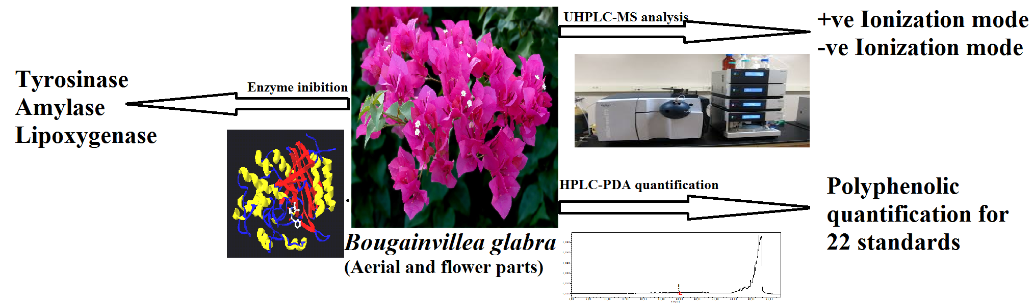
HPLC–PDA Polyphenolic Quantification, UHPLC–MS Secondary Metabolite Composition, and In Vitro Enzyme Inhibition Potential of Bougainvillea glabra

 , ,
, ,  ,
,  and
and
Abstract
:
1. Introduction
2. Results and Discussion
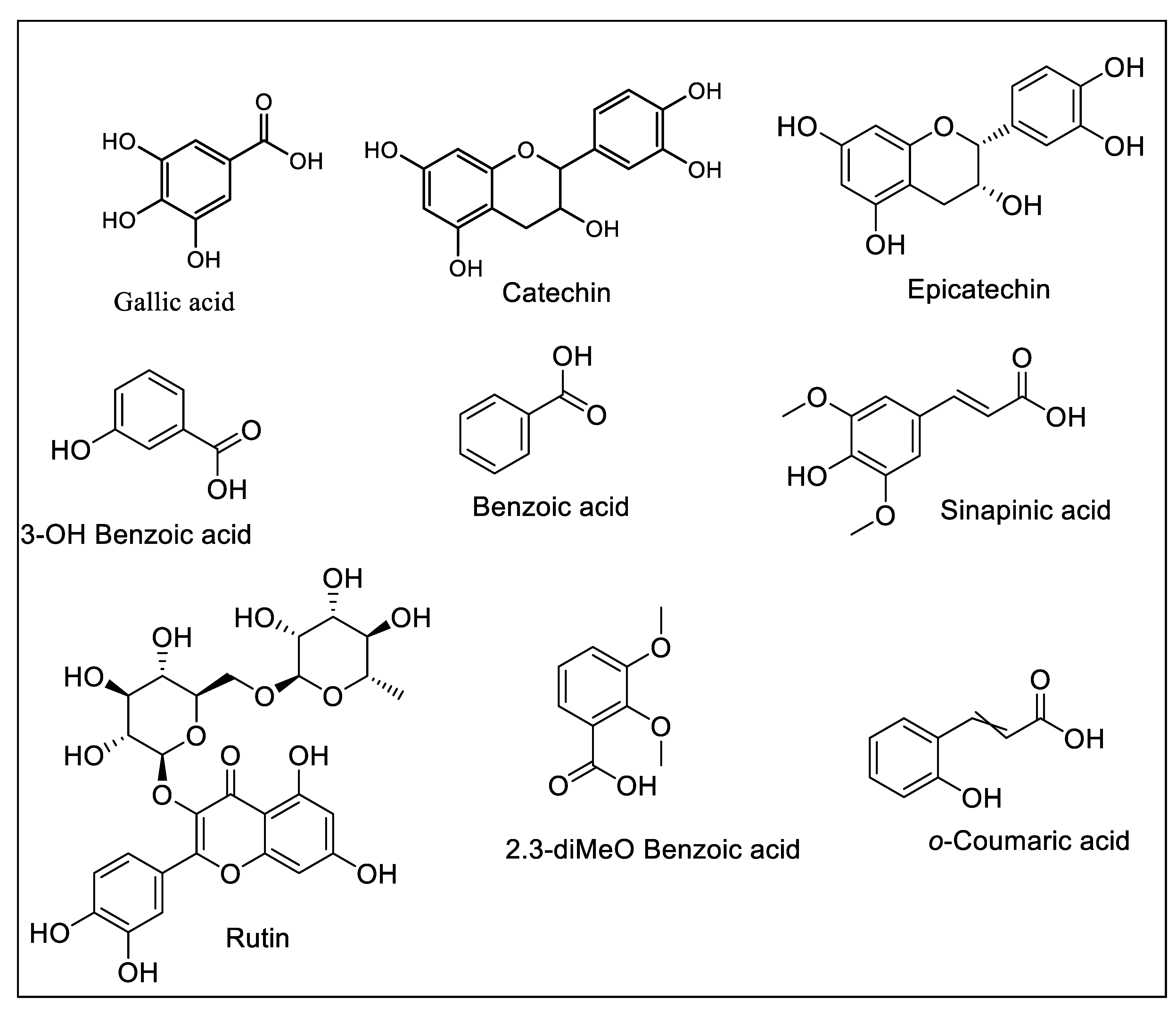
2.1. HPLC–PDA Polyphenolic Quantification
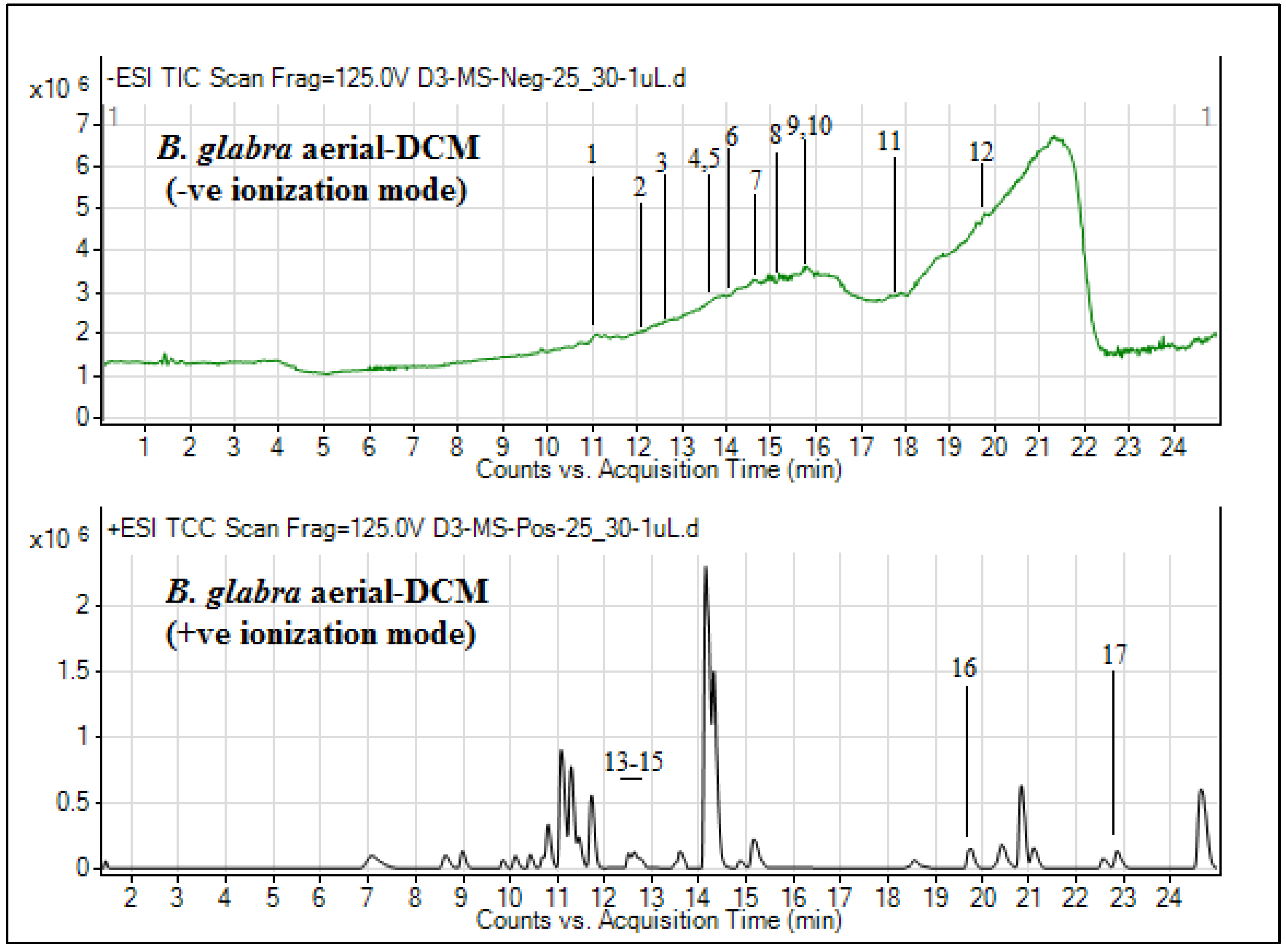
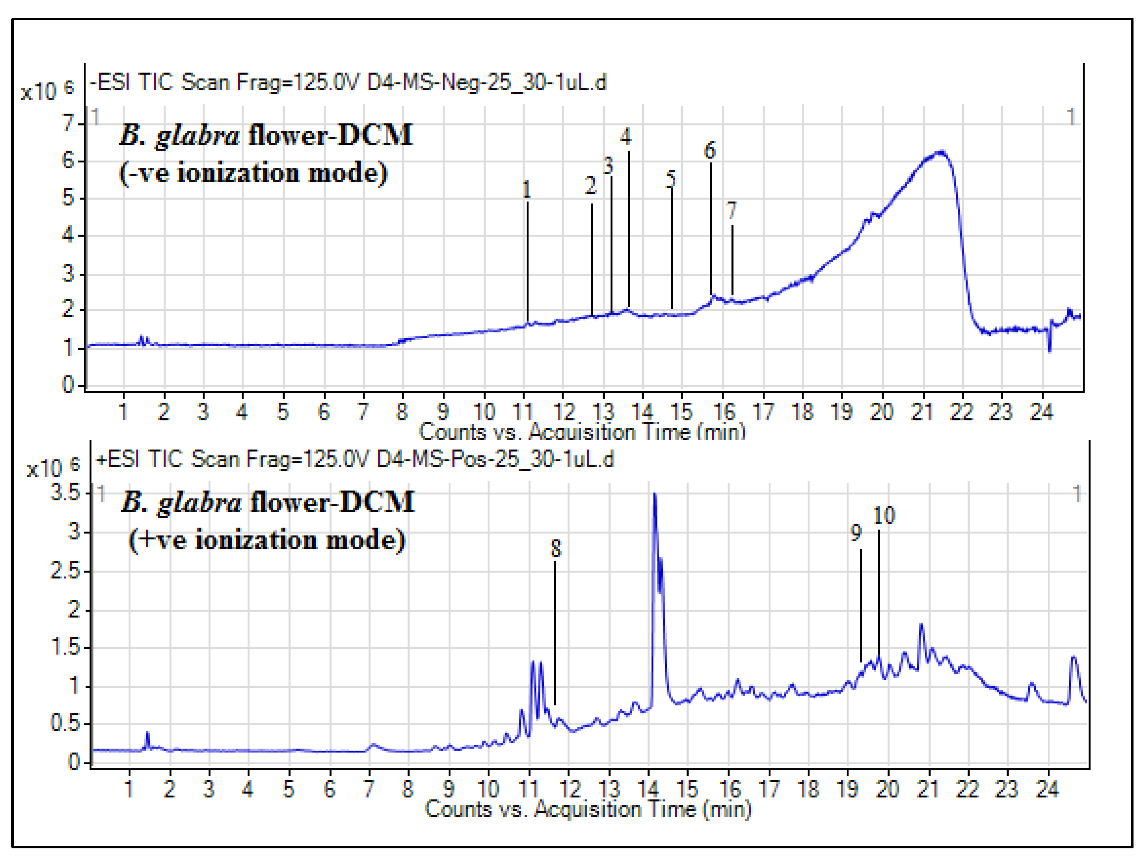
2.2. UHPLC–MS Secondary Metabolite Analysis
2.3. Enzyme Inhibition Potential
3. Materials and Methods
3.1. Plant Material and Extraction
3.2. Phytochemical Composition
3.2.1. HPLC–PDA Polyphenolic Quantification
3.2.2. UHPLC–MS Secondary Metabolites Identification
3.3. Enzyme Assays
3.3.1. α-Amylase Inhibition Assay
3.3.2. Tyrosinase Inhibition Assay
3.3.3. Lipoxygenase Inhibition Assay
3.4. Statistical Analysis
4. Conclusions
Supplementary Materials
Author Contributions
Acknowledgments
Conflicts of Interest
References
- Newman, D.J.; Cragg, G.M. Natural products as sources of new drugs from 1981 to 2014. J. Nat. Prod. 2016, 79, 629–661. [Google Scholar] [CrossRef] [PubMed] [Green Version]
- Dall’Acqua, S.; Sieniawska, E.; Senkardes, I.; Picot-Allain, M.C.N.; Sinan, K.I.; Mahomoodally, M.F. Antioxidant abilities, key enzyme inhibitory potential and phytochemical profile of Tanacetum poteriifolium Grierson. Ind. Crop. Prod. 2019, 140, 111629. [Google Scholar]
- Saleem, H.; Zengin, G.; Ahmad, I.; Lee, J.T.B.; Htar, T.T.; Mahomoodally, F.M.; Naidu, R.; Ahemad, N. Multidirectional insights into the biochemical and toxicological properties of Bougainvillea glabra (Choisy.) aerial parts: A functional approach for bioactive compounds. J. Pharm. Biomed. Anal. 2019, 170, 132–138. [Google Scholar] [CrossRef] [PubMed]
- Saleem, H.; Htar, T.T.; Naidu, R.; Zengin, G.; Ahmad, I.; Ahemad, N. Phytochemical profiling, antioxidant, enzyme inhibition and cytotoxic potential of Bougainvillea glabra flowers. Nat. Prod. Res. 2019, 1–5. [Google Scholar] [CrossRef] [PubMed]
- Markandan, S.; Abdullah, A.; Musa, K.H.; Subramaniam, V.; Stockham, K. Determination of antioxidant activities, total phenolic and flavanoid contents in Bougainvillea glabra bracts at various methanol concentrations. In Proceedings of the 2016 postgraduate colloquium: Proceedings of the Universiti Kebangsaan Malaysia, Faculty of Science and Technology 2016 Postgraduate Colloquium; AIP Publishing: Melville, NY, USA, 2016; Volume 1784, p. 30038. [Google Scholar]
- Edwin, E.; Sheeja, E.; Toppo, E.; Tiwari, V.; Dutt, K. Efecto antimicrobiano, antiulceroso y antidiarreico de las hojas de buganvilla (Bougainvillea glabra Choisy). Ars Pharm. 2007, 48, 135–144. [Google Scholar]
- Edwin, E.; Edwin, S.; Amalraj, A.; Soni, R.; Smita, G.; Gupta, V. Antihyperglycemic activity of Bougainvillea glabra, Choisy. Planta Indica 2006, 2, 25–26. [Google Scholar]
- Simon, A.; Tóth, G.; Duddeck, H.; Soliman, H.; Mahmoud, I.I.; Samir, H. Glycosides from Bougainvillea glabra. Nat. Prod. Res. 2006, 20, 63–67. [Google Scholar] [CrossRef]
- Rashid, S.A.; Rehmani, F.S.; Arman, M.; Ibrahim, M.; Shafique, S. Estimation of Moisture Content & Metal Ions in White Flowers of Bougainvillea spectabilis and Purple Flowers of Bougainvillea glabra in Pakistan. Pak. J. Chem. 2011, 1, 190–192. [Google Scholar]
- Gupta, V.; George, M.; Singhal, M.; Joseph, L.; Arya, H. Pharmacognostical evaluation of the leaf of Bougainvillea glabra ‘Snow White’. J. Pharm. Res. 2009, 2, 1775–1779. [Google Scholar]
- Kaisoon, O.; Konczak, I.; Siriamornpun, S. Potential health enhancing properties of edible flowers from Thailand. Food Res. Int. 2012, 46, 563–571. [Google Scholar] [CrossRef]
- Ahmed, K.I.; Opu, S.A.; Muttaki, A.A.; Al-Mamun, M.; Islam, M.T.; Das, P.R.; Rahmatullah, M. Plant remedies of a unani medicinal practitioner in Bhola district, Bangladesh. World J. Pharm. Pharm. Sci. 2015, 4, 186–198. [Google Scholar]
- Tanase, C.; Coșarcă, S.; Muntean, D.-L. A critical review of phenolic compounds extracted from the bark of woody vascular plants and their potential biological activity. Molecules 2019, 24, 1182. [Google Scholar] [CrossRef] [PubMed] [Green Version]
- Soares, J.J.; Rodrigues, D.T.; Gonçalves, M.B.; Lemos, M.C.; Gallarreta, M.S.; Bianchini, M.C.; Gayer, M.C.; Puntel, R.; Roehrs, R.; DeNardin, E. Paraquat exposure-induced Parkinson’s disease-like symptoms and oxidative stress in Drosophila melanogaster: Neuroprotective effect of Bougainvillea glabra Choisy. Biomed. Pharmacother. 2017, 95, 245–251. [Google Scholar] [CrossRef] [PubMed]
- Ozer, M.S.; Sarikurkcu, C.; Tepe, B. Phenolic composition, antioxidant and enzyme inhibitory activities of ethanol and water extracts of Chenopodium botrys. RSC Adv. 2016, 6, 64986–64992. [Google Scholar] [CrossRef]
- Melucci, D.; Locatelli, M.; Locatelli, C.; Zappi, A.; De Laurentiis, F.; Vinci, G.; Campestre, C.; Leporini, L.; Dall’Acqua, S.; Picot-Allain, M.C.N.; et al. A comparative assessment of biological effects and chemical profile of italian Asphodeline lutea extracts. Molecules 2018, 23, 461. [Google Scholar] [CrossRef] [Green Version]
- Mocan, A.; Zengin, G.; Simirgiotis, M.; Schafberg, M.; Mollica, A.; Vodnar, D.C.; Crişan, G.; Rohn, S. Functional constituents of wild and cultivated Goji (L. barbarumL.) leaves: phytochemical characterization, biological profile, and computational studies. J. Enzym. Inhib. Med. Chem. 2017, 32, 153–168. [Google Scholar] [CrossRef] [Green Version]
- Picot, M.C.N.; Mahomoodally, M.F. Effects of Aphloia theiformis on key enzymes related to diabetes mellitus. Pharm. Boil. 2017, 55, 864–872. [Google Scholar] [CrossRef] [Green Version]
- Sari, A.C.; Elya, B.; Katrin, Z. Antioxidant activity and lipoxygenase enzyme inhibition assay with total flavonoid assay of Garcinia porrecta Laness. stem bark extracts. Pharmacogn. J. 2017, 9, 257–266. [Google Scholar] [CrossRef] [Green Version]
- Dall’Acqua, S.; Cvetanović, A.; Gašić, U.; Stupar, A.; Bulut, G.; Senkardes, I.; Dogan, A.; Seebaluck-Sandoram, R.; Rengasamy, K.R.; Sinan, K.I.; et al. Chemical composition and bio-functional perspectives of Erica arborea L. extracts obtained by different extraction techniques: Innovative insights. Ind. Crop. Prod. 2019, 142, 111843. [Google Scholar]
- Locatelli, M.; Zengin, G.; Uysal, A.; Carradori, S.; De Luca, E.; Bellagamba, G.; Aktumsek, A.; Lazarova, I. Multicomponent pattern and biological activities of seven Asphodeline taxa: potential sources of natural-functional ingredients for bioactive formulations. J. Enzym. Inhib. Med. Chem. 2016, 32, 60–67. [Google Scholar] [CrossRef] [Green Version]
- Vinci, G.; Di Giacomo, S.; Amatore, D.; Locatelli, M.; Vitalone, A.; Toniolo, C.; Rotino, G.L.; Scalzo, R.L.; Palamara, A.T.; Marcocci, M.E.; et al. A polyphenol rich extract from Solanum melongena L. DR2 peel exhibits antioxidant properties and anti-herpes simplex virus type 1 activity in vitro. Molecules 2018, 23, 2066. [Google Scholar]
- Saleem, H.; Htar, T.T.; Naidu, R.; Nawawi, N.S.; Ahmad, I.; Ashraf, M.; Ahemad, N. Biological, chemical and toxicological perspectives on aerial and roots of Filago germanica (L.) huds: Functional approaches for novel phyto-pharmaceuticals. Food Chem. Toxicol. 2019, 123, 363–373. [Google Scholar] [CrossRef] [PubMed]
- Savran, A.; Dall’Acqua, S.; Aktumsek, A.; Mocan, A.; Glamočlija, J.; Ćirić, A.; Soković, M. Phenolic compounds and biological effects of edible Rumex scutatus and Pseudosempervivum sempervivum: potential sources of natural agents with health benefits. Food Funct. 2016, 7, 3252–3262. [Google Scholar] [CrossRef] [PubMed]
- Baylac, S. Inhibition of 5-lipoxygenase by essential oils and other natural fragrant extracts. Int. J. Aromather. 2003, 13, 138–142. [Google Scholar] [CrossRef]


| Phenolic Compounds | B. glabra extracts | |||
|---|---|---|---|---|
| Aerial-MeOH | Aerial-DCM | Flower-MeOH | Flower-DCM | |
| Gallic acid | nd | nd | 2.39 ± 0.41 | nd |
| Catechin | nd | nd | 6.31 ± 0.74 | 0.26 ± 0.02 |
| Epicatechin | 0.62 ± 0.05 | nd | nd | nd |
| 3-OH benzoic acid | 0.37 ± 0.04 | nd | nd | nd |
| Rutin | 0.60 ± 0.05 | nd | 1.26 ± 0.11 | nd |
| Sinapinic acid | nd | nd | nd | 0.23 ± 0.02 |
| 2,3-diMeO benzoic acid | nd | nd | nd | 0.52 ± 0.05 |
| Benzoic acid | nd | nd | nd | 0.26 ± 0.03 |
| o-Coumaric acid | nd | nd | BLD | nd |
| S. no | RT (min) | B. Peak (m/z) | Tentative Identification | Comp. Class | Mol. Formula | Mol. Mass |
|---|---|---|---|---|---|---|
| Negative ionization mode | ||||||
| 1 | 11.015 | 579.2176 | (+)-Syringaresinol O-beta-d-glucoside | Phenol | C28 H36 O13 | 580.2176 |
| 2 | 12.238 | 261.014 | 2-Hydroxy-4-methoxyacetophenone 5-sulfate | Phenol | C9 H10 O7 S | 262.014 |
| 3 | 12.61 | 342.145 | N-trans-Feruloyl-4-O-methyldopamine | Alkaloid | C19 H21 N O5 | 343.145 |
| 4 | 13.637 | 329.2406 | 5,8,12-trihydroxy-9-octadecenoic acid | Fatty acid | C18 H34 O5 | 330.2406 |
| 5 | 13.637 | 369.2002 | 5-Megastigmen-7-yne-3,9-diol 9-glucoside | Terpenoid | C19 H30 O7 | 370.2002 |
| 6 | 14.008 | 399.2101 | Cincassiol B | Diterpenoid | C20 H32 O8 | 400.2101 |
| 7 | 14.64 | 695.4067 | Momordicoside E | Triterpenoid | C37 H60 O12 | 696.4067 |
| 8 | 15.259 | 286.246 | Prosopinine | Alkaloid | C16 H33 N O3 | 287.246 |
| 9 | 15.77 | 407.1776 | 3-O-Methylniveusin A | Sesquiterpene | C21 H28 O8 | 408.1776 |
| 10 | 15.772 | 356.1801 | Uplandicine | Alkaloid | C17 H27 N O7 | 357.1801 |
| 11 | 17.826 | 519.3402 | Cucurbitacin P | Triterpene | C30 H48 O7 | 520.3402 |
| 12 | 19.783 | 347.1795 | Schizonepetoside E | Terpene glycoside | C16 H28 O8 | 348.1795 |
| Positive ionization mode | ||||||
| 13 | 12.516 | 292.148 | (S)-Edulinine | Alkaloid | C16 H21 N O4 | 291.148 |
| 14 | 12.648 | 227.1206 | 12-hydroxyjasmonic acid | Plant hormone | C12 H18 O4 | 226.1206 |
| 15 | 12.783 | 344.1433 | N-trans-Feruloyl-4-O-methyldopamine | Lactam alkaloid | C19 H21 N O5 | 343.1433 |
| 16 | 19.744 | 279.1528 | Emmotin A | Terpenoid | C16 H22 O4 | 278.1528 |
| 17 | 22.863 | 593.2689 | Pheophorbide A | Chlorophyll derivative | C35 H36 N4 O5 | 592.2689 |
| S. No | RT (min) | B. Peak (m/z) | Tentative Identification | Comp. Class | Mol. Formula | Mol. Mass |
|---|---|---|---|---|---|---|
| Negative ionization mode | ||||||
| 1 | 11.086 | 415.2055 | Ethyl 7-epi-12-hydroxyjasmonate glucoside | Terpene glycoside | C20 H32 O9 | 416.2055 |
| 2 | 12.699 | 263.1365 | Geigerin | Sesquiterpene | C15 H20 O4 | 264.1365 |
| 3 | 13.279 | 225.1206 | 12-Hydroxyjasmonic acid | Plant hormone | C12 H18 O4 | 226.1206 |
| 4 | 13.644 | 329.2411 | 5,8,12-trihydroxy-9-octadecenoic acid | Fatty acid | C18 H34 O5 | 330.2411 |
| 5 | 14.877 | 221.125 | Annuionone B | Apocarotenoids | C13 H18 O3 | 222.125 |
| 6 | 15.775 | 356.1798 | Uplandicine | Alkaloid | C17 H27 N O7 | 357.1798 |
| 7 | 16.205 | 307.2 | Methylgingerol | Phenol | C18 H28 O4 | 308.2 |
| Positive ionization mode | ||||||
| 8 | 11.73 | 197.1096 | 4-(2-hydroxypropoxy)-3,5-dimethyl-Phenol | Phenol | C11 H16 O3 | 196.1096 |
| 9 | 19.322 | 293.1675 | 9-Acetoxyfukinanolide | Terpene lactone | C17 H24 O4 | 292.1675 |
| 10 | 19.738 | 279.152 | Emmotin A | Alkaloid | C16 H22 O4 | 278.152 |
| Extracts | Tyrosinase (mg KAE/g extract) | α-amylase (mmol ACAE/g extract) | Lipoxygenase | |
|---|---|---|---|---|
| (% Inhibition; 0.5 mg/mL) | IC50 (µg/mL) | |||
| Aerial MeOH | 27.12 ± 0.26 | 0.10 ± 0.01 | 35.4 ± 1.3 | >500 ** |
| Aerial DCM | 25.64 ± 0.87 | 0.12 ± 0.02 | 6.3 ± 1.2 | >500 |
| Flower MeOH | 25.47 ± 0.04 | 0.09 ± 0.01 | 7.2 ± 1.5 | >500 |
| Flower DCM | 26.22 ± 0.27 | 0.12 ± 0.02 | 24.3 ± 1.7 | >500 |
| Quercetin | nt | nt | 89.2 ± 0.6 | 2.3 ± 0.3 (µM) |
© 2020 by the authors. Licensee MDPI, Basel, Switzerland. This article is an open access article distributed under the terms and conditions of the Creative Commons Attribution (CC BY) license (http://creativecommons.org/licenses/by/4.0/).
Share and Cite
Saleem, H.; Htar, T.T.; Naidu, R.; Anwar, S.; Zengin, G.; Locatelli, M.; Ahemad, N. HPLC–PDA Polyphenolic Quantification, UHPLC–MS Secondary Metabolite Composition, and In Vitro Enzyme Inhibition Potential of Bougainvillea glabra. Plants 2020, 9, 388. https://doi.org/10.3390/plants9030388
Saleem H, Htar TT, Naidu R, Anwar S, Zengin G, Locatelli M, Ahemad N. HPLC–PDA Polyphenolic Quantification, UHPLC–MS Secondary Metabolite Composition, and In Vitro Enzyme Inhibition Potential of Bougainvillea glabra. Plants. 2020; 9(3):388. https://doi.org/10.3390/plants9030388
Chicago/Turabian StyleSaleem, Hammad, Thet Thet Htar, Rakesh Naidu, Sirajudheen Anwar, Gokhan Zengin, Marcello Locatelli, and Nafees Ahemad. 2020. "HPLC–PDA Polyphenolic Quantification, UHPLC–MS Secondary Metabolite Composition, and In Vitro Enzyme Inhibition Potential of Bougainvillea glabra" Plants 9, no. 3: 388. https://doi.org/10.3390/plants9030388