Review

Post-Translational Modification of Proteins Mediated by Nitro-Fatty Acids in Plants: Nitroalkylation

by
Lorena Aranda-Caño
,
Beatriz Sánchez-Calvo
,
Juan C. Begara-Morales
,
Mounira Chaki
,
Capilla Mata-Pérez
,
María N. Padilla
,
Raquel Valderrama
and
Juan B. Barroso
*
Group of Biochemistry and Cell Signaling in Nitric Oxide, Department of Experimental Biology, Center for Advanced Studies in Olive Grove and Olive Oils, Faculty of Experimental Sciences, University Campus Las Lagunillas, University of Jaén, E-23071 Jaén, Spain
*
Author to whom correspondence should be addressed.
Plants 2019, 8(4), 82; https://doi.org/10.3390/plants8040082
Submission received: 26 February 2019 / Revised: 25 March 2019 / Accepted: 26 March 2019 / Published: 29 March 2019
(This article belongs to the Special Issue Nitric Oxide Signaling in Plants)

Abstract

:
Nitrate fatty acids (NO2-FAs) are considered reactive lipid species derived from the non-enzymatic oxidation of polyunsaturated fatty acids by nitric oxide (NO) and related species. Nitrate fatty acids are powerful biological electrophiles which can react with biological nucleophiles such as glutathione and certain protein–amino acid residues. The adduction of NO2-FAs to protein targets generates a reversible post-translational modification called nitroalkylation. In different animal and human systems, NO2-FAs, such as nitro-oleic acid (NO2-OA) and conjugated nitro-linoleic acid (NO2-cLA), have cytoprotective and anti-inflammatory influences in a broad spectrum of pathologies by modulating various intracellular pathways. However, little knowledge on these molecules in the plant kingdom exists. The presence of NO2-OA and NO2-cLA in olives and extra-virgin olive oil and nitro-linolenic acid (NO2-Ln) in Arabidopsis thaliana has recently been detected. Specifically, NO2-Ln acts as a signaling molecule during seed and plant progression and beneath abiotic stress events. It can also release NO and modulate the expression of genes associated with antioxidant responses. Nevertheless, the repercussions of nitroalkylation on plant proteins are still poorly known. In this review, we demonstrate the existence of endogenous nitroalkylation and its effect on the in vitro activity of the antioxidant protein ascorbate peroxidase.

Graphical Abstract

1. Introduction

Reactive lipid species (RLS), or so-called lipid-derived electrophiles (LDEs), are caused by polyunsaturated fatty acids (PUFAs) peroxidation [1,2,3,4]. Reactive lipid species have been identified in sanguine fluid, plasma, urine, human tissues, and animal models using array techniques. Recently, they have also been detected in plant systems with the aid of mass spectrometry. A rise in RLS abundance under pathological and stress circumstances has been broadly reported [4,5,6,7,8,9,10].
Polyunsaturated fatty acids are targets of peroxidation due to their unsaturated double bonds [4,11]. The main mechanisms of PUFA peroxidation are non-enzymatic autocatalytic oxidation reactions [1,12], while enzymatic oxidation reactions involving three heme-containing metallo-enzyme families (lipoxygenases (LOXs), cyclooxigenases (COXs) [1,13], and cytochromes P450 (CYPs) [1]), as well as NADP+-dependent dehydrogenases [1,14] which can also occur. Non-enzymatic mechanisms include PUFA nitration triggered by reactive nitrogen species (RNS) such as nitric oxide (NO) and its derived molecules [1,15,16]. A preferential target for lipid peroxidation is arachidonic acid, whose oxidation yields several products. The non-enzymatic oxidation reactions of PUFAs yield aldehydes such as 4-hydroxynonenal (HNE) and malondialdehyde (MDA), as well as the J- and A-series of isoprostanes [4,17]. Prostaglandins (15-deoxy-Δ12,14-prostaglandin J2) and lipoxins are generated by enzymatic oxidation reactions catalyzed by COX and LOX, respectively [4,18,19]. The oxidation of arachidonic acid by NO-derived species yields 12-nitroarachidonic acid (12-NO2-AA) [4,20].
The addition of aldehyde, α-β-unsaturated carbonyl, epoxide or nitroalkene substituents to PUFAs during the peroxidation process causes the formation of lipid-derived species with electrophilic properties. From a chemical perspective, electrophilic molecules contain an electron-poor moiety, which makes them chemically reactive with nucleophiles (electron-rich molecules) [1]. Nucleophiles and electrophiles can be classified according to a hard/soft acid–base (HSAB) model [21]. Hard electrophiles, whose outer layer electrons are not readily excited, are difficult to polarize. Conversely, soft electrophiles have a more diffused electron distribution or partial positive charges due to the possession of electron-withdrawing substituents such as nitro groups. Nucleophiles can be characterized in a similar manner. Hard nucleophiles are highly electronegative and difficult to polarize, in contrast to soft nucleophiles, which have empty, low-lying electron orbitals. The softest biological nucleophiles, cysteine thiols, which integrate proteins, are also present in the antioxidant tripeptide glutathione (GSH). Primary and secondary amines of lysine, arginine, and histidine residues are regarded as hard nucleophiles [22]. The reactivity of nucleophiles does not only depend on the presence of hard and soft electrophiles in their vicinity, other factors such as their microenvironment (including hydrogen bonding reactions with neighboring amino residues) can influence nucleophile ionization too. For instance, as the reactivity of thiolate anions is higher than that of protonated thiols, the decrease in cysteine pKa induced by protein conformation increases its nucleophilicity [23,24]. As a general rule, hard electrophiles preferentially react with hard nucleophiles, while soft electrophiles interact with soft nucleophiles [1,25].
The importance of RLS resides in their electrophilic reactivity, which enables them to establish covalent adducts with GSH and nucleophilic amino acid residues of proteins such as cysteine, histidine, and lysine, generating post-translational modifications (PTMs) of proteins [4,26,27,28,29,30]. The endogenous occurrence of electrophilic fatty acids has been detected at low concentrations in plasma and animal tissues, whose biological significance is still little known [1,31]. Due to their innate reactivity, the rapid adduction process of RLS with susceptible GSH and nucleophilic residues of proteins may be functionally significant in relation to signaling responses [1,32]. However, it should be mentioned that an equilibrium between adducted and free forms exists in the milieu [1,33].
Pathological conditions promote the enzymatic and non-enzymatic generation of endogenous RLS. In these situations, an increase in the expression of oxidases and oxygenases and in the non-enzymatic production of reactive oxygen and nitrogen species (ROS and RNS), such as reduced oxygen species and oxides of nitrogen (NO, peroxynitrite (ONOO), nitrogen dioxide (·NO2), nitronium cation (NO2+), takes place. All these species could react with PUFAs yielding RLS. Macrophage, eosinophil, and neutrophil cells in the immune system alter lipase activation, causing the scission of fatty acids from membranes. Thus, these disengaged fatty acids may be substrates for subsequent RLS formation [1,22,34]. The electrophilic nature of RLS induces the nucleophilic attack of proteins, leading to modifications in tertiary and quaternary structures, in catalytic activities, in charge and hydrophobicity, in subcellular localization, and in protein cross-linking. The main proteins susceptible to adduction perform metabolic functions such as cytoskeletal function, transcriptional regulation, host defense, ion and macromolecule transport, and enzyme catalysis. These proteins are involved in manifold physiological processes comprising resolution of inflammation, cell death, and induction of cellular antioxidants. In this respect, the anti-inflammatory and antioxidant responses stimulated by RLS adduction suggest the existence of an equilibrium between prompting events, electrophile production, protein adduction, and adaptive cellular responses. Therefore, RLS adduction allows organisms to cope with alterations generated under conditions of metabolic stress, inflammation, and modification in cells and tissues [1,4,35,36,37,38,39].
In plant systems, PUFA peroxidation caused by non-enzymatic or/and enzymatic (LOX-mediated) reactions generates some products with cytotoxic effects and others with protective anti-stress effects. The LOX pathway yields RLS related to plant defense responses to pathogen infections [40] and wounding [41], and in the regulation of hypersensitive programmed cell death [42] and senescence [43]. Non-enzymatic processes can generate both harmful products with damaging actions [44] and phytoprostanes, which have biological properties similar to those of jasmonic acid [45]. Recent knowledge has illustrated the formation of RLS that perform signaling roles and are implicated in antioxidant responses as a result of the oxidation of NO-derived molecules [9].
This review focuses on the study of reactive lipids species called nitroalkenes. Specifically, we will argue the biological properties of nitroalkenes both in animal and plant systems, as well as their signaling potential generated by a post-translational modification of proteins called nitroalkylation.

2. Nitro-Fatty Acids in Animals

The reactive lipids species resulting from the interaction of unsaturated fatty acids with NO and derived species, such as NO2 and ONOO, are called nitro-fatty acids (NO2-FAs), nitrolipids or nitroalkenes [46].
Although the interaction between unsaturated fatty acids and RNS has been widely studied, two distinct mechanisms have been suggested to explain the in vivo nitration of fatty acids, a process which remains unknown [47]. One mechanism involves the generation of an alkyl radical through a radical hydrogen abstraction from a bis-allylic carbon followed by a double-bond rearrangement and the incorporation of a NO2 radical (Figure 1A) [48,49]. The other mechanism consists on the generation of a carbon-centered radical through the direct addition of NO2, which can be further oxidized either with or without a second insertion of NO2 in order to form the nitro-fatty acid. When the carbon-centered radical reacts with the second NO2, an unstable nitro-nitrite or dinitro compound appears which rapidly decomposes and releases nitrous acid (HNO2), yielding the nitro-fatty acid (Figure 1B) [49,50].
In recent years, important progress in the endogenous detection of NO2-FAs has been achieved in animal and human models. In animal systems, it is worth highlighting the detection of nitrated oleic (NO2-OA) and linoleic acid (NO2-LA) in the murine model of focal cardiac ischemia-reperfusion (I/R). The formation of these NO2-FAs was due to reoxygenation-induced tissue damage which generated acidification, hypoxia, as well as ROS and RNS [49,51]. It should be mentioned that other NO2-FAs were detected in an experimental rat model of ischemic preconditioning (IPC) [49,52]. High-resolution liquid chromatography mass spectrometry (LC-MS/MS) procedures have revealed a preferential nitration of conjugated linoleic acid (cLA) in animal systems. This fatty acid presents positional and geometric isomers of linoleic acid which have conjugated dienes in cis and trans configurations. These species have conjugated double bonds which are not separated by a methylene group [53]. Conjugated linoleic acid, which displays more reactivity with ·NO2 than bis-allylic fatty-acids, is the main in vivo endogenous nitration target [47,49]. The formation of nitrated cLA has been detected in activated macrophages under inflammatory conditions and in the gastric compartment following the ingest of cLA and NO2 [47,49,54].
Advances in chromatography mass spectrometry techniques, in vitro nitration, and animal model studies have increased our understanding of the nitration of unsaturated fatty acids in humans. Dietary products such as oils and seeds are the principal sources of unsaturated fatty acids such as oleic acid (OA), conjugated linoleic (cLA), and linolenic (cLn) acids. Pomegranates are regarded as sources of cLn, while dairy products and meat are a source of cLA. Interestingly, cLA is absorbed at much higher levels than cLn [49,55]. Dietary products such as vegetables and herbs are sources of nitrate (NO3) and nitrite (NO2) [49,56,57]. These NO-derived species are necessary to generate nitrated PUFAs, as nitrite is a nitrating compound derived from nitrate. However, the low level of nitrite in basal metabolic conditions is increased through the conversion of nitrate by commensal bacteria in the gastrointestinal tract [58]. As with animal models, NO2-cLA is the principal nitroalkene generated in humans (Table 1) [47].
In addition to those mentioned above, other NO2-FAs, such as nitro-oleic acid (NO2-OA), nitro-linoleic acid (NO2-LA), conjugated nitro-linoleic acid (NO2-cLA), nitro-arachidonic acid (NO2-AA), and cholesteryl nitrolinoleate (NO2-CL) have been detected in vivo through quantitative analyses of blood and urine under both healthy and inflammatory conditions (Table 1) [59,60].
Nitrate fatty acids are endowed with a specific chemical reactivity which facilitates cellular signaling events. In addition, these molecules have potent biological properties such as a NO-releasing capacity which was observed for the first time in aqueous milieu in animal systems [15,61,62,63]. Two possible NO-releasing mechanisms have been proffered. The first one consists of a modified Nef reaction which generates a nitrous intermediate with an especially weak C–N bond that quickly decays to yield NO and a radical stabilized by conjugation with alkene and the OH group (Figure 2) [15,46]. The second mechanism involves the rearrangement of the nitroalkene to a nitrite ester followed by a process of homolysis to form NO and an enol group (Figure 3) [46,64,65]. Another biological property of these compounds is their hydrophobic stability in cell membranes and lipoproteins, which act as endogenous NO2-FA reservoirs which can be supplied to other locations in the cell in order to act as signaling molecules [15]. An additional biological property of NO2-FAs is their capacity to mediate post-translational modifications through nitroalkylation, which will be discussed below [46,51,66,67,68].
Following the discovery of the presence of endogenous NO2-FAs and their biological properties in animal and human systems, their metabolism and distribution have been examined. In this regard, NO2-FAs have been shown to bind carrier proteins such as albumin, may be subjected to the normal lipid metabolism processes such as saturation and β-oxidation and can be esterified into complex lipids [22,49,69,70]. A recent study has shown that prostaglandin reductase leads to the reduction of NO2-FA to electrophilic nitroalkanes and that both alkenes and nitroalkanes are subjected to β-oxidation [71]. On the other hand, gastric digestion and inflammatory conditions lead to the formation of complex lipids containing NO2-FAs, as the formation of triglycerides (TGs) containing NO2-FAs has been detected in adipocytes and rat plasma following the in vitro acidic gastric digestion of TGs with NO2-OA supplementation [69]. Phospholipids containing NO2-FAs have also been uncovered in cardiac mitochondria and cardiomyoblasts from a diabetes mellitus animal model [72]. All these studies illustrate the presence of NO2-FAs and their metabolites in complex lipids. Lipase action can cause these NO2-FA-containing complex lipids to release electrophilic species. In addition, free electrophilic species may travel to remote tissues to regulate cell homeostasis and tissue signaling [49].

3. Nitro-Fatty Acids in Plants
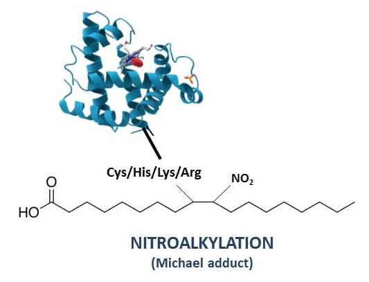
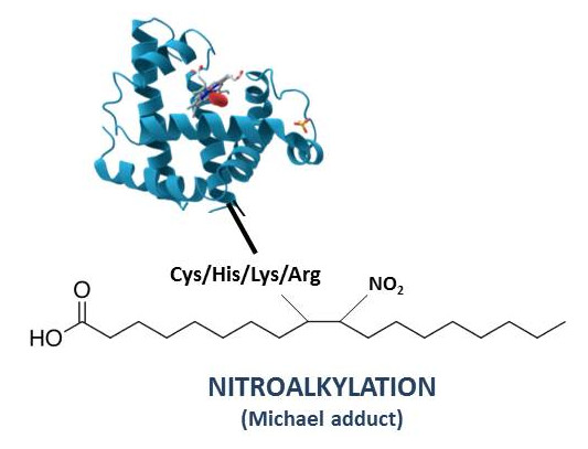
Nitrate fatty acids have been widely regarded as novel mediators of cell signaling in animal organisms. However, the knowledge about them in the plant kingdom is limited. The constitutive presence of NO2-FAs in plant systems was initially characterized in extra-virgin olive oil (EVOO) (a basic component of the Mediterranean diet) in which oleic acid, followed by palmitic (PA), linoleic (LA), and linolenic (Ln) acids are present [73,74,75]. Given their properties mentioned above, the inherent occurrence of NO2-FAs in EVOO and olives was analyzed using mass spectrometry techniques. Different endogenous NO2-cLA isomers were detected in EVOO, while intrinsic NO2-OA-cysteine adducts (higher levels in the olive peel) were found in olives. These reports demonstrate that both EVOO and olives are sources and endogenous reservoirs of NO2-FAs, which could be responsible of the anti-inflammatory and anti-hypertensive properties of EVOO [10,70].
Additionally, the presence of NO2-FAs has been recently reported in both cell–suspension cultures (ACSC) and seedlings of the model plant Arabidopsis thaliana. Originally, the model plant’s lipid composition was analyzed, with a predominance of Ln, followed by LA and OA [10]. The biological occurrence of NO2-Ln (Table 1) was only detected in ACSC (0.28 pmol/g FW) and seedlings (3.84 pmol/g FW) [9,10], while a modulation in NO2-Ln levels was detected during plant growth. Seeds, 14-day-old seedlings and leaves from 30- and 45-day-old Arabidopsis plants were used. The higher NO2-Ln content (11.18 pmol/g FW) was quantified at the seeds stage, with a continuous decline observed in the final vegetative and reproductive stages of the life cycle (0.54 pmol/g FW) [9,10]. In addition, the potential of NO2-Ln to emit NO has been recently evidenced [9,76], and the high NO2-Ln content in seeds could provide an additional source of NO which could favor germination and the onset of vegetative development [9,77,78,79].
Mass spectrometry techniques were also used to analyze the profile of NO2-FAs in other plant species. In this sense, NO2-Ln was detected in rice (Oryza sativa) leaves (0.748 pmol/g FW). The same type of NO2-FA was identified in pea leaves (Pisum sativum) mitochondria (0.084 pmol/g FW) and peroxisomes (0.282 pmol/g FW) and roots (0.072 pmol/g FW). These analyses show the wide spread of NO2-FAs in plant organisms [10,76]. Furthermore, the levels of NO2-Ln detected in plants are similar to those found in animal systems [31], which reinforces their essential role as signaling contributors in plants [10,80].
On the other hand, the NO2-Ln abundance was also quantified in Arabidopsis under adverse environmental conditions such as mechanical wounding, salinity, low temperature, and heavy-metal stress. Under these stress situations, a meaningful rise in NO2-Ln content was monitored accompanied by an induction of genes associated with oxidative stress and oxygen-containing compound responses [9,80,81].
After demonstrating its relationship with plant development and plant adverse situations, a transcriptomic analysis by RNA-seq technology allowed us to analyze the signaling role played by NO2-Ln. Initially, ACSC treated with increasing concentrations of NO2-Ln (10 µM and 100 µM) showed this molecule’s clear signaling response in terms of plant physiology and dose-dependence responses [9] previously described in animal systems [47,82]. A set of overexpressed genes related to abiotic and oxidative stress responses were detected after treatment with NO2-Ln, while other genes implicated in biological procedures, such as biosynthesis of cellular metabolites, were downregulated, with a similar pattern being observed in seedlings. It is important to highlight the involvement of upregulated genes in protein folding as well as in responses to heat and H2O2 stress. Unexpectedly, around 40% of the genes which responded to NO2-Ln were involved in heat-shock responses (HSRs) [9]. In animal systems, the treatment with NO2-OA also activates a considerable number of genes related to HSRs, which reveals the presence of a conserved mechanism of response to NO2-FAs in both animal and plant systems [9,82].
Among the upregulated genes which responded to reactive oxygen species (ROS) is a gene encoding for cytosolic ascorbate peroxidase 2 (APX2), which is a relevant enzyme involved in defending plants against H2O2. Additionally, under abiotic stress situations such as high temperatures and light intensity, interactions between APX2 and the heat shock transcription factor (HSFA2) have been detected [10,83].
Although the participation of NO2-Ln in plant biology and responses to abiotic stress conditions has been previously described, the mechanisms involved in NO2-Ln’s defense responses to stress in plants are still little known. As with animal systems, the release of NO by NO2-Ln in aqueous medium, which could be a signaling mechanism, has been demonstrated in Arabidopsis cell cultures by various in vitro experimental techniques such as ozone chemiluminescence, 4,5-diaminofluorescein (DAF-2) spectrofluorometric probes, confocal laser scanning microscopy, and the oxyhemoglobin oxidation method. Ozone chemiluminescence showed that NO-releasing from NO2-FA was not propitious in acidic locations, since at neutral pH (7.4) the maximum releasing of NO was achieved. This finding may be of considerable importance inside the cells, as mitochondria, peroxisomes, and the cytosol have a basic or neutral pH [10,76]. In addition, when the leaves and roots of Arabidopsis seedlings were treated with NO2-Ln, green fluorescence arose as a consequence of the increase in NO content, thus demonstrating the in vivo capability of NO2-Ln to provide NO. In addition, the subsequent treatment of samples with the NO scavenger 2-(4-carboxyphenyl)-4,4,5,5-tetramethylimidazoline-1-oxyl-3-oxide (cPTIO) causes a decrease in fluorescence [9,84]. These results emphasize the important role of NO2-Ln as a NO reservoir, and thus, the indirect involvement of NO2-FAs in plant growth, in the response to (a)biotic stress processes and in a variety of NO-related post-translational modifications (NO-PTMs) [80,85,86,87].

4. Nitroalkylation

Nitro-fatty acids, which are potent electrophiles owing to the presence of electron-withdrawing nitro (-NO2) substituents in the beta carbon, mainly act via post-translational modifications. For this reason, they are able to react with nucleophiles like glutathione or target amino acid residues, which affects their protein structure and eventually their function and subcellular localization [67,88]. The nitroalkylation PTM involves the establishment of a nitro-lipid-protein adduct with the cession of a couple of electrons from the nucleophile to the electrophile (NO2-FA) to form a covalent bond, via a Michael adduction. This process generates lipoxidation adducts (Figure 4). Nitroalkylation provokes a chain of signaling phenomena that concludes with anti-inflammatory, anti-hypersensitive, anti-tumorigenic, cytoprotective, and antioxidant effects arbitrated by NO2-FAs [46,89].
Diverse studies have displayed the reversible character of nitroalkylation which enables it to act as a selective signaling pathway in stressful environments. Under these conditions, the rise in the ROS and RNS levels could affect the stability of nitroalkylation. Reactive oxygen and nitrogen species (ROS and RNS) can cause the oxidation of the bond between the sulfur residues and the α-carbon of the NO2-FAs (Michael adduct) resulting both in the generation of sulfoxides and derived species and the scission of the Michael adduct. This process results in the releasing of the nitroalkene which enables the protein to recover its initial state [22,66,81,88,90,91]. The reversible possibilities of nitroalkylation in biological processes are of considerable importance, as irreversible PTMs usually lead to permanent loss of function, and thus protein degradation [22,46,68]. Although the main nucleophiles which react with NO2-FA are cysteine thiols (Cys-SH), and not all are able to react with electrophiles, in this sense, the deprotonated cysteine thiolate (Cys-S) is specifically the most prone to react [92,93]. Other nucleophiles are the amino substituents of lysine and arginine residues and the imidazole moiety of histidine [89].

4.1. Nitroalkylation in Animals

Nitrate fatty acids act as signaling mediators, since a scant amount of them act as powerful signal transduction cascade mediators that carry out changes in protein function through PTMs [1,66,68,94]. As mentioned above, processes such as digestion and inflammation lead to the genesis of NO2-FAs, predominantly NO2-cLA. In animal systems, NO2-FAs protect against a broad cluster of diseases such as atherosclerosis, restenosis, ischemia-reperfusion, renal injury, diabetes, metabolic syndrome, endotoxemia, and triple-negative breast cancer [95,96]. Their pluripotent cell signaling capacity enables NO2-FAs to modulate various intracellular pathways. In this line, the capacity of NO2-FAs to release NO via the Nef reaction generates low concentrations of NO which modulates cyclic monophosphate guanosine (cGMP)-dependent cell signaling activity. Nitrate fatty acids also control the generation of NO by regulating endothelial and inducible nitric oxide synthase (eNOS and iNOS) independently of cGMP mechanisms [34,62,76].
In addition, NO2-FAs can regulate the expression levels of differentiation-related, key inflammation, and cell proliferation genes [82,97,98,99,100,101]. Signaling via the Kelch-like ECH-associated protein 1 (Keap 1)-nuclear factor erythroid-derived 2-like 2 (Nrf2) pathway is a primary regulator of cellular responses to oxidative stress. The transcription factor Nrf2, which controls antioxidant protein expression, is located in the cytosol in its inactive form due to Keap1 activity which promotes Nrf2 ubiquitination and subsequent degradation by the ubiquitin–proteasome system. Keap 1 contains reactive cysteines (Cys 151, 273, and 288) which can be modified by oxidation or alkylation and used as redox state sensors. When electrophiles such as NO2-OA, NO2-LA, and NO2-AA are formed, the interaction between Nrf2 and Keap1 is interrupted. This facilitates the transfer of Nrf2 to the nucleus, where it will link to specific cis targets and activate the regulation of antioxidant response element (ARE) genes [1,55,97,102,103,104,105]. The NO2-FA-sensitive system involving heat-shock responses (HSRs) is a complex alliance of regulatory proteins and transcription factors which promotes cytoprotective and anti-inflammatory target gene expression [46]. Heat-shock proteins (HSPs) are chaperones whose expression is triggered by stress conditions, including heat, as well as by electrophilic and reactive species caused under inflammatory injury. Chaperones prevent the aggregation of denatured or oxidized proteins, collaborate in the transfer of these proteins to intracellular locations, and thus contribute to cellular redox homeostasis [106]. Nitro-oleic acid in human endothelial cells has been reported to activate HSF1 (Heat Shock Factor 1), the most important regulator of HSRs, followed by a remarkable induction of a large group of heat shock genes (Table 2) [82,102,107].
Nitro-fatty acid can also activate the peroxisome proliferator-activating receptor (PPAR), particularly PPARƴ, which is included in the family of nuclear hormone receptors. This receptor plays a marked role in the expression of transcription factors associated with lipid generation, lipid and glucose metabolism, macrophage differentiation, and immune responses. The PPARƴ regulatory domain is located in the C-terminal side which coincides with the ligand binding domain. The location of a cysteine at position 285 makes this hydrophobic region susceptible to nitroalkylation by NO2-FAs such as NO2-OA and NO2-LA (Table 2) [1,101,108,109,110].
Another example is the nuclear factor kappa betta (NF-kβ) involved in transcriptional regulation under inflammatory and immune processes. The nuclear factor kappa betta is a protein complex with two subunits (p50 and p65) [1,98,111,112]. Experimental studies have shown that NF-kβ is regulated by NO2-FAs at multiple levels including the inhibition of Toll-like receptor 4 (TLR4) by NO2-OA. Toll-like receptor 4 is a transmembrane protein which pertains to the pattern recognition receptor (PRR) family which is able to recognize bacterial lipopolysaccharide (LPS). Its activation triggers the intracellular NF-κB signaling pathway and inflammatory cytokine production which activate the innate immune system. Thus, the inhibition of TLR4 by NO2-FAs also triggers the inhibition of NF-kβ [101,113]. Another level of regulation is the inhibition of NF-kβ by nitroalkylation, specifically, the residue Cys38, placed in the DNA-binding domain of the p65 subunit, is susceptible to nitroalkylation [96,98]. The final level of regulation is the activation of PPAR by NO2-FAs which causes the trans-repression of inflammatory genes such as NF-kβ (Table 2) [101,114].
In animal systems, nitroalkylation is considered to be a decisive signaling resource in anti-inflammatory processes. Nitrate fatty acids modify the anti-inflammatory response at multiple levels including gene expression, protein translation (acting on transcription factors and lipid receptors), as well as cell function, as many inflammatory proteins contain numerous nucleophilic amino acid residues which can be nitroalkylation targets. Table 2 shows a summary list of NO2-FA protein targets in animal systems and how they are affected by nitroalkylation.

4.2. Nitroalkylation in Plants

Although the effects of nitroalkylation have been extensively studied in animal organisms, the impact of NO2-FA action in plants, which has not been fully explored, constitutes an emerging area of interesting research work. Probably, the signaling function of NO2-Ln is due to nitroalkylation processes. In this context, the endogenous presence of 37 proteins adducted with NO2-Ln in Arabidopsis cell cultures has been identified. However, cell cultures treated with 100 µM NO2-Ln showed an increase in the number of nitroalkylated proteins (342), belonging to different areas of cell metabolism, which included APX2 (unpublished results), whose encoding gene expression, according to the transcriptomic studies mentioned above, was induced [9].
Ascorbate peroxidase (APX2) is one of the primary antioxidant systems in plants. This enzyme belongs to the ascorbate–glutathione cycle, which detoxifies hydrogen peroxide and contains non-enzymatic antioxidants (ascorbate and glutathione) and enzymatic antioxidants such as monodehydroascorbate reductase (MDAR), glutathione reductase (GR), and dehydroascorbate reductase (DHAR), as well as the reductive coenzyme NADPH [127,128].
In this study, the APX recombinant protein from Arabidospsis thaliana was incubated with increasing concentrations of NO2-Ln (1 µM and 10 µM). The enzymatic activity was spectrophotometrically monitored [129]. Furthermore, the nitroalkylation targeted residues of the treated recombinant protein were detected and characterized using LC-MS/MS. Thus, the protein was digested by trypsin and desalted by C18 columns to obtain the peptide fraction which was analyzed using an Exactive Q mass spectrometer attached to a nano-flow liquid chromatograph (nanoLC) (Thermo Fisher Scientific). The LC-MS/MS spectrum deconvolution was carried out employing Proteome Discoverer version 1.4. bioinformatics software (Thermo Fisher Scientific). The Percolator node was used to filter the peptides at a 1% false discovery rate (FDR) at the peptide-spectrum matches (PSMs).
In order to identify the position of the nitroalkylation-targeted nucleophilic residues, an in silico modeling was carried out using Raptor X bioinformatics software (http://raptorx.uchicago.edu/). The APX model was based on the structure of isoniazid (INH) bound to cytosolic soybean ascorbate peroxidase (PDB:2VCF) [130].
The treatment of recombinant APX with NO2-Ln modulates its enzymatic activity, showing a significant decrease in the presence of 10 µM NO2Ln (Figure 5). This decreased activity was associated with the post-translational modification caused by nitroalkylation, which was detected by mass spectrometry. Comparison of the spectra of control and NO2-Ln-treated samples displayed a rise in the mass of nucleophilic residues due to treatment with NO2-Ln. The electrophilic attack by NO2-Ln generated the nitroalkylation of the residues showed in Figure 6 and Figure 7A. with histidine 43 and histidine 163 being preferentially nitroalkylated. This could have functional implications (Figure 7B), as histidine 43 and histidine 163 are located at the active and metal-binding site, respectively. This fact suggests that the nitroalkylation of these residues blocks APX enzymatic activity, modulating protein function.
Figure 8 explains the model of the nitro-lipid-protein adducts signaling mechanism in plants. Nitro-lipid-protein adducts stability can be affected by the accumulation of ROS and RNS, which could cause the oxidation of sulfhydryl substituents in proteins, and consequently the scission of the Michael adduct releasing NO2-Ln. As was previously mentioned, the nitroalkylation of APX by NO2-Ln generates function loss. Under nitro-oxidative conditions, the function of APX would be reactivated due to the reversibility of the nitroalkylation PTM. On the other hand, the levels of free NO2-FA increase, being able to stimulate the expression of heat shock proteins (HSPs) and certain antioxidant systems such as APX and methionine sulfoxide reductase B (MSRB). Another possibility is that NO2-FA could donate ·NO in the cellular aqueous environment which could act in a broad set of plant activities such as plant development, (a)biotic disorders, antioxidant responses, and NO-PTMs.
The ability of NO2-Ln to trigger pleiotropic signaling actions mainly depends on the nitroalkylation of regulatory proteins involved in plant biology and numerous types of (a)biotic-stress. Being a reversible post-translational modification, which can affect a large number of target amino acid residues (Cys, His, Lys, and Arg), together with the features outlined above, render nitroalkylation an important cell signaling mechanism mediated by NO2-FAs.

5. Conclusions and Future Perspectives

The potent electrophilic molecules NO2-FAs, whose electrophilicity triggers potential signaling mechanisms via nitroalkylation, were recently discovered in both animal and plant systems. This NO2-FA-mediated PTM can be considered a NO-PTM similar to S-nitrosylation, because NO2-FAs are RLS formed as a result of the oxidation of PUFA by NO-derived species. The importance of nitroalkylation resides in its reversibility and in the presence of a considerable amount of target amino acids residues that generate the formation of nitro-lipid-protein adducts, which enables this NO-PTM to trigger pleiotropic signaling actions. In animal systems, nitroalkylation is associated with signaling mechanisms in anti-inflammatory processes. However, in plant systems, this little-known NO-PTM constitutes an emerging area of research which should be developed through advances in mass spectrometry techniques.

Funding

The study was supported by an ERDF grant co-financed by the Spanish Ministry of Economy and Competitiveness (Project BIO2015-66390-P) and the Junta de Andalucía (Group BIO-286).

Acknowledgments

L.A.-C. wishes to thank the University of Jaén for funding her PhD fellowship.

Conflicts of Interest

The authors declare no conflicts of interest.

References

  1. Schopfer, F.J.; Cipollina, C.; Freeman, B.A. Formation and signaling actions of electrophilic lipids. Chem. Rev. 2011, 111, 5997–6021. [Google Scholar] [CrossRef] [PubMed]
  2. Beavers, W.N.; Rose, K.L.; Galligan, J.J.; Mitchener, M.M.; Rouzer, C.A.; Tallman, K.A.; Lamberson, C.R.; Wang, X.; Hill, S.; Ivanova, P.T. Protein modification by endogenously generated lipid electrophiles: Mitochondria as the source and target. ACS Chem. Biol. 2017, 12, 2062–2069. [Google Scholar] [CrossRef] [PubMed]
  3. Wang, C.; Weerapana, E.; Blewett, M.M.; Cravatt, B.F. A chemoproteomic platform to quantitatively map targets of lipid-derived electrophiles. Nat. Methods 2013, 11, 79. [Google Scholar] [CrossRef] [PubMed]
  4. Higdon, A.; Diers, A.R.; Oh, J.Y.; Landar, A.; Darley-Usmar, V.M. Cell signalling by reactive lipid species: New concepts and molecular mechanisms. Biochem. J. 2012, 442, 453–464. [Google Scholar] [CrossRef] [PubMed]
  5. Marwah, S.; Blann, A.; Rea, C.; Phillips, J.; Wright, J.; Bareford, D. Reduced vitamin E antioxidant capacity in sickle cell disease is related to transfusion status but not to sickle crisis. Am. J. Hematol. 2002, 69, 144–146. [Google Scholar] [CrossRef] [PubMed]
  6. Yin, H.; Porter, N.A. New insights regarding the autoxidation of polyunsaturated fatty acids. Antioxid. Redox Signal. 2005, 7, 170–184. [Google Scholar] [CrossRef]
  7. Poon, H.F.; Calabrese, V.; Scapagnini, G.; Butterfield, D.A. Free radicals: Key to brain aging and heme oxygenase as a cellular response to oxidative stress. J. Gerontol. A Biol. Sci. Med. Sci. 2004, 59, M478–M493. [Google Scholar] [CrossRef]
  8. Morrow, J.D. Quantification of isoprostanes as indices of oxidant stress and the risk of atherosclerosis in humans. Arterioscler. Thromb. Vasc. Biol. 2005, 25, 279–286. [Google Scholar] [CrossRef]
  9. Mata-Pérez, C.; Sánchez-Calvo, B.; Padilla, M.N.; Begara-Morales, J.C.; Luque, F.; Melguizo, M.; Jiménez-Ruiz, J.; Fierro-Risco, J.; Peñas-Sanjuán, A.; Valderrama, R. Nitro-fatty acids in plant signaling: Nitro-linolenic acid induces the molecular chaperone network in Arabidopsis. Plant Physiol. 2016, 170, 686–701. [Google Scholar] [CrossRef]
  10. Mata-Perez, C.; Sanchez-Calvo, B.; Padilla, M.N.; Begara-Morales, J.C.; Valderrama, R.; Corpas, F.J.; Barroso, J.B. Nitro-fatty acids in plant signaling: New key mediators of nitric oxide metabolism. Redox Biol. 2017, 11, 554–561. [Google Scholar] [CrossRef]
  11. Porter, N.A.; Caldwell, S.E.; Mills, K.A. Mechanisms of free radical oxidation of unsaturated lipids. Lipids 1995, 30, 277–290. [Google Scholar] [CrossRef] [PubMed]
  12. Addis, P. Occurrence of lipid oxidation products in foods. Food Chem. Toxicol. 1986, 24, 1021–1030. [Google Scholar] [CrossRef]
  13. Hwa Lee, S.; Rangiah, K.; Williams, M.V.; Wehr, A.Y.; DuBois, R.N.; Blair, I.A. Cyclooxygenase-2-mediated metabolism of arachidonic acid to 15-oxo-eicosatetraenoic acid by rat intestinal epithelial cells. Chem. Res. Toxicol. 2007, 20, 1665–1675. [Google Scholar] [CrossRef]
  14. Chiang, N.; Serhan, C.N.; Dahlén, S.-E.; Drazen, J.M.; Hay, D.W.; Rovati, G.E.; Shimizu, T.; Yokomizo, T.; Brink, C. The lipoxin receptor ALX: Potent ligand-specific and stereoselective actions in vivo. Pharmacol. Rev. 2006, 58, 463–487. [Google Scholar] [CrossRef] [PubMed]
  15. Baker, P.R.; Schopfer, F.J.; O’Donnell, V.B.; Freeman, B.A. Convergence of nitric oxide and lipid signaling: Anti-inflammatory nitro-fatty acids. Free Radic. Biol. Med. 2009, 46, 989–1003. [Google Scholar] [CrossRef] [PubMed]
  16. Rubbo, H.; Radi, R.; Trujillo, M.; Telleri, R.; Kalyanaraman, B.; Barnes, S.; Kirk, M.; Freeman, B.A. Nitric oxide regulation of superoxide and peroxynitrite-dependent lipid peroxidation. Formation of novel nitrogen-containing oxidized lipid derivatives. J. Biol. Chem. 1994, 269, 26066–26075. [Google Scholar] [PubMed]
  17. Davies, S.S.; Amarnath, V.; Roberts, L.J., II. Isoketals: Highly reactive γ-ketoaldehydes formed from the H2-isoprostane pathway. Chem. Phys. Lipids 2004, 128, 85–99. [Google Scholar] [CrossRef]
  18. Ueno, N.; Murakami, M.; Tanioka, T.; Fujimori, K.; Tanabe, T.; Urade, Y.; Kudo, I. Coupling between cyclooxygenase, terminal prostanoid synthase, and phospholipase A2. J. Biol. Chem. 2001, 276, 34918–34927. [Google Scholar] [CrossRef]
  19. Prigge, S.; Boyington, J.; Faig, M.; Doctor, K.; Gaffney, B.; Amzel, L. Structure and mechanism of lipoxygenases. Biochimie 1997, 79, 629–636. [Google Scholar] [CrossRef]
  20. O’donnell, V.B.; Freeman, B.A. Interactions between nitric oxide and lipid oxidation pathways: Implications for vascular disease. Circ. Res. 2001, 88, 12–21. [Google Scholar] [CrossRef]
  21. Swain, C.G.; Scott, C.B. Quantitative correlation of relative rates. Comparison of hydroxide ion with other nucleophilic reagents toward alkyl halides, esters, epoxides and acyl halides1. J. Am. Chem. Soc. 1953, 75, 141–147. [Google Scholar] [CrossRef]
  22. Rudolph, T.K.; Freeman, B.A. Transduction of redox signaling by electrophile-protein reactions. Sci. Signal. 2009, 2, re7. [Google Scholar] [CrossRef] [PubMed]
  23. Nagahara, N.; Matsumura, T.; Okamoto, R.; Kajihara, Y. Protein cysteine modifications: (2) reactivity specificity and topics of medicinal chemistry and protein engineering. Curr. Med. Chem. 2009, 16, 4490–4501. [Google Scholar] [CrossRef]
  24. Turell, L.; Steglich, M.; Alvarez, B. The chemical foundations of nitroalkene fatty acid signaling through addition reactions with thiols. Nitric Oxide 2018. [Google Scholar] [CrossRef] [PubMed]
  25. Pearson, R.G. Hard and soft acids and bases. J. Am. Chem. Soc. 1963, 85, 3533–3539. [Google Scholar] [CrossRef]
  26. Reed, T.T. Lipid peroxidation and neurodegenerative disease. Free Radic. Biol. Med. 2011, 51, 1302–1319. [Google Scholar] [CrossRef]
  27. Di Domenico, F.; Pupo, G.; Tramutola, A.; Giorgi, A.; Schininà, M.E.; Coccia, R.; Head, E.; Butterfield, D.A.; Perluigi, M. Redox proteomics analysis of HNE-modified proteins in Down syndrome brain: Clues for understanding the development of Alzheimer disease. Free Radic. Biol. Med. 2014, 71, 270–280. [Google Scholar] [CrossRef]
  28. Butterfield, D.A.; Gu, L.; Domenico, F.D.; Robinson, R.A. Mass spectrometry and redox proteomics: Applications in disease. Mass Spectrom. Rev. 2014, 33, 277–301. [Google Scholar] [CrossRef]
  29. Sauriasari, R.; Andrajati, R.; Saputri, D.; Muris, R.; Manfaatun, A.; Amanda, O.; Setiawan, H.; Sakano, N.; Wang, D.; Ogino, K. Marker of lipid peroxidation related to diabetic nephropathy in Indonesian type 2 diabetes mellitus patients. Diabetes Res. Clin. Pract. 2015, 108, 193–200. [Google Scholar] [CrossRef] [PubMed]
  30. Uchida, K. Role of reactive aldehyde in cardiovascular diseases. Free Radic. Biol. Med. 2000, 28, 1685–1696. [Google Scholar] [CrossRef]
  31. Bell-Parikh, L.C.; Ide, T.; Lawson, J.A.; McNamara, P.; Reilly, M.; FitzGerald, G.A. Biosynthesis of 15-deoxy-Δ 12, 14-PGJ 2 and the ligation of PPARγ. J. Clin. Investig. 2003, 112, 945–955. [Google Scholar] [CrossRef] [PubMed]
  32. Oh, J.Y.; Giles, N.; Landar, A.; Darley-Usmar, V. Accumulation of 15-deoxy-Δ12, 14-prostaglandin J2 adduct formation with Keap1 over time: Effects on potency for intracellular antioxidant defence induction. Biochem. J. 2008, 411, 297–306. [Google Scholar] [CrossRef]
  33. Groeger, A.L.; Cipollina, C.; Cole, M.P.; Woodcock, S.R.; Bonacci, G.; Rudolph, T.K.; Rudolph, V.; Freeman, B.A.; Schopfer, F.J. Cyclooxygenase-2 generates anti-inflammatory mediators from omega-3 fatty acids. Nat. Chem. Biol. 2010, 6, 433–441. [Google Scholar] [CrossRef]
  34. Khoo, N.K.; Freeman, B.A. Electrophilic nitro-fatty acids: Anti-inflammatory mediators in the vascular compartment. Curr. Opin. Pharmacol. 2010, 10, 179–184. [Google Scholar] [CrossRef]
  35. Wong, H.L.; Liebler, D.C. Mitochondrial protein targets of thiol-reactive electrophiles. Chem. Res. Toxicol. 2008, 21, 796–804. [Google Scholar] [CrossRef]
  36. Vila, A.; Tallman, K.A.; Jacobs, A.T.; Liebler, D.C.; Porter, N.A.; Marnett, L.J. Identification of protein targets of 4-hydroxynonenal using click chemistry for ex vivo biotinylation of azido and alkynyl derivatives. Chem. Res. Toxicol. 2008, 21, 432–444. [Google Scholar] [CrossRef]
  37. Szapacs, M.E.; Kim, H.-Y.H.; Porter, N.A.; Liebler, D.C. Identification of proteins adducted by lipid peroxidation products in plasma and modifications of apolipoprotein A1 with a novel biotinylated phospholipid probe. J. Proteome Res. 2008, 7, 4237–4246. [Google Scholar] [CrossRef]
  38. Shin, N.-Y.; Liu, Q.; Stamer, S.L.; Liebler, D.C. Protein targets of reactive electrophiles in human liver microsomes. Chem. Res. Toxicol. 2007, 20, 859–867. [Google Scholar] [CrossRef] [PubMed]
  39. Dennehy, M.K.; Richards, K.A.; Wernke, G.R.; Shyr, Y.; Liebler, D.C. Cytosolic and nuclear protein targets of thiol-reactive electrophiles. Chem. Res. Toxicol. 2006, 19, 20–29. [Google Scholar] [CrossRef] [PubMed]
  40. Gomi, K.; Yamamoto, H.; Akimitsu, K. Characterization of a lipoxygenase gene in rough lemon induced by Alternaria alternata. J. Gen. Plant Pathol. 2002, 68, 21–30. [Google Scholar] [CrossRef]
  41. Kim, E.-S.; Choi, E.; Kim, Y.; Cho, K.; Lee, A.; Shim, J.; Rakwal, R.; Agrawal, G.K.; Han, O. Dual positional specificity and expression of non-traditional lipoxygenase induced by wounding and methyl jasmonate in maize seedlings. Plant Mol. Biol. 2003, 52, 1203–1213. [Google Scholar] [CrossRef] [PubMed]
  42. Montillet, J.-L.; Agnel, J.-P.; Ponchet, M.; Vailleau, F.; Roby, D.; Triantaphylidès, C. Lipoxygenase-mediated production of fatty acid hydroperoxides is a specific signature of the hypersensitive reaction in plants. Plant Physiol. Biochem. 2002, 40, 633–639. [Google Scholar] [CrossRef]
  43. He, Y.; Fukushige, H.; Hildebrand, D.F.; Gan, S. Evidence supporting a role of jasmonic acid in Arabidopsis leaf senescence. Plant Physiol. 2002, 128, 876–884. [Google Scholar] [CrossRef] [PubMed]
  44. Uchida, K. 4-Hydroxy-2-nonenal: A product and mediator of oxidative stress. Prog. Lipid Res. 2003, 42, 318–343. [Google Scholar] [CrossRef]
  45. Thoma, I.; Loeffler, C.; Sinha, A.K.; Gupta, M.; Krischke, M.; Steffan, B.; Roitsch, T.; Mueller, M.J. Cyclopentenone isoprostanes induced by reactive oxygen species trigger defense gene activation and phytoalexin accumulation in plants. Plant J. 2003, 34, 363–375. [Google Scholar] [CrossRef] [PubMed]
  46. Geisler, A.C.; Rudolph, T.K. Nitroalkylation—a redox sensitive signaling pathway. BBA-Gen. Subj. 2012, 1820, 777–784. [Google Scholar] [CrossRef] [PubMed]
  47. Bonacci, G.; Baker, P.R.; Salvatore, S.R.; Shores, D.; Khoo, N.K.; Koenitzer, J.R.; Vitturi, D.A.; Woodcock, S.R.; Golin-Bisello, F.; Watkins, S. Conjugated linoleic acid is a preferential substrate for fatty acid nitration. J. Biol. Chem. 2012, 287, 44071–44082. [Google Scholar] [CrossRef] [PubMed]
  48. Pryor, W.A.; Lightsey, J.W.; Church, D.F. Reaction of nitrogen dioxide with alkenes and polyunsaturated fatty acids: Addition and hydrogen-abstraction mechanisms. J. Am. Chem. Soc. 1982, 104, 6685–6692. [Google Scholar] [CrossRef]
  49. Buchan, G.R.; Bonacci, G.; Fazzari, M.; Salvatore, S.; Wendell, S.G. Nitro-fatty acid formation and metabolism. Nitric Oxide 2018, 79, 38–44. [Google Scholar] [CrossRef] [PubMed]
  50. d’Ischia, M.; Napolitano, A.; Manini, P.; Panzella, L. Secondary targets of nitrite-derived reactive nitrogen species: Nitrosation/nitration pathways, antioxidant defense mechanisms and toxicological implications. Chem. Res. Toxicol. 2011, 24, 2071–2092. [Google Scholar] [CrossRef]
  51. Rudolph, V.; Rudolph, T.K.; Schopfer, F.J.; Bonacci, G.; Woodcock, S.R.; Cole, M.P.; Baker, P.R.; Ramani, R.; Freeman, B.A. Endogenous generation and protective effects of nitro-fatty acids in a murine model of focal cardiac ischaemia and reperfusion. Cardiovasc. Res. 2009, 85, 155–166. [Google Scholar] [CrossRef] [PubMed]
  52. Nadtochiy, S.M.; Baker, P.R.; Freeman, B.A.; Brookes, P.S. Mitochondrial nitroalkene formation and mild uncoupling in ischaemic preconditioning: Implications for cardioprotection. Cardiovasc. Res. 2008, 82, 333–340. [Google Scholar] [CrossRef] [PubMed]
  53. Reynolds, C.; Roche, H. Conjugated linoleic acid and inflammatory cell signalling. Prostaglandins Leukot. Essent. Fat. Acids 2010, 82, 199–204. [Google Scholar] [CrossRef]
  54. Vitturi, D.A.; Minarrieta, L.; Salvatore, S.R.; Postlethwait, E.M.; Fazzari, M.; Ferrer-Sueta, G.; Lancaster, J.R., Jr.; Freeman, B.A.; Schopfer, F.J. Convergence of biological nitration and nitrosation via symmetrical nitrous anhydride. Nat. Chem. Biol. 2015, 11, 504. [Google Scholar] [CrossRef] [PubMed]
  55. Suzuki, T.; Yamamoto, M. Stress-sensing mechanisms and the physiological roles of the Keap1-Nrf2 system during cellular stress. J. Biol. Chem. 2017, 292, 16817–16824. [Google Scholar] [CrossRef] [PubMed]
  56. Weitzberg, E.; Lundberg, J.O. Novel aspects of dietary nitrate and human health. Annu. Rev. Nutr. 2013, 33, 129–159. [Google Scholar] [CrossRef]
  57. Lundberg, J.O.; Weitzberg, E. NO generation from inorganic nitrate and nitrite: Role in physiology, nutrition and therapeutics. Arch. Pharm. Res. 2009, 32, 1119–1126. [Google Scholar] [CrossRef] [PubMed]
  58. Moreno-Vivián, C.; Cabello, P.; Martínez-Luque, M.; Blasco, R.; Castillo, F. Prokaryotic nitrate reduction: Molecular properties and functional distinction among bacterial nitrate reductases. J. Bacteriol. 1999, 181, 6573–6584. [Google Scholar]
  59. Ferreira, A.M.; Ferrari, M.I.; Trostchansky, A.; Batthyany, C.; Souza, J.M.; Alvarez, M.N.; López, G.V.; Baker, P.R.; Schopfer, F.J.; O’Donnell, V. Macrophage activation induces formation of the anti-inflammatory lipid cholesteryl-nitrolinoleate. Biochem. J. 2009, 417, 223–238. [Google Scholar] [CrossRef]
  60. Freeman, B.A.; Baker, P.R.; Schopfer, F.J.; Woodcock, S.R.; Napolitano, A.; d’Ischia, M. Nitro-fatty acid formation and signaling. J. Biol. Chem. 2008, 283, 15515–15519. [Google Scholar] [CrossRef]
  61. Faine, L.A.; Cavalcanti, D.; Rudnicki, M.; Ferderbar, S.; Macedo, S.; Souza, H.P.; Farsky, S.; Boscá, L.; Abdalla, D. Bioactivity of nitrolinoleate: Effects on adhesion molecules and CD40-CD40L system. J. Nutr. Biochem. 2010, 21, 125–132. [Google Scholar] [CrossRef]
  62. Schopfer, F.J.; Baker, P.R.; Giles, G.; Chumley, P.; Batthyany, C.; Crawford, J.; Patel, R.P.; Hogg, N.; Branchaud, B.P.; Lancaster, J.R. Fatty acid transduction of nitric oxide signaling Nitrolinoleic acid is a hydrophobically stabilized nitric oxide donor. J. Biol. Chem. 2005, 280, 19289–19297. [Google Scholar] [CrossRef] [PubMed]
  63. Su, Y.H.; Wu, S.S.; Hu, C.H. Release of nitric oxide from nitrated fatty acids: Insights from computational chemistry. J. Chin. Chem. Soc. 2019, 66, 41–48. [Google Scholar] [CrossRef]
  64. Lima, É.S.; Bonini, M.G.; Augusto, O.; Barbeiro, H.V.; Souza, H.P.; Abdalla, D.S. Nitrated lipids decompose to nitric oxide and lipid radicals and cause vasorelaxation. Free Radic. Biol. Med. 2005, 39, 532–539. [Google Scholar] [CrossRef]
  65. Gorczynski, M.J.; Huang, J.; Lee, H.; King, S.B. Evaluation of nitroalkenes as nitric oxide donors. Bioorg. Med. Chem. Lett. 2007, 17, 2013–2017. [Google Scholar] [CrossRef]
  66. Batthyany, C.; Schopfer, F.J.; Baker, P.R.; Durán, R.; Baker, L.M.; Huang, Y.; Cerveñansky, C.; Braunchaud, B.P.; Freeman, B.A. Reversible post-translational modification of proteins by nitrated fatty acids in vivo. J. Biol. Chem. 2006, 281, 20450–20463. [Google Scholar] [CrossRef]
  67. Baker, L.M.; Baker, P.R.; Golin-Bisello, F.; Schopfer, F.J.; Fink, M.; Woodcock, S.R.; Branchaud, B.P.; Radi, R.; Freeman, B.A. Nitro-fatty acid reaction with glutathione and cysteine Kinetic analysis of thiol alkylation by a Michael addition reaction. J. Biol. Chem. 2007, 282, 31085–31093. [Google Scholar] [CrossRef] [PubMed]
  68. Rubbo, H.; Radi, R. Protein and lipid nitration: Role in redox signaling and injury. BBA-Gen. Subj. 2008, 1780, 1318–1324. [Google Scholar] [CrossRef]
  69. Fazzari, M.; Khoo, N.; Woodcock, S.R.; Li, L.; Freeman, B.A.; Schopfer, F.J. Generation and esterification of electrophilic fatty acid nitroalkenes in triacylglycerides. Free Radic. Biol. Med. 2015, 87, 113–124. [Google Scholar] [CrossRef]
  70. Fazzari, M.; Trostchansky, A.; Schopfer, F.J.; Salvatore, S.R.; Sánchez-Calvo, B.; Vitturi, D.; Valderrama, R.; Barroso, J.B.; Radi, R.; Freeman, B.A. Olives and olive oil are sources of electrophilic fatty acid nitroalkenes. PLoS ONE 2014, 9, e84884. [Google Scholar] [CrossRef]
  71. Vitturi, D.A.; Chen, C.-S.; Woodcock, S.R.; Salvatore, S.R.; Bonacci, G.; Koenitzer, J.R.; Stewart, N.A.; Wakabayashi, N.; Kensler, T.W.; Freeman, B.A. Modulation of nitro-fatty acid signaling prostaglandin reductase-1 is a nitroalkene reductase. J. Biol. Chem. 2013, 288, 25626–25637. [Google Scholar] [CrossRef] [PubMed]
  72. Melo, T.N.; Domingues, P.; Ferreira, R.; Milic, I.; Fedorova, M.; Santos, S.R.M.; Segundo, M.A.; Domingues, M.R.r.M. Recent advances on mass spectrometry analysis of nitrated phospholipids. Anal. Chem. 2016, 88, 2622–2629. [Google Scholar] [CrossRef] [PubMed]
  73. Catharino, R.R.; Haddad, R.; Cabrini, L.G.; Cunha, I.B.; Sawaya, A.C.; Eberlin, M.N. Characterization of vegetable oils by electrospray ionization mass spectrometry fingerprinting: Classification, quality, adulteration, and aging. Anal. Chem. 2005, 77, 7429–7433. [Google Scholar] [CrossRef]
  74. Rastrelli, L.; Passi, S.; Ippolito, F.; Vacca, G.; De Simone, F. Rate of degradation of α-tocopherol, squalene, phenolics, and polyunsaturated fatty acids in olive oil during different storage conditions. J. Agric. Food Chem. 2002, 50, 5566–5570. [Google Scholar] [CrossRef] [PubMed]
  75. Pérez-Camino, M.C.; Moreda, W.; Mateos, R.; Cert, A. Determination of esters of fatty acids with low molecular weight alcohols in olive oils. J. Agric. Food Chem. 2002, 50, 4721–4725. [Google Scholar] [CrossRef]
  76. Mata-Pérez, C.; Sánchez-Calvo, B.; Begara-Morales, J.C.; Carreras, A.; Padilla, M.N.; Melguizo, M.; Valderrama, R.; Corpas, F.J.; Barroso, J.B. Nitro-linolenic acid is a nitric oxide donor. Nitric Oxide 2016, 57, 57–63. [Google Scholar] [CrossRef]
  77. Beligni, M.V.; Lamattina, L. Nitric oxide stimulates seed germination and de-etiolation, and inhibits hypocotyl elongation, three light-inducible responses in plants. Planta 2000, 210, 215–221. [Google Scholar] [CrossRef] [PubMed]
  78. Bethke, P.C.; Libourel, I.G.; Jones, R.L. Nitric oxide reduces seed dormancy in Arabidopsis. J. Exp. Bot. 2005, 57, 517–526. [Google Scholar] [CrossRef]
  79. Libourel, I.G.; Bethke, P.C.; De Michele, R.; Jones, R.L. Nitric oxide gas stimulates germination of dormant Arabidopsis seeds: Use of a flow-through apparatus for delivery of nitric oxide. Planta 2006, 223, 813–820. [Google Scholar] [CrossRef] [PubMed]
  80. Mata-Pérez, C.; Padilla, M.N.; Sánchez-Calvo, B.; Begara-Morales, J.C.; Valderrama, R.; Chaki, M.; Barroso, J.B. Biological properties of nitro-fatty acids in plants. Nitric Oxide 2018. [Google Scholar] [CrossRef] [PubMed]
  81. Padilla, M.N.; Mata-Pérez, C.; Melguizo, M.; Barroso, J.B. In vitro nitro-fatty acid release from Cys-NO2-fatty acid adducts under nitro-oxidative conditions. Nitric Oxide 2017, 68, 14–22. [Google Scholar] [CrossRef] [PubMed]
  82. Kansanen, E.; Jyrkkanen, H.-K.; Volger, O.L.; Leinonen, H.; Kivela, A.M.; Hakkinen, S.-K.; Woodcock, S.R.; Schopfer, F.J.; Horrevoets, A.J.; Yla-Herttala, S. Nrf2-dependent and-independent responses to nitro-fatty acids in human endothelial cells: Identification of heat shock response as the major pathway activated by nitro-oleic acid. J. Biol. Chem. 2009, 284, 33233–33241. [Google Scholar] [CrossRef]
  83. Nishizawa, A.; Yabuta, Y.; Yoshida, E.; Maruta, T.; Yoshimura, K.; Shigeoka, S. Arabidopsis heat shock transcription factor A2 as a key regulator in response to several types of environmental stress. Plant J. 2006, 48, 535–547. [Google Scholar] [CrossRef] [PubMed]
  84. Sánchez-Calvo, B.; Barroso, J.B.; Corpas, F.J. Hypothesis: Nitro-fatty acids play a role in plant metabolism. Plant Sci. 2013, 199, 1–6. [Google Scholar] [CrossRef] [PubMed]
  85. Astier, J.; Kulik, A.; Koen, E.; Besson-Bard, A.; Bourque, S.; Jeandroz, S.; Lamotte, O.; Wendehenne, D. Protein S-nitrosylation: What’s going on in plants? Free Radic. Biol. Med. 2012, 53, 1101–1110. [Google Scholar] [CrossRef] [PubMed]
  86. Begara-Morales, J.C.; Chaki, M.; Sánchez-Calvo, B.; Mata-Pérez, C.; Leterrier, M.; Palma, J.M.; Barroso, J.B.; Corpas, F.J. Protein tyrosine nitration in pea roots during development and senescence. J. Exp. Bot. 2013, 64, 1121–1134. [Google Scholar] [CrossRef] [PubMed]
  87. Chaki, M.; Shekariesfahlan, A.; Ageeva, A.; Mengel, A.; von Toerne, C.; Durner, J.; Lindermayr, C. Identification of nuclear target proteins for S-nitrosylation in pathogen-treated Arabidopsis thaliana cell cultures. Plant Sci. 2015, 238, 115–126. [Google Scholar] [CrossRef]
  88. Rudolph, V.; Schopfer, F.J.; Khoo, N.K.; Rudolph, T.K.; Cole, M.P.; Woodcock, S.R.; Bonacci, G.; Groeger, A.L.; Golin-Bisello, F.; Chen, C.-S. Nitro-fatty acid metabolome: Saturation, desaturation, β-oxidation, and protein adduction. J. Biol. Chem. 2009, 284, 1461–1473. [Google Scholar] [CrossRef]
  89. Melo, T.; Montero-Bullón, J.-F.; Domingues, P.; Domingues, M.R. Discovery of bioactive nitrated lipids and nitro-lipid-protein adducts using mass spectrometry-based approaches. Redox Biol. 2019, 2019, 101106. [Google Scholar] [CrossRef]
  90. Lin, W.-W.; Jang, Y.-J.; Wang, Y.; Liu, J.-T.; Hu, S.-R.; Wang, L.-Y.; Yao, C.-F. An improved and easy method for the preparation of 2, 2-disubstituted 1-nitroalkenes. J. Org. Chem. 2001, 66, 1984–1991. [Google Scholar] [CrossRef]
  91. Jang, Y.-J.; Lin, W.-W.; Shih, Y.-K.; Liu, J.-T.; Hwang, M.-H.; Yao, C.-F. A one-pot, two step synthesis of 2, 2-disubstituted 1-nitroalkenes. Tetrahedron 2003, 59, 4979–4992. [Google Scholar] [CrossRef]
  92. Sawa, T.; Arimoto, H.; Akaike, T. Regulation of redox signaling involving chemical conjugation of protein thiols by nitric oxide and electrophiles. Bioconjugate Chem. 2010, 21, 1121–1129. [Google Scholar] [CrossRef]
  93. Winterbourn, C.C.; Hampton, M.B. Thiol chemistry and specificity in redox signaling. Free Radic. Biol. Med. 2008, 45, 549–561. [Google Scholar] [CrossRef] [PubMed]
  94. Trostchansky, A.; Rubbo, H. Nitrated fatty acids: Mechanisms of formation, chemical characterization, and biological properties. Free Radic. Biol. Med. 2008, 44, 1887–1896. [Google Scholar] [CrossRef] [PubMed]
  95. Delmastro-Greenwood, M.; Freeman, B.A.; Wendell, S.G. Redox-dependent anti-inflammatory signaling actions of unsaturated fatty acids. Annu. Rev. Physiol. 2014, 76, 79–105. [Google Scholar] [CrossRef] [PubMed]
  96. Woodcock, C.-S.C.; Huang, Y.; Woodcock, S.R.; Salvatore, S.R.; Singh, B.; Golin-Bisello, F.; Davidson, N.E.; Neumann, C.A.; Freeman, B.A.; Wendell, S.G. Nitro-fatty acid inhibition of triple-negative breast cancer cell viability, migration, invasion, and tumor growth. J. Biol. Chem. 2018, 293, 1120–1137. [Google Scholar] [CrossRef]
  97. Villacorta, L.; Zhang, J.; Garcia-Barrio, M.T.; Chen, X.-l.; Freeman, B.A.; Chen, Y.E.; Cui, T. Nitro-linoleic acid inhibits vascular smooth muscle cell proliferation via the Keap1/Nrf2 signaling pathway. Am. J. Physiol. Heart Circ. Physiol. 2007, 293, H770–H776. [Google Scholar] [CrossRef]
  98. Cui, T.; Schopfer, F.J.; Zhang, J.; Chen, K.; Ichikawa, T.; Baker, P.R.; Batthyany, C.; Chacko, B.K.; Feng, X.; Patel, R.P. Nitrated fatty acids: Endogenous anti-inflammatory signaling mediators. J. Biol. Chem. 2006, 281, 35686–35698. [Google Scholar] [CrossRef]
  99. Ambrozova, G.; Martiskova, H.; Koudelka, A.; Ravekes, T.; Rudolph, T.K.; Klinke, A.; Rudolph, V.; Freeman, B.A.; Woodcock, S.R.; Kubala, L. Nitro-oleic acid modulates classical and regulatory activation of macrophages and their involvement in pro-fibrotic responses. Free Radic. Biol. Med. 2016, 90, 252–260. [Google Scholar] [CrossRef]
  100. Ambrozova, G.; Fidlerova, T.; Verescakova, H.; Koudelka, A.; Rudolph, T.K.; Woodcock, S.R.; Freeman, B.A.; Kubala, L.; Pekarova, M. Nitro-oleic acid inhibits vascular endothelial inflammatory responses and the endothelial-mesenchymal transition. BBA-Gen. Subj. 2016, 1860, 2428–2437. [Google Scholar] [CrossRef]
  101. Rom, O.; Khoo, N.K.; Chen, Y.E.; Villacorta, L. Inflammatory signaling and metabolic regulation by nitro-fatty acids. Nitric Oxide 2018, 78, 140–145. [Google Scholar] [CrossRef] [PubMed]
  102. Deen, A.J.; Sihvola, V.; Härkönen, J.; Patinen, T.; Adinolfi, S.; Levonen, A.-L. Regulation of stress signaling pathways by nitro-fatty acids. Nitric Oxide 2018, 78, 170–175. [Google Scholar] [CrossRef] [PubMed]
  103. Kansanen, E.; Bonacci, G.; Schopfer, F.J.; Kuosmanen, S.M.; Tong, K.I.; Leinonen, H.; Woodcock, S.R.; Yamamoto, M.; Carlberg, C.; Ylä-Herttuala, S. Electrophilic nitro-fatty acids activate NRF2 by a KEAP1 cysteine 151-independent mechanism. J. Biol. Chem. 2011, 286, 14019–14027. [Google Scholar] [CrossRef]
  104. Dinkova-Kostova, A.T.; Holtzclaw, W.D.; Cole, R.N.; Itoh, K.; Wakabayashi, N.; Katoh, Y.; Yamamoto, M.; Talalay, P. Direct evidence that sulfhydryl groups of Keap1 are the sensors regulating induction of phase 2 enzymes that protect against carcinogens and oxidants. Proc. Natl. Acad. Sci. USA 2002, 99, 11908–11913. [Google Scholar] [CrossRef] [PubMed]
  105. Diaz-Amarilla, P.; Miquel, E.; Trostchansky, A.; Trias, E.; Ferreira, A.M.; Freeman, B.A.; Cassina, P.; Barbeito, L.; Vargas, M.R.; Rubbo, H. Electrophilic nitro-fatty acids prevent astrocyte-mediated toxicity to motor neurons in a cell model of familial amyotrophic lateral sclerosis via nuclear factor erythroid 2-related factor activation. Free Radic. Biol. Med. 2016, 95, 112–120. [Google Scholar] [CrossRef] [PubMed]
  106. Benjamin, I.J.; McMillan, D.R. Stress (heat shock) proteins: Molecular chaperones in cardiovascular biology and disease. Circ. Res. 1998, 83, 117–132. [Google Scholar] [CrossRef] [PubMed]
  107. Vihervaara, A.; Sistonen, L. HSF1 at a glance. J. Cell Sci. 2014, 127, 261–266. [Google Scholar] [CrossRef] [PubMed]
  108. Li, Y.; Zhang, J.; Schopfer, F.J.; Martynowski, D.; Garcia-Barrio, M.T.; Kovach, A.; Suino-Powell, K.; Baker, P.R.; Freeman, B.A.; Chen, Y.E. Molecular recognition of nitrated fatty acids by PPARγ. Nat. Struct. Mol. Biol. 2008, 15, 865. [Google Scholar] [CrossRef]
  109. Baker, P.; Schopfer, F.; Batthyany, C.; Groeger, A.; Branchaud, B.; Chen, Y.; Freeman, B. multiple Nitrated Unsaturated Fatty Acid Derivatives Exist In Human Blood And Urine And Serve As Endogenous Ppar Ligands: 261. Free Radic. Biol. Med. 2005, 39, S97. [Google Scholar]
  110. Schopfer, F.J.; Cole, M.P.; Groeger, A.L.; Chen, C.-S.; Khoo, N.K.; Woodcock, S.R.; Golin-Bisello, F.; Motanya, U.N.; Li, Y.; Zhang, J. Covalent peroxisome proliferator-activated receptor γ adduction by nitro-fatty acids selective ligand activity and anti-diabetic signaling actions. J. Biol. Chem. 2010, 285, 12321–12333. [Google Scholar] [CrossRef] [PubMed]
  111. Villacorta, L.; Minarrieta, L.; Salvatore, S.R.; Khoo, N.K.; Rom, O.; Gao, Z.; Berman, R.C.; Jobbagy, S.; Li, L.; Woodcock, S.R. In situ generation, metabolism and immunomodulatory signaling actions of nitro-conjugated linoleic acid in a murine model of inflammation. Redox Biol. 2018, 15, 522–531. [Google Scholar] [CrossRef] [PubMed]
  112. Khoo, N.K.; Li, L.; Salvatore, S.R.; Schopfer, F.J.; Freeman, B.A. Electrophilic fatty acid nitroalkenes regulate Nrf2 and NF-κB signaling: A medicinal chemistry investigation of structure-function relationships. Sci. Rep. 2018, 8, 2295. [Google Scholar] [CrossRef] [PubMed]
  113. Villacorta, L.; Chang, L.; Salvatore, S.R.; Ichikawa, T.; Zhang, J.; Petrovic-Djergovic, D.; Jia, L.; Carlsen, H.; Schopfer, F.J.; Freeman, B.A. Electrophilic nitro-fatty acids inhibit vascular inflammation by disrupting LPS-dependent TLR4 signalling in lipid rafts. Cardiovasc. Res. 2013, 98, 116–124. [Google Scholar] [CrossRef]
  114. Ricote, M.; Glass, C.K. PPARs and molecular mechanisms of transrepression. Biochim. Biophys. Acta Mol. Cell Biol. Lipids 2007, 1771, 926–935. [Google Scholar] [CrossRef] [PubMed]
  115. Bonacci, G.; Schopfer, F.J.; Batthyany, C.I.; Rudolph, T.K.; Rudolph, V.; Khoo, N.K.; Kelley, E.E.; Freeman, B.A. Electrophilic fatty acids regulate matrix metalloproteinase activity and expression. J. Biol. Chem. 2011, 286, 16074–16081. [Google Scholar] [CrossRef] [PubMed]
  116. Artim, D.; Bazely, F.; Daugherty, S.; Sculptoreanu, A.; Koronowski, K.; Schopfer, F.; Woodcock, S.; Freeman, B.; de Groat, W. Nitro-oleic acid targets transient receptor potential (TRP) channels in capsaicin sensitive afferent nerves of rat urinary bladder. Exp. Neurol. 2011, 232, 90–99. [Google Scholar] [CrossRef]
  117. Sculptoreanu, A.; Kullmann, F.; Artim, D.; Bazley, F.; Schopfer, F.; Woodcock, S.; Freeman, B.; De Groat, W. Nitro-oleic acid inhibits firing and activates TRPV1-and TRPA1-mediated inward currents in dorsal root ganglion neurons from adult male rats. J. Pharmacol. Exp. Ther. 2010, 333, 883–895. [Google Scholar] [CrossRef]
  118. Zhang, J.; Villacorta, L.; Chang, L.; Fan, Z.; Hamblin, M.; Zhu, T.; Chen, C.S.; Cole, M.P.; Schopfer, F.J.; Deng, C.X. Nitro-oleic acid inhibits angiotensin II–induced hypertension. Circ. Res. 2010, 107, 540–548. [Google Scholar] [CrossRef] [PubMed]
  119. Gil, M.; Graña, M.; Schopfer, F.J.; Wagner, T.; Denicola, A.; Freeman, B.A.; Alzari, P.M.; Batthyány, C.; Durán, R. Inhibition of Mycobacterium tuberculosis PknG by non-catalytic rubredoxin domain specific modification: Reaction of an electrophilic nitro-fatty acid with the Fe–S center. Free Radic. Biol. Med. 2013, 65, 150–161. [Google Scholar] [CrossRef]
  120. Kelley, E.E.; Batthyany, C.I.; Hundley, N.J.; Woodcock, S.R.; Bonacci, G.; Del Rio, J.M.; Schopfer, F.J.; Lancaster, J.R.; Freeman, B.A.; Tarpey, M.M. Nitro-oleic acid, a novel and irreversible inhibitor of xanthine oxidoreductase. J. Biol. Chem. 2008, 283, 36176–36184. [Google Scholar] [CrossRef]
  121. Nadtochiy, S.M.; Zhu, Q.; Urciuoli, W.; Rafikov, R.; Black, S.M.; Brookes, P.S. Nitroalkenes confer acute cardioprotection via adenine nucleotide translocase 1. J. Biol. Chem. 2012, 287, 3573–3580. [Google Scholar] [CrossRef] [PubMed]
  122. Turell, L.; Vitturi, D.A.; Coitiño, E.L.; Lebrato, L.; Möller, M.N.; Sagasti, C.; Salvatore, S.R.; Woodcock, S.R.; Alvarez, B.; Schopfer, F.J. The chemical basis of thiol addition to nitro-conjugated linoleic acid, a protective cell-signaling lipid. J. Biol. Chem. 2017, 292, 1145–1159. [Google Scholar] [CrossRef] [PubMed]
  123. Trostchansky, A.; Bonilla, L.; Thomas, C.P.; O’Donnell, V.B.; Marnett, L.J.; Radi, R.; Rubbo, H. Nitroarachidonic acid, a novel peroxidase inhibitor of prostaglandin endoperoxide H synthases 1 and 2. J. Biol. Chem. 2011, 286, 12891–12900. [Google Scholar] [CrossRef] [PubMed]
  124. Bonilla, L.; O‘Donnell, V.; Clark, S.; Rubbo, H.; Trostchansky, A. Regulation of protein kinase C by nitroarachidonic acid: Impact on human platelet activation. Arch. Biochem. Biophys. 2013, 533, 55–61. [Google Scholar] [CrossRef]
  125. González-Perilli, L.; Álvarez, M.N.; Prolo, C.; Radi, R.; Rubbo, H.; Trostchansky, A. Nitroarachidonic acid prevents NADPH oxidase assembly and superoxide radical production in activated macrophages. Free Radic. Biol. Med. 2013, 58, 126–133. [Google Scholar] [CrossRef] [PubMed]
  126. González-Perilli, L.; Mastrogiovanni, M.; de Castro Fernandes, D.; Rubbo, H.; Laurindo, F.; Trostchansky, A. Nitroarachidonic acid (NO2AA) inhibits protein disulfide isomerase (PDI) through reversible covalent adduct formation with critical cysteines. BBA-Gen. Subj. 2017, 1861, 1131–1139. [Google Scholar] [CrossRef]
  127. Asada, K. Ascorbate peroxidase—A hydrogen peroxide-scavenging enzyme in plants. Physiol. Plant. 1992, 85, 235–241. [Google Scholar] [CrossRef]
  128. Noctor, G.; Foyer, C.H. Ascorbate and glutathione: Keeping active oxygen under control. Annu. Rev. Plant Biol. 1998, 49, 249–279. [Google Scholar] [CrossRef]
  129. Hossain, M.A.; Asada, K. Inactivation of ascorbate peroxidase in spinach chloroplasts on dark addition of hydrogen peroxide: Its protection by ascorbate. Plant Cell Physiol. 1984, 25, 1285–1295. [Google Scholar]
  130. Metcalfe, C.; Macdonald, I.K.; Murphy, E.J.; Brown, K.A.; Raven, E.L.; Moody, P.C. The tuberculosis prodrug isoniazid bound to activating peroxidases. J. Biol. Chem. 2008, 283, 6193–6200. [Google Scholar] [CrossRef]
Figure 1. Possible mechanisms of nitrate fatty acid (NO2-FA) formation. (A) Alkyl radical generation through a radical hydrogen abstraction from a bis-allylic carbon followed by the insertion of NO2. (B) NO2-FA formation by the direct addition of NO2 and its oxidation (modified from Reference [49]).
Plants 08 00082 g001
Figure 2. Mechanism of NO release through the modified Nef reaction. This mechanism consists of the generation of a nitrous intermediate which can homolyze in the aqueous medium to yield a carbon radical and nitric oxide (modified from Reference [62]).
Plants 08 00082 g002
Figure 3. Release of nitric oxide from nitroalkenes through a rearrangement process. A nitrite ester is formed and homolyzed to yield NO and an enol radical (modified from Reference [15]).
Plants 08 00082 g003
Figure 4. Nitroalkylation or formation of nitro-lipid-protein adducts. The attack of electrophilic nitro-fatty acids by nucleophilic protein residues leads to the establishment of a Michael adduct.
Plants 08 00082 g004
Figure 5. Modulation of the enzymatic activity of cytosolic recombinant APX following the treatment with increasing concentrations of NO2-Ln. The negative controls methanol (NO2-FA vehicle) and linolenic acid (non-nitrated fatty acid) were used. Vertical bars represent the mean ± standard deviation of at least three replicates. Statistically significant differences p < 0.05 (*) and p < 0.01 (**). (Ascorbate peroxidase: APX).
Plants 08 00082 g005
Figure 6. Detection of nitroalkylated residues in cytosolic recombinant APX by mass spectrometry (LC-MS/MS). The number on each column represents the number of PSMs of the unmodified residue related to the nitroalkylated residue. PSM: peptide-spectrum match.
Plants 08 00082 g006
Figure 7. (A) In silico molecular model of cytosolic recombinant APX and localization of nitroalkylated residues. (B) Zoomed in illustration of the in silico molecular model where nitroalkylated histidines 43 and 163 located in the active site and in a metal-binding site, respectively, are highlighted.
Plants 08 00082 g007
Figure 8. Model of the NO2-FA signaling mechanism by nitro-lipid-protein adduct in plants. Nitro oxidative conditions triggers the oxidation of the protein, the subsequent scission of the Michael adduct and the releasing of the NO2-FA. Free NO2-FAs display signaling actions by activating the chaperone network expression and several antioxidant systems. Moreover, NO2-FAs, which can also act as NO donors, are involved in NO signaling processes. ROS: reactive oxygen species; RNS: reactive nitrogen species; NO: nitric oxide.
Plants 08 00082 g008
Table 1. Principal nitro-fatty acids detected in animal and plant systems. The lines on the middle of the double bond indicate that the nitro group could be bounded in any of the adjacent carbons. Although double bonds can generate the corresponding cis- and trans-isomers, only the cis forms are shown.
NameFormulaChemical Structure
Nitro-oleic acid
(9-, and 10-nitro-all-cis-octadecaenoic acid)
NO2-OA
(18:1)
Plants 08 00082 i001
Nitro-linoleic acid
(9-, 10-, 12-, and 13-nitro-all-cis octadecadienoic acid)
NO2-LA
(18:2)
Plants 08 00082 i002
Nitro-linolenic acid
(9-, 10-, 12-, 13-, 15- and 16-nitro-all-cis-octadecatrienoic acid)
NO2-Ln
(18:3)
Plants 08 00082 i003
Nitro-arachidonic acid
(5-, 6-, 8-, 9-, 11-, 12-, 14- and 15-nitro-all-cis-eicosatetraenoic acid)
NO2-AA
(20:4)
Plants 08 00082 i004
Table 2. NO2-FA protein targets in animal systems and their effects on protein function (modified from Reference [24]).
Nitro-Fatty AcidProteinNucleophile SiteEffectReferences
NO2-OAGAPDHCatalytic Cys, other Cys and HisInhibition, increase in hydrophobicity and change in subcellular distribution[66]
Pro-MMP7 and Pro-MMP9Zinc coordination Cys in the active siteZinc release, autocatalytic cleavage of the pro-domain. MMP activation[115]
TRPV1 and TRPA1Not detectedActivation of TRP channels[116,117]
AT1RNot detectedDecrease in coupling with G-protein, inhibition of downstream signaling[118]
PknGIron coordination Cys in non-catalytic domain and HisInhibition of kinase activity[119]
XORPterindithiolene which coordinates molybdenumInhibition of electron transfer reactions at the molybdenum cofactor[120]
HSF1Not detectedActivation of HSFA1 and subsequent robust induction of heat shock genes[82,107]
NO2-LAANT1CysCardio-protection[121]
NO2-cLAHSACys and non-covalent binding [122]
NO2-AAPGHSDisruption of heme binding to the proteinInhibition of PGHS-1 cyclooxygenase activity and both PGHS-1 and -2 peroxidase activity[123]
PKCProbable covalent modificationInhibitory effect on PKC activation[124]
NOX2Inhibition of assemblyInhibition of superoxide production[125]
PDICys at active site Inhibition of reductase and chaperone activities[126]
NO2-OA and NO2-LANF-κB p65DNA binding domain CysInhibition of NF-κB DNA binding, abolition of pro-inflammatory responses[98]
PPARγCys in ligand-binding domainAgonist activation of PPARγ[110]
NO2-OA, NO2-LA and NO2-AAKeap 1CysStabilization of the complex with Nrf2, newly synthesized Nrf2 translocated to the nucleus[97,103,104,105]
Abbreviations: Glyceraldehyde-3-phosphate dehydrogenase (GAPDH); Pro-matrix metalloproteinases, (Pro-MMP7 and Pro-MMP9); Transient receptor potential (TRPV1, TRPA1); Angiotensin II receptor (AT1R); Protein kinase G (PknG); Xanthine oxidoreductase (XOR); Heat Shock Factor 1 (HSF1); Adenine nucleotide translocase 1 (ANT1); Human serum albumin (HSA); Prostaglandin endoperoxide H synthase (PGHS); Protein kinase C (PKC); NADPH oxidase 2 (NOX2); Protein disulfide isomerase (PDI); Nuclear factor κB subunit p65 (NF-κB p65); Peroxisome proliferator-activated receptor (PPARγ); Kelch-like ECH-associating protein 1 (Keap 1).

