Article

Ectopic Expression of Perilla frutescens WRI1 Enhanced Storage Oil Accumulation in Nicotiana benthamiana Leaves

1
Department of Life Science, Sogang University, Seoul 04107, Republic of Korea
2
Department of Agricultural Biotechnology, National Institute of Agricultural Sciences, Jeonju 54875, Republic of Korea
*
Author to whom correspondence should be addressed.
Plants 2023, 12(5), 1081; https://doi.org/10.3390/plants12051081
Submission received: 19 December 2022 / Revised: 14 February 2023 / Accepted: 24 February 2023 / Published: 28 February 2023
(This article belongs to the Special Issue Lipid Genes and Biotechnology in Plants)

Abstract

:
Vegetable oils are indispensable in human and animal diets and have been widely used for the production of detergents, lubricants, cosmetics, and biofuels. The seeds of an allotetraploid Perilla frutescens contain approximately 35 to 40% oils with high levels of polyunsaturated fatty acids (PUFAs). WRINKELD1 (WRI1) encoding an AP2/ERF-type transcription factor is known to upregulate the expression of genes involved in glycolysis and fatty acid biosynthesis and TAG assembly. In this study, two WRI1 isoforms, PfWRI1A, and PfWRI1B were isolated from Perilla and predominantly expressed in developing Perilla seeds. The fluorescent signals from PfWRI1A:eYFP and PfWRI1B:eYFP driven by the CaMV 35S promoter were detected in the nucleus of the Nicotiana benthamiana leaf epidermis. Ectopic expression of each of PfWRI1A and PfWRI1B increased the levels of TAG by approximately 2.9- and 2.7-fold in N. benthamiana leaves and particularly, the enhanced levels (mol%) of C18:2, and C18:3 in the TAGs were prominent with the concomitant reduction in the amounts of saturated fatty acids. The expression levels of NbPl-PKβ1, NbKAS1, and NbFATA, which were known to be target genes of WRI1, significantly increased in tobacco leaves overexpressing PfWRI1A or PfWRI1B. Therefore, newly characterized PfWRI1A and PfWRI1B can be potentially useful for the enhanced accumulation of storage oils with increased PUFAs in oilseed crops.

1. Introduction

Plants accumulate triacylglycerols (TAGs), carbon, and energy storage components during seed or fruit development [1]. Storage oils produced from oil palm trees (Elaeis guineesis), soybean (Glycine max), corn (Zea mays), and rapeseed (Brassica napus) have been used for the production of edible oils and as industrial raw materials for manufacturing paints, detergents, lubricants, and cosmetics [2,3]. Recently, the usage of sustainable vegetable oils has been expanded tremendously to dissolve global warming caused by rapid human population growth and the increased ratio of carbon dioxide emitted from the consumption of large-scale fossil fuels [4,5]. To meet the increased demand for vegetable oils, oilseed crops with enhanced storage oil contents have been developed using genetic engineering technologies [6,7,8,9,10]. Therefore, it is a critical step to isolate novel genetic resources to contribute to the enhancement of TAG levels in oilseed crops.
Fatty acid biosynthesis exclusively occurs in the plastids [11,12]. When pyruvate is produced from glucose through glycolysis, the pyruvate is converted to acetyl-CoA by pyruvate dehydrogenase complex (PDHC) by a process called pyruvate decarboxylation [13]. The PDHC contains pyruvate dehydrogenase α and β subunits (α-PDH and β-PDH), dihydrolipoamide acetyltransferase (DHLAT), and dihydrolipoamide dehydrogenase (DPD). The generated acetyl-CoA is converted to malonyl-CoA by the acetyl-CoA carboxylase (ACCase), which contains carboxyltransferase α and β subunit (α-CT and β-CT), biotin carboxylase (BC), and biotin carboxyl carrier protein (BCCP) [14,15]. Malonyl-CoA is converted to malonyl-acyl-carrier-protein (ACP) by malonyl-CoA: ACP malonyltransferase (MCAMT) [16]. Notably, 3-ketoacyl-ACP is formed through a condensation reaction between malonyl-ACP and acetyl-CoA catalyzed by 3-ketoacyl-ACP synthase III (KAS III) [17]. After that, it undergoes a series of two-carbon elongation reactions by the fatty acid synthase complex to C16 or C18 fatty acyl-ACPs. Fatty acyl-ACP thioesterases (FATA/FATB) hydrolyze the thioester bond to release the fatty acyl group from ACP [18]. The generated C16 and C18 fatty acids are exported to the cytosol in a form of fatty acyl-CoAs by long-chain acyl-CoA synthetases (LACS) [19].
For TAG biosynthesis in the endoplasmic reticulum (ER), the C16 and C18 fatty acyl-CoAs are esterified to the glycerol backbone through the Kennedy or glycerol-3-phosphate pathway [16]. Glycerol-3-phosphate acyltransferase (GPAT) causes the first acylation at sn-1 of G3P to produce lysophosphatidic acid (LPA), and then the second acylation of an acyl-CoA is processed by 2-lysophosphatidic acid acyltransferase (LPAAT) to sn-2 of LPA to form phosphatidic acid (PA). Next, diacylglycerol (DAG) is produced by the dephosphorylation of PA via phosphatidate phosphatase (PAP). Finally, DAG acyltransferase (DGAT) generates TAG by acylation of sn-3 of DAG using a fatty acyl-CoA. DAG is also acylated by phospholipid: diacylglycerol acyltransferase (PDAT) using phosphatidyl choline (PC) as an acyl donor [20,21,22]. In addition, TAG is also synthesized by a cytosolic DGAT-dependent pathway, which is sequentially catalyzed by soluble GPAT, LPA phosphatase, monoacylglycerol acyltransferase (MGAT), and DGAT3 [23,24,25,26,27].
It was first reported that the Arabidopsis wrinkled1 (wri1) mutant impaired starch accumulation, seed germination, seedling development, and storage oil deposition [28]. Later, Arabidopsis WRI1, which belongs to the plant-specific APETALA2/ethylene-responsive element binding factor (AP2/ERF) family was identified to upregulate the expression of genes involved in glycolysis and fatty acid biosynthesis by directly binding to the promoters of pyruvate kinase β subunit 1 (PI-PKβ1), pyruvate dehydrogenase (PDHE1α), biotin carboxyl carrier protein 2 (BCCP2), and KAS I [29,30,31,32,33,34,35,36]. The reduced oil content in Arabidopsis wri1 mutants was rescued to the wild-type levels by the expression of several WRI1 orthologs isolated from Zea mays, camelina (Camelina sativa), jatropha (Jatropha curcas), Elaeis guineensis, and Glycine max [34,35,36,37,38,39]. In Arabidopsis, camelina, and rapeseed (Brassica napus) seeds overexpressing AtWRI1 or BnWRI1, the oil content increased by approximately 7 to 20% compared with the control [37,38,40,41,42,43,44]. When Arabidopsis, potato (Solanum tuberosum), oat (Avena sativa), nutsedge (Cyperus esculentus), or poplar (Populus trichocarpa) WRI1 was transiently expressed in Nicotiana benthamiana leaves, the levels of TAGs was also elevated [37,42].
Annual herbaceous seed oil crop, Perilla frutescens belongs to the Lamiaceae family and contains 35–45% seed storage oils harboring approximately 70 to 80% ω-3 fatty acids (α-linolenic acid, C18:3) and ω-6 fatty acids (linoleic acid, C18:2) [43]. Perilla is an allotetraploid plant (2n = 40), which is a hybrid of P. citriodora (2n = 20) and another diploid species (2n = 20) [44,45]. Transcriptome analysis from developing Perilla seeds provided genetic resources involved in storage oil biosynthesis and enabled the functional characterization of oleate desaturase (fatty acid desaturase 2; FAD2) and linoleate desaturase (FAD3), which are involved in the biosynthesis of linoleic and linolenic acids, respectively, and DGAT1 required for TAG biosynthesis [8,46]. Recently, Perilla genome sequencing revealed that the acyl-CoA: lysophosphatidylcholine acyltransferase (LPCAT) gene contributes to the high α-linolenic acid content in seed storage oils [47].
In this study, we isolated WRI1A and WRI1B isoforms, which were predominantly expressed in developing Perilla seeds. The fluorescent signals from the PfWRI1: enhanced yellow fluorescence protein (eYFP) construct were localized to the nucleus in the N. benthamiana leaf epidermal cells. When the PfWRI1:eYFP driven by the cauliflower mosaic virus (CaMV) 35S promoter was expressed in N. benthamiana leaves, oil body formation was detected by Nile red staining, the levels of TAGs were examined by thin-layer chromatography (TLC) and gas chromatography (GC) and the transcript levels of NbPl-PKβ1, NbKASI, and NbFATA were investigated. This study revealed that PfWRI1s harbor the potential for the enhancement of storage oil biosynthesis and can be useful for the development of oilseed crops with increased seed oils containing high levels of linoleic and α-linolenic acids.

2. Materials and Methods

2.1. Plant Materials and Growth Conditions

N. benthamiana (Nb) and P. frutescens var. frutesces (Pf) were used in this study. Seeds were sown in the soil (soil: perlite = 3:1) and then plants were grown in the growth condition, which was maintained at 23 ± 2 °C, 50–60% humidity, and light and dark cycles (16 h/8 h, L/D).

2.2. Isolation and Subcellular Localization of PfWRI1A and PfWRI1B

Total RNA was isolated from developing Perilla seeds 2 to 3 weeks after flowering by modifying the method of [48]. Total RNA was extracted from the finely ground developing seeds under liquid nitrogen in extraction buffer (8 M LiCl, 1 M Tris-HCl, pH 8.0, 0.5 M EDTA, and 10% sodium dodecyl sulfate) and chloroform, cleaned up by phenol:chloroform: isoamyl alcohol (25:24:1 = v/v/v) extraction, and precipitated in 8 M LiCl at 4 °C. DNase I (Qiagen, Hilden, Germany)-treated RNAs were converted to cDNAs in GoScriptTM 5X reaction buffer, 2.5 mM MgCl2, 0.5 mM dNTP, and GoScriptTM reverse transcriptase according to the manufacturer protocol (Promega, Madison, WI, USA). The generated cDNAs were used for PfWRI1 cDNA isolation, reverse transcription-polymerase chain reaction (RT-PCR), and RT-quantitative PCR analyses.
To isolate PfWRI1A and PfWRI1B cDNAs and further examine their subcellular localization, the generated cDNAs were amplified by PCR using SacI-PfWRI1_F1 and XmaI-PfWRI1_R1 primers (Supplementary Table S1). The amplified PfWRI1s PCR products were digested with SacI (5′ terminus) and XmaI (3′ terminus) and then cloned into the binary vector pPZP212. Two WRI1 isoforms, PfWRI1A, and PfWRI1B were identified after sequencing of the clones. PfWRI1s were translationally fused with eYFP in the pPZP212 vector and expressed under the control of the CaMV promoter. Subsequently, the binary constructs were transformed into Agrobacterium tumefaciens GV3101 using the freeze-thaw method [49]. The transformed Agrobacteria suspended in infiltration media (10 mM MES, pH5.7, 10 mM MgCl2, 200 µM acetosyringone) were infiltrated into N. benthamiana leaves. The infiltrated leaves were observed using a confocal laser scanning microscope (Leica TCS SPE, Weitzlar, Germany) 48 h after infiltration. The YFP fluorescence signal was observed at 488 nm excitation and 532 nm emission wavelengths. To visualize the nucleus in the infiltrated leaves, the leaves were stained with DAPI solution (5 µg/mL in PBS) for 5 min, washed 1–2 times with DW, and then the fluorescent signals were obtained at 359 nm excitation and 461 nm emission.

2.3. Protein Sequence Alignment and Phylogenetic Tree Analysis of WRI1 Orthologs from Various Plant Species

The deduced amino acid sequence similarity of PfWRI1A, PfWRI1B, and AtWRI1 was analyzed using Multiple Sequence Alignment by CLUSTALW (http://www.genome.jp/tools/clustalw/ (accessed on 25 May 2018) and GeneDoc (https://genedoc.software.informer.com/) programs. Phylogenetic tree analysis of interspecies obtained through Basic Local Alignment Search Tool (http://blast.ncbi.nlm.nih.gov/Blast.cgi (accessed on 17 March 2022) and Phytozome (https://phytozome-next.jgi.doe.gov/) was performed. Dendrograms were constructed using MEGA X software to prepare a phylogenetic tree [50]. The Bootstrap method was set to 1000 replicas.

2.4. RT-PCR and RT-qPCR Analyses

To investigate the expression of PfWRI1s in various Perilla organs, total RNA was extracted from leaves, stems, roots, and open flowers of approximately 3-month-old Perilla plants using a total RNA isolation kit (Qiagen). RNA was reverse transcribed into cDNA in the same manner as described above, and then the generated cDNA was subjected to the RT-PCR and RT-qPCR analyses using gene-specific primers (Table S1). RT-PCR was performed in a volume of 20 μL with Prime Taq Premix (GENET BIO, Daejeon, Republic of Korea); 1 cycle at 94 °C for 5 min, 28 cycles at 94 °C for 30 s, 61 °C for 30 s, and 72 °C for 30 s, and 1 cycle at 72 °C for 7 min. RT-qPCR was performed in a volume of 20 μL with TOPrealTM SYBR Green qPCR PreMix (Enzynomics, Daejeon, Republic of Korea); 1 cycle at 95 °C for 12 min, 30 cycles at 95 °C for 20 s, 60 °C for 20 s, 72 °C for 20 s, and 1 cycle at 95 °C for 10 s, 65 °C for 60 s, 97 °C for 1 s (CFX96 real-time PCR system, Bio-Rad, Heracles, CA, USA).
To examine the expression of PfWRI1s and their target genes in N. benthamiana leaves expressing PfWRI1A or PfWRI1B, total RNA was isolated from the transformed leaves using a total RNA isolation kit (Qiagen) and gene-specific primers of NbACT, NbBCCP, NbKAS1, NbENR1, NbFATA, NbPI-PKβ1, and NbPDH-E1α genes were designed by BLAST searches of the N. benthamiana genome sequence database (Sol Genomics Network, https://solgenomics.net/organism/Nicotiana_benthamiana/genome (accessed on 26 November 2022) [51]) using their Arabidopsis orthologs as queries and based on the previous report (Supplementary Table S1) [52]. RT-PCR and RT-qPCR were performed as described above with some modifications. N. benthamiana Actin 2 gene (Niben101Scf06087g02002) was used to determine quantity and quality of cDNAs.

2.5. Transient Expression of PfWRI1A and PfWRI1B in N. benthamiana Leaves and Nile Red Staining

Agrobacterium containing PfWRI1A:eYFP, PfWRI1B:eYFP, or empty vector (pPZP212) were cultured to OD600 = 0.8. Each Agrobacterium was mixed with Agrobacterium cell containing p19 in the infiltration solution, and then infiltrated into 4–5 week-old N. benthamiana leaves. The transformed leaves were stained with Nile red solution (10 µg/mL in 0.1 M Tris-HCl, pH 8.0) at room temperature for 30 min in dark conditions. It was washed twice for 5 min with Tris-HCl buffer (pH 8.0), and oil body formation was observed at 560 nm excitation and 615 nm emission wavelength using a confocal laser scanning microscope (TCS SPE, Leica Microsystems, Weitzlar, Germany).

2.6. TLC and GC Analyses

Six days after Agrobacterium infiltration, N. benthamiana leaves were finely ground under liquid nitrogen and freeze-dried and then used for TLC analysis. The dry residues (~5 mg) were mixed with glyceryl triheptadecanoate (C17:0) internal standard (15 μg/mL) in 1.5 mL of chloroform: methanol (2:1 = v/v) by vigorous vortexing and then mixed with 500 μL of 0.1 M KCl. After centrifugation, the lower lipid phase was concentrated under nitrogen gas and dissolved in 100 μL of chloroform. The extracted lipids were loaded on a TLC plate (Kieselgel 60, MERC) and separated in hexane: diethyl ether: acetic acid (70:30:1, v/v/v). After the TLC plate was sprayed with 80% acetone containing 0.01% primuline, the bands corresponding to TAGs were visualized under UV light. The marked TAG bands were scraped off, mixed with toluene and 1 mL of 5% H2SO4 in methanol, and then methyl-esterified at 90 °C for 90 min. After 1.5 mL of aqueous 0.9% NaCl (w/v) was added, fatty acid methyl esters (FAMEs) were extracted with 2 mL of hexane three times. The concentrated FAMEs were analyzed on a GC-2010 (Shimadzu, Kyoto, Japan) equipped with flame ionization detector (FID). DB-23 column (30 mm × 0.25 mm, 0.25 μm film thickness; J&W Scientific, Folsom. CA, USA) was used and GC conditions were set to increase at a rate of 2.5 °C per minute from 160 °C to 220 °C. For each FAMEs, retention time and peak areas of internal standards were compared and analyzed. The composition and amount of FAMEs were analyzed by comparison of their retention times and peak areas with those of internal and individual standards.

3. Results

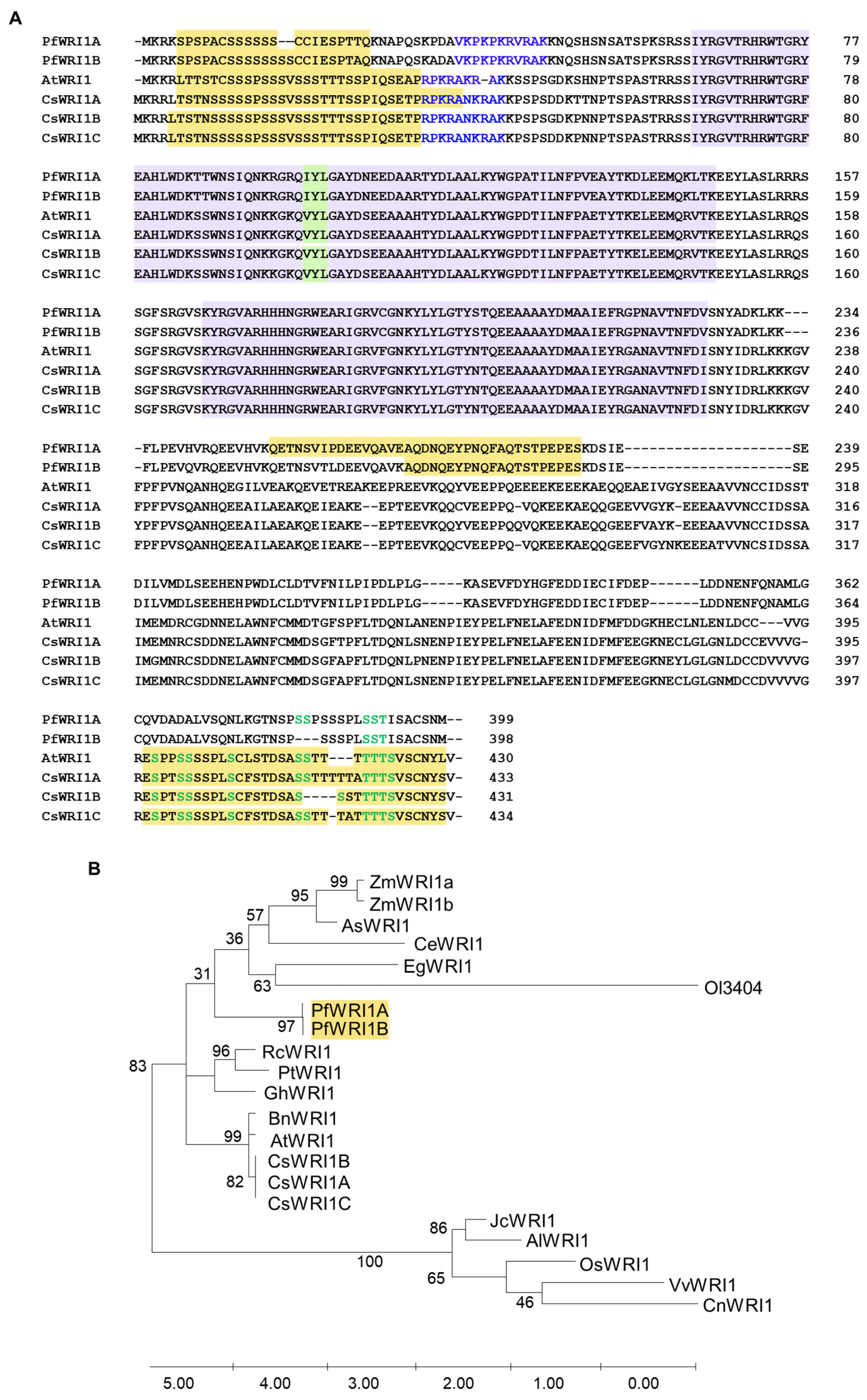
3.1. Nucleotide and Amino Acid Sequences of P. frutescens WRI1 Isoforms and Phylogenetic Tree in WRI1 Orthologs from Various Plant Resources

To isolate WRI1 orthologs in P. frutescens, total RNAs were isolated from developing Perilla seeds and then the converted cDNAs were subjected to the RT-PCR using PfWRI1 gene-specific primers (Supplementary Table S1). When the PCR products were sequenced, two 1200-nucleotide-long and 1197-nucleotide-long WR1 isoforms were isolated and named PfWRI1A and PfWRI1B, respectively (Figure S1). The nucleotide sequence similarity between PfWRI1A and PFWRI1B was very similar (about 97%), suggesting that the two isoforms are very well conserved (Figure S2). Next, the deduced amino acid sequences of PfWRI1A and PfWRI1B were compared with those of WRI1 orthologs from various plant resources including an Arabidopsis WRI1, and their phylogenetic tree was constructed. As shown in an Arabidopsis WRI1, both PfWRI1A and PfWRI1B harbor two AP2/EREBP DNA binding motifs and an ‘IYL’ motif instead of ‘VYL’, which is known to be a transcriptional activation motif in AtWRI1 (Figure 1A and Figure S2) [32]. The 17th and 18th serine residues in PfWRI1B were deleted in PfWRI1A, whereas thee amino acid residues corresponding to proline, serine, and serine at 384th to 386th in PfWRI1A were not present in PfWRI1B (Figure 1A). The proline and serine motifs (or PEST motifs), which were reported to be involved in the regulation of WRI1 [53] were searched by ePESTfind (https://emboss.bioinformatics.nl/cgi-bin/emboss/epestfind (accessed on 1 June 2000) and then the PEST motifs were detected in the N- and C-terminal regions of Arabidopsis and Camelina WRI1s, but in the middle region of Perilla WRI1A and WRI1B (Figure 1A). The putative amino acid residues, which correspond to the phosphorylation sites in the PEST motifs of AtWRI1 [53] were also shown in PfWRIs and CsWRI1s (Figure 1A), but their regulatory roles in PfWRIs and CsWRI1s remain to be further investigated. In the search of plant WRI1s, we found that one WRI1 isoform was detected from various crops examined, except Zea mays (ZmWRI1a and ZmWRI1b) and Camelina sativa (CsWRI1 A, B, and C). In the phylogenetic tree of plants, PfWRI1s and the clade including ZmWRI1, Avena sativa AsWRI1, Cyperus esculentus CeWRI1, EgWRI1, and Ostreococcus lucimarinus Ol3404 were divided after the clade containing Brassica WRI1s were divided from the ancestor (Figure 1B). The deduced amino acid sequence similarity of WRIs orthologs from various plant species are shown in Supplementary Table S2.

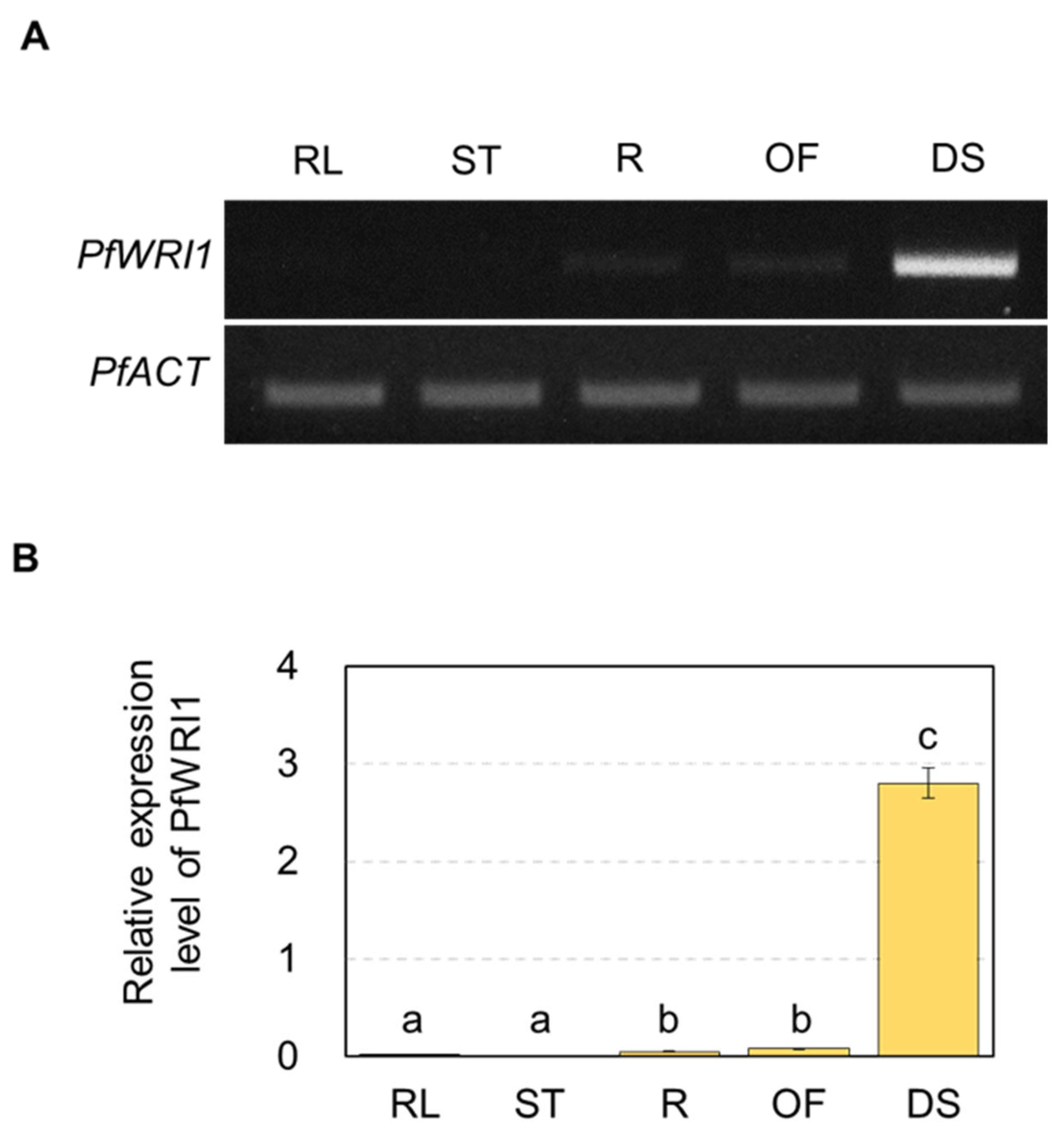
3.2. Expression of PfWRI1A and PfWRI1B in Various Perilla Organs

To examine the transcript levels of PfWRI1A and PfWRI1B in various Perilla organs, total RNAs were isolated from rosette leaves, stems, roots, open flowers, and developing seeds of P. frutescens plants, and subjected to the RT-PCR and RT-quantitative PCR analyses. Both RT-PCR and RT-qPCR analyses revealed that PfWRI1s were predominantly ex-pressed in developing Perilla seeds (Figure 2A,B). Initially, we tried to design PfWRI1A- or PfWRI1B-specific primers. The design of PfWRI1B-specific primers was successful, but it failed to design PfWRI1A-specific primers due to a high sequence homology between PfWRI1A and PfWRI1B (Figure S2). Thus, the total transcript levels of PfWRI1A and PfWRI1B were shown in Figure 2B.

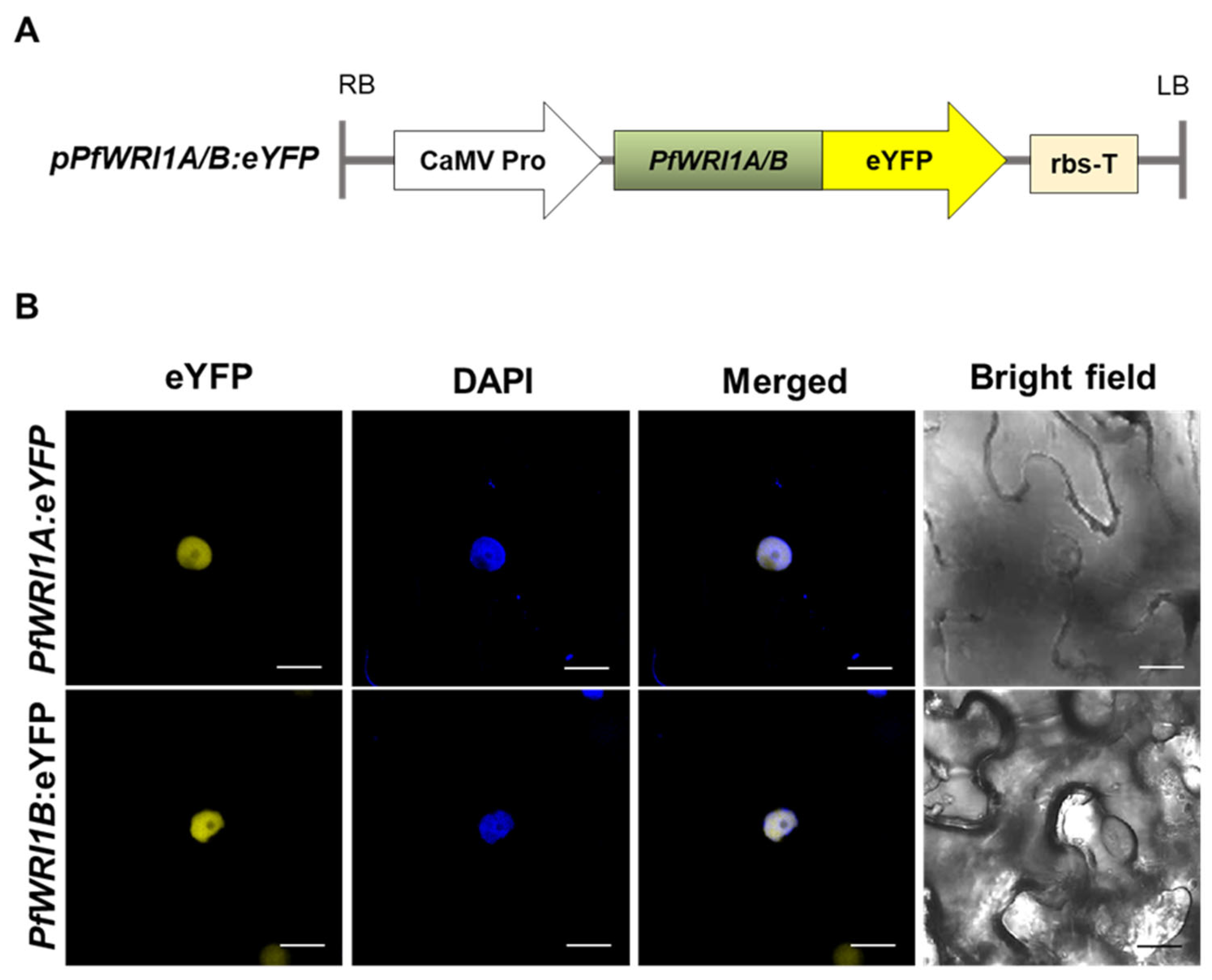
3.3. Subcellular Localization of PfWRI1A:eYFP and PfWRI1B:eYFP in N. benthamiana Leaves

To obtain the binary vector constructs containing PfWRI1A:eYFP or PfWRI1B:eYFP, the coding regions of the isolated PfWRI1A and PfWRI1B were fused in-frame eYFP under the control of CaMV 35S promoter in the pPZP212 binary vector (Figure 3A). The PfWRI1A:eYFP and PfWRI1B:eYFP constructs were transformed into Agrobacterium (GV3101 strain), and the transformed Agrobacteria were infiltrated into the N. benthamiana leaves. The leaves containing the Agrobacterium-infiltrated sites were stained with a DAPI solution 48 h after infiltration and then observed using a confocal laser microscope. The fluorescent signals from both PfWRI1A:eYFP or PfWRI1B:eYFP constructs were merged with the DAPI signals, indicating that PfWRI1A and PfWRI1B are localized in the nucleus in N. benthamiana leaf epidermal cells (Figure 3B) and might play a role as a transcription factor.

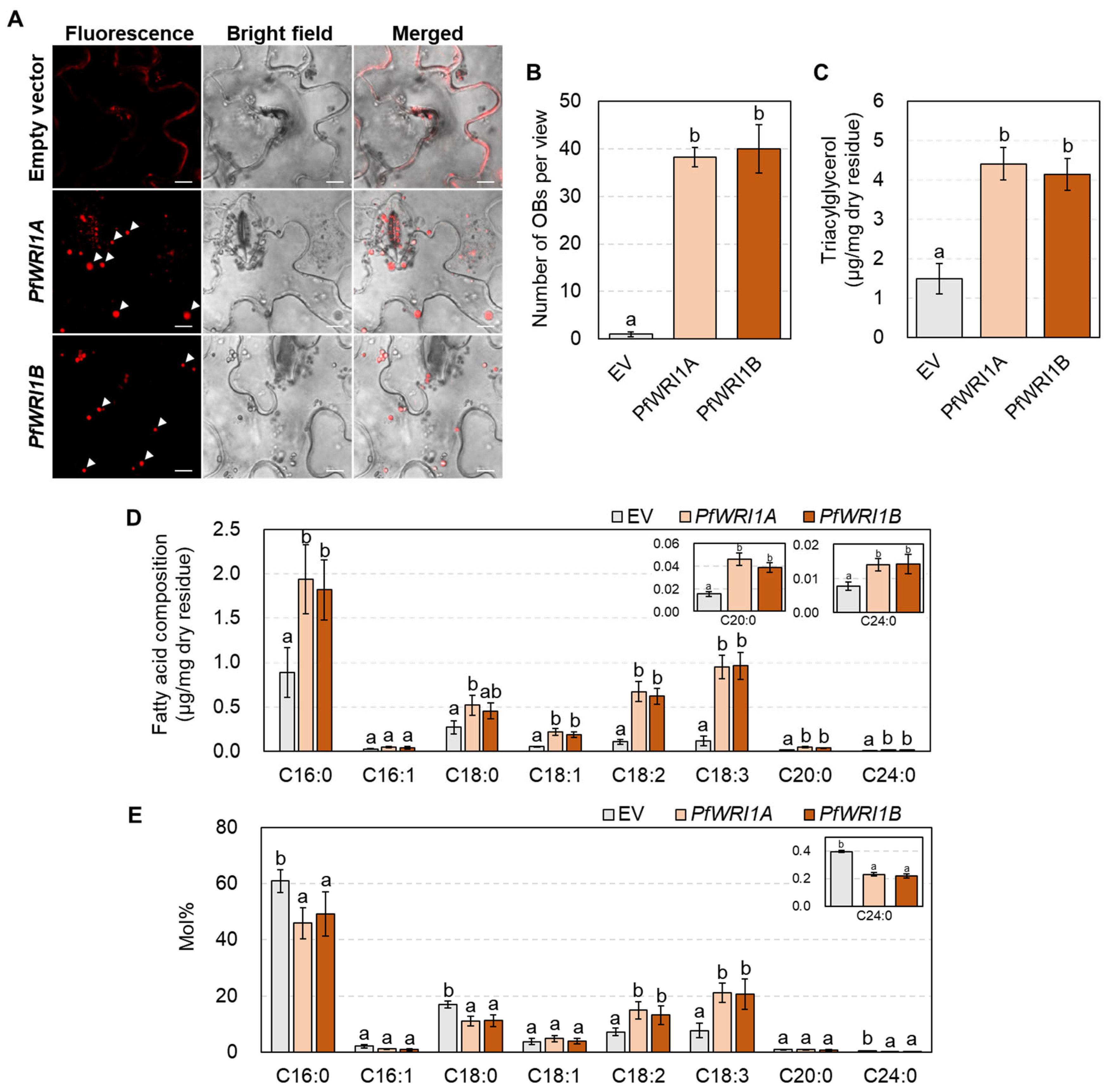
3.4. Oil Body Formation and TAG Measurement in N. benthamiana Leaves Expressing PfWRI1A or PfWRI1B

To investigate the functional activities of PfWIR1A and PfWRI1B, Agrobacteria containing PfWRI1A:eYFP or PfWRI1B:eYFP driven by the CaMV 35S promoter were infiltrated in N. benthamiana leaves. The leaves containing the Agrobacterium-infiltrated sites were stained with Nile red solution 6 d after infiltration and then oil bodies were observed under confocal microscopy. Ectopic expression of PfWIR1A or PfWRI1B caused the formation of oil bodies in N. benthamiana leaves, but no signals corresponding to oil bodies were observed in N. benthamiana leaves infiltrated with the pPZP212 binary vector without PfWRI1 (Figure 4A). The number of oil bodies significantly increased in the leaves expressing PfWIR1A or PfWRI1B relative to the control, indicating that the PfWRI1A and PfWRI1B may contribute to the accumulation of storage oils in N. benthamiana leaves (Figure 4B).
Subsequently, the amount of TAGs accumulated in the oil bodies in N. benthamiana leaves expressing PfWRI1A or PfWRI1B was measured by TLC and GC with a flame ionization detector. Briefly, the N. benthamiana leaves expressing PfWRI1A or PfWRI1B were lyophilized and then subjected to the extraction of total lipids. After the chloroform-extracted lipids were separated by TLC and visualized with 0.01% primuline under UV light (Supplementary Figure S3), the bands corresponding to the TAGs were eluted, and then further analyzed by GC. The levels of total FAMEs in N. benthamiana leaves expressing PfWRI1A and PfWRI1B increased approximately 2.9- and 2.7-fold relative to the control transformed with the pPZP212 binary vector without PfWRI1, respectively (Figure 4C). In fatty acid composition analysis, the levels of all fatty acid components except C16:1 significantly increased in the leaves expressing PfWRI1A:eYFP or PfWRI1B:eYFP compared with the control expressing eYFP (Figure 4D). The ratio of polyunsaturated fatty acids, C18:2 and C18:3 were elevated by approximately 2- and 3-fold, but the proportions of saturated fatty acids, C16:0, C18:0, and C24:0 decreased by approximately 22%, 34%, and 50% in N. benthamiana leaves expressing PfWRI1A and PfWRI1B compared with the control, respectively (Figure 4E). Taken together, ectopic expression of each of PfWRI1A and PfWRI1B enhanced the levels of TAG with unsaturated fatty acids (C18:2, and C18:3) in N. benthamiana leaves.

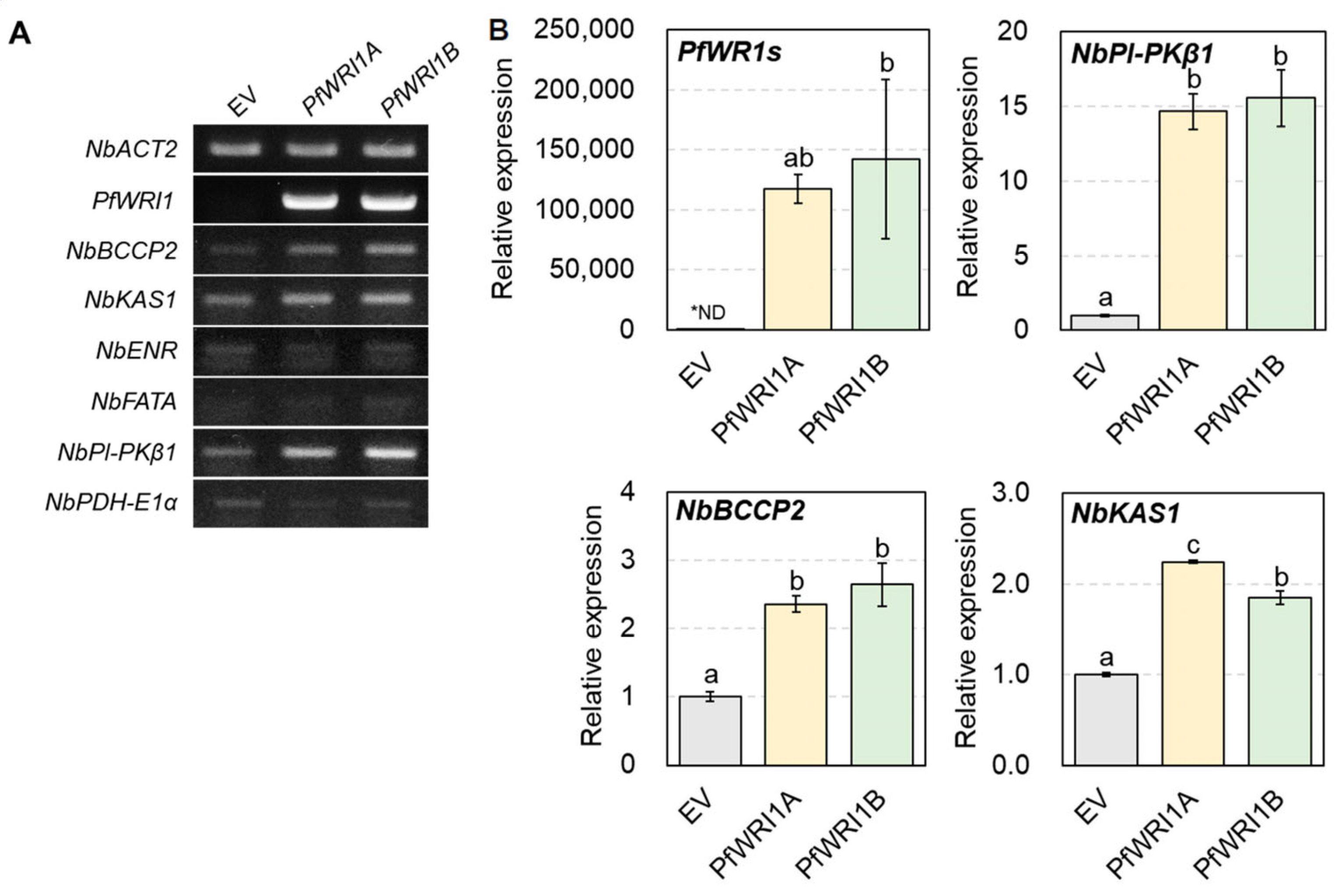
3.5. Induced Expzression of WRI1′s Target Genes in N. benthamiana Leaves Expressing PfWRI1A or PfWRI1B

To investigate whether or not the ectopic expression of PfWRI1A and PfWRI1B upregulates the expression of WRI1’s target genes, total RNAs were extracted from N. benthamiana leaves expressing PfWRI1A or PfWRI1B 2 d after Agrobacterium infiltration and converted to the cDNAs. The synthesized cDNAs were subjected to RT-PCR and RT-qPCR analyses using the gene-specific primers, which were searched from the N. benthamiana transcriptome database (https://solgenomics.net/organism/Nicotiana_benthamiana/genome (accessed on 26 November 2014). N. benthamiana leaves transformed with the pPZP212 binary vector without PfWRI1 were used as a control. In RT-PCR analysis, we observed the induced expression of NbBCCP2, NbKAS1, and NbPl-PKβ1 in N. benthamiana leaves expressing PfWRI1A or PfWRI1B relative to the control, but no significant differences in the levels of NbENR, NbFATA, and NbPDH-E1α transcripts were observed between N. benthamiana leaves expressing PfWRI1A or PfWRI1B and the control leaves (Figure 5A). We further measured the levels of PfWRI1, NbBCCP2, NbKAS1, and NbPl-PKβ1 transcripts by RT-qPCR. The NbACT was used for the determination of the quantity and quality of cDNAs. As shown in Figure 5B, the expression of NbPl-PKβ1 transcripts was upregulated by approximately 15- and 16-fold in N. benthamiana leaves expressing PfWRI1A and PfWRI1B relative to the control, respectively. The expression of NbBCCP2 and NbKAS1 was also elevated by approximately two- to three-fold in N. benthamiana leaves expressing PfWRI1A or PfWRI1B compared with the control. The results revealed that PfWRI1A and PfWRI1B are able to accumulate storage oils in N. benthamiana leaves by the upregulation of their target genes such as NbBCCP2, NbKAS1, and NbPl-PKβ1.

4. Discussion

Since the usage of vegetable oils has increased tremendously as a sustainable and alternative energy resource, it is known to be the critical step in elevating oil content in oilseed crops [2,3,4]. Vanhercke et al. [54] reported that the “Push-Pull-Protect” module was suggested to increase storage oil deposition in genetically engineered seeds or leaves by an increase in the production of fatty acids in the plastids (Push), an increase in TAG assembly and accumulation (Pull), and a decrease in TAG hydrolysis or catabolism (Protect). WRINKLED1 (WRI1) is well known as a “Push” factor to activate fatty acid biosynthesis by the upregulation of genes involved in glycolysis and fatty acid biosynthesis and thereby enhances TAG accumulation [28,29,30]. In this study, we identified a noble genetic resource, P. frutescens WRI1 genes, which contribute to the increased accumulation of storage oils in N. benthamiana leaves.
Since the WRI1 was first reported from Arabidopsis (Cernac and Benning, 2004), several WRI1 orthologs have been characterized from various monocot and dicot plants including Brassica napus [55], Camelina sativa [37], Zea mays [35,56], Jatropha curcas [38], and Oryza sativa [57]. In the previous report [42], the WRI1 orthologs from Arabidopsis (AtWRI1), potato (StWRI1), oat (AsWRI1), poplar (PtWRI1), and nutsedge (CeWRI1) were transiently expressed in tobacco leaves and then a significant increase in TAG accumulation was observed in all transformed leaves relative to the control. Ectopic expression of C. sativa WRI1A, B, or C induced the formation of oil bodies in N. benthamiana leaves and eventually increased TAG levels by approximately 2.5- to 4.0-fold in the leaves compared to the control [37]. The formation of oil bodies was also observed in N. benthamiana leaves expressing PfWRI1A or PfWRI1B by the Nile red staining (Figure 4A). In the tobacco leaves expressing PfWRI1A and PfWRI1B, the levels of TAGs were significantly elevated relative to the control (Figure 4B), indicating that the PfWRI1A and PfWRI1B enhance storage oil deposition in leaves, which have been used for the production of vegetable oils as an alternative non-seed organ [48].
In the fatty acid composition in the TAG fractions accumulated in N. benthamiana leaves expressing AtWRI1, StWRI1, AsWRI1, PtWRI1, or CeWRI1, interestingly the ratio of C18:3 were increased, but the proportions of C18:0 were remarkably decreased in all transgenic leaves compared to transformed control [42]. An et al. [37] reported that the levels (mol%) of C18:1 were increased, but the amounts of C18:0 were decreased in the TAG fractions isolated from leaves expressing CsWRI1A, B, or C. Interestingly, we observed an increase and a decrease in the levels (mol%) of polyunsaturated fatty acid (C18:2 and C18:3) and saturated fatty acids (C16:0, C18:0, and C24:0), respectively, in the TAG fractions induced by the overexpression of PfWRI1A or PfWRI1B compared to the transformed control (Figure 4C). A similar observation was also reported in N. benthamiana leaves expressing RcWRIA or RcWRIB [52]. Therefore, the overexpression of plant WRI1s can alter the ratio of saturated and unsaturated fatty acids in the TAGs accumulated in leaves and they are possibly useful for the production of storage oils with high unsaturated fatty acids in vegetative organs.
Several WRI1 orthologs including PfWRI1 showed their predominant expression in developing seeds or oil-rich non-seed tissues (Figure 2) [58], indicating that they play a crucial role in the deposition of seed storage materials. WRI1s were known to be a key transcriptional regulator, which activates the expression of several genes involved in late steps of glycolysis, fatty acid biosynthesis, and TAG assembly such as BCCP2, Pl-PKβ1, PDHE1α, enoyl-ACP reductase (EAR), ACP1, KAS1, and DGAT1 [29,30,31,38]. The expression of CsWRIs caused the upregulation of BCCP2 and Pl-PKβ1 in developing seeds of Arabidopsis wri1-3 [37]. Transient expression of RcWRI1-A or RcWRI1-B in N. benthamiana leaves significantly activated the expression of ACP1, PDHE1α, KAS1, BCCP2, Pl-PKβ1, and PlPKα [52]. We also observed that the expression of Pl-PKβ1, BCCP2, and KAS1 was significantly enhanced in N. benthamiana leaves by the expression of PfWRI1s (Figure 5).
Arabidopsis WRI1 and its orthologs were reported to be localized in the nucleus [36,37,38,40,59,60], and the fluorescent signals from PfWRI1A:eYFP and PfWRI1B:eYFP were also observed in the nucleus (Figure 3). When the nuclear localization signal (NLS) sequences were searched on the website (https://www.novoprolabs.com/tools/nls-signal-prediction)), the “PRPKRAKRA” motif was predicted in AtWRI1 protein. In the case of PfWRI1A and PfWRI1B, the “VKPKPKRVRAK” motif might be important to be localized in the nucleus. In addition, mutations in any of the three “VYL” residues, which are present in the first AP2 domain of AtWRI1 partially rescued the low seed oil phenotype of wri1-1 to the wild type and mutations in all three residues failed to restore the fatty acid content of wri1-1, indicating that the motif is essential for the function of AtWRI1 [36]. However, Ji et al. [52] reported that both RcWRI1A with the VYL motif and RcWRI1B without the VYL motif were functionally active and restored the wrinkled seed phenotype of wri1-1. Although PfWRI1s also contain the “IYL” motif instead of “VYL” (Figure 1A), the role of the “IYL” motif remains to be further investigated in the transcriptional regulation.

5. Conclusions

Transcriptome and genetic resources from Perilla [46,47] enabled us to isolate two WRI1 isoforms, PfWRI1A and PfWRI1B genes from developing Perilla seeds. Ectopic expression upregulated the expression of their target genes in N. benthamiana leaves and stimulated storage oil accumulation. These findings can be applied to the production of sustainable and renewable storage oils in leafy biomass to meet the increasing demand for their usage.

Supplementary Materials

The following are available online at https://www.mdpi.com/article/10.3390/plants12051081/s1, Table S1: Primers used in this study. Table S2: The deduced amino acid sequence similarity of WRIs orthologs from various plant species. Figure S1: Nucleotide and deduced amino acid sequences of P. frutescens WRI1A and WRI1B isoforms. Figure S2: Design of PfWRI1A- or PfWRI1B-specific primers. Figure S3: Thin layer chromatography (TLC) of neutral lipids on a silica gel plate developed with a solvent mixture of hexane/ethyl ether/acetic acid (70:30:1, v/v/v).

Author Contributions

M.C.S. designed the research. S.K. performed the experiments and analyzed the data with M.C.S. and K.-R.L.; S.K., K.-R.L. and M.C.S. wrote the paper. All authors have read and agreed to the published version of the manuscript.

Funding

This work was supported by grants from the BioGreen21 Agri-Tech Innovation Program (Project No. PJ015709 to M.C.S) of the Rural Development Administration, Republic of Korea and the National Research Foundation of Korea (Project No. NRF-2021R1A2C1006049).

Data Availability Statement

All data supporting the finding of this study are available within the paper and its Supplementary Materials published online.

Acknowledgments

We appreciate Hyun Uk Kim (Sejong University) for providing raw RNA sequencing data from developing Perilla seeds and Ryeo Jin Kim for technical support in TLC and GC analyses.

Conflicts of Interest

The authors declare no conflict of interest.

References

  1. Graham, I.A. Seed storage oil mobilization. Annu. Rev. Plant Biol. 2008, 59, 115–142. [Google Scholar] [CrossRef] [PubMed]
  2. Durrett, T.P.; Benning, C.; Ohlrogge, J. Plant triacylglycerols as feedstocks for the production of biofuels. Plant J. 2008, 54, 593–607. [Google Scholar] [CrossRef] [PubMed]
  3. Dyer, J.M.; Stymne, S.; Green, A.G.; Carlsson, A.S. High-value oils from plants. Plant J. 2008, 54, 640–655. [Google Scholar] [CrossRef] [PubMed]
  4. Foley, J.A. Can we feed the world & sustain the planet? Sci. Am. 2011, 305, 60–65. [Google Scholar] [PubMed]
  5. Goncalves, E.C.; Wilkie, A.C.; Kirst, M.; Rathinasabapathi, B. Metabolic regulation of triacylglycerol accumulation in the green algae: Identification of potential targets for engineering to improve oil yield. Plant Biotechnol. J. 2016, 14, 1649–1660. [Google Scholar] [CrossRef] [PubMed]
  6. Cahoon, E.B.; Shockey, J.M.; Dietrich, C.R.; Gidda, S.K.; Mullen, R.T.; Dyer, J.M. Engineering oilseeds for sustainable production of industrial and nutritional feedstocks: Solving bottlenecks in fatty acid flux. Curr. Opin. Plant Biol. 2007, 10, 236–244. [Google Scholar] [CrossRef]
  7. Vigeolas, H.; Waldeck, P.; Zank, T.; Geigenberger, P. Increasing seed oil content in oil-seed rape (Brassica napus L.) by over-expression of a yeast glycerol-3-phosphate dehydrogenase under the control of a seed-specific promoter. Plant Biotechnol. J. 2007, 5, 431–441. [Google Scholar] [CrossRef]
  8. Kim, H.; Park, J.; Kim, D.; Kim, A.; Suh, M. Functional analysis of diacylglycerol acyltransferase1 genes from Camelina sativa and effects of CsDGAT1B overexpression on seed mass and storage oil content in C. sativa. Plant Biotechnol. Rep. 2016, 10, 141–153. [Google Scholar] [CrossRef]
  9. Chhikara, S.; Abdullah, H.M.; Akbari, P.; Schnell, D.; Dhankher, O.P. Engineering Camelina sativa (L.) Crantz for enhanced oil and seed yields by combining diacylglycerol acyltransferase1 and glycerol-3-phosphate dehydrogenase expression. Plant Biotechnol. J. 2018, 16, 1034–1045. [Google Scholar] [CrossRef] [Green Version]
  10. Jing, G.; Tang, D.; Yao, Y.; Su, Y.; Shen, Y.; Bai, Y.; Jing, W.; Zhang, Q.; Lin, F.; Guo, D.; et al. Seed specifically over-expressing DGAT2A enhances oil and linoleic acid contents in soybean seeds. Biochem. Biophys. Res. Comm. 2021, 568, 143–150. [Google Scholar] [CrossRef]
  11. Browse, J.; Slack, C.R. Fatty-acid synthesis in plastids from maturing safflower and linseed cotyledons. Planta 1985, 166, 74–80. [Google Scholar] [CrossRef]
  12. Rawsthorne, S. Carbon flux and fatty acid synthesis in plants. Prog. Lipid Res. 2002, 41, 182–196. [Google Scholar] [CrossRef]
  13. DeBrosse, S.D.; Kerr, D.S. Chapter 12—Pyruvate dehydrogenase complex deficiency. In Mitochondrial Case Studies; Saneto, R.P., Parikh, S., Cohen, B.H., Eds.; Academic Press: Boston, MA, USA, 2016; pp. 93–101. [Google Scholar]
  14. Chapman, K.D.; Ohlrogge, J.B. Compartmentation of triacylglycerol accumulation in plants. J. Biol. Chem. 2012, 287, 2288–2294. [Google Scholar] [CrossRef] [Green Version]
  15. Jung, S.H.; Kim, R.J.; Kim, K.J.; Lee, D.H.; Suh, M.C. Plastidial and mitochondrial malonyl CoA-ACP malonyltransferase is essential for cell division and its overexpression increases storage oil content. Plant Cell Physiol. 2019, 60, 1239–1249. [Google Scholar] [CrossRef]
  16. Kennedy, E.P. Biosynthesis of complex lipids. Fed. Proc. 1961, 20, 934–940. [Google Scholar]
  17. Clough, R.C.; Matthis, A.L.; Barnum, S.R.; Jaworski, J.G. Purification and characterization of 3-ketoacyl-acyl carrier protein synthase III from spinach. A condensing enzyme utilizing acetyl-coenzyme A to initiate fatty acid synthesis. J. Biol. Chem. 1992, 267, 20992–20998. [Google Scholar] [CrossRef]
  18. Bonaventure, G.; Salas, J.J.; Pollard, M.R.; Ohlrogge, J.B. Disruption of the FATB gene in Arabidopsis demonstrates an essential role of saturated fatty acids in plant growth. Plant Cell 2003, 15, 1020–1033. [Google Scholar] [CrossRef] [Green Version]
  19. Shockey, J.M.; Fulda, M.S.; Browse, J.A. Arabidopsis contains nine long-chain acyl-Coenzyme A synthetase genes that participate in fatty acid and glycerolipid metabolism. Plant Physiol. 2002, 129, 1710–1722. [Google Scholar] [CrossRef] [Green Version]
  20. Li-Beisson, Y.; Shorrosh, B.; Beisson, F.; Andersson, M.X.; Arondel, V.; Bates, P.D.; Baud, S.; Bird, D.; Debono, A.; Durrett, T.P.; et al. Acyl-lipid metabolism. In The Arabidopsis Book; American Society of Plant Biologists: Rockville, MD, USA, 2013; Volume 11, p. e0161. [Google Scholar]
  21. Dahlqvist, A.; Stahl, U.; Lenman, M.; Banas, A.; Lee, M.; Sandager, L.; Ronne, H.; Stymne, S. Phospholipid:diacylglycerol acyltransferase: An enzyme that catalyzes the acyl-CoA-independent formation of triacylglycerol in yeast and plants. Proc. Natl. Acad. Sci. USA 2000, 97, 6487–6492. [Google Scholar] [CrossRef] [Green Version]
  22. Zhang, M.; Fan, J.; Taylor, D.C.; Ohlrogge, J.B. DGAT1 and PDAT1 acyltransferases have overlapping functions in Arabidopsis triacylglycerol biosynthesis and are essential for normal pollen and seed development. Plant Cell. 2009, 21, 3885–3901. [Google Scholar] [CrossRef] [Green Version]
  23. Tumaney, A.W.; Shekar, S.; Rajasekharan, R. Identification, purification, and characterization of monoacylglycerol acyltransferase from developing peanut cotyledons. J. Biol. Chem. 2001, 276, 10847–10852. [Google Scholar] [CrossRef] [PubMed] [Green Version]
  24. Saha, S.; Enugutti, B.; Rajakumari, S.; Rajasekharan, R. Cytosolic triacylglycerol biosynthetic pathway in oilseeds. Molecular cloning and expression of peanut cytosolic diacylglycerol acyltransferase. Plant Physiol. 2006, 141, 1533–1543. [Google Scholar] [CrossRef] [PubMed] [Green Version]
  25. Hernández, M.L.; Whitehead, L.; He, Z.; Gazda, V.; Gilday, A.; Kozhevnikova, E.; Vaistij, F.E.; Larson, T.R.; Graham, I.A. A cytosolic acyltransferase contributes to triacylglycerol synthesis in sucrose-rescued Arabidopsis seed oil catabolism mutants. Plant Physiol. 2012, 160, 215–225. [Google Scholar] [CrossRef] [PubMed] [Green Version]
  26. Cao, H.; Shockey, J.M.; Klasson, K.T.; Chapital, D.C.; Mason, C.B.; Scheffler, B.E. Developmental regulation of diacylglycerol acyltransferase family gene expression in tung tree tissues. PLoS ONE 2013, 8, e76946. [Google Scholar] [CrossRef] [PubMed]
  27. Lee, K.R.; Kim, Y.M.; Yeo, Y.; Kim, S.; Suh, M.C. Camelina cytosol-localized diacylglycerol acyltransferase 3 contributes to the accumulation of seed storage oils. Ind. Crop. Prod. 2022, 189, 115808. [Google Scholar] [CrossRef]
  28. Focks, N.; Benning, C. wrinkled1: A novel, low-seed-oil mutant of Arabidopsis with a deficiency in the seed-specific regulation of carbohydrate metabolism. Plant Physiol. 1998, 118, 91–101. [Google Scholar] [CrossRef] [Green Version]
  29. Baud, S.; Mendoza, M.S.; To, A.; Harscoët, E.; Lepiniec, L.; Dubreucq, B. WRINKLED1 specifies the regulatory action of LEAFY COTYLEDON2 towards fatty acid metabolism during seed maturation in arabidopsis. Plant J. 2007, 50, 825–838. [Google Scholar] [CrossRef]
  30. Baud, S.; Wuillème, S.; To, A.; Rochat, C. Lepiniec, Role of WRINKLED1 in the transcriptional regulation of glycolytic and fatty acid biosynthetic genes in Arabidopsis. Plant J. 2009, 60, 933–947. [Google Scholar] [CrossRef]
  31. Maeo, K.; Tokuda, T.; Ayame, A.; Mitsui, N.; Kawai, T.; Tsukagoshi, H.; Ishiguro, S.; Nakamura, K. An AP2-type transcription factor, WRINKLED1, of Arabidopsis thaliana binds to the AW-box sequence conserved among proximal upstream regions of genes involved in fatty acid synthesis. Plant J. 2009, 60, 476–487. [Google Scholar] [CrossRef]
  32. Fukuda, N.; Ikawa, Y.; Aoyagi, T.; Kozaki, A. Expression of the genes coding for plastidic acetyl-CoA carboxylase subunits is regulated by a location-sensitive transcription factor binding site. Plant Mol. Biol. 2013, 82, 473–483. [Google Scholar] [CrossRef] [Green Version]
  33. Marchive, C.; Nikovics, K.; To, A.; Lepiniec, L.; Baud, S. Transcriptional regulation of fatty acid production in higher plants: Molecular bases and biotechnological outcomes. Eur. J. Lipid Sci. Technol. 2014, 116, 1332–1343. [Google Scholar] [CrossRef]
  34. Cernac, A.; Benning, C. WRINKLED1 encodes an AP2/EREB domain protein involved in the control of storage compound biosynthesis in Arabidopsis. Plant J. 2004, 40, 575–585. [Google Scholar] [CrossRef]
  35. Pouvreau, B.; Baud, S.; Vernoud, V.; Morin, V.; Py, C.; Gendrot, G.; Pichon, J.P.; Rouster, J.; Paul, W.; Rogowsky, P.M. Duplicate maize Wrinkled1 transcription factors activate target genes involved in seed oil biosynthesis. Plant Physiol. 2011, 156, 674–686. [Google Scholar] [CrossRef] [Green Version]
  36. Ma, W.; Kong, Q.; Arondel, V.; Kilaru, A.; Bates, P.D.; Thrower, N.A.; Benning, C.; Ohlrogge, J.B. WRINKLED1, a ubiquitous regulator in oil accumulating tissues from Arabidopsis embryos to oil palm mesocarp. PLoS ONE 2013, 8, e68887. [Google Scholar] [CrossRef]
  37. An, D.; Kim, H.; Ju, S.; Go, Y.S.; Kim, H.U.; Suh, M.C. Expression of camelina WRINKLED1 isoforms rescue the seed phenotype of the Arabidopsis wri1 mutant and increase the triacylglycerol content in tobacco leaves. Front. Plant Sci. 2017, 8, 34. [Google Scholar] [CrossRef] [Green Version]
  38. Ye, J.; Wang, C.; Sun, Y.; Qu, J.; Mao, H.; Chua, N.-H. Overexpression of a transcription factor increases lipid content in a woody perennial Jatropha curcas. Front. Plant Sci. 2018, 9, 1479. [Google Scholar] [CrossRef] [Green Version]
  39. Chen, B.; Zhang, G.; Li, P.; Yang, J.; Guo, L. Multiple GmWRI1s are redundantly involved in seed filling and nodulation by regulating plastidic glycolysis, lipid biosynthesis and hormone signalling in soybean (Glycine max). Plant Biotechnol. J. 2020, 18, 155–171. [Google Scholar] [CrossRef] [Green Version]
  40. Wu, X.L.; Liu, Z.H.; Hu, Z.H.; Huang, R.Z. BnWRI1 coordinates fatty acid biosynthesis and photosynthesis pathways during oil accumulation in rapeseed. J. Integr. Plant Biol. 2014, 56, 582–593. [Google Scholar] [CrossRef]
  41. An, D.; Suh, M.C. Overexpression of Arabidopsis WRI1 enhanced seed mass and storage oil content in Camelina sativa. Plant Biotechnol. Rep. 2015, 9, 137–148. [Google Scholar] [CrossRef]
  42. Grimberg, Å.; Carlsson, A.S.; Marttila, S.; Bhalerao, R.; Hofvander, P. Transcriptional transitions in Nicotiana benthamiana leaves upon induction of oil synthesis by WRINKLED1 homologs from diverse species and tissues. BMC Plant Biol. 2015, 15, 192. [Google Scholar] [CrossRef] [Green Version]
  43. Asif, M. Health effects of omega-3,6,9 fatty acids: Perilla frutescens is a good example of plant oils. Orient. Pharm. Exp. Med. 2011, 11, 51–59. [Google Scholar] [CrossRef] [PubMed] [Green Version]
  44. Honda, G.; Yuba, A.; Kojima, T.; Tabata, M. Chemotaxonomic and cytogenetic studies on Perilla frutescens var. citriodora (‘Lemon Egoma’). Nat. Med. 1994, 48, 185–190. [Google Scholar]
  45. Nitta, M.; Lee, J.K.; Kang, C.W.; Katsuta, M.; Yasumoto, S.; Liu, D.; Nagamine, T.; Ohnishi, O. The distribution of Perilla species. Genet. Resour. Crop Evol. 2005, 52, 797–804. [Google Scholar] [CrossRef]
  46. Lee, K.R.; Lee, Y.; Kim, E.H.; Lee, S.B.; Roh, K.H.; Kim, J.B.; Kang, H.C.; Kim, H.U. Functional identification of oleate 12-desaturase and ω-3 fatty acid desaturase genes from Perilla frutescens var. frutescens. Plant Cell Rep. 2016, 35, 2523–2537. [Google Scholar] [CrossRef] [PubMed]
  47. Zhang, Y.; Shen, Q.; Leng LZhang, D.; Chen, S.; Shi, Y.; Ning, Z.; Chen, S. Incipient diploidization of the medicinal plant Perilla within 10,000 years. Nat. Comm. 2021, 12, 5508. [Google Scholar] [CrossRef]
  48. Oñate-Sánchez, L.; Vicente-Carbajosa, J. DNA-free RNA isolation protocols for Arabidopsis thaliana, including seeds and siliques. BMC Res. Notes. 2008, 1, 93. [Google Scholar] [CrossRef] [Green Version]
  49. Holsters, M.; De Waele, D.; Depicker, A.; Messens, E.; Van Montagu, M.; Schell, J. Transfection and transformation of Agrobacterium tumefaciens. Mol. Gen. Genet. 1978, 163, 181–187. [Google Scholar] [CrossRef]
  50. Kumar, S.; Stecher, G.; Li, M.; Knyaz, C.; Tamura, K. MEGA X: Molecular Evolutionary Genetics Analysis across Computing Platforms. Mol. Biol. Evol. 2018, 35, 1547–1549. [Google Scholar] [CrossRef]
  51. Fernandez-Pozo, N.; Menda, N.; Edwards, J.D.; Saha, S.; Tecle, I.Y.; Strickler, S.R.; Bombarely, A.; Fisher-York, T.; Pujar, A.; Foerster, H.; et al. The sol genomics network (SGN)--from genotype to phenotype to breeding. Nucleic Acids Res. 2015, 43, 1036–1041. [Google Scholar] [CrossRef]
  52. Ji, X.J.; Mao, X.; Hao, Q.T.; Liu, B.L.; Xue, J.A.; Li, R.Z. Splice variants of the castor WRI1 gene upregulate fatty acid and oil biosynthesis when expressed in tobacco leaves. Int. J. Mol. Sci. 2018, 19, 146. [Google Scholar] [CrossRef] [Green Version]
  53. Ma, W.; Kong, Q.; Grix, M.; Mantyla, J.J.; Yang, Y.; Benning, C.; Ohlrogge, J.B. Deletion of a C-terminal intrinsically disordered region of WRINKLED1 affects its stability and enhances oil accumulation in Arabidopsis. Plant J. 2015, 83, 864–874. [Google Scholar] [CrossRef]
  54. Vanhercke, T.; Tahchy, A.E.; Shrestha, P.; Zhou, X.R.; Singh, S.P.; Petrie, J.R. Synergistic effect of WRI1 and DGAT1 coexpression on triacylglycerol biosynthesis in plants. FEBS Lett. 2013, 587, 364–369. [Google Scholar] [CrossRef] [Green Version]
  55. Liu, J.; Hua, W.; Zhan, G.; Wei, F.; Wang, X.; Liu, G.; Wang, H. Increasing seed mass and oil content in transgenic Arabidopsis by the overexpression of wri1-like gene from Brassica napus. Plant Physiol. Biochem. 2010, 48, 9–15. [Google Scholar] [CrossRef]
  56. Shen, B.; Allen, W.B.; Zheng, P.; Li, C.; Glassman, K.; Ranch, J.; Tarczynski, M.C. Expression of ZmLEC1 and ZmWRI1 increases seed oil production in maize. Plant Physiol. 2010, 153, 980–987. [Google Scholar] [CrossRef] [Green Version]
  57. Mano, F.; Aoyanagi, T.; Kozaki, A. Atypical splicing accompanied by skipping conserved micro-exons produces unique WRINKLED1, An AP2 domain transcription factor in rice plants. Plants 2019, 8, 207. [Google Scholar] [CrossRef] [Green Version]
  58. Kong, Q.; Yuan, L.; Ma, W. WRINKLED1, a “master regulator” in transcriptional control of plant oil biosynthesis. Plants 2019, 8, 238. [Google Scholar] [CrossRef] [Green Version]
  59. Chen, L.; Lee, J.H.; Weber, H.; Tohge, T.; Witt, S.; Roje, S.; Hellmann, H. Arabidopsis BPM proteins function as substrate adaptors to a CULLIN3-based E3 ligase to affect fatty acid metabolism in plants. Plant Cell 2013, 25, 2253–2264. [Google Scholar] [CrossRef] [Green Version]
  60. Li, Q.; Shao, J.; Tang, S.; Shen, Q.; Wang, T.; Chen, W.; Hong, Y. Wrinkled1 accelerates flowering and regulates lipid homeostasis between oil accumulation and membrane lipid anabolism in Brassica napus. Front Plant Sci. 2015, 6, 1015. [Google Scholar] [CrossRef] [Green Version]
Figure 1. Alignment of the deduced amino acid sequences of WRI1 isoforms from P. frutescens, A. thaliana, and C. sativa and phylogenetic tree of PfWRI1 homologs from diverse plant species. (A) Whitish purple and green backgrounds indicate the conserved AP2/ERF DNA binding motifs and the transcriptional activation motif in WRI1, respectively. The predicted nuclear localization signals (NLS) sequences of AtWRI1A (PRPKRAKRAK, 334−42 aa), PfWRI1A (VKPKPKRVRAK, 36−46 aa), PfWRI1B (VKPKPKRVRAK, 38−48 aa), and CsWRI1A to C (RPKRANKRAK, 35−43 aa) are shown in blue letters. PEST motifs were analyzed by ePESTfind (https://emboss.bioinformatics.nl/cgi-bin/emboss/epestfind (accessed on 1 June 2000) and are shown in yellow backgrounds. Ten phosphorylation sites of Arabidopsis WRI1 are involved in its regulatory function [53], and their putative corresponding sites in PfWRI1s and CsWRI1s are shown in green letters. (B) Phylogenetic tree of PfWRI1 homologs from diverse plant species. The phylogenetic tree was generated using MEGA X by the maximum likelihood method. Bootstrap value percentages of 1000 replicates are shown at the branching points. AtWRI1, Arabidopsis thaliana (824599); PfWRI1s, Perilla frutescens var. frutescens in this study; AsWRI1, Avena sativa (SRX1079426); BnWRI1, Brassica napus (ABD16282.1); CsWRI1A, CsWRI1B and CsWRI1C, Camelina sativa; EgWRI1, Elaeis guineensis; GhWRI1, Gossypium hirsutum (TC200263); OsWRI1, Oryza sativa (CAE00853.1); PtWRI1, Populus trichocarpa (SRX1079428); Ol34044, Ostreococcus lucimarinus (34044); RcWRI1, Ricinus communis (AB774159.1, AB774160.1); ZmWRI1, Zea mays (ACF83189.1, ACF80269.1); JcWRI1, Jatropha curcas (AIA57945.1); AlWRI1, Arabidopsis lyrata (EFH52510.1); VvWRI1, Vitis vinifera (CBI32013.3); CnWRI1, Cocos nucifera (JQ040545).
Plants 12 01081 g001
Figure 2. Expression of Perilla WRI1s in various P. frutescens organs. (A) RT-PCR analysis of PfWRI1s in various P. frutescens organs. Total RNAs were isolated from rosette leaves (RL), stems (ST), roots (R), and open flowers (OF) of 3-month-old Perilla plants and developing seeds (DS) for 2 to 3 weeks after flowering. P. frutescens ACTIN (PfACT) was used to determine cDNA quantity and quality. (B) RT-qPCR analysis of PfWRIs1 in various P. frutescens organs. The PfACT was used to normalize the levels of PfWRI1 transcripts. Values are the means of the ± SE of three replicates. Data were statistically analyzed using ANOVA test (p < 0.05).
Plants 12 01081 g002
Figure 3. Subcellular localization of PfWRI1s:eYFP in N. benthamiana epidermis. (A) Schematic diagram of PfWRI1A:eYFP and PfWRI1B:eYFP constructs. CaMV Pro, cauliflower mosaic virus 35S promoter; LB, left border; RB, right border; rbs-T, the terminator of ribulose-1,5-bisphosphate carboxylase and oxygenase small subunit from pea (Pisum sativum). (B) Agrobacterium harboring the PfWRI1A:eYFP (upper) or PfWRI1B:eYFP (bottom) construct was infiltrated into N. benthamiana leaves and then the fluorescent signals were visualized under laser confocal scanning microscopy. The nucleus was visualized by staining with DAPI under the UV filter. The fluorescent signals from eYFP and DAPI were merged. Bright-field images visualized epidermal cells. Bars = 20 µm.
Plants 12 01081 g003
Figure 4. Oil body formation (A), oil body counts (B) and TAG accumulation (CE) in N. benthamiana leaves expressing PfWRI1A:eYFP or PfWRI1B:eYFP. (A) Oil body formation in N. benthamiana leaves expressing PfWRI1A:eYFP or PfWRI1B:eYFP. Agrobacterium harboring the PfWRI1A:eYFP, PfWRI1B:eYFP, or empty vector without PfWRI1 construct was infiltrated into N. benthamiana leaves. The transformed N. benthamiana leaf disks were stained with Nile red solution and then the fluorescent signals were visualized under laser confocal scanning microscopy. The white arrows indicate oil bodies (OBs). Bars = 10 μm. (B) Oil body (OB) counts in N. benthaminana leaves expressing empty vector (pPZP212), PfWRI1A, or PfWIR1B. Values are averages and SD of four individual images. Data were statistically analyzed using ANOVA test (p < 0.05). (CE) Total TAG levels (C) and fatty acid levels (D) and composition (E) in the TAG fractions in N. benthamiana leaves expressing PfWRI1A:eYFP, PfWRI1B:eYFP, or empty vector. Total lipids were extracted from the transformed N. benthamiana leaf disks and fractionated by thin-layer chromatography. The TAG fractions were eluted, transmethylated, and then the fatty acid methyl esters were analyzed by gas chromatography. Values are averages and SD of four individual experiments. Data were statistically analyzed using ANOVA test (p < 0.05).
Plants 12 01081 g004
Figure 5. Expression of its target genes in N. benthamiana leaves expressing PfWRI1A or PfWRI1B. (A) RT-PCR analysis of its target genes in N. benthamiana leaves expressing PfWRI1A or PfWRI1B. Total RNA was isolated from N. benthamiana leaves expressing PfWRI1A, PfWRI1B, or empty vector control and converted to the cDNA, which was subjected to the RT-PCR analysis. N. benthamiana Actin2 was used to determine cDNA quantity and quality. (B) RT-qPCR analysis of its target genes in N. benthamiana leaves expressing PfWRI1A or PfWRI1B. NbActin2 was used to normalize the levels of PfWRI1s and their target genes’ transcripts. Each value is the mean of three independent measurements ± standard error. Data were statistically analyzed using ANOVA test (p < 0.01). * ND indicates non-detected.
Plants 12 01081 g005
Disclaimer/Publisher’s Note: The statements, opinions and data contained in all publications are solely those of the individual author(s) and contributor(s) and not of MDPI and/or the editor(s). MDPI and/or the editor(s) disclaim responsibility for any injury to people or property resulting from any ideas, methods, instructions or products referred to in the content.

