Article

Natural Variation in OASC Gene for Mitochondrial O-Acetylserine Thiollyase Affects Sulfate Levels in Arabidopsis

1
Institute for Plant Sciences, Cluster of Excellence on Plant Sciences, University of Cologne, Zülpicher Str. 47b, 50674 Cologne, Germany
2
John Innes Centre, Norwich Research Park, Colney, Norwich NR4 7UH, UK
3
Department of Biology, University of York, Heslington, York YO10 5DD, UK
*
Author to whom correspondence should be addressed.
Plants 2023, 12(1), 35; https://doi.org/10.3390/plants12010035
Submission received: 11 December 2022 / Revised: 18 December 2022 / Accepted: 19 December 2022 / Published: 21 December 2022
(This article belongs to the Special Issue Plant Sulfur Network)

Abstract

:
Sulfur plays a vital role in the primary and secondary metabolism of plants, and carries an important function in a large number of different compounds. Despite this importance, compared to other mineral nutrients, relatively little is known about sulfur sensing and signalling, as well as about the mechanisms controlling sulfur metabolism and homeostasis. Sulfur contents in plants vary largely not only among different species, but also among accessions of the same species. We previously used associative transcriptomics to identify several genes potentially controlling variation in sulfate content in the leaves of Brassica napus, including an OASC gene for mitochondrial O-acetylserine thiollyase (OAS-TL), an enzyme involved in cysteine synthesis. Here, we show that loss of OASC in Arabidopsis thaliana lowers not only sulfate, but also glutathione levels in the leaves. The reduced accumulation is caused by lower sulfate uptake and translocation to the shoots; however, the flux through the pathway is not affected. In addition, we identified a single nucleotide polymorphism in the OASC gene among A. thaliana accessions that is linked to variation in sulfate content. Both genetic and transgenic complementation confirmed that the exchange of arginine at position 81 for lysine in numerous accessions resulted in a less active OASC and a lower sulfate content in the leaves. The mitochondrial isoform of OAS-TL is, thus, after the ATPS1 isoform of sulfurylase and the APR2 form of APS reductase 2, the next metabolic enzyme with a role in regulation of sulfate content in Arabidopsis.

1. Introduction

The essential nutrient sulfur possesses versatile functions in the plant by forming the amino acids cysteine (Cys) and methionine (Met), different coenzymes, and prosthetic groups, and participating in the structure of defense molecules, such as glucosinolates (GLS) or camalexin [1]. Cysteine acts as a key molecule in the participation of sulfur in metabolism and connects sulfur, nitrogen, and carbon assimilation [2,3]. The bioavailable form of sulfur for plants is sulfate [1,4]. Sulfate taken up from the soil solution by roots is translocated to the leaves by the nutrient flow of the xylem, facilitated by sulfate transporters [5]. For cysteine synthesis, sulfate must be reduced. The sulfate transported into the cells is first activated to APS by the ATP sulfurylase (ATPS) enzyme [6,7]. Sulfide is formed by the reduction of APS by the subsequent action of APS reductase (APR) and sulfide reductase (SIR) [8]. The carbon backbone of cysteine comes from O-acetylserine (OAS), which is formed after the reaction of serine with Coenzyme A, catalyzed by serine-acetyl transferase (SAT) [5,9]. The O-acetylserine(thiol)lyase (OAS-TL) replaces the acetyl group in the OAS with sulfide [9]. SAT and OAS-TL enzymes form the cysteine synthase complex (CSC) [10]. Various isoforms of SAT and OAS-TL enzymes are found in different subcellular structures of the cell [5,11,12]. For example, the well-studied OAS-TL isoforms OAS-TL A, OAS-TL B, and OAS-TL C are localized in the cytoplasm, plastids, and mitochondria, respectively [6,10,13]. Similarly, SAT also is present in these compartments; therefore, all three major subcellular structures possess cysteine synthase complexes and have the potential to synthesize cysteine [10]. However, the contribution of the three compartments to total cysteine synthesis in the cell is not the same, and the main site for cysteine synthesis is the cytosol [14]. Interestingly, this requires a precise coordination of the metabolite fluxes in the cell, because sulfate reduction is specific to plastids, and the majority of OAS is produced in the mitochondria [3].
While there is a good understanding of the functions of genes involved in sulfur metabolism, identifying genes regulating sulfur homeostasis, sensing, and signalling still lags. However, given the importance of sulfur for crop plants, finding such regulatory genes is critical to underpin the development of crop varieties with improved sulfur nutrition. Using natural variation in sulfur content is a promising approach to reveal genes that contribute to controlling sulfur homeostasis. Indeed, in experiments with the model plant Arabidopsis thaliana, genetic variations in genes encoding the APR2 isoform of APS reductase and the ATPS1 isoform of ATPS were found to underlie differences in sulfate and/or total sulfur content [7,15,16]. In addition, varieties of the crop plant Brassica napus were investigated for variation in nutrient content using associative transcriptomics (AT) [17]. The analysis suggested that the gene encoding OAS-TL C (OASC) controlled sulfate levels in B. napus [17]. This was confirmed by showing that the T-DNA mutant of the OASC gene in Arabidopsis resulted in significantly reduced sulfate accumulation in the leaves [17]. Here, we show that the natural variation in the OASC gene is also linked to variation in sulfate content in Arabidopsis accessions. We identified a non-synonymous single nucleotide polymorphism responsible for this variation, as evidenced by both genetic and transgenic complementations. However, the mechanism by which OASC affects sulfate levels still needs to be elucidated.

2. Results

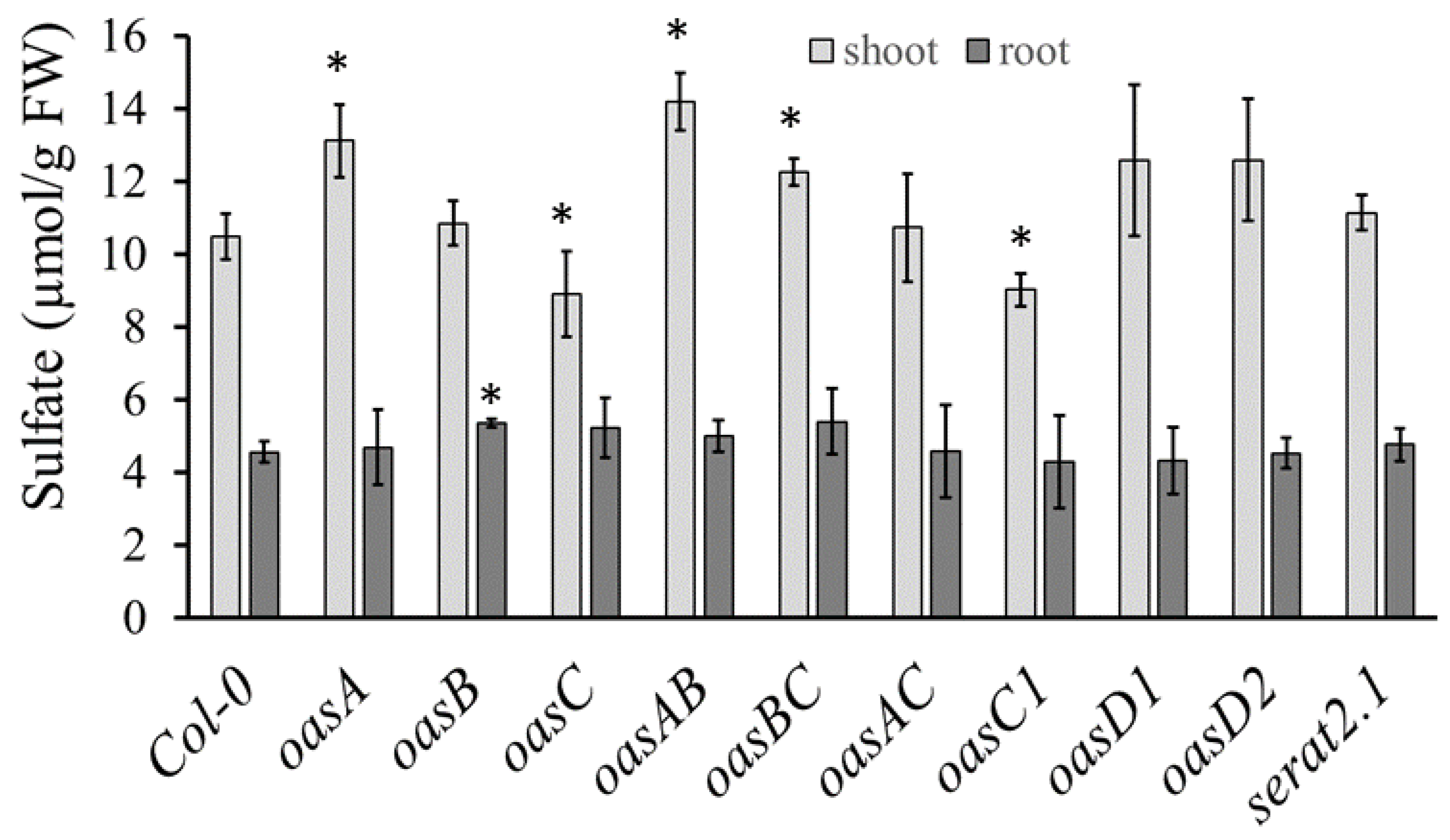
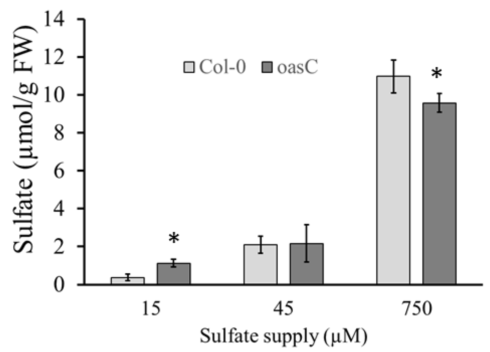
2.1. Characterization of the oasC Mutant

We previously showed that disruption of the OASC gene in Arabidopsis leads to reduced accumulation of sulfate [17]. Further metabolite analyses were, therefore, performed in the roots and leaves of Col-0 and oasC to understand how the disruption of the OASC gene affects the content of sulfur-containing biomolecules. As observed previously with greenhouse grown plants and also in seedlings grown on agarose plates, the loss of OASC resulted in significantly less sulfate accumulation in the leaf than in the wild-type Col-0 (Figure 1A). However, the absence of functional OASC in the root had no effect on sulfate accumulation (Figure 1E). Loss of OASC did not affect Cys content in the leaves; however, there was a decrease in glutathione concentration (Figure 1B,C). As for sulfate, the decrease in GSH content was not observed in the root (Figure 1F). These results strongly indicated that the sulfate assimilation in oasC is affected, specifically in leaves. However, the GLS levels were not affected (Figure 1D); hence, the alterations might be limited to primary sulfate assimilation.
To determine the cause of the lower accumulation of sulfate and GSH in the mutant, sulfate uptake and flux through the sulfate assimilation pathway were determined. Indeed, oasC showed a dramatic reduction in sulfate uptake of almost 30% compared to Col-0 (Figure 2A). The reduction of uptake in oasC also directly affected the translocation of sulfate to leaves, which was reduced to a similar extent (Figure 2B). Measurement of flux through the pathway, determined as incorporation of 35S into thiols and proteins, however, revealed no alteration in the oasC mutant compared to the wild type (Figure 2C,D). Therefore, the reduction in sulfate and GSH contents seem to result from the considerable drop in sulfate uptake. These results also showed that the loss of the oasC gene affects sulfate uptake.
Expression analysis was performed using RNA isolated from roots and leaves to test whether genes for sulfur metabolism were affected by the loss of the OASC (Figure 3). The transcript levels of the three isoforms of APR, the key enzyme in the reduction of sulfate, were not significantly affected by the disruption of the OASC gene, neither in the shoot nor in the root (Figure 3). The expression of two markers of sulfur deficiency, SDI1, which is involved in GLS regulation, and GGTC2;1, which contributes to GSH degradation, were also not different in oasC compared to Col-0 [4,18]. In addition, the expression levels of the major isoforms of OAS-TL were compared. As expected, no expression of the OAS-TL C isoform was detected in the shoots or roots of the oasC mutant (Figure 3). The transcript level of the cytosolic OAS-TL A was slightly, but significantly, elevated in the shoot, but not in the root. In contrast, no changes in the transcript levels of the plastidic OAS-TL B or the mitochondrial OAS-TL C1 isoforms were observed in oasC compared to Col-0 (Figure 3). Thus, in general, the loss of the oasC gene had almost no effect on the expression of genes for sulfate assimilation.
Next, we wanted to see if the reduction in sulfate levels in the oasC mutant is specific to this OAS-TL isoform, or whether loss of other isoforms might have the same effect. We, therefore, measured sulfate concentrations in the shoot of mutants in six OAS-TL isoforms in Arabidopsis and the double mutants of the main A, B, and C forms, and, in addition, in the sat2;1 mutant in the mitochondrial form of SAT. Among all the mutants, only oasC and oasC1 caused a significant decrease in sulfate accumulation (Figure 4). In contrast, in oasA, oasAB, oasBC and oasD2, sulfate accumulation in the shoot was slightly increased compared to Col-0. Thus, only changes in mitochondrial OAS-TL have a negative effect on sulfate accumulation. In roots, only oasB showed a small, but significant, increase in sulfate accumulation compared to Col-0. The mitochondrial serat2.1 knockout mutant had no significant effect on sulfate levels compared to Col-0, indicating that the changes in sulfate are not caused by the cysteine synthase complex, but are specific to OAS-TL. To test whether the effect of loss of OASC on sulfate levels is dependent on external sulfate supply, various sulfate concentrations were supplied to Col-0 and oasC. Interestingly, the sulfate accumulation in oasC and Col-0 was affected differently by low and high sulfur supply. As in previous experiments, sulfate accumulation in the oasC was reduced compared to Col-0 when 750 µM sulfate was supplied. However, when 15 µM sulfate was supplied, the loss of OASC resulted in significantly higher sulfate accumulation than in Col-0 (Figure 5). Thus, OASC seems to have different regulatory roles depending on the sulfate status of the plant.

2.2. Amino Acid Variation in OASC Responsible for Variation in Sulfate Level

The OASC gene A_JCVI_8073 was identified in an associative transcriptomics with B. napus as linked to variation in sulfate content [17]. To find out the responsible genetic variation, the SNP markers within the OASC gene in 84 B. napus varieties used for the AT analysis were examined and connected to the sulfate content measured in Koprivova et al. [17] (Supplementary Table S1). Six haplotypes were found to be associated with a difference in sulfate content. However, only one of them, JCVI_8073:377, was a non-synonymous SNP, changing a Q335 (the position corresponding to AtOASC) to R, and, therefore, likely to be the causative SNP (Supplementary Table S2). While other SNPs were linked to the JCVI_8073:377, they represented either synonymous SNPs or were located in the 3’non-translated region on the transcript. Thus, it seems that a Q335R amino acid change in the OASC gene might be responsible for at least part of the variation of sulfate levels in B. napus varieties.
We then interrogated the sequences of Arabidopsis accessions from the 1001 genome project [19] for variation in the OASC gene. Interestingly, another non-synonymous SNP, leading to a K81R amino acid alteration, was found in Ha-0 and other accessions. We collected 34 Arabidopsis accessions differing in the K81R haplotype and measured their foliar sulfate level (Figure 6A). While there was clearly a variation among the different genotypes, on average, the haplotypes with the Ha-0 allele had significantly lower sulfate content than haplotypes with the Col-0 allele (Figure 6B). The 18% reduction in sulfate levels between the two haplotypes was very similar to the 16% reduction in sulfate level in oasC compared to Col-0 (Figure 1A). The K81R variation may, thus, be associated with the low sulfate content in oasC.
To confirm that the K81R causes the variation in sulfate between the two haplotypes, we employed genetic complementation. oasC and Col-0 were reciprocally crossed with Ha-0 and Col-0, and the F1 plants were examined. The sulfate levels of plants obtained by crossing oasC with Col-0 accumulated significantly more sulfate than plants obtained by crossing of oasC and Ha-0 (Figure 6C). Thus, clearly, the Ha-0 allele was not able to complement the loss of the OASC gene in the oasC mutant. To confirm that it is indeed the K81R variation that underlies the different functionality of the Col-0 and Ha-0 OASC genes, we performed a transgenic complementation of the oasC mutant. To avoid confounding the effects of the other variations between the accessions, we engineered the K81R SNP in the OASC gene from Col-0. Complementing oasC with the Col-0 allele of OASC (K81) significantly increased the sulfate level compared to oasC reaching those in the Col-0 wild type (Figure 7). In contrast, the Ha-0 allele with an R81 did not significantly increase sulfate compared to oasC. Thus, the K81R amino acid variation reduces the function of the OASC gene, resulting in a decrease in the sulfate level. However, since the K81 is part of the predicted organellar targeting peptide, the mechanism by which this variation affects OASC still needs to be elucidated, as well as the mechanism by which sulfate content is decreased in plants with the less active OASC alleles.

3. Discussion

Sulfur has an indispensable place in plant development in general and in the productivity of crops in particular [20,21]. For example, the sulfur-containing amino acid methionine is essential for animal and human nutrition, sulfur-containing secondary metabolites often promote health, and sulfur deficiency leads to a higher susceptibility to diseases, as well as to the accumulation of acrylamide in baked products from such crops [1,8,20,21,22]. B. napus is used in many different sectors, from livestock to cosmetics, and needs higher amounts of sulfur than other crop plants [23,24]. Therefore, B. napus with insufficient sulfur content shows a yield penalty. This is potentially a serious problem, because in the not-too-distant future, it is expected that abiotic and biotic stress factors, especially global warming, will decrease the availability of various nutrients, such as sulfur in the soil [25]. Therefore, there is a need to elucidate the regulation of genes involved in sulfur homeostasis for the production of B. napus (and other) crop plants with improved sulfur content. Indeed, several studies assessed the variation of ionome in B. napus, either in different varieties [26] or in response to perturbations in nutrient supply [27,28]. Another approach to learning about the control of nutrient homeostasis in B. napus was an associative transcriptomics study that identified several candidate genes controlling variation in nitrate, phosphate, and sulfate contents [17]. One of the candidate genes was the OASC for mitochondrial OAS-TL, which was the object of this study aimed at understanding how variation in OASC affects sulfate levels.
In Arabidopsis, OAS-TL is encoded by a multigene family with isoforms in different compartments [11,29]. Interestingly, the impact of loss-of-function mutations on growth is not consistent. While Watanabe et al. [11] did not observe any visible symptoms, Heeg et al. [29] described a reduction in growth of oasB and oasC mutants. Both studies agree on the loss of cytosolic OAS-TL A having the greatest impact on the total enzyme activity and concentration of thiols [11,29]. Watanabe et al. [11], but not Heeg et al. [29], showed a reduction in GSH content in oasC mutants, as measured also in our experiments (Figure 1). In contrast, the reduction in sulfate levels in oasC observed already in Koprivova et al. [17] and confirmed here (Figure 1) has not been observed before. Corresponding to the literature, however, oasA showed lower sulfate accumulation in this study (Figure 4) and in Heeg et al. [29]. Importantly, however, the reduced sulfate content in oasC was observed both in seedlings grown on nutrient solution and in plants grown in a greenhouse, pointing to a robustness of the phenotype.
The significantly decreased sulfate accumulation in oasC could be explained by reduced sulfate uptake or by increased sulfate utilisation. Given that GSH content was also reduced in oasC mutants (Figure 1), the latter explanation is most probably not correct. Indeed, feeding radioactively marked sulfate revealed not only reduced sulfate uptake and translocation to leaves, but also no changes in the rate of sulfate reduction (Figure 2). Therefore, the loss of OASC affects sulfate uptake, which in turn results in lower sulfate accumulation. This regulation is specific for OASC and is not compensated by other major OAS-TL isoforms (Figure 3). The reduction in sulfate, however, does not seem to be substantial enough to trigger a sulfate deficiency response, as the markers for sulfate deficiency, upregulated in other mutants with lower sulfate content [30], were not affected in this study (Figure 3). In addition, the effect of loss of OASC on sulfate is reversed during sulfate deficiency, when the mutant contains higher sulfate levels than WT. Although the interplay of the deficiency and the mutation still needs to be elucidated, this points to a better sulfur-use efficiency of the mutants. However, before it could be considered a way to improve the efficiency in crops the effects of the loss of OASC on fitness needs to be carefully assessed. Interestingly, among the SAT isoforms, it is also the mitochondrial SERAT2;2 that has the greatest impact on plant sulfur metabolism and growth [31]. Cysteine synthesis is a critical step in the incorporation of sulfate into sulfur-containing molecules [1,2,6]. The exact mechanism how the mitochondrial cysteine synthase regulates plant sulfur homeostasis, however, still needs to be elucidated.
Apart from the mechanisms explaining the effect of oasC mutation on sulfate content, the question on the exact nature of the natural variation in this gene leading to the variation in sulfate was addressed. The haplotype analysis on B. napus identified only one non-synonymous SNP, correlating with the sulfate content (Supplementary Table S2). Whether this sequence alteration indeed changes the enzymatic properties of OASC, however, needs to be determined. We instead turned to Arabidopsis and identified another SNP linked to variation in sulfate content. Accessions with a K at position 81 of the OASC sequence (Col-0-like) accumulated sulfate to higher levels than those with R (Ha-0-like) at this position (Figure 6). This result was confirmed by genetic and transgenic complementation analyses (Figure 6 and Figure 7), unequivocally showing that the K81R alteration affects the function of OASC. However, the K81 is located in the transit peptide, and, therefore, it is not clear what functional impact this variation may have. Possibly, it may affect the transport of the OASC precursor protein to the mitochondria. However, this hypothesis still needs to be tested.
In conclusion, we revealed that the loss of the OASC gene for the mitochondrial isoform of OAS-TL results in a significant decrease in sulfate content, unlike other OAS-TL mutations. The reduction in sulfate content is caused by a direct drop in the rate of sulfate uptake and translocation to the leaves. We also identified a K81R variation in OASC among Arabidopsis accessions that affects sulfate accumulation. Our findings, thus, pave the way towards a better understanding of sulfur homeostasis and the role of the OASC gene in sulfur metabolism.

4. Materials and Methods

4.1. Plant Materials and Growth Conditions

Arabidopsis thaliana L. ecotype Columbia-0 (Col-0) and T-DNA insertion line SALK_000860 that disrupts OASC (At3g59760; Supplementary Figure S1), as well as additional oastl and serat2;1 T-DNA insertion lines, were obtained from the Nottingham Arabidopsis Stock Centre (NASC) (Supplementary Table S1). Seeds were surface-sterilized with chlorine gas for 4 h. Under sterile conditions, seeds were placed on modified Long Ashton Medium agarose plates with 0.75 mM MgSO4 and stratified for 3 days at 4 °C in the dark. For sulfate deficiency experiments, the medium contained 15 µM or 45 µM sulfate, and the Mg2+ concentration was kept constant by adding 735 µM or 705 µM MgCl2, respectively. Afterward, the plates were incubated in Sanyo growth cabinets at 22 °C in long-day conditions, with a 16/8 h light cycle and 100 µmol photons m−2 s−1. After 18 days, shoot and root samples were collected and immediately frozen in liquid nitrogen. Each biological replicate was collected from seedlings grown on different plates. Results were obtained from at least two independent experiments.
The SNP molecular markers in OASC gene sequences of 84 varieties of B. napus were obtained from Harper et al. [32]. Arabidopsis accessions with Col-0 or Ha-0 alleles of the OASC gene were selected from the 1001 genome project and obtained from NASC. F1 plants were obtained after crossing Col-0 and oasC with Ha-0 and Col-0. For transgenic complementation, OASC gene sequence including 1500 bp upstream promotor sequence was amplified from A. thaliana Col-0 genomic DNA by PCR, cloned into pENTR-TOPO, and completely sequenced to exclude PCR artefacts. The A242 nucleotide was mutated to G by site-directed mutagenesis to create the K81R Ha-0 like allele (AAG into AGG). Both constructs were transferred into pGWB3 vector by LR clonase reaction. Transgenic plants were selected by hygromycin, and three independent transgenic lines were further analysed. For sulfate measurements, the plants were grown in the soil in the greenhouse for four weeks.

4.2. Measurements of Sulfur-Containing Metabolites

Sulfate levels were measured in root and leaf material by ion chromatography, exactly as described in Dietzen et al. [33]. The plant material was homogenized in 1 mL deionized H2O, shaken for 1 h at 4 °C, and then heated at 95 °C for 15 min. Inorganic anions were measured with the Dionex ICS-1100 chromatography system and separated on a Dionex IonPac AS22 RFIC 4 × 250 mm analytic column (Thermo Scientific, Darmstadt, Germany), using 4.5 mM Na2CO3/1.4 mM NaHCO3 as running buffer [33]. Cysteine and GSH concentrations were measured after conjugation to monobromobimane, using HPLC as described in Dietzen et al. [33]. The thiols were extracted from ca. 20 mg plant material in the 10-fold volume of 0.1 M HCl. Then, 25 μL extract were incubated with 25 µL of 0.1 M NaOH and 1 µL of 100 mM dithiothreitol (DTT) for 15 min at 37 °C in the dark. Subsequently, 10 μL 1 M Tris/HCl pH 8.0, 35 μL water, and 5 μL 100 mM monobromobimane (Thiolyte® MB, Calbiochem) were added for 15 min at 37 °C in the dark. Next, 100 μL of 9% acetic acid was used to stabilize the bimane conjugates, which were separated and measured via high-performance liquid chromatography (HPLC; Spherisorb™ ODS2, 250 × 4.6 mm, 5 µm, Waters, Eschborn, Germany) and detected fluorimetrically (excitation: 390 nm, emission:480 nm), using a linear gradient of methanol in 0.25% acetic acid pH 3.9 [33]. GLS levels in leaves were determined as described in Huseby et al. [34]. The leaf material was extracted with 2 times 250 µL hot 70% MeOH, with the addition of 10 µL sinigrin as internal standard, and incubated at 70 °C for 45 min. After centrifugation, the extracts were loaded on DEAE Sephadex A-25 columns and washed twice with 0.5 mL dH2O and twice with 0.5 mL 0.02 M sodium acetate buffer. Then, 75 µL sulfatase solution was added on the surface of the column, and the columns were incubated overnight at room temperature. The produced desulfo-glucosinolates were eluted twice with water and were measured via HPLC (Spherisorb™ ODS2, 250 × 4.6 mm, 5 µm, Waters, Eschborn, Germany) by UV absorption at 229 nm, using a gradient of acetonitrile in water (5–30% in 8 min, 30–50% in 7 min), identified by retention time of the peaks, and quantified with the help of the internal standard sinigrin [34].

4.3. Analysis of Sulfate Uptake and Flux

Sulfate uptake and flux through the assimilation pathway were measured in 18-day-old seedlings grown on full sulfur supply using [35S] sulfate. The seedlings were incubated in 24-well plates in 1 mL nutrient solution containing 0.2 mM sulfate, supplemented with 12 µCi [35S] sulfuric acid, for 3 h in the light. Shoots and roots were extracted separately in a 10-fold volume of 0.1 M HCl. Ten microliters of extract were used to determine sulfate uptake, and 50 µL aliquots of the extracts were collected for quantification of [35S] incorporation into thiols, proteins, and glucosinolates, exactly as in [35].

4.4. RNA Extraction and Quantitative PCR Analysis

Total RNA was isolated by standard phenol/chloroform/isoamyl alcohol extraction and LiCl precipitation. First, strand cDNA was synthesized from 800 ng of total RNA, using QuantiTect Reverse transcription Kit (Qiagen, Hilden, Germany). Quantitative real time RT-PCT (qPCR) was carried out, using gene specific primers (Supplementary Table S3) and the fluorescent dye SYBR Green (Promega, Walldorf, Germany), as described in Koprivova et al. [36]. The expression level of genes was normalized, according to the TIP41 (AT4G34270) gene. The qPCR reactions were performed in duplicate for each of the 4 independent samples.

Supplementary Materials

The following supporting information can be downloaded at: https://www.mdpi.com/article/10.3390/plants12010035/s1, Figure S1: Scheme of oasC T-DNA line; Figure S2: Protein sequence comparison of OASC gene from B. napus and A. thaliana; Table S1: OASC haplotypes and sulfate content in B. napus varieties; Table S2: SNP markers in OASC gene in B. napus; Table S3: Primers used for qPCR and cloning.

Author Contributions

Conceptualization, S.K.; Formal analysis, A.K.; Investigation, A.K., S.C.G., A.L.H. and I.B.; Resources, M.T., A.L.H. and I.B.; Supervision, S.K.; Writing—original draft, B.E.; Writing—review & editing, A.K., A.L.H., I.B. and S.K. All authors have read and agreed to the published version of the manuscript.

Funding

This research was funded by the Deutsche Forschungsgemeinschaft (DFG) under Germany´s Excellence Strategy—EXC 2048/1—project 390686111.

Data Availability Statement

All data is contained within the article or Supplementary Material. Additional material can be obtained on request to skopriva@uni-koeln.de.

Acknowledgments

The authors thank Irene Klinkhammer for technical assistance.

Conflicts of Interest

The authors declare no conflict of interest.

References

  1. Li, Q.; Gao, Y.; Yang, A. Sulfur Homeostasis in Plants. Int. J. Mol. Sci. 2020, 21, 8926. [Google Scholar] [CrossRef]
  2. Scheerer, U.; Haensch, R.; Mendel, R.R.; Kopriva, S.; Rennenberg, H.; Herschbach, C. Sulphur Flux through the Sulphate Assimilation Pathway Is Differently Controlled by Adenosine 5’-Phosphosulphate Reductase under Stress and in Transgenic Poplar Plants Overexpressing Gamma-ECS, SO, or APR. J. Exp. Bot. 2010, 61, 609–622. [Google Scholar] [CrossRef]
  3. Jobe, T.O.; Zenzen, I.; Karvansara, P.R.; Kopriva, S. Integration of Sulfate Assimilation with Carbon and Nitrogen Metabolism in Transition from C3 to C4 Photosynthesis. J. Exp. Bot. 2019, 70, 4211–4221. [Google Scholar] [CrossRef]
  4. Aarabi, F.; Kusajima, M.; Tohge, T.; Konishi, T.; Gigolashvili, T.; Takamune, M.; Sasazaki, Y.; Watanabe, M.; Nakashita, H.; Fernie, A.R.; et al. Sulfur Deficiency-Induced Repressor Proteins Optimize Glucosinolate Biosynthesis in Plants. Sci. Adv. 2016, 2, e1601087. [Google Scholar] [CrossRef] [Green Version]
  5. Krueger, S.; Donath, A.; Lopez-Martin, M.C.; Hoefgen, R.; Gotor, C.; Hesse, H. Impact of Sulfur Starvation on Cysteine Biosynthesis in T-DNA Mutants Deficient for Compartment-Specific Serine-Acetyltransferase. Amino. Acids 2010, 39, 1029–1042. [Google Scholar] [CrossRef] [PubMed]
  6. Birke, H.; Heeg, C.; Wirtz, M.; Hell, R. Successful Fertilization Requires the Presence of at Least One Major O-Acetylserine(Thiol)Lyase for Cysteine Synthesis in Pollen of Arabidopsis. Plant Physiol. 2013, 163, 959–972. [Google Scholar] [CrossRef] [Green Version]
  7. Koprivova, A.; Giovannetti, M.; Baraniecka, P.; Lee, B.R.; Grondin, C.; Loudet, O.; Kopriva, S. Natural Variation in the ATPS1 Isoform of ATP Sulfurylase Contributes to the Control of Sulfate Levels in Arabidopsis. Plant Physiol. 2013, 163, 1133–1141. [Google Scholar] [CrossRef] [Green Version]
  8. Takahashi, H.; Kopriva, S.; Giordano, M.; Saito, K.; Hell, R. Sulfur Assimilation in Photosynthetic Organisms: Molecular Functions and Regulations of Transporters and Assimilatory Enzymes. Annu. Rev. Plant Biol. 2011, 62, 157–184. [Google Scholar] [CrossRef] [PubMed]
  9. Riemenschneider, A.; Riedel, K.; Hoefgen, R.; Papenbrock, J.; Hesse, H. Impact of Reduced O-Acetylserine(Thiol)Lyase Isoform Contents on Potato Plant Metabolism. Plant Physiol. 2005, 137, 892–900. [Google Scholar] [CrossRef] [PubMed] [Green Version]
  10. Wirtz, M.; Hell, R. Functional Analysis of the Cysteine Synthase Protein Complex from Plants: Structural, Biochemical and Regulatory Properties. J. Plant Physiol. 2006, 163, 273–286. [Google Scholar] [CrossRef] [PubMed]
  11. Watanabe, M.; Kusano, M.; Oikawa, A.; Fukushima, A.; Noji, M.; Saito, K. Physiological Roles of the Beta-Substituted Alanine Synthase Gene Family in Arabidopsis. Plant Physiol. 2008, 146, 310–320. [Google Scholar] [CrossRef] [PubMed] [Green Version]
  12. Brunold, C.; Suter, M. Intracellular Localization of Serine Acetyltransferase in Spinach Leaves. Planta 1982, 155, 321–327. [Google Scholar] [CrossRef]
  13. Álvarez, C.; Calo, L.; Romero, L.C.; García, I.; Gotor, C. An O-Acetylserine(Thiol)Lyase Homolog with l-Cysteine Desulfhydrase Activity Regulates Cysteine Homeostasis in Arabidopsis. Plant Physiol. 2010, 152, 656–669. [Google Scholar] [CrossRef] [PubMed] [Green Version]
  14. Hesse, H.; Lipke, J.; Altmann, T.; Höfgen, R. Molecular Cloning and Expression Analyses of Mitochondrial and Plastidic Isoforms of Cysteine Synthase (O-Acetylserine(Thiol)Lyase) FromArabidopsis Thaliana. Amino. Acids 1999, 16, 113–131. [Google Scholar] [CrossRef] [PubMed]
  15. Loudet, O.; Saliba-Colombani, V.; Camilleri, C.; Calenge, F.; Gaudon, V.; Koprivova, A.; North, K.A.; Kopriva, S.; Daniel-Vedele, F. Natural Variation for Sulfate Content in Arabidopsis Thaliana Is Highly Controlled by APR2. Nat. Genet. 2007, 39, 896–900. [Google Scholar] [CrossRef]
  16. Chao, D.Y.; Baraniecka, P.; Danku, J.; Koprivova, A.; Lahner, B.; Luo, H.; Yakubova, E.; Dilkes, B.; Kopriva, S.; Salt, D.E. Variation in Sulfur and Selenium Accumulation Is Controlled by Naturally Occurring Isoforms of the Key Sulfur Assimilation Enzyme ADENOSINE 5′-PHOSPHOSULFATE REDUCTASE2 across the Arabidopsis Species Range. Plant Physiol. 2014, 166, 1593–1608. [Google Scholar] [CrossRef] [Green Version]
  17. Koprivova, A.; Harper, A.L.; Trick, M.; Bancroft, I.; Kopriva, S. Dissection of the Control of Anion Homeostasis by Associative Transcriptomics in Brassica napus. Plant Physiol. 2014, 166, 442–450. [Google Scholar] [CrossRef] [Green Version]
  18. Ito, T.; Kitaiwa, T.; Nishizono, K.; Umahashi, M.; Miyaji, S.; Agake, S.-i.; Kuwahara, K.; Yokoyama, T.; Fushinobu, S.; Maruyama-Nakashita, A.; et al. Glutathione Degradation Activity of γ-Glutamyl Peptidase 1 Manifests Its Dual Roles in Primary and Secondary Sulfur Metabolism in Arabidopsis. Plant J. 2022, 111, 1626–1642. [Google Scholar] [CrossRef]
  19. 1001 Genomes Consortium. 1,135 Genomes Reveal the Global Pattern of Polymorphism in Arabidopsis thaliana. Cell 2016, 166, 481–491. [Google Scholar] [CrossRef] [Green Version]
  20. Chorianopoulou, S.N.; Bouranis, D.L. The Role of Sulfur in Agronomic Biofortification with Essential Micronutrients. Plants 2022, 11, 1979. [Google Scholar] [CrossRef]
  21. Koprivova, A.; Kopriva, S. Sulfur metabolism and its manipulation in crops. J. Genet. Genom. 2016, 43, 623–629. [Google Scholar] [CrossRef] [PubMed]
  22. Jobe, T.O.; Karvansara, P.R.; Zenzen, I.; Kopriva, S. Ensuring Nutritious Food Under Elevated CO2 Conditions: A Case for Improved C4 Crops. Front. Plant Sci. 2020, 11, 1267. [Google Scholar] [CrossRef] [PubMed]
  23. Raboanatahiry, N.; Li, H.; Yu, L.; Li, M. Rapeseed (Brassica Napus): Processing, Utilization, and Genetic Improvement. Agronomy 2021, 11, 1776. [Google Scholar] [CrossRef]
  24. Weese, A.; Pallmann, P.; Papenbrock, J.; Riemenschneider, A. Brassica Napus L. Cultivars Show a Broad Variability in Their Morphology, Physiology and Metabolite Levels in Response to Sulfur Limitations and to Pathogen Attack. Front. Plant Sci. 2015, 6, 9. [Google Scholar] [CrossRef]
  25. Feller, U.; Kopriva, S.; Vassileva, V. Plant Nutrient Dynamics in Stressful Environments: Needs Interfere with Burdens. Agriculture 2018, 8, 97. [Google Scholar] [CrossRef] [Green Version]
  26. Bus, A.; Körber, N.; Parkin, I.A.P.; Samans, B.; Snowdon, R.J.; Li, J.; Stich, B. Species- and Genome-Wide Dissection of the Shoot Ionome in Brassica Napus and Its Relationship to Seedling Development. Front. Plant Sci. 2014, 5, 485. [Google Scholar] [CrossRef] [PubMed] [Green Version]
  27. Maillard, A.; Etienne, P.; Diquélou, S.; Trouverie, J.; Billard, V.; Yvin, J.-C.; Ourry, A. Nutrient Deficiencies Modify the Ionomic Composition of Plant Tissues: A Focus on Cross-Talk between Molybdenum and Other Nutrients in Brassica Napus. J. Exp. Bot. 2016, 67, 5631–5641. [Google Scholar] [CrossRef] [Green Version]
  28. Courbet, G.; D’Oria, A.; Lornac, A.; Diquélou, S.; Pluchon, S.; Arkoun, M.; Koprivova, A.; Kopriva, S.; Etienne, P.; Ourry, A. Specificity and Plasticity of the Functional Ionome of Brassica Napus and Triticum Aestivum Subjected to Macronutrient Deprivation. Front. Plant Sci. 2021, 12, 641648. [Google Scholar] [CrossRef]
  29. Heeg, C.; Kruse, C.; Jost, R.; Gutensohn, M.; Ruppert, T.; Wirtz, M.; Hell, R. Analysis of the Arabidopsis O-Acetylserine(Thiol)Lyase Gene Family Demonstrates Compartment-Specific Differences in the Regulation of Cysteine Synthesis. Plant Cell 2008, 20, 168–185. [Google Scholar] [CrossRef] [Green Version]
  30. Lee, B.-R.; Huseby, S.; Koprivova, A.; Chételat, A.; Wirtz, M.; Mugford, S.T.; Navid, E.; Brearley, C.; Saha, S.; Mithen, R.; et al. Effects of Fou8/Fry1 Mutation on Sulfur Metabolism: Is Decreased Internal Sulfate the Trigger of Sulfate Starvation Response? PLoS ONE 2012, 7, e39425. [Google Scholar] [CrossRef]
  31. Haas, F.H.; Heeg, C.; Queiroz, R.; Bauer, A.; Wirtz, M.; Hell, R. Mitochondrial Serine Acetyltransferase Functions as a Pacemaker of Cysteine Synthesis in Plant Cells. Plant Physiol. 2008, 148, 1055–1067. [Google Scholar] [CrossRef] [Green Version]
  32. Harper, A.L.; Trick, M.; Higgins, J.; Fraser, F.; Clissold, L.; Wells, R.; Hattori, C.; Werner, P.; Bancroft, I. Associative Transcriptomics of Traits in the Polyploid Crop Species Brassica Napus. Nat. Biotechnol. 2012, 30, 798–802. [Google Scholar] [CrossRef] [PubMed]
  33. Dietzen, C.; Koprivova, A.; Whitcomb, S.J.; Langen, G.; Jobe, T.O.; Hoefgen, R.; Kopriva, S. The Transcription Factor EIL1 Participates in the Regulation of Sulfur-Deficiency Response. Plant Physiol. 2020, 184, 2120–2136. [Google Scholar] [CrossRef] [PubMed]
  34. Huseby, S.; Koprivova, A.; Lee, B.R.; Saha, S.; Mithen, R.; Wold, A.B.; Bengtsson, G.B.; Kopriva, S. Diurnal and Light Regulation of Sulphur Assimilation and Glucosinolate Biosynthesis in Arabidopsis. J. Exp. Bot. 2013, 64, 1039–1048. [Google Scholar] [CrossRef] [PubMed] [Green Version]
  35. Mugford, S.G.; Lee, B.R.; Koprivova, A.; Matthewman, C.; Kopriva, S. Control of Sulfur Partitioning between Primary and Secondary Metabolism. Plant J. 2011, 65, 96–105. [Google Scholar] [CrossRef]
  36. Koprivova, A.; Schuck, S.; Jacoby, R.P.; Klinkhammer, I.; Welter, B.; Leson, L.; Martyn, A.; Nauen, J.; Grabenhorst, N.; Mandelkow, J.F.; et al. Root-Specific Camalexin Biosynthesis Controls the Plant Growth-Promoting Effects of Multiple Bacterial Strains. Proc. Natl. Acad. Sci. USA 2019, 116, 15735–15744. [Google Scholar] [CrossRef]
Figure 1. Disruption of OASC affects sulfur-containing metabolites. Col-0 (light grey) and oasC (dark grey) plants were grown for 2.5 weeks on MS-agarose plates. The content in the leaves of (A) sulfate, (B) cysteine, (C) glutathione, and (D) glucosinolates, as well as of (E) sulfate and (F) glutathione in roots was measured. Data are presented as means ± S.D. from four biological replicates. Asterisks mark values significantly different from the wild-type Col-0 at p < 0.05 (Student’s t-test).
Plants 12 00035 g001
Figure 2. Sulfate uptake and translocation decreased in oasC mutant. Col-0 (light grey) and oasC (dark grey) seedlings grown on MS-agarose plates for 2.5 weeks were fed with the solution containing 35SO42− for 4 h. (A) Sulfate uptake and (B) translocation to shoots was determined by scintillation counting. Relative flux was measured as incorporation of [35S] in (C) thiols and proteins and (D) from the [35S] sulfate taken up. Data are presented as means ± S.D. from four biological replicates. Asterisks mark values significantly different from the wild-type Col-0 at p < 0.05 (Student’s t-test).
Plants 12 00035 g002
Figure 3. Disruption of OASC has no effect on expression of genes involved in sulfur metabolism. Col-0 (light grey) and oasC (dark grey) were grown for 2.5 weeks on MS-agarose plates. RNA was extracted from (A) shoots and (B) roots and relative transcript levels of nine genes involved in sulfur metabolism were analyzed by qPCR. The TIP4 gene was used as an internal control to normalize expression levels. Data are presented as means ± S.D. from four biological replicates analyzed in duplicates. Asterisks mark values significantly different from the wild-type Col-0 at p < 0.05 (Student’s t-test).
Plants 12 00035 g003
Figure 4. Sulfate accumulation is affected by the mutation of genes for OAS-TL isoforms. Col-0, oasA, oasB, oasC, oasAB, oasAC, oasBC, oasC1, oasD1, oasD2 and serat2.1 mutant plants were grown on MS-agarose plates for 2.5 weeks. The sulfate concentration of root and shoot was determined. Data are presented as means ± S.D. from four biological replicates. Asterisks mark values significantly different from the wild-type Col-0 at p < 0.05 (Student’s t-test).
Plants 12 00035 g004
Figure 5. Exogenous sulfate supply affects sulfate levels in oasC mutant. Col-0 and oasC were grown on MS-agarose plates containing 15 µM, 45 µM and 750 µM sulfate. Sulfate levels of the leaves were measured. Data are presented as means ± S.D. from four biological replicates. Asterisks mark values significantly different from the wild-type Col-0 at p < 0.05 (Student’s t-test).
Plants 12 00035 g005
Figure 6. Sulfate content is affected by the Ha-0 allele of OASC. Plants were grown in soil in the greenhouse for four weeks. Shoots were harvested, and sulfate levels were measured. (A) Sulfate levels in different Arabidopsis accessions. Accessions with Col-0-like allele of OASC are dark grey, those with Ha-0-like allele are black. (B) Mean sulfate levels from Arabidopsis accessions with the two alleles of OasC from (A). Asterisks mark significantly different values at p < 0.05 (Student’s t-test). (C) Genetic complementation. Col-0 and oasC plants were crossed with Ha-0 and Col-0, F1 plants were grown in greenhouse for four weeks and sulfate was measured in the leaves. Data are presented as means ± S.D. from four biological replicates.
Plants 12 00035 g006
Figure 7. Transgenic complementation. Homozygous oasC mutants complemented either with Ha-0 or Col-0 allele of OASC were grown in soil in the greenhouse for four weeks. Sulfate levels in leaves were measured. Three lines were used for each construct. Data are presented as means ± S.D. from four biological replicates. Different letters denote significantly different values at p < 0.05 (Student’s t-test).
Plants 12 00035 g007
Disclaimer/Publisher’s Note: The statements, opinions and data contained in all publications are solely those of the individual author(s) and contributor(s) and not of MDPI and/or the editor(s). MDPI and/or the editor(s) disclaim responsibility for any injury to people or property resulting from any ideas, methods, instructions or products referred to in the content.

