Article

Phytochemical Profiling, Antimicrobial and α-Glucosidase Inhibitory Potential of Phenolic-Enriched Extracts of the Aerial Parts from Echium humile Desf.: In Vitro Combined with In Silico Approach

1
Department of Chemistry, College of Science, Qassim University, Buraidah 51452, Saudi Arabia
2
Department of Chemistry, Faculty of Sciences of Monastir, University of Monastir, Avenue of the Environment, Monastir 5019, Tunisia
3
Research Unit Valorization and Optimization of Resource Exploitation (UR16ES04), Faculty of Science and Technology of Sidi Bouzid, Campus University Agricultural City, University of Kairouan, Sidi Bouzid 9100, Tunisia
4
Regional Center of Agricultural Research (CRRA) Sidi Bouzid, Gafsa Road Km 6, PB 357, Sidi Bouzid 9100, Tunisia
5
Research Laboratory, Valorization of Non-Conventional Waters, University of Carthage, Road Hedi EL Karray, El Menzah IV, PB 10, Ariana 2080, Tunisia
6
Department of Biology, College of Science, Hail University, Ha’il 2440, Saudi Arabia
7
Laboratory of Genetic, Biodiversity and Valorization of Bioressources, Higher Institute of Bio-Technology of Monastir, University of Monastir, Avenue Taher Hadded, B.P. 74, Monastir 5000, Tunisia
8
Department of Chemistry, Faculty of Science of Sfax, University of Sfax, B.P. 1171, Sfax 3000, Tunisia
9
Faculty of Science and Arts in Baljurashi, Albaha University, Albaha 65527, Saudi Arabia
*
Author to whom correspondence should be addressed.
These authors contributed equally to this work.
Plants 2022, 11(9), 1131; https://doi.org/10.3390/plants11091131
Submission received: 7 April 2022 / Revised: 16 April 2022 / Accepted: 19 April 2022 / Published: 21 April 2022
(This article belongs to the Special Issue Pharmacological and Toxicological Study of Medicinal Plants)

Abstract

:
The current study aimed to evaluate the naturally occurring antimicrobial and antidiabetic potential of various Echium humile (E. humile) solvent extracts (hexane, dichloromethane, ethyl acetate, methanol and aqueous). The bioactive compounds were identified using HPLC–MS, revealing the presence of sixteen phytochemical compounds, with the most abundant being p-coumaric acid, followed by 4,5-di-O-caffeoylquinic acid, trans-ferulic acid and acacetin. Furthermore, E. humile extracts showed marked antimicrobial properties against human pathogen strains, with MIC values for the most relevant extracts (methanol and ethyl acetate) ranging from 0.19 to 6.25 mg/mL and 0.39 to 12.50 mg/mL, respectively. Likewise, methanol was found to be bactericidal towards S. aureus, B. cereus and M. luteus, fungicidal against P. catenulatum and F. oxysporum and have a bacteriostatic/fungicidal effect for the other strains. In addition, the E. humile methanolic extract had the greatest α-glucosidase inhibitory effect (IC50 = 0.06 ± 0.29 mg/mL), which is higher than the standard drug, acarbose (IC50 = 0.80 ± 1.81 mg/mL) and the aqueous extract (IC50 = 0.70 ± 0.67 mg/mL). A correlation study between the major phytochemicals and the evaluated activities was investigated. Docking studies evidenced that most of the identified phenolic compounds showed strong interactions into the binding sites of S. aureus tyrosyl-tRNA synthetase and human lysosomal acid-α-glucosidase, confirming their suitable inhibitory effect. In summary, these results may provide rational support to explore the clinical efficacy of E. humile and its secondary metabolites in the treatment of dual diabetes and infections.

1. Introduction

Diabetes mellitus (DM) is a degenerative chronic metabolic disorder associated with various types of infections, with the most frequent being type 2 diabetes mellitus (T2DM). T2DM is an autoimmune and preventable disease, characterized by impaired/absence of insulin signaling, and is associated with various degrees of pancreatic β-cell failure in hepatic, adipose and muscle tissues, as well as reduced insulin sensitivity, leading to endocrine abnormalities and persistent hyperglycemia [1,2]. According to the IDF Middle East and North Africa (MENA) estimation, the prevalence of diabetes in the Kingdom of Saudi Arabia reached 18.3% in June 2020, including 4,275,200 total cases of diabetes in adults. The Saudi Scientific Diabetes Society indicates that more than 52% of patients with DM2 die of cardiovascular causes [3]. On the other hand, pathogenic infections have threatened human health for many years due to the excessive abuse and misuse of antimicrobial agents [4]. Diabetic patients are prone to antimicrobial-resistant bacterial pathogens, which may be associated with a higher risk of mortality [5]. The risk to be diabetic increases dramatically (two-fold) for patients suffering from infectious disease (naturally taking more antibiotics), which alters and weakens their immunity [6]. In addition, some pathogens, such as Staphylococcus spp., Salmonella enterica and Mycobacterium tuberculosis, are associated, and Klebsiella spp. is linked to DM [4]. Akash et al. [6] showed the abundance of some bacterial strains identified in diabetic patients such as Enterococcus spp. (4%), Staphylococcus spp. (5%), Klebsiella spp. (6%) and E. coli (71%). More Enterococcus spp. than Klebsiella spp. and Staphylococcus spp. was found in patients without DM. Thus, searching for preventive and therapeutic strategies has become urgent in order to avoid the undesirable adverse effects caused by current synthetic drugs such as voglibose, miglitol and acarbose [7,8]. These drugs serve as oral hypoglycemic medications to inhibit α-amylase and α-glucosidase and, therefore, decrease the postprandial blood sugar levels in borderline patients [9,10,11]. α-Glucosidase is released from intestine cells and hydrolyzes oligosaccharides and polysaccharides to the small monosaccharides. Thus, α-glucosidase inhibitors play a pivotal role in controlling T2DM [12]. α-Glucosidase is capable of ameliorating hyperglycemia, especially postprandial hyperglycemia over α-amylase inhibitors, and its inhibition facilitates the maintenance of circulating glucose levels by decreasing the rate of blood sugar absorption [13]. As aforementioned, the high emergence of multidrug-resistant bacterial and fungal strains, as well as the increased attention to inhibiting the digestive enzymes linked to T2DM, has encouraged researchers to pay more interest to plant-based phytochemicals, although optimal treatment effects are yet to be achieved [14,15]. Therefore, medicinal plant-based therapies have gained great attention, given their rich constituents and metabolites [16,17]. They have been used to cure infections and different types of diseases by offering attractive, effective and holistic drug action without side effects [18,19,20]. Additionally, they can avoid excessive free radicals and exert many positive health benefits due to their richness in phenolic compounds acting as antioxidant, antimicrobial and antidiabetic agents, among others [21,22,23,24,25,26].
Echium humile Desf. (syn., Echium pycnanthum ssp.) is a wild plant species belonging to the Boraginaceae family. It is a small hispid biennial to perennial herb commonly found in dry and desert places, well recognized as a traditional remedy and largely used to treat liver disease, digestive ailments and hepatitis [27].
In the continuing effort to develop an effective therapeutic solution against target proteins [28,29,30,31], and due to the increasing global prevalence of diabetes associated with high mortality from infection in diabetic patients, the present work aimed to assess, for the first time, the antimicrobial and antidiabetic inhibitory effect of E. humile aerial parts using different extraction solvents (hexane, dichloromethane, ethyl acetate, methanol and aqueous). Furthermore, the phytochemical compounds of the most active extract(s) were investigated by HPLC–MS analysis, correlated with the tested activities and their possible binding interaction at the active site of TyrRS for S. aureus tyrosyl-tRNA synthetase (PDB ID: 1JIJ) and human lysosomal acid-α-glucosidase (PDB ID: 5NN8) in order to describe the “best-fit” orientation of a ligand that binds to a particular protein of interest.

2. Results

2.1. Biological Properties

2.1.1. Antimicrobial Activity

Plant-based phytochemicals offer attractive, effective and holistic drug action against microorganisms with minimal side effects. The antimicrobial susceptibility assays were initially performed to determine the inhibitory effect of the various extracts. Data revealed that all studied extracts showed significant antimicrobial activity against the tested strains and that the activity varied from strains (Table 1). Except for S. aureus, E. coli and those from fungal strains, the aqueous extract was found to be active towards all tested strains, with recorded IZDs in the range of 9.00 ± 0.00–15.00 ± 0.00 mm. Likewise, M. luteus and B. subtilis exhibited high resistance to hexane and dichloromethane extracts. In contrast, the methanolic extract inhibited the growth of E. faecalis (15.50 ± 1.00 mm) and F. oxysporum (20.00 ± 0.00 mm), respectively, with values not significantly different (p > 0.05) from those of the standards, chloramphenicol and cycloheximide. The statistical analysis shows significant differences (p < 0.05) in the most susceptible strains, i.e., M. luteus (hexane and aqueous extracts), E. coli (dichloromethane extract), S. aureus, M. luteus and F. oxysporum (ethyl acetate extract), E. faecalis and F. oxysporum (methanolic extract) and others.
In addition, all extracts were examined for their ability to inhibit growth (MIC) or cause death (MBC/MIC and MFC/MIC) of strains (Table 2). Results outlined that MIC values for the most relevant methanolic and ethyl acetate extracts ranged from 0.19 to 6.25 mg/mL and 0.39 to 12.5 mg/mL, respectively. Methanol was found to be bactericidal (MBC/MIC = 2–4) against S. aureus, B. cereus and M. luteus, fungicidal (MBC/MFC = 2–4) against P. catenulatum and F. oxysporum and have a bacteriostatic or fungicidal effect for the other strains. Hexane and dichloromethane extracts seemed to exert a bactericidal effect towards all strains (except B. cereus); however, ethyl acetate was found to be bactericidal only against M. luteus, K. pneumoniae and S. Enteritidis (MBC/MIC = 2–4) and fungicidal against P. catenulatum and F. oxysporum (MBC/MFC = 4), while aqueous extract had a bacteriostatic effect against only M. luteus and E. faecalis (MBC/MIC = 16–32).

2.1.2. Antidiabetic Activity

T2DM is a metabolic disorder of protein, fat and carbohydrate metabolism characterized by persistent hyperglycemia with serious complications. α-Glucosidase is one of the enzymes that inhibit the digestion of carbohydrates into glucose and promotes glucose conversion; therefore, its inhibition has been used for the treatment of T2DM. Inhibition of the α-glucosidase enzyme can help in delaying the digestion of carbohydrates, thereby reducing the levels of glucose in the blood. In this study, an antidiabetic bioassay was carried out for the first time to test the potentiality of E. humile extracts as an α-glucosidase inhibitor. As shown, among the different extracts, methanolic extract exhibited maximum inhibitory activity (IC50 = 0.06 ± 0.29 mg/mL), which is about 12-fold higher than the commercial standard, acarbose (IC50 = 0.80 ± 1.81 mg/mL) and the aqueous extract (IC50 = 0.70 ± 0.67 mg/mL), which were non-significantly different (p > 0.05), while ethyl acetate displayed moderate activity, and hexane and dichloromethane were inactive (Table 3).

2.2. HPLC–MS Analysis

The phenolic compounds of the most active extract (methanol extract) were tentatively identified by HPLC–MS analysis. As shown in Table 4, a total of sixteen components with their identities, retention times (Rts), pseudomolecular ions [M−H]- and levels were found with p-coumaric acid (1335.48 µg/g), 4,5-di-O-caffeoylquinic acid (319.373 µg/g), trans-ferulic acid (125.522 µg/g) and acacetin (107.462 µg/g) obtained with a [M−H] ion at m/z 163, 515, 193 and 283, respectively, which were found to be the major phenolics in methanol E. humile extract. Additionally, medium levels of salvianolic acid (47.832 µg/g), cirsiliol (41.623 µg/g), rosmarinic acid (41.154 µg/g), (+)-catechin (39.286 µg/g), epicatechin (26.868 µg/g) and 1,3-di-O-caffeoylquinic acid (22.367 µg/g) with a pseudomolecular ion at m/z 717, 329, 359, 289, 289 and 515, and lower amounts of caffeic acid (9.673 µg/g), 3,4-di-O-caffeoylquinic acid (6.7645 µg/g), protocatechuic acid (4.204 µg/g) hyperoside (quercetin 3-O-galactoside) (2.992 µg/g), apigenin 7-O-glucoside (2.481µg/g) and apigenin (0.973µg/g), were detected as well.

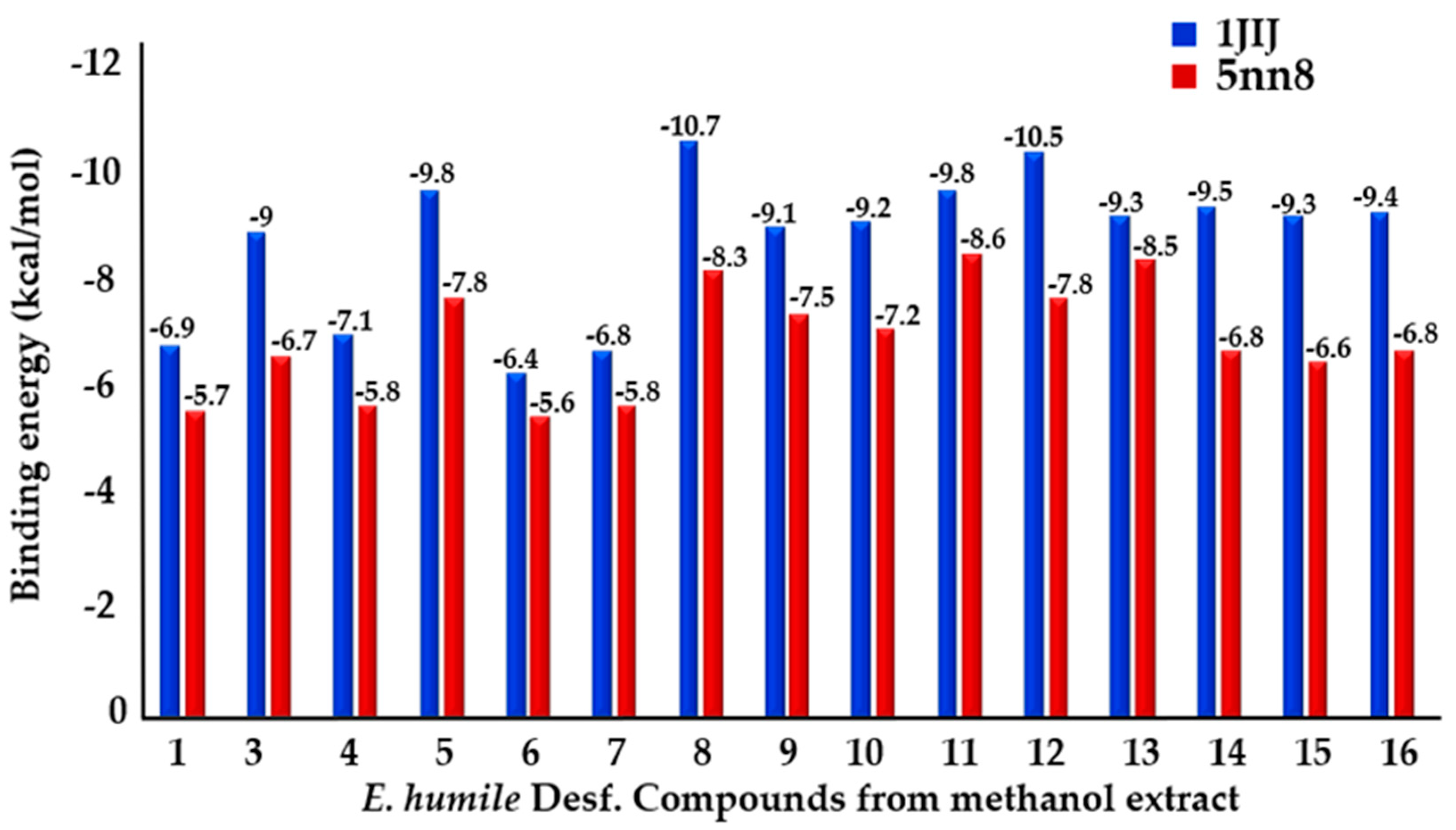
2.3. Computational Study

2.3.1. Binding Energies

In order to provide insight into which of the identified phytocompound(s) may be responsible for dual antimicrobial and antidiabetic activities, a molecular docking study was carried out on the following different protein structures: TyrRS from S. aureus (PDB: 1JIJ) and human lysosomal acid-α-glucosidase (PDB: 5NN8) enzymes, to modulate the inhibition potential of phytochemicals found in the most active extract against these target enzymes. The results revealed that all identified compounds exhibited good affinity (−10.7 kcal/mol ≤ phytocompounds-1JIJ ≤ −6.4 kcal/mol; −8.5 kcal/mol ≤ phytocompounds-5NN8 ≤ −5.6 kcal/mol) towards the investigated target enzymes (Figure 1).

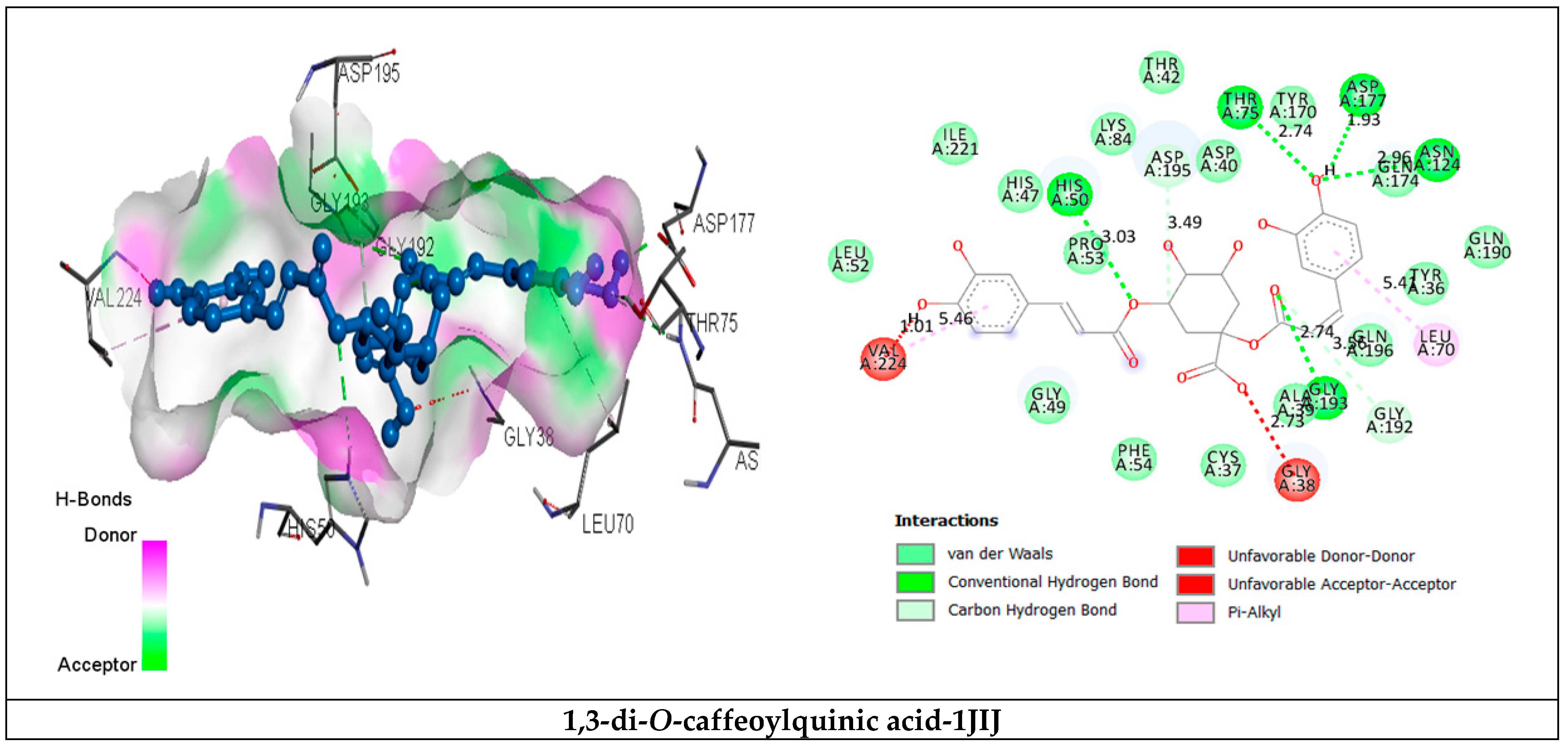
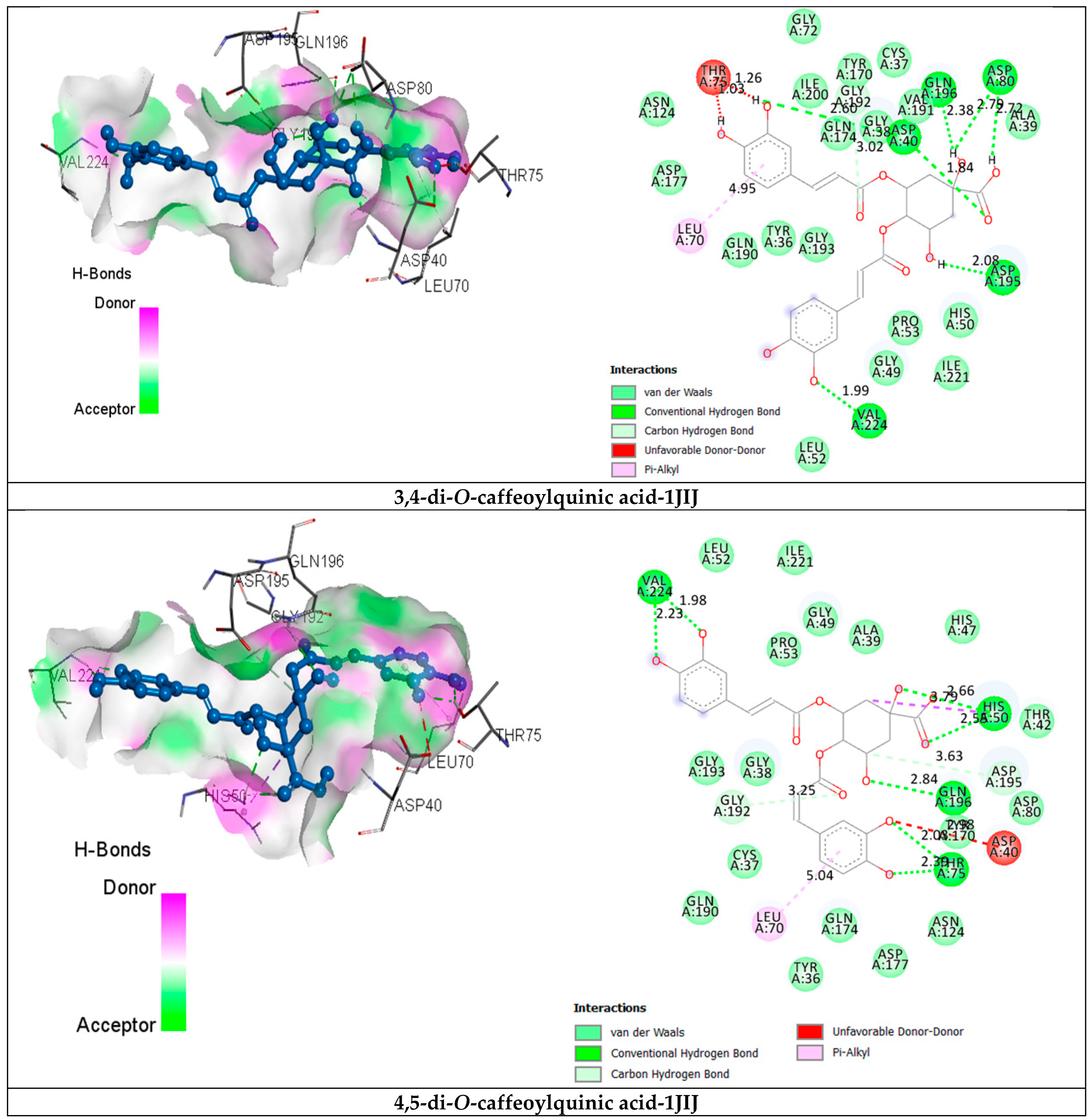
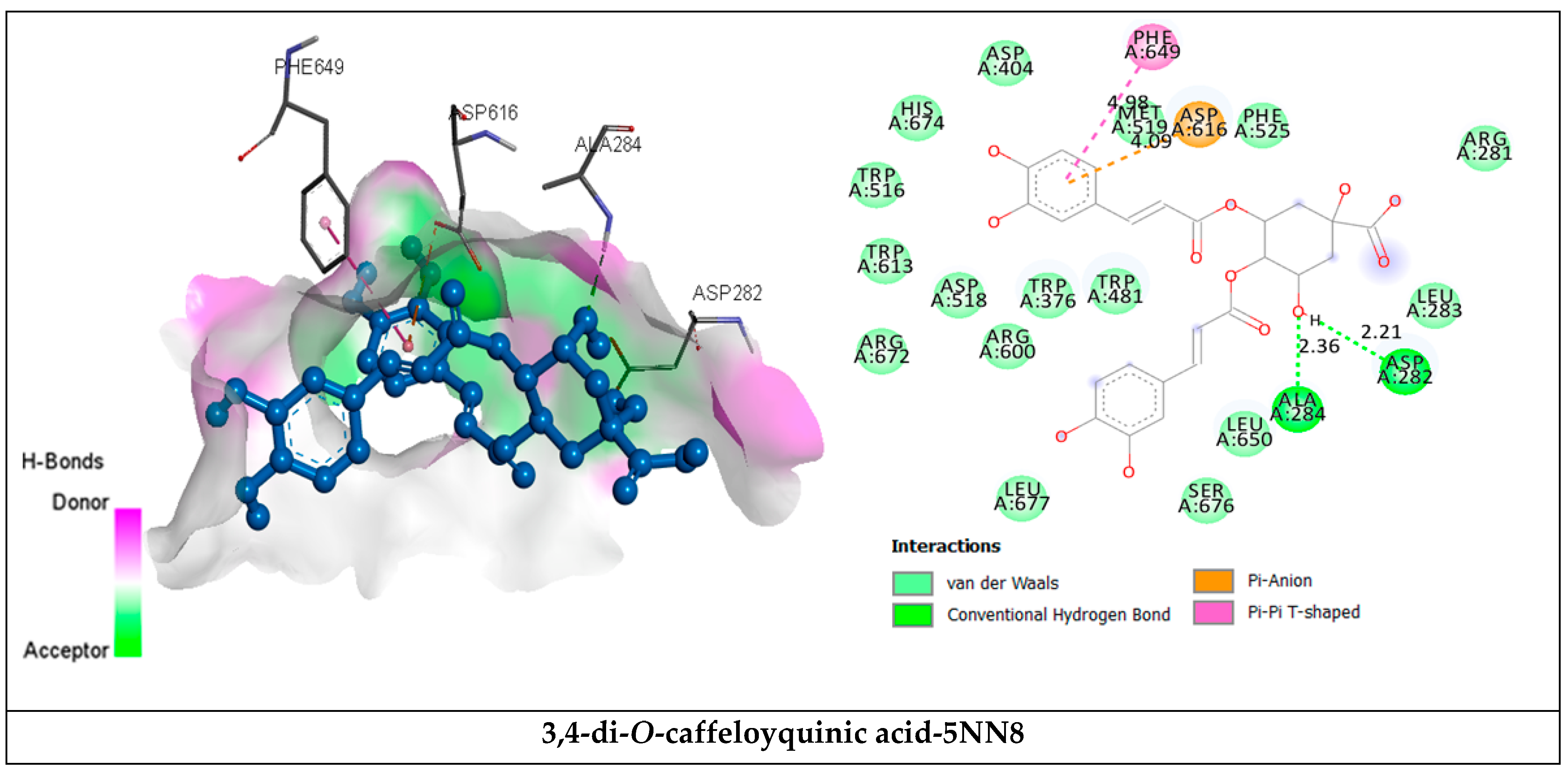
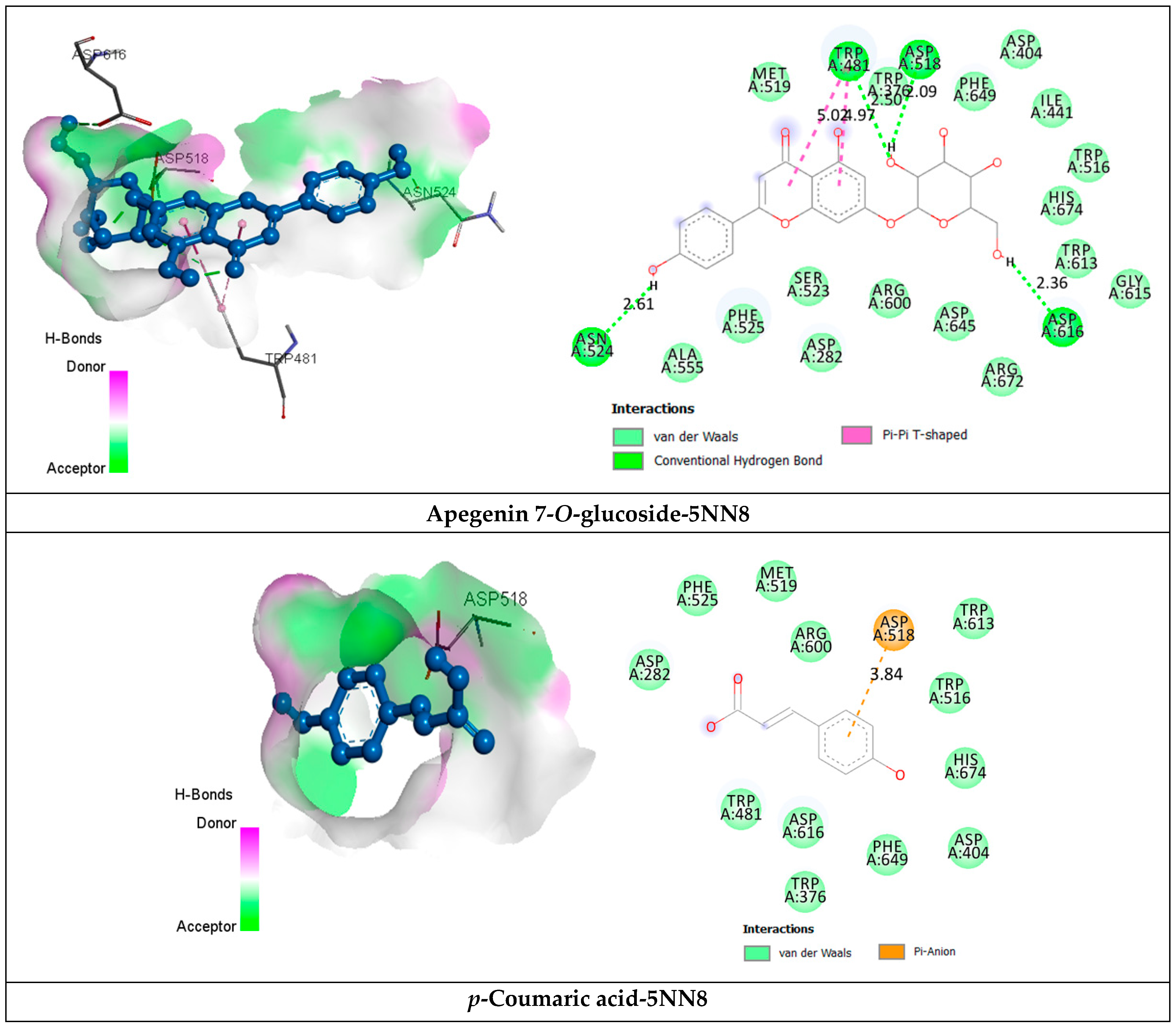
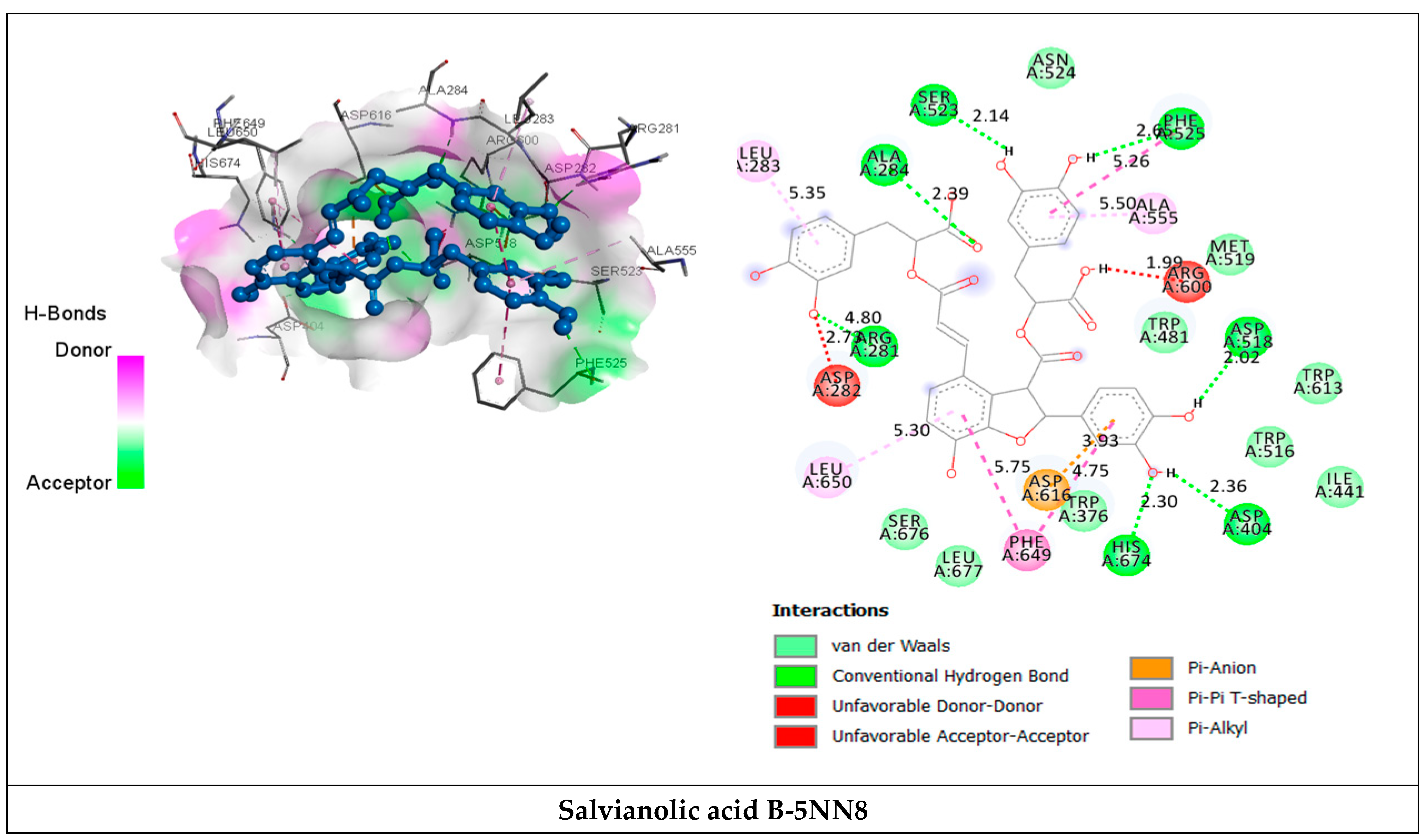
2.3.2. Receptor–Ligand Interaction Analysis

Phytochemicals were selected based on their high concentration in the extract and/or lowest binding energy values in order to identify the key molecular scaffold responsible for the antimicrobial and antidiabetic activities. Their interaction types and attachment positions are shown in Table 5 and Figure 2. Considering the lowest binding energy, the phytochemicals 1,3-di-O-caffeoylquinic acid, 3,4-di-O-caffeoylquinic acid and 4,5-di-O-caffeoylquinic acid were the most potent scaffolds, with binding energies of −9.8 kcal/mol, −10.7 kcal/mol and -10.5 kcal/mol, respectively, bound in the active site and able to adopt several favorable H-bond contacts, as well as other interactions with the common catalytic residues involved in the ternary structure of TyrRS from S. aureus (Cys37, Gly38, Ala39, Asp40, His47, Gly49, His50, Leu70, Thr75, Gln174, Asp177, Gln190, Gly192, Asp195 and Pro222).
Interactions of selected phytocompounds inside α-glucosidase (Table 6 and Figure 3) show that there are two hydrogen bonds (Asp282 at 2.21 Å, Ala284 at 2.36 Å) formed with 3,4-di-O-caffeoylquinic acid, seven hydrogen bonds with salvianolic acid and enzyme residues Arg281 (4.80 Å), Ala284 (2.39 Å), Asp404 (2.36 Å), Asp518 (2.02 Å), Ser523 (2.14 Å), Phe525 (2.6 Å), His674 (2.30 Å) and four hydrogen bonds with apigenin 7-O-glucoside and Trp481 (2.50 Å), Asp518 (2.09 Å), Asn524 (2.61 Å), Asp616 (2.36 Å) amino acids with, respectively, predicted binding energy of −8.3 kcal/mol, −8.6 kcal/mol and −8.5 kcal/mol.

3. Discussion

A structure–activity relationship (SAR) study was carried out to emphasize the importance of the discovery of new alternative drugs and highlight the different contributors to the powerful activity of various E. humile extracts. The biological activities of plant extracts depended on their identified phytocomponents. It is well known that ethanol, methanol and water are effective solvents, commonly used for the preliminary screening of plant extracts and withdrawal of most antimicrobial compounds containing aromatic and saturated organic compounds [31]. As recently verified by our team [26], E. humile extracts are very rich in polyphenols and flavonoid contents, which favor membrane disruption, followed by leakage of cellar components from the microbial membrane [32,33]. The authors also mentioned that methanolic extract exhibited a high amount of phenolic contents [26]. In this study, the high susceptibility of Gram-positive bacteria may be attributed to the composition difference in the bacteria’s cell wall, where the hydrophilic surface of the extra outer membrane in Gram-negative bacteria limits membrane permeability, acts as a barrier to the percolation of different antibacterial substations and prevents antimicrobial agents from penetrating the cell wall [31]. We mentioned that isolates of E. faecalis and E. faecium are among the most prevalent nosocomial pathogens worldwide displaying high resistance, particularly to penicillin/ampicillin and aminoglycosides (high-level resistance). Therefore, therapeutic alternatives to treat infections will require careful attention and remain essential for health care provision [34]. Based on the HPLC/MS phytochemical analysis in the docking study relationships, the main contributors to the strongest antimicrobial activity were identified as follows. As a secondary metabolite, the first major compound in the methanolic E. humile extract was p-coumaric acid (3-[4-hydroxyphenyl]-2-propenoic acid or 4-hydroxycinnamic acid), which has been well confirmed for its antioxidative properties. Indeed, Lou et al. [35] suggested that p-coumaric acid possesses dual mechanisms of bactericidal activity, disrupting bacterial cell membranes and binding to bacterial genomic DNA to inhibit cellular functions, ultimately leading to cell death. The same authors revealed in their study that the effectiveness of p-coumaric acid against E. coli, S. typhimurium and S. dysenteriae via the compound changes the permeability of the cell membrane in parallel with its capacity to bind to DNA-inhibiting cell function. Some authors have reported that the scavenging ability of p-coumaric acid is mainly attributed to its phenyl hydroxyl group (−OH) [36]. Amalan et al. [37] stated that the antidiabetic potential of p-coumaric acid provides a protective role in pancreatic β-cells of diabetic rats by reducing ROS-induced oxidative stress and improving antioxidant status. The authors demonstrated also that p-coumaric acid significantly decreases the blood glucose level and gluconeogenic enzymes such as glucose-6-phosphatase and fructose-1,6-bisphosphatase, but enhances the activities of hexokinase, glucose-6 phosphatase dehydrogenase and GSH via increasing the level of insulin. Additionally, it reduces the total cholesterol and triglycerides in both plasma and tissues, i.e., liver and kidney [37]. Recently, Shen et al. revealed that p-coumaric acid displayed potential therapeutic effects on hyperlipidemia [38]. Ferulic acid (4-hydroxy,3-methoxy cinnamic acid) is known to exert beneficial effects by scavenging free radicals and activating antioxidant enzymes, resulting in a reduction in cellular redox imbalance in a DM animal model. Georgiev et al. [39] reported that ferulic acid displayed potent inhibitory growth towards S. aureus and S. pyogenes 10535. Moreover, it has been mentioned that ferulic acid can alleviate the high-glucose-induced oxidative stress and decreased cell apoptosis in hepatocytes and cardiomyocytes, which are associated with the Keap1-Nrf2-ARE signaling pathway [40]. Ferulic acid possesses beneficial health effects against oxidative stress, hypertension and late-stage diabetes in obese rats, as well as significantly improving insulin sensitivity and lipid profiles and reducing elevated blood pressure [41,42]. Salvianolic acids, known as the most abundant water-soluble compounds extracted from Salvia miltiorrhiza (Danshen), are a powerful antioxidative agent that protects cells from peroxidation and free radical damage [43]. Salvianolic acid B can protect pancreatic beta-cells against cytotoxicity and prevent high-glucose-induced apoptosis [10]. Raoufi et al. [10] suggested that treatment of diabetic rats with salvianolic B induces antidiabetic effects by protecting pancreatic β-256 cells against chemical insult and ameliorates the insulin secretory function of β-cells. Rosmarinic phenylpropanoid acid is an ester of caffeic acid. 3,4-Dihydroxyphenyl lactic acid is known as the strongest antioxidant of all hydroxycinnamic acid derivatives, and its antibacterial activity against S. aureus and the clinical isolate of methicillin-resistant Staphylococcus aureus (MRSA) has been shown [44,45]. Rosmarinic acid has also been shown to exert antidiabetic effects [46,47].
Acacetin (5,7-dihydroxy-4-methoxyflavone) is a naturally occurring flavonoid (synonym linarigenin) that displayed moderate antimicrobial activities against several Gram-positive bacteria (Actinomyces naeslundii, Actinomyces Israelii, Streptococcus mutans), as well as against a variety of Gram-negative bacteria (Prevotella intermedia, Porphyromonas gingivalis, Aggregatibacter actinomycete mcomitans) [48]. As a bioactive constituent of Combretum vendae (Combretaceae), acacetin exhibited strong activity against Staphylococcus aureus, Enterococcus faecalis, Escherichia coli and Pseudomonas aeruginosa [49]. It has been shown to exhibit antihyperglycemic activity in streptozotocin-induced diabetic mice at oral doses of 3 and 31.6 mg/kg by decreasing blood glucose levels in healthy and hyperglycemic mice when compared to an untreated group [50]. Acacetin displayed stable binding to the active site of aldose reductase, a primary mediator of diabetes-induced oxidative stress in retinopathy [51].
The obtained data are in good agreement with previous work, justifying the potential increased inhibitory effect against the α-glucosidase enzyme with increasing polarity of plant extracts [52]. Jesus et al. [53] reported that the infusion extract of Prunus avium vegetal stems parts is the most active. Additionally, methanolic Cornus capitata Wall. extract exhibited maximum inhibitory activity with 98.37% inhibition (IC50 = 12.5 μg/mL), better than that shown by acarbose. Likewise, Arbutus pavarii has been reported to exhibit potent α-glucosidase inhibitory activities [54].
The findings confirm the powerful use of E. humile extract to treat dual diabetes and infections.

4. Materials and Methods

4.1. Plant Material and Extraction

The aerial parts of E. humile were collected during the flowering stage from the region of Douz (Kebilli, South of Tunisia). Extracts were prepared following the same protocol as described by Aouadi et al. [26]. Briefly, extracts were obtained by maceration of 200 g of powder plant material with 600 mL of each solvent (hexane, dichloromethane, ethyl acetate, methanol and aqueous) for 72 h, filtered with Whatman filter paper and then collected and concentrated using a rotary evaporator at 35–55 °C. After that, the obtained extracts were kept in the dark at 4 °C until further use.

4.2. Antimicrobial Activity

The microorganisms tested in this study belonged to 18 reference bacterial strains and 5 fungal strains, which are presented in Table 3 and Table 4, respectively. Experimental details are the same as Hajlaoui et al. [13].

4.3. α-Glucosidase Inhibitory Assay

The α-glucosidase assay of the tested extract was conducted according to the previous protocol with slight modification [55]. The enzyme inhibition rate expressed as a percentage of inhibition was calculated using the following formula:
Percentage inhibitory activity (%) = (1 − A/B) × 100
where A is the absorbance in the presence of the test substance, and B is the absorbance in the presence of phosphate buffer (control).

4.4. HPLC–MS Analysis of Phenolic Compounds

The identification of polyphenolics was performed using the Shimadzu HPLC-MS 2020 system. Detailed experiments were the same as per the method reported by Hajlaoui et al. [26].

4.5. Molecular Docking Approach

Interactions between the selected identified bioactive phytocompounds and the receptor human peroxiredoxin 5 were assessed by in silico molecular docking, in order to explore the preferred orientation of the ligands in the binding site of receptors. We used the same protocol as described previously by our team [26]. The structures of the natural compounds were minimized as done previously [26].

4.6. Statistical Analysis

All experiments were performed in triplicates, and average values were calculated using the SPSS 25.0 statistical package for Windows. Differences in means were calculated using Duncan’s multiple range tests for means with a 95% confidence interval (p ≤ 0.05).

5. Conclusions

Our findings suggest that E. humile extracts showed significant antimicrobial potential against human pathogen strains, especially those of methanolic and ethyl acetate extracts. Additionally, E. humile methanolic extract exhibited a potent α-glucosidase inhibitory effect with respect to the commercial drug acarbose. The structure–activity relationship between the identified phytochemicals and the studied activities was assessed. Docking analysis indicated that the majority of compounds from the most active (methanolic) extract interact with the tested target enzymes through their preferential binding to the active site. Nevertheless, broad research should be conducted to isolate the main compounds from E. humile methanolic extract and explore their clinical efficacy in the treatment of diabetes and infections.

Author Contributions

Conceptualization S.A., A.K., M.S., H.H., K.A. and S.G.; methodology, S.A., A.K., M.S., H.H., K.A. and S.G.; formal analysis, H.H., K.A. and S.A.; software, M.S. and A.K.; investigation, S.A., A.K., M.S., H.H., K.A. and S.G.; writing—original draft preparation, S.A., A.K., M.S., H.H., K.A. and S.G.; writing—review and editing, S.A., H.H., A.K., K.A. and M.S.; supervision, H.H., A.K. and M.S.; funding acquisition, K.A.; project administration, K.A. All authors have read and agreed to the published version of the manuscript.

Funding

The author(s) gratefully acknowledge Qassim University, represented by the Deanship of Scientific “Research, on the financial support for this research under the number (10201-cos-2020-1-3-I) during the academic year 1442 AH/2020 AD”.

Institutional Review Board Statement

Not applicable.

Informed Consent Statement

Not applicable.

Data Availability Statement

The data generated and analyzed during this study are included in this article.

Conflicts of Interest

The authors declare no conflict of interest.

References

  1. Zaky, A.S.; Kandeil, M.; Abdel-Gabbar, M.; Fahmy, E.M.; Almehmadi, M.M.; Ali, T.M.; Ahmed, O.M. The Antidiabetic Effects and Modes of Action of the Balanites aegyptiaca Fruit and Seed Aqueous Extracts in NA/STZ-Induced Diabetic Rats. Pharmaceutics 2022, 14, 263. [Google Scholar] [CrossRef] [PubMed]
  2. Pottathil, S.; Nain, P.; Morsy, M.A.; Kaur, J.; Al-Dhubiab, B.E.; Jaiswal, S.; Nair, A.B. Mechanisms of Antidiabetic Activity of Methanolic Extract of Punica granatum Leaves in Nicotinamide/Streptozotocin-Induced Type 2 Diabetes in Rats. Plants 2020, 9, 1609. [Google Scholar] [CrossRef] [PubMed]
  3. Asirvatham, R.A.; Mohamed, D.A. Cardiovascular Disease among Patients with Diabetes: The Current Scenario in Saudi Arabia. Curr. Diabetes Rev. 2021, 17, 180. [Google Scholar]
  4. Huang, C.H.; Chiu, C.H.; Chen, I.W.; Hung, S.Y.; Lin, C.W.; Hsu, B.R.S.; Huang, Y.Y. Antimicrobial resistance and outcomes of community-onset bacterial bloodstream infections in patients with type 2 diabetes. J. Glob. Antimicrob. Resist. 2018, 15, 271. [Google Scholar] [CrossRef]
  5. Akash, M.S.H.; Rehman, K.; Fiayyaz, F.; Sabir, S.; Kurshid, M. Diabetes-associated infections: Development of antimicrobial resistance and possible treatment strategies. Arch. Microbiol. 2020, 202, 953. [Google Scholar] [CrossRef]
  6. Xiong, Y.; Ng, K.; Zhang, P.; Warner, R.D.; Shen, S.; Tang, H.-Y.; Liang, Z.; Fang, Z. In Vitro α-Glucosidase and α-Amylase Inhibitory Activities of Free and Bound Phenolic Extracts from the Bran and Kernel Fractions of Five Sorghum Grain Genotypes. Foods 2020, 9, 1301. [Google Scholar] [CrossRef]
  7. Akinyede, K.A.; Oyewusi, H.A.; Hughes, G.D.; Ekpo, O.E.; Oguntibeju, O.O. In Vitro Evaluation of the Anti-Diabetic Potential of Aqueous Acetone Helichrysum petiolare Extract (AAHPE) with Molecular Docking Relevance in Diabetes Mellitus. Molecules 2022, 27, 155. [Google Scholar] [CrossRef]
  8. Saddique, F.A.; Ahmad, M.; Ashfaq, U.A.; Muddassar, M.; Sultan, S.; Zaki, M.E.A. Identification of Cyclic Sulfonamides with an N-Arylacetamide Group as α-Glucosidase and α-Amylase Inhibitors: Biological Evaluation and Molecular Modeling. Pharmaceuticals 2022, 15, 106. [Google Scholar] [CrossRef]
  9. Yuan, S.; Ming-Wei, L.; Qi-Qiang, H.; Larsson, S.C. Egg, cholesterol and protein intake and incident type 2 diabetes mellitus: Results of repeated measurements from a prospective cohort study. Clin. Nutr. 2021, 40, 4180. [Google Scholar] [CrossRef]
  10. Raoufi, S.; Baluchnejadmojarad, T.; Roghani, M.; Ghazanfari, T.; Khojasteh, F.; Mansouri, M. Antidiabetic potential of salvianolic acid B in multiple low-dose streptozotocin-induced diabetes, Pharm. Biol. 2015, 53, 1803. [Google Scholar]
  11. Hajlaoui, H.; Arraouadi, S.; Mighri, H.; Ghannay, S.; Aouadi, K.; Adnan, M.; Elasbali, A.M.; Noumi, E.; Snoussi, M.; Kadri, A. HPLC-MS Profiling, Antioxidant, Antimicrobial, Antidiabetic, and Cytotoxicity Activities of Arthrocnemum indicum (Willd.) Moq. Extracts. Plants 2022, 11, 232. [Google Scholar] [CrossRef] [PubMed]
  12. Bhatia, A.; Singh, B.; Arora, R.; Arora, S. In vitro evaluation of the α-glucosidase inhibitory potential of methanolic extracts of traditionally used antidiabetic plants. BMC Complement. Altern Med. 2019, 19, 74. [Google Scholar] [CrossRef] [PubMed] [Green Version]
  13. Hajlaoui, H.; Arraouadi, S.; Noumi, E.; Aouadi, K.; Adnan, M.; Khan, M.A.; Kadri, A.; Snoussi, M. Antimicrobial, antioxidant, anti-acetylcholinesterase, antidiabetic, and pharmacokinetic properties of Carum carvi L. and Coriandrum sativum L. essential oils alone and in combination. Molecules 2021, 26, 3625. [Google Scholar] [CrossRef] [PubMed]
  14. Mseddi, K.; Alimi, F.; Noumi, E.; Veettil, V.N.; Deshpande, S.; Adnan, M.; Hamdi, A.; Elkahoui, S.; Alghamdi, A.; Kadri, A.; et al. Thymus musilii Velen. as a promising source of potent bioactive compounds with its pharmacological properties: In vitro and in silico analysis. Arab. J. Chem. 2020, 13, 6782–6801. [Google Scholar] [CrossRef]
  15. Ben Mefteh, F.; Daoud, A.; Chenari Bouket, A.; Thissera, B.; Kadri, Y.; Cherif-Silini, H.; Eshelli, M.; Alenezi, F.N.; Vallat, A.; Oszako, T.; et al. Date palm trees root-derived endophytes as fungal cell factories for diverse bioactive metabolites. Int. J. Mol. Sci. 2018, 19, 1986. [Google Scholar] [CrossRef] [Green Version]
  16. Daoud, A.; Ben Mefteh, F.; Mnafgui, K.; Turki, M.; Jmal, S.; Ben Amar, R.; Ayadi, F.; ElFeki, A.; Abid, L.; Rateb, M.E.; et al. Cardiopreventive effect of ethanolic extract of date palm pollen against isoproterenol induced myocardial infarction in rats through the inhibition of the angiotensin-converting enzyme. Exp. Toxicol. Pathol. 2017, 69, 656–665. [Google Scholar] [CrossRef] [Green Version]
  17. Hajlaoui, H.; Mighri, H.; Aouni, M.; Gharsallah, N.; Kadri, A. Chemical composition and in vitro evaluation of antioxidant antimicrobial cytotoxicity and anti-acetylcholinesterase properties of Tunisian Origanum majorana L. essential oil. Microb. Pathog. 2016, 95, 86–94. [Google Scholar] [CrossRef]
  18. Kadri, A.; Zarai, Z.; Chobba, I.B.; Gharsallah, N.; Damak, M.; Békir, A. Chemical composition and in vitro antioxidant activities of Thymelaea hirsuta L: Essential oil from Tunisia. Afr. J. Biotechnol. 2011, 10, 2930–2935. [Google Scholar]
  19. Felhi, S.; Saoudi, M.; Daoud, A.; Hajlaoui, H.; Ncir, M.; Chaabane, R.; El Feki, A.; Gharsallah, N.; Kadri, A. Investigation of phytochemical contents, in vitro antioxidant and antibacterial behavior and in vivo anti-inflammatory potential of Ecballium elaterium methanol fruits extract. Food Sci. Technol. 2017, 37, 558–563. [Google Scholar] [CrossRef] [Green Version]
  20. Bakari, S.; Daoud, A.; Felhi, S.; Smaoui, S.; Gharsallah, N.; Kadri, A. Proximate analysis, mineral composition, phytochemical contents antioxidant and antimicrobial activities and GC-MS investigation of various solvent extracts of Cactus cladode. Food Sci Technol. 2017, 27, 286–293. [Google Scholar] [CrossRef] [Green Version]
  21. Bakari, S.; Hajlaoui, H.; Daoud, A.; Mighri, H.; Ross-Garcia, J.M.; Gharsallah, N.; Kadri, A. Phytochemicals, antioxidant and antimicrobial potentials and LC-MS analysis of hydroalcoholic extracts of leaves and flowers of Erodium glaucophyllum collected from Tunisian Sahara. Food Sci. Technol. 2018, 38, 310–317. [Google Scholar] [CrossRef] [Green Version]
  22. Felhi, S.; Hajlaoui, H.; Ncir, M.; Bakari, S.; Ktari, N.; Saoudi, M.; Gharsallah, N.; Kadri, A. Nutritional, phytochemical and antioxidant evaluation and FT-IR analysis of freeze-dried extracts of Ecballium elaterium fruit juice from three localities. Food Sci. Technol. 2016, 36, 646–655. [Google Scholar] [CrossRef] [Green Version]
  23. Bakari, S.; Ncir, M.; Felhi, S.; Hajlaoui, H.; Saoudi, M.; Gharsallah, N.; Kadri, A. Chemical composition and in vitro evaluation of total phenolic, flavonoid, and antioxidant properties of essential oil and solvent extract from the aerial parts of Teucrium polium grown in Tunisia. Food Sci Biotechnol. 2015, 24, 1943–1949. [Google Scholar] [CrossRef]
  24. Alminderej, F.; Bakari, S.; Almundarij, T.I.; Snoussi, M.; Aouadi, K.; Kadri, A. Antioxidant activities of a new chemotype of Piper cubeba L. fruit essential oil (Methyleugenol/Eugenol): In Silico molecular docking and ADMET studies. Plants 2020, 9, 1534. [Google Scholar] [CrossRef]
  25. Noumi, E.; Snoussi, M.; Anouar, E.H.; Alreshidi, M.; Veettil, V.N.; Elkahoui, S.; Adnan, M.; Patel, M.; Kadri, A.; Aouadi, K.; et al. HR-LCMS-Based Metabolite Profiling, Antioxidant, and Anticancer Properties of Teucrium polium L. Methanolic Extract: Computational and In Vitro Study. Antioxidants 2020, 9, 1089. [Google Scholar] [CrossRef]
  26. Aouadi, K.; Hajlaoui, H.; Arraouadi, S.; Ghannay, S.; Snoussi, M.; Kadri, A. HPLC/MS phytochemical profiling with antioxidant activities of Echium humile Desf. extracts: ADMET prediction and computational study targeting human peroxiredoxin 5 receptor. Agronomy 2021, 11, 2165. [Google Scholar] [CrossRef]
  27. Alminderej, F.; Bakari, S.; Almundarij, T.I.; Snoussi, M.; Aouadi, K.; Kadri, A. Antimicrobial and wound healing potential of a new chemotype from Piper cubeba L. essential oil and in silico study on S. aureus tyrosyl-tRNA synthetase protein. Plants 2021, 10, 205. [Google Scholar] [CrossRef]
  28. Othman, I.M.M.; Gad-Elkareem, M.A.M.; Anouar, E.H.; Aouadi, K.; Kadri, A.; Snoussi, M. Design, synthesis ADMET and molecular docking of new imidazo[4,5-b]pyridine-5-thione derivatives as potential tyrosyl-tRNA synthetase inhibitors. Bioorg. Chem. 2020, 102, 104105. [Google Scholar] [CrossRef]
  29. Othman, I.M.M.; Gad-Elkareem, M.A.M.; Anouar, E.H.; Aouadi, K.; Snoussi, M.; Kadri, A. New substituted pyrazolones and dipyrazolotriazines as promising tyrosyl-tRNA synthetase and peroxiredoxin-5 inhibitors: Design, synthesis, molecular docking and structure-activity relationship (SAR) analysis. Bioorg. Chem. 2021, 109, 104704. [Google Scholar] [CrossRef]
  30. Othman, I.M.M.; Gad-Elkareem, M.A.M.; Anouar, E.H.; Snoussi, M.; Aouadi, K.; Kadri, A. Novel fused pyridine derivatives containing pyrimidine moiety as prospective tyrosyl-tRNA synthetase inhibitors: Design, synthesis, pharmacokinetics and molecular docking studies. J. Mol. Struct. 2020, 1219, 128651. [Google Scholar] [CrossRef]
  31. Wong, J.X.; Ramli, S. Antimicrobial activity of different types of Centella asiatica extracts against foodborne pathogens and food spoilage microorganisms. LWT 2021, 142, 111026. [Google Scholar] [CrossRef]
  32. Kalita, D.; Saikia, J. Ethnomedicinal, antibacterial and antifungal potentiality of Centella asiatica, Nerium indicum and Cuscuta reflexa widely used in Tiwa Tribe of Morigaon district of Assam, India. Int. J. Phytomed. 2012, 4, 380. [Google Scholar]
  33. Rattanakom, S.; Yasurin, P. Chemical profiling of Centella asiatica under different extraction solvents and its antibacterial activity, antioxidant activity. Orient. J. Chem. 2015, 31, 2453. [Google Scholar] [CrossRef]
  34. Werner, G.; Coque, T.M.; Franz, C.M.A.P.; Grohmann, E.; Hegstad, K.; Jensen, L.; Schaik, W.; Weaver, K. Antibiotic resistant enterococci—Tales of a drug resistance gene trafficker. Int. J. Med. Microbiol. 2013, 303, 360. [Google Scholar] [CrossRef] [PubMed]
  35. Lou, Z.; Wang, H.; Rao, S.; Sun, J.; Ma, C.; Li, J. p-Coumaric acid kills bacteria through dual damage mechanisms. Food Control 2012, 25, 550. [Google Scholar] [CrossRef]
  36. Roychoudhury, S.; Sinha, B.; Choudhury, B.P.; Jha, N.K.; Palit, P.; Kundu, S.; Mandal, S.C.; Kolesarova, A.; Yousef, M.I.; Ruokolainen, J.; et al. Scavenging Properties of Plant-Derived Natural Biomolecule Para-Coumaric Acid in the Prevention of Oxidative Stress-Induced Diseases. Antioxidants 2021, 10, 1205. [Google Scholar] [CrossRef]
  37. Amalan, V.; Vijayakumar, N.; Indumathi, D.; Ramakrishnan, A. Antidiabetic and antihyperlipidemic activity of p-coumaric acid in diabetic rats, role of pancreatic GLUT 2: In vivo approach. Biomed. Pharmacother. 2016, 84, 230. [Google Scholar] [CrossRef]
  38. Shen, Y.; Song, X.; Li, L.; Sun, J.; Jaiswal, Y.; Huang, J.; Liu, C.; Yang, W.; Williams, L.; Zhang, H.; et al. Protective effects of p-coumaric acid against oxidant and hyperlipidemia-an in vitro and in vivo evaluation. Biomed. Pharmacother. 2019, 111, 579. [Google Scholar] [CrossRef]
  39. Georgiev, L.; Chochkova, M.; Ivanova, G.; Najdenski, H.; Ninova, M.; Milkova, T. Radical scavenging and antimicrobial activities of cinnamoyl amides of biogenic monoamines. Riv. Ital. Sost. Grasse 2012, 89, 91–102. [Google Scholar]
  40. Song, Y.; Wen, L.; Sun, J.; Bai, W.; Jiao, R.; Hu, Y.; Peng, X.; He, Y.; Ou, S. Cytoprotective mechanism of ferulic acid against high glucose-induced oxidative stress in cardiomyocytes and hepatocytes. Food Nut. Res. 2016, 60, 30323. [Google Scholar] [CrossRef] [Green Version]
  41. Senaphan, K.; Kukongviriyapan, U.; Sangartit, W.; Pakdeechote, P.; Pannangpetch, P.; Prachaney, P.; Greenwald, S.E.; Kukongviriyapan, V. Ferulic Acid Alleviates Changes in a Rat Model of Metabolic Syndrome Induced by High-Carbohydrate, High-Fat Diet. Nutrients 2015, 7, 6446–6464. [Google Scholar] [CrossRef] [PubMed]
  42. Song, Y.; Wu, T.; Yang, Q.; Chen, X.; Wang, M.; Wang, Y.; Peng, X.; Ou, S. Ferulic acid alleviates the symptoms of diabetes in obese rats. J. Funct. Foods. 2014, 9, 141. [Google Scholar] [CrossRef]
  43. Ma, L.; Tang, L.; Yi, Q. Salvianolic Acids: Potential Source of Natural Drugs for the Treatment of Fibrosis Disease and Cancer. Front. Pharmacol. 2019, 10, 97. [Google Scholar] [CrossRef] [PubMed] [Green Version]
  44. Ekambaram, S.P.; Perumal, S.S.; Balakrishnan, A.; Marappan, N.; Gajendran, S.S.; Viswanathan, V. Antibacterial synergy between rosmarinic acid and antibiotics against methicillin-resistant Staphylococcus aureus. J. Intercult. Ethnopharmacol. 2016, 5, 358–363. [Google Scholar] [CrossRef]
  45. Amoah, S.K.; Sandjo, L.P.; Kratz, J.M.; Biavatti, M.W. Rosmarinic Acid—Pharmaceutical and Clinical Aspects. Planta Med. 2016, 82, 388–406. [Google Scholar] [CrossRef] [Green Version]
  46. Mushtaq, N.; Schmatz, R.; Ahmed, M.; Pereira, L.B.; da Costa, P.; Reichert, K.P.; Dalenogare, D.; Pelinson, L.P.; Vieira, J.M.; Stefanello, N.; et al. Protective e_ect of rosmarinic acid against oxidative stress biomarkers in liver and kidney of strepotozotocin-induced diabetic rats. J. Physiol. Biochem. 2015, 71, 743–751. [Google Scholar] [CrossRef]
  47. Sotnikova, R.; Kaprinay, B.; Navarova, J. Rosmarinic acid mitigates signs of systemic oxidative stress in streptozotocin-induced diabetes in rats. Gen. Physiol. Biophys. 2015, 34, 449–452. [Google Scholar]
  48. Kuete, V.; More, G.; Lall, N.; Hussein, A.; Tshikalange, T.E. Antimicrobial Constituents of Artemisia afra Jacq. ex Willd. against Periodontal Pathogens. Evid Based Complement. Alternat. Med. 2012, 2012, 252758. [Google Scholar]
  49. Komape, N.P.M.; Aderogba, M.; Bagla, V.P.; Masoko, P.; Eloff, J.N. Antibacterial and antioxidant activities of leaf extracts of Combretum vendae (combretecacea) and the isolation of an antibacterial compound. Afr. J. Tradit Complement. Altern. Med. 2014, 11, 73. [Google Scholar] [CrossRef] [Green Version]
  50. Juárez-Reyes, K.; Brindis, F.; Medina-Campos, O.N.; Pedraza-Chaverri, J.; Bye, R.; Linares, E.; Mata, R. Hypoglycemic, antihyperglycemic, and antioxidant effects of the edible plant Anoda cristata. J. Ethnopharmacol. 2015, 161, 36. [Google Scholar] [CrossRef]
  51. Semwal, R.B.; Semwal, D.K.; Combrinck, S.; Trill, J.; Gibbons, S.; Viljoen, A. Acacetin—A simple flavone exhibiting diverse pharmacological activities. Phytochem. Lett. 2019, 32, 56. [Google Scholar] [CrossRef]
  52. Brown, A.; Anderson, D.; Racicot, K.; Pilkenton, S.J.; Apostolidis, E. Evaluation of phenolic phytochemical enriched commercial plant extracts on the in vitro inhibition of α-glucosidase. Front. Nutr. 2017, 4, 56. [Google Scholar] [CrossRef] [PubMed] [Green Version]
  53. Jesus, F.; Gonçalves, A.C.; Alves, G.; Silva, L.R. Exploring the phenolic profile, antioxidant, antidiabetic and anti-hemolytic potential of Prunus avium vegetal parts. Food Res. Int. 2019, 116, 600–610. [Google Scholar] [CrossRef] [PubMed]
  54. El Shibani, F.A.E.S. A Pharmacognostical Study of Arbutus pavarii Pampan. Family Ericaceae and Sarcopoterium spinosum L. Family Rosaceae Growing in Libya. Ph.D. Thesis, Cairo University, Cairo, Egypt, 2017. [Google Scholar]
  55. Kadri, A.; Aouadi, K. In vitro antimicrobial and α-glucosidase inhibitory potential of enantiopure cycloalkylglycine derivatives: Insights into their in silico pharmacokinetic, druglikeness, and medicinal chemistry properties. J. App. Pharm. Sci. 2020, 10, 107–115. [Google Scholar]
Figure 1. Binding energies of E. humile Desf. compounds complexed with TyrRS from S. aureus (PDB: 1JIJ) and human lysosomal acid-α-glucosidase (PDB: 5NN8) enzymes.
Plants 11 01131 g001
Figure 2. Interactions of tyrosyl-tRNA synthetase TyrRS from S. aureus (PDB: 1JIJ) with the selected major phytochemicals, with the top two having the lowest binding energies of E. humile methanolic extract.
Plants 11 01131 g002aPlants 11 01131 g002bPlants 11 01131 g002c
Figure 3. Interactions of human lysosomal acid-α-glucosidase (PDB: 5NN8) with the selected major phytochemicals, with the top two having the lowest binding energies of E. humile Desf. methanolic extract.
Plants 11 01131 g003aPlants 11 01131 g003bPlants 11 01131 g003c
Table 1. Antibacterial and antifungal activities of E. humile extracts.
StrainsIZD (mm)
ExtractsHexaneDichloromethaneEthyl AcetateMethanolAqueousChloramphenicol
Gram-positive bacteria
Staphylococcus aureus11.50 ± 1.00 cdC14.00 ± 2.00 abB10.00 ± 0.00 dCD9.00 ± 0.00 cD16.50 ± 1.00 dA
Enterococcus faecalis10.50 ± 1.00 cC13.00 ± 1.00 bcB12.50 ± 1.00 bcBC15.50 ± 1.00 aA10.50 ± 1.00 bC12.00 ± 1.00 eBC
Bacillus cereus14.00 ± 1.00 bB14.00 ± 1.00 bB12.00±1.00 cC14.00±1.00 bB11.50 ±1.00 bC26.00 ± 1.00 aA
Bacillus subtilis11.50 ± 1.00 cC14.50 ± 1.00 bB15.00 ± 1.00 aB12.50 ± 1.00 cC24.00 ± 0.00 bA
Micrococcus luteus18.50 ± 0.86 aA_14.5 ± 1.00 aB14.00 ± 0.00 bB15.00 ± 0.00 aB20.00 ± 2.00 cA
Gram-negative bacteria
Escherichia coli10.33 ± 0.76 cC18.00 ± 1.00 aB8.00 ± 0.00 eD23.50 ± 1.50 bA
Klebsiella pneumoniae11.00 ± 0.00 c11.00 ± 0.00 dB11.50 ± 1.50 cdB10.50±0.50 bB22.00 ± 1.00 bA
Salmonella Enteritidis8.00 ± 0.00 dE11.50 ± 0.50 cdB10.00 ± 0.00 dC11.50 ± 0.50 cB9.00 ± 0.00 cD16.00 ± 0.00 dA
Fungal strains Cycloheximide
Fusarium sp. 11.5 ± 0.71 bC14.5 ± 0.71 bB18.00 ± 1.50 abA
Pythium catenulatum9.00 ± 0.00 cB10.50 ± 0.71 cB17.50 ± 1.50 bA
Fusarium oxysporum13.00 ± 1.00 aB20.00 ± 0.00 aA20.00 ± 2.00 aA
Values are mean ± standard deviation of three separate experiments. Diameter of inhibition zones of extract including diameter of well is 6 mm; –: no inhibition; a,b,c,d,e,f,g,h,A,B,C,D,E: each value represents the average of 3 repetitions. Small letters are used to compare each extract means between different strains, while capital letters are used to compare means between extracts for the same strain.
Table 2. Determination of MIC (MBC or MFC) in mg/mL and {MBC/MIC or MFC/MIC} of E. humile extracts.
StrainsHexaneDichloromethaneEthyl AcetateMethanolAqueous
Gram-positive bacteria
Staphylococcus aureus12.5 (25) {2}1.56 (12.5) {8}3.12 (6.25) {2}3.12 (6.25) {2}
Enterococcus faecalis6.25 (12.5) {2}12.5 (50) {4}1.56 (12.5) {8}0.19 (3.12) {16}0.19 (6.25) {32}
Bacillus cereus0.39 (6.25) {16}6.25 (25) {4}12.5 (100) {8}6.25 (12.5) {2}
Bacillus subtilis6.25 (25) {4}50 (100) {2}1.56 (12.5) {8}0.39 (6.25) {16}0.19 (3.12) {16}
Micrococcus luteus12.5 (50) {4}1.56 (6.25) {4}6.25 (25) {4}3.12 (6.25) {2}
Gram-negative bacteria
Escherichia coli3.12 (12.5) {4}1.56 (6.25) {4}1.56 (12.5) {8}
Klebsiella pneumoniae50 (100) {2}25 (50) {2}12.5 (25) {2}3.12 (6.25) {2}
Salmonella Enteritidis1.56 (3.12) {2}3.12 (12.5) {4}1.56 (3.12) {2}0.39 (3.12) {8}1.56 (6.25) {4}
Fungal strains
Fusarium sp. 0.39 (3.12) {8}0.39 (3.12) {8}
Pythium catenulatum__1.56 (6.25) {4}0.195 (0.78) {4}
Fusarium oxysporum0.39 (1.56) {4}3.12 (6.25) {2}
Table 3. IC50 values of various extracts of E. humile against α-glucosidase.
ExtractsIC50 (mg/mL)
Hexane
Dichloromethane
Ethyl acetate11.17 ± 0.62 a
Methanol0.06 ± 0.29 c
Aqueous0.80 ± 1.81 b
Acarbose0.70 ± 0.67 b
Means followed by the same letters are not significantly different at p = 0.05 based on Duncan’s multiple range test.
Table 4. HPLC–MS analysis of E. humile methanolic extract from aerial parts.
PeakRetention Time (min)MS [M-H]-m/zCompoundsConcentration (µg/g)
17.385153.00Protocatechuic acid4.204 ± 0.06
29.189289.00(+)-Catechin 39.286 ± 2.46
313.795289.00Epicatechin26.868 ± 0.82
412.993179.00Caffeic acid9.673 ± 0.1
514.960515.001,3-di-O-caffeoylquinic acid22.367 ± 0.15
617.087163.00p-Coumaric acid1335.48 ± 5.22
718.744193.00Trans-Ferulic acid125.522 ± 10.3
821.779515.003,4-di-O-caffeoylquinic acid6.7645 ± 0.41
922.209359.00Rosmarinic acid41.154 ± 0.12
1022.910463.00Hyperoside 2.992 ± 0.01
1123.754717.00Salvianolic acid B47.832 ± 1.51
1223.754515.004,5-di-O-caffeoylquinic acid319.373 ± 0.41
1324.302431.00Apegenin 7-O-glucoside2.481 ± 0.48
1423.451329.00Cirsiliol41.623 ± 0.19
1531.852269.00Apegenin0.973 ± 0.25
1637.061283.00Acacetin107.462 ± 17.4
Table 5. Major phytochemicals, with the top two having the lowest binding energies with their interaction residues with TyrRS from S. aureus (PDB: 1JIJ).
CompoundsInteractions TypeInteracting Residues (1JIJ)Binding Energy (kcal/mol)
1,3-di-O-caffeoylquinic
acid
van der Waals
 
H bond
C-H bond
Unfavorable Donor-Donor/
Acceptor-Acceptor
Pi-Alkyl
Tyr36, Cys37, Ala39, Thr42, His47, Gly49, Leu52, Pro53, Phe54, Lys84,
Tyr170, Gln174, Gln190, Gln196, Ile221.
His50 (3.03), Thr75 (2.74), Asp177 (1.93), Asn124 (2.96), Gly193 (2.74).
Gly192 (3.56), Asp195 (3.38).
Gly38 (2.77), Val224 (2.01).
 
Leu70 (5.41), Val224 (5.46).
−9.8
3,4-di-O-caffeoylquinic
acid
 
van der Waals
 
H bond
 
C-H bond
Unfavorable Donor-Donor
Pi-Alkyl
Tyr36, Cys37, Gly38, Ala39, Gly49, His50, Leu52, Pro53, Gly72, Asn124,
Tyr170, Gln174, Asp177, Gln190, Val191, Gly193, Ile200, Ile221.
Asp40 (1.84) (2.60), Asp80 (2.72) (2.79), Asp195 (2.08), Gln196 (2.38), Val224 (1.99).
Gly192 (3.02).
Thr75 (1.03) (1.26).
Leu70 (4.95).
−10.7
4,5-di-O-caffeoylquinic
acid
van der Waals
 
H bond
C-H bond
Unfavorable Donor-Donor
Pi-Alkyl
Tyr36, Cys37, Gly38, Ala39, Thr42, His47, Gly49, Leu52, Pro53, Asp80, Asn124,
Tyr170, Gln174, Asp177, Gln190, Gly193, Ile221.
His50 (2.55) (2.66), Thr75 (2.03) (2.39), Gln196 (2.84), Val224 (1.98) (2.23).
Asp195 (3.36)
Asp40 (2.98)
Leu70 (5.04)
−10.5
p-Coumaric
acid
van der Waals
 
C-H bond
Pi-Cation
Pi-Alkyl
Tyr36, Gly38, Ala39, Asp40, Thr75, Asp80, Asn124, Tyr170, Gln174,
Gln190, Asp195.
Asp177 (2.15)
Gln196 (2.99)
Leu70 (5.22)
−6.4
Salvianolic
Acid B
van der Waals
 
H bond
C-H bond
Pi-Cation/Anion
Pi-Alkyl
Tyr36, Ala39, Asp40, Thr42, His47, Gly49, Phe54, Ile71, Gly72, Gly79, Asn124,
Tyr170, Gln174, Asp177, Gln190, Val191, Gly193, Gln196, Ile200, Val224.
Cys37 (3.71), His50 (2.59), Thr75 (2.16), Lys84 (2.02) (2.94), Arg88 (2.36).
Gly38 (3.38), His50 (3.46), Gly192 (3.65).
Asp80 (4.13), Lys84 (4.28), Asp195 (3.97)
Pro53 (5.15), Leu70 (5.24)
−9.8
Table 6. Major phytochemicals, with the top two having the lowest binding energies with their interaction residues with human lysosomal acid-α-glucosidase (PDB: 5NN8).
CompoundsInteractions TypeInteracting Residues (5NN8) (Å)Binding Energy (kcal/mol)
3,4-di-O-caffeoylquinic acid 
van der Waals
 
H bond
Pi-Anion
Pi-Pi T shaped
Arg281, Leu283, Trp376, Asp404, Trp481, Asp518, Met519, Phe525, Trp516, Arg600, Trp613, Arg672, leu650, His674, Ser676, Leu677.
Asp282(2.21), Ala284 (2.36).
Asp616 (4.09)
Phe649 (4.98)
−8.3
p-Coumaric acidvan der Waals
 
Pi-Anion
Asp282, Trp376, Asp404, Trp481, Trp516, Met519, Phe525, Arg600,
Trp613, Asp616, Phe649, His674.
Asp518 (3.84).
−5.6
Salvianolic acid Bvan der Waals
H bond
 
Unfavorable Donor-Donor/
Acceptor-Acceptor
Pi-Anion
Pi-Pi T shaped
Trp376, Ile441, Trp481, Trp516, Met519, Asn524, Trp613, Ser676, Leu677.
Arg281 (4.80), Ala284 (2.39), Asp404 (2.36), Asp518 (2.02),
Ser523 (2.14), Phe525 (2.6), His674 (2.30).
Asp282 (2.73), Arg600 (1.99).
 
Asp616 (3.93).
Phe649 (4.75) (5.75).
−8.6
Apegenin 7-O-glucosidevan der Waals
 
H bond
Pi-Pi T shaped
Pi-Alkyl
Asp282, Trp376, Ile441, Trp516, Met519, Ser523, Phe525, Ala555,
Arg600, Trp613, Gly615, Asp645, Phe649, Arg672, His674.
Trp481 (2.50), Asp518 (2.09), Asn524 (2.61), Asp616 (2.36).
Trp481 (4.97) (5.02).
Leu283 (5.35), Ala555 (5.50), Leu650 (5.30).
−8.5
Publisher’s Note: MDPI stays neutral with regard to jurisdictional claims in published maps and institutional affiliations.

