Article

The Biodiversity of Grapevine Bacterial Endophytes of Vitis amurensis Rupr.

by
Olga A. Aleynova
*,
Nikolay N. Nityagovsky
,
Alexandra S. Dubrovina
and
Konstantin V. Kiselev
Laboratory of Biotechnology, Federal Scientific Center of the East Asia Terrestrial Biodiversity, Far Eastern Branch of the Russian Academy of Sciences, Vladivostok 690022, Russia
*
Author to whom correspondence should be addressed.
Plants 2022, 11(9), 1128; https://doi.org/10.3390/plants11091128
Submission received: 18 March 2022 / Revised: 12 April 2022 / Accepted: 19 April 2022 / Published: 21 April 2022

Abstract

:
In this paper, the composition profiles of bacterial endophytes in wild-growing Amur grape Vitis amurensis Rupr. grown in the south of the Russian Far East were analyzed using both a cultivation-dependent (sowing bacteria) and a cultivation-independent (next generation sequencing, NGS) approach. Both methods revealed the prevalent endophytes in V. amurensis were represented by Gammaproteobacteria—40.3–75.8%, Alphaproteobacteria—8.6–18.7%, Actinobacteria—9.2–15.4%, and Bacilli—6.1–6.6%. NGS also showed a large proportion of Bacteroidia (12.2%) and a small proportion of other classes (less than 5.7%). In general, NGS revealed a greater variety of classes and genera in the endophytic bacterial community due to a high number of reads (574,207) in comparison with the number of colonies (933) obtained after the cultivation-dependent method. A comparative analysis performed in this study showed that both wild grape V. amurensis from Russia and domesticated cultivars of V. vinifera from Germany and California (USA) exhibit the same basic composition of endophytic bacteria, while the percentages of major taxa and minor taxa showed some differences depending on the plant organ, grape individuals, environmental conditions, and sampling time. Furthermore, the obtained data revealed that lower temperatures and increased precipitation favored the number and diversity of endophytic bacteria in the wild Amur grape. Thus, this study firstly described and analyzed the biodiversity of endophytic bacteria in wild grapevine V. amurensis.

1. Introduction

Grapes from the genus Vitis have been widely recognized as economically important fruit crops used for grape, wine, raisins, and juice production [1]. The Vitaceae family includes about 15 genera and ca. 900 species mostly in pantropical regions of Asia, Africa, Australia and the Pacific islands, with a few genera in temperate regions of the Northern Hemisphere [2]. It is well known that grape yield and fruit quality depend highly on external factors, such as the vineyard location, weather conditions, agricultural practices, and various biotic factors, such as microbial pathogens and endophytic microbiome [3].
Plant endophytes include bacteria, archaea, fungi, and protists that colonize the plant interior regardless of the outcome of the association [4]. Some endophytes are known to confer mutually beneficial effects to the host plant [3]. Some endophytes promote plant growth via nitrogen fixation, phytohormone production, or nutrient acquisition, and are also known to confer tolerance to abiotic and biotic stresses [5]. Endophytes possess considerable potential for application in agriculture as natural agents for biological control, plant growth promotion, crop yield improvement, and environmental stress control.
In the last decade, researchers have been actively studying the microbiome assemblage in grape species. Both general composition of endophytic communities and the endophyte biodiversity in healthy vs. diseased grapevine cultivars were studied to isolate microorganisms capable of grapevine pathogen biocontrol and fruit yield improvement.
The main body of research on the biodiversity of grapevine endophytic bacteria has been devoted to the European wine grape (Vitis vinifera) [6,7,8,9,10,11]. Most available studies show Gammaproteobacteria represent the dominant class of endophytic bacteria in V. vinifera [6,8]. It has been shown that the main bacteria included Bacillus spp., Pseudomonas spp., Erwinia spp., Pantoea spp., and Curtobacterium spp. In addition, there were studies on the endophytic bacterial communities of the grapevine varieties grown across the North American continent [12,13]. Proteobacteria were reported as the main representative of the bacterial community in American grape [12]. According to Deyett and Rolshausen [12], the core microbiome of grapevine sap from California was primarily composed of seven bacterial taxa (Streptococcus, Micrococcus, Pseudomonas, Bacteroides, Massilia, Acinetobacter and Bacillus) that were present throughout the growing season [13]. Moreover, Campisano et al. [14] conducted a comparative analysis of bacterial endophytes detected in wild and domesticated European grapevine V. vinifera. According to the study, wild grapevines were inhabited by a much more diverse endophytic bacterial community than domesticated counterparts. Thus, studying the bacterial microbiome of wild grapes represents an important area of research with a potential to detect new bacteriome variations that are not typical to cultivated varieties.
Currently, little is known about the microbiome of grapes growing in the Far East of Russia. The main representative of the Far Eastern wild grapes is the Amur grape Vitis amurensis Rupr. This species of grape exhibits a high resistance to low temperatures and microbial diseases, such as powdery mildew, grape white rot and anthracnose [15,16,17]. Furthermore, V. amurensis is used as rootstock to generate grape varieties with a high resistance to biotic and abiotic stress. Moreover, this species contains a high number of bioactive compounds (i.e., resveratrol and other stilbenes) with beneficial effects to human health [18]. It has been shown that stilbenes found in the stem of V. amurensis are capable of suppressing pathogenic bacteria such as Pseudomonas aeruginosa, Xanthomonas axonopodis, Streptococcus mutans and Streptococcus sanguis [19,20,21].
In addition, there are many examples where endophytic bacteria from wild relatives of crops, such as olive, rice, maize, barley and others, have been used to increase crop yields and resistance to environmental stresses. For example, bacterial endophytes from wild maize suppressed Fusarium graminearum and Sclerotinia homoeocarpa in cultivated maize and inhibited mycotoxin accumulation [22,23]. Moreover, endophytic bacteria from wild rice (Oryza meridionalis) bear considerable potential for promoting plant growth and degradation of phthalates [24,25].
Therefore, studying endophytic bacteriome of wild grape V. amurensis could contribute to the production of natural endophyte-containing agents that can be used to increase grapevine stress resistance and to improve the quality of grape-derived products. In this study, we aimed to analyze the biodiversity of endophytic bacteria in V. amurensis growing in natural population. The composition of bacterial endophytic community in V. amurensis was determined using bacterial sowing and NGS. This paper presents data on the biodiversity of bacterial endophytes in V. amurensis depending on the sample collection times, individual grape varieties, and different grapevine organs.

2. Results

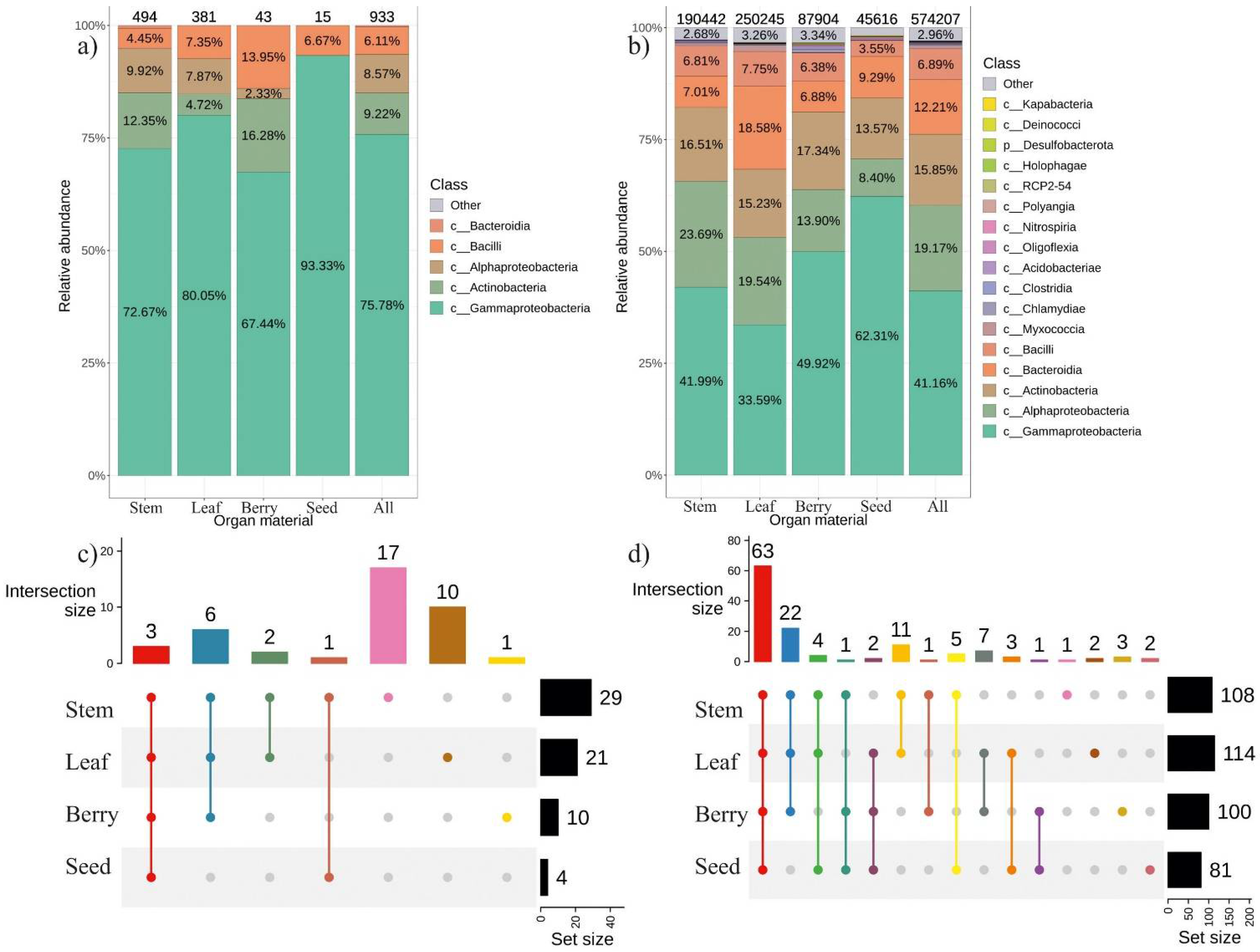
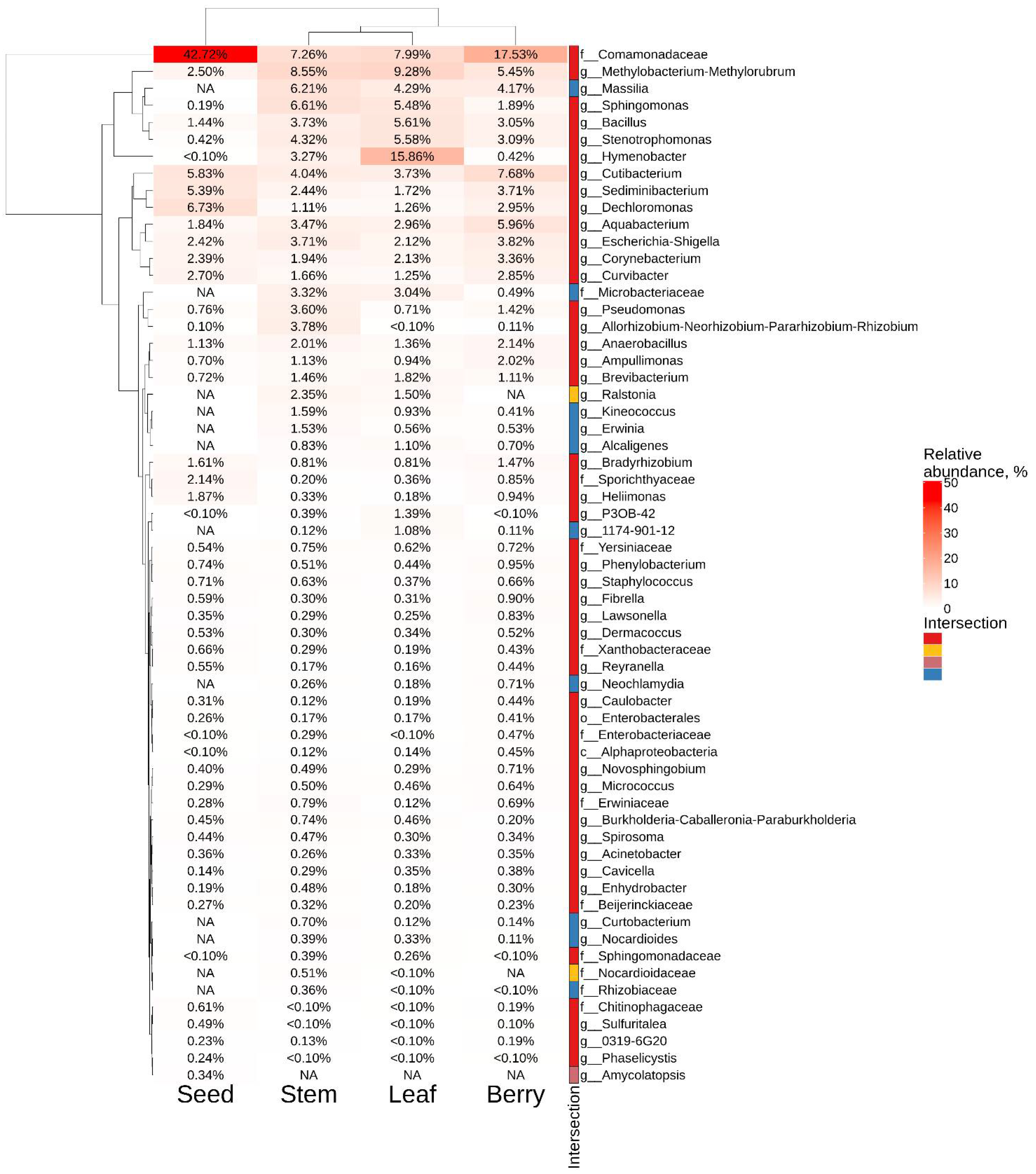
2.1. The General Composition of Endophytic Bacterial Community in Different Organ of V. amurensis

We selected and identified the main representatives of the V. amurensis bacteriome. V. amurensis tissues were collected from two different plants in July and September for four years from 2018 to 2021. Then, a cultivation-dependent approach (bacterial seeding) was employed to analyze the endophytic bacteriome of V. amurensis using surface-sterilized plant tissues. We analyzed a total of 933 bacterial strains obtained as a result of the microbiological seeding procedure performed over the 4-year period. These strains were divided into four classes of bacteria. Gammaproteobacteria was the dominant class—76%. In addition, we detected Actinobacteria—9.2%, Alphaproteobacteria—8.6%, Bacilli—6.1%, and Bacteroidia less than 1% (Figure 1a).
Moreover, we used surface-sterilized tissues of the same two wild V. amurensis plants collected in July and September 2021 for NGS analysis of the endophytic bacterial community. We obtained 3,108,452 paired reads of 16s rRNA gene. A total of 574,207 amplicon sequence variants (ASVs) were obtained after pre-processing for further analysis (Figure 1b). According to the analysis, 17 taxa were presented in different grapevine organs (stem, leaf, berry, seed) with the relative representation above 0.1%. Among the 17 taxa, ASVs belonging to 5 classes were the most represented: Gammaproteobacteria—40.3%, Alphaproteobacteria—18.7%, Actinobacteria—15.4%, Bacteroidia—12.2% and Bacilli—6.6% (Figure 1b). Thus, the metagenomic analysis generally confirmed the data of the cultivation-dependent approach. Notably, a total of 22 common genera of endophytic bacteria were detected by both cultivation-dependent and cultivation-independent approaches, while 18 unique genera were detected by the cultivation-dependent method and 59 unique genera–by NGS (Supporting Information S1).
According to the cultivation-dependent method, the predominant bacterial genera were Pantoae—29.5% and Erwinia—25%, while these bacterial genera were either not present or detected in trace amounts (less than 1%) in the metagenomics study (Figure 2). The top five taxa in NGS-analysis included Comamonadaceae (12%), Methylobacterium (8%), Hymenobacter (8%), Sphingomonas (5%) and Cutibacretium (5%). The Comamonadaceae, Hymenobacter, and Cutibacterium taxa accounted for 12%, 8%, and 4.6%, respectively, for all analyzed ASVs according to the cultivation-independent method, while they were not detected after bacterial seeding (Figure 2).
The analysis of endophytic bacteriome of the different V. amurensis organs revealed that the highest number of strains and obtained ASVs were detected in the leaves and stems, while the seeds showed much lower diversity of endophytic bacteria (Figure 1c,d). The Gammaproteobacteria class was dominant in the seeds, while the Alphaproteobacteria and Bacilli classes were less represented. The leaves of V. amurensis were predominantly colonized by the Bacteroidia and Myxococcia compared to other plant organs. ASVs were represented by 128 taxa in the samples of plant organs. Among them, 63 taxa were found in all analyzed grapevine organs (Figure 1d and Supporting Information S2). The beta diversity data showed diffuse clustering and no significant difference between organ samples performed by PERMANOVA test (R2: 13.6%, p = 0.521) (Supporting Information S3).
The most common taxa for the leaves of V. amurensis were Hymenobacter (16%), Methylobacterium-Methylorubrum (9%), and Comamonadaceae (8%), while the most common taxa for the stem were represented by Sphingomonas (6.6%), Methylobacterium-Methylorubrum (8.5%), and Comamonadaceae (7.2%) (Figure 3). The largest number of ASVs in grape berries and seeds belonged to the taxa Comamonadaceae (17.5% and 43%, respectively). Moreover, genera Cutibacterium (5.8%) and Dechloromonas (6.7%) were most often found in grape seeds (Figure 3).

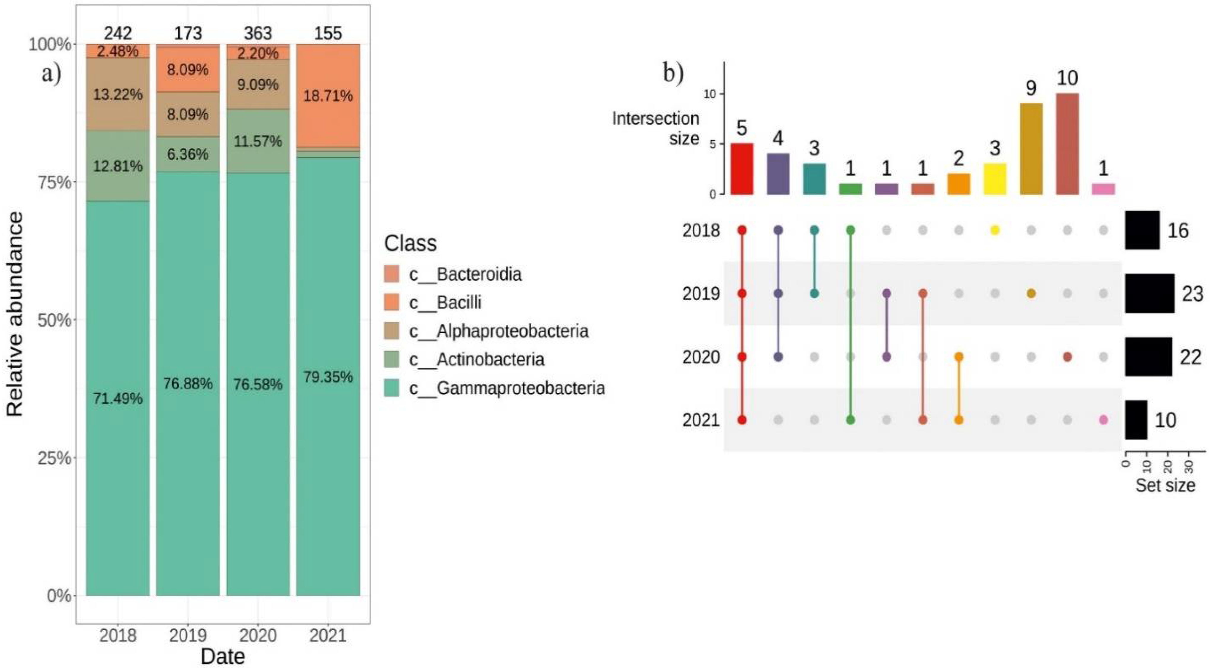
2.2. Differences in the Composition of Endophytic Bacterial Community in V. amurensis Depending on the Year of Tissue Collection

We used the cultivation-dependent approach to isolate individual endophytic bacteria from the tissues of two V. amurensis plants collected every year from 2018 to 2021. In 2018, we isolated and identified 242 strains of endophytic bacteria, in 2019–173 strains, in 2020–363 strains and in 2021–155 strains (Figure 4a). Most strains of endophytic bacteria (71.5–79.4%) collected each year belonged to Gammaproteobacteria. The distribution among other classes of endophytic bacteria varied depending on the year of sampling. In 2018 and 2020, the percentage ratio of the endophytic bacteria classes were similar. In 2020, Bacteroidia class were detected (0.55%), while in 2018 this class was not present. The incidence of the Actinobacteria class in 2019 decreased by half and reached 6% compared to 2018 and 2020, respectively, and dropped to less than 1.3% in 2021. The incidence of Bacilli was 2% in 2018 and 2020, while it increased to 8% and 19% in 2019 and 2021, respectively (Figure 4a). The generic biodiversity was the richest in 2019 and 2020 (23 and 22 genera), and there were 9 and 10 unique genera for 2019 and 2020, respectively (Figure 4b). Common genera that were detected every year of the bacterial seeding were Bacillus, Erwinia, Pantoea, Pseudomonas and Sphingomonas (Supporting Information S4).

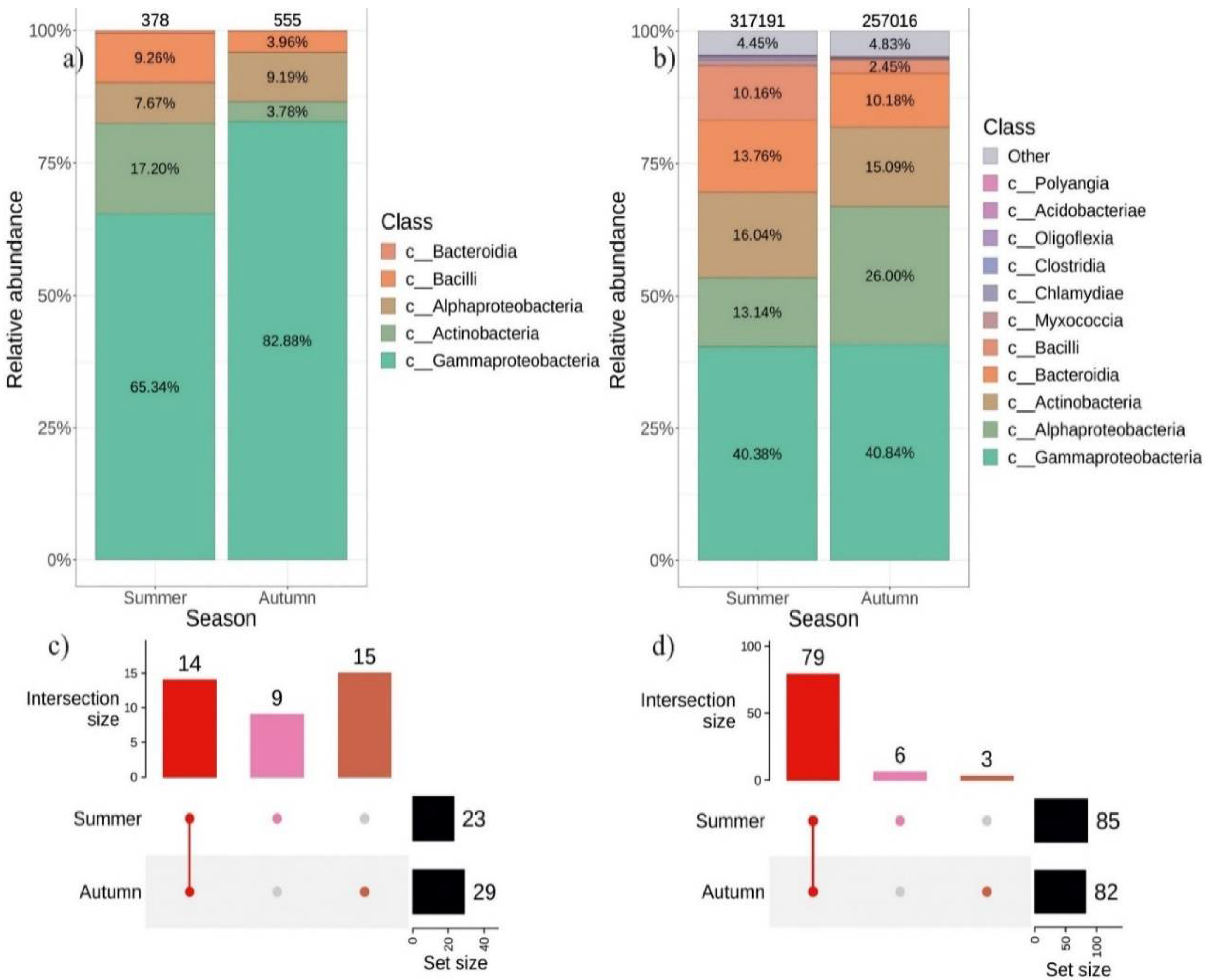
2.3. Seasonal Variations in the Composition of Endophytic Bacteriome in V. amurensis

We also analyzed the biodiversity profile of endophytic bacteria in V. amurensis depending on the collection season. The leaves and stems of V. amurensis were collected in the first half of July and in the second half of September. The cultivation-dependent approach resulted in a greater number of strains sown in autumn (555) than in summer–(378) (Figure 5a). In total, 14 genera of bacteria were common for summer and autumn, while 9 genera were unique for summer and 15 for autumn (Figure 5c and Supporting Information S5).
A total of 257,016 ASVs were obtained in the autumn of 2021 and 317,191 ASVs in the summer of 2021 using the cultivation-independent approach. The percentage of Alphaproteobacteria increased two-fold in the autumn, while the percentage of Bacilli decreased four-fold (Figure 5b). A total of 6 genera were identified as unique for the summer, and 3 genera were unique for the autumn (Figure 5d and Supporting Information S6). PERMANOVA test showed no significant difference between summer and autumn samples (R2: 8.8%, p = 0.068) (Supporting Information S3).

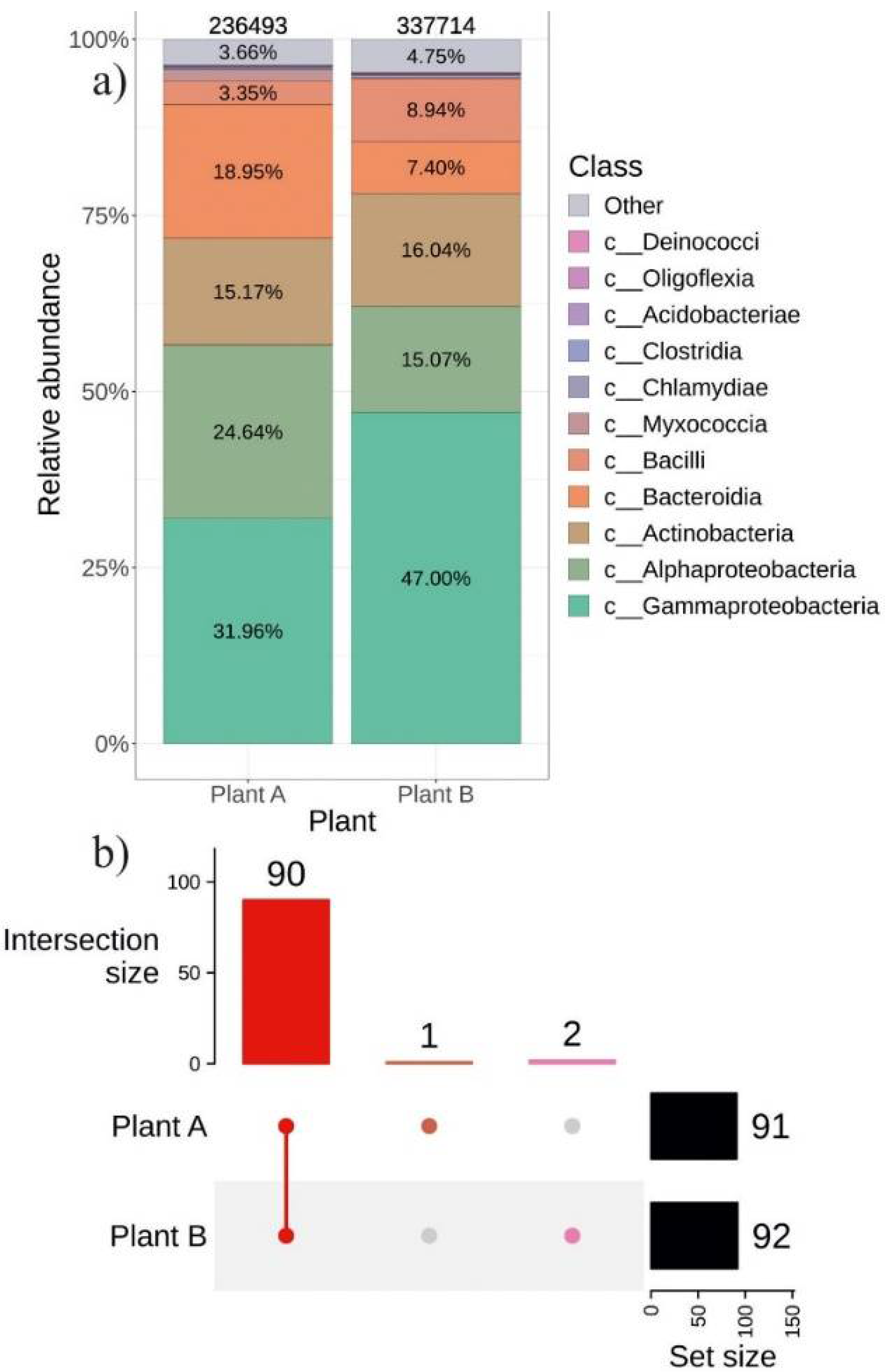
2.4. The Composition of Endophytic Bacterial Community in Different Representatives of V. amurensis Grapevine

In addition, we compared the percentage of the community of endophytic bacteria in individual plants of V. amurensis. The two representative V. amurensis plants differed only in their place of growth, while their approximate ages were the same. We detected no significant differences in beta diversity between samples collected from plant A and plant B (R2: 8.9%, p = 0.064) (Supporting Information S3). The incidence of Bacteroidia was 2-fold higher in plant A than in plant B (Figure 6a). There were only two unique taxa in plant B belonging to Gammaproteobacteria and Intrasporangiaceae and one unique genus Frondihabitans in plant A (Figure 7d and Supporting Information S7).

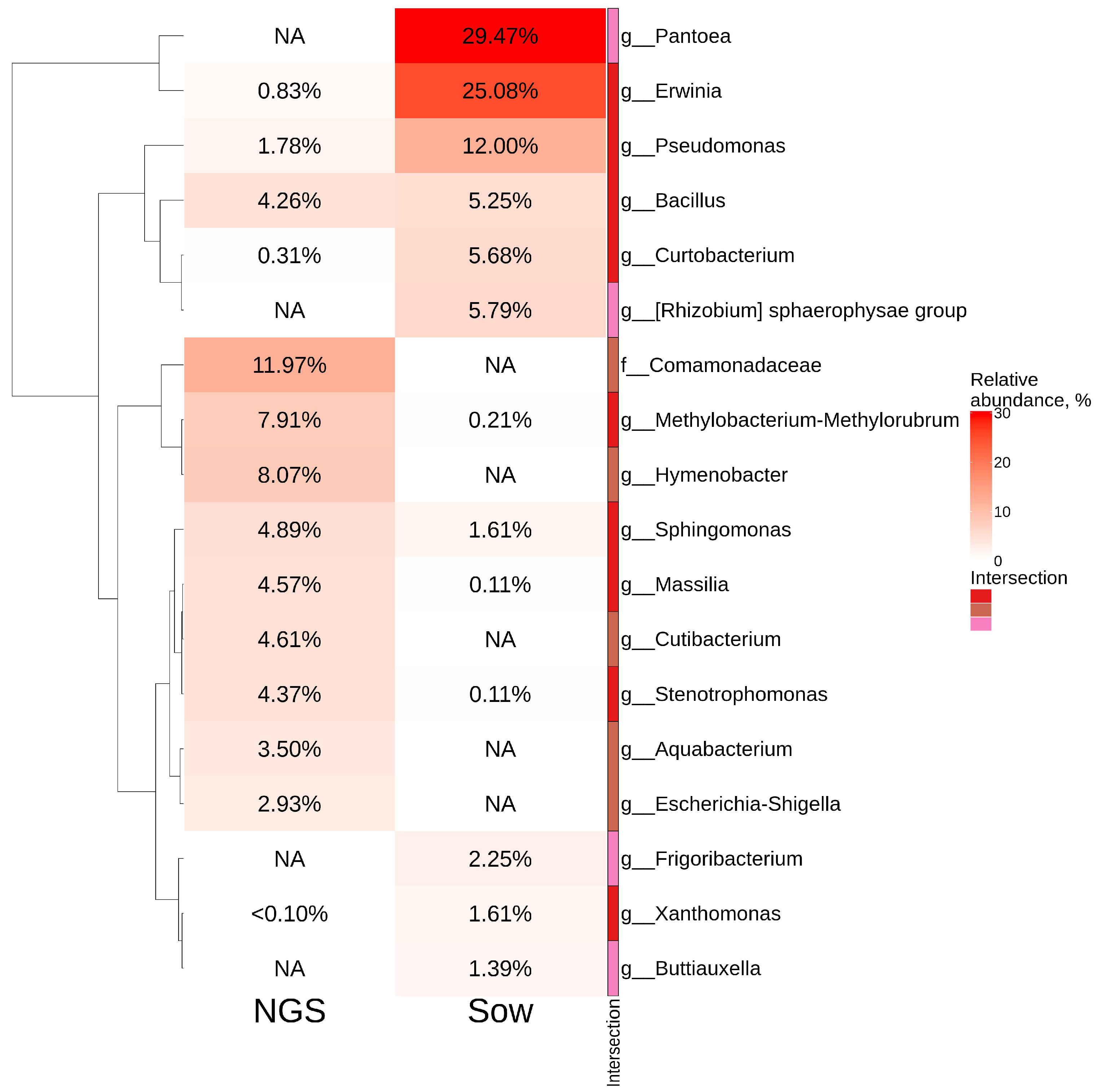
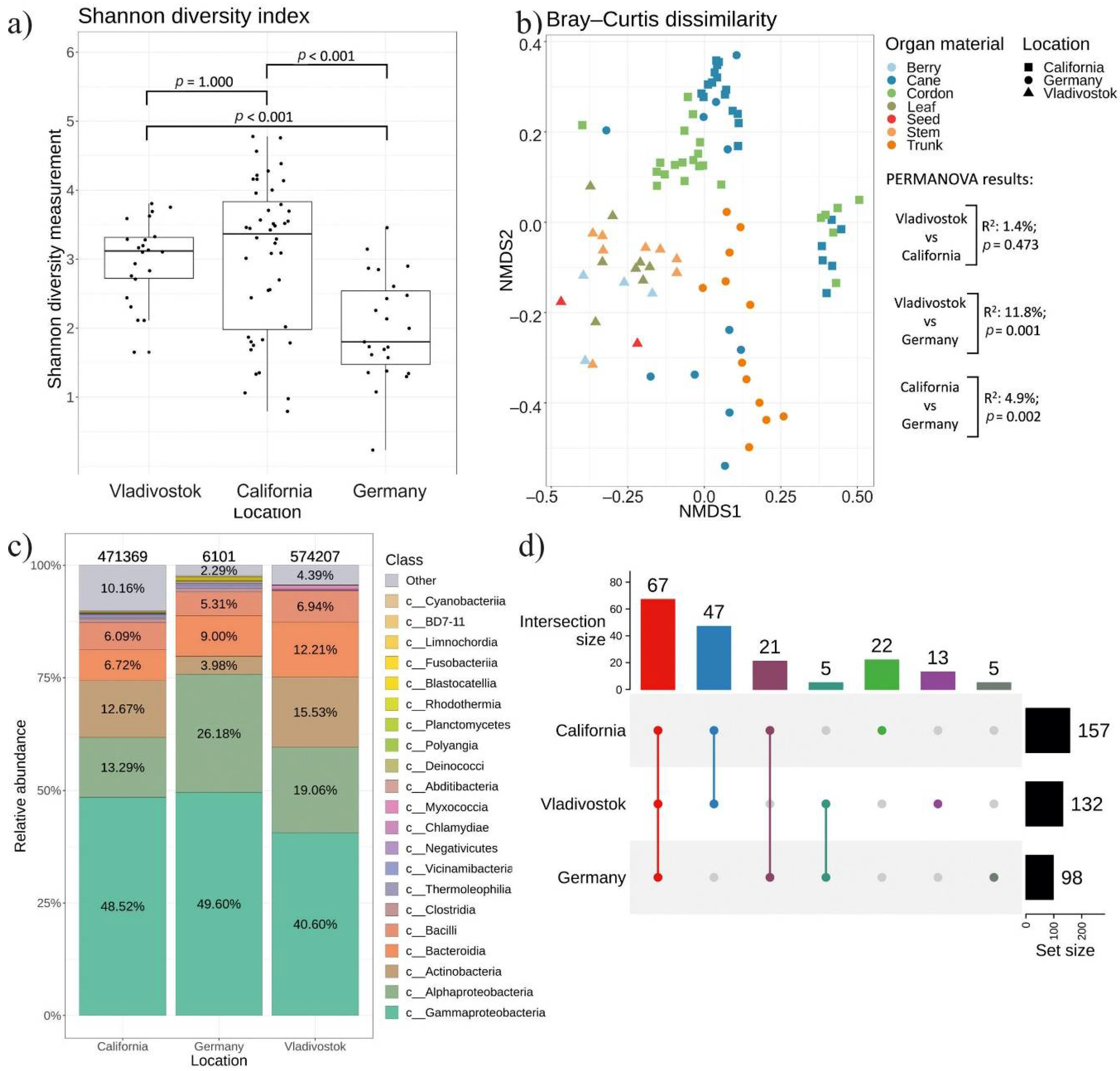
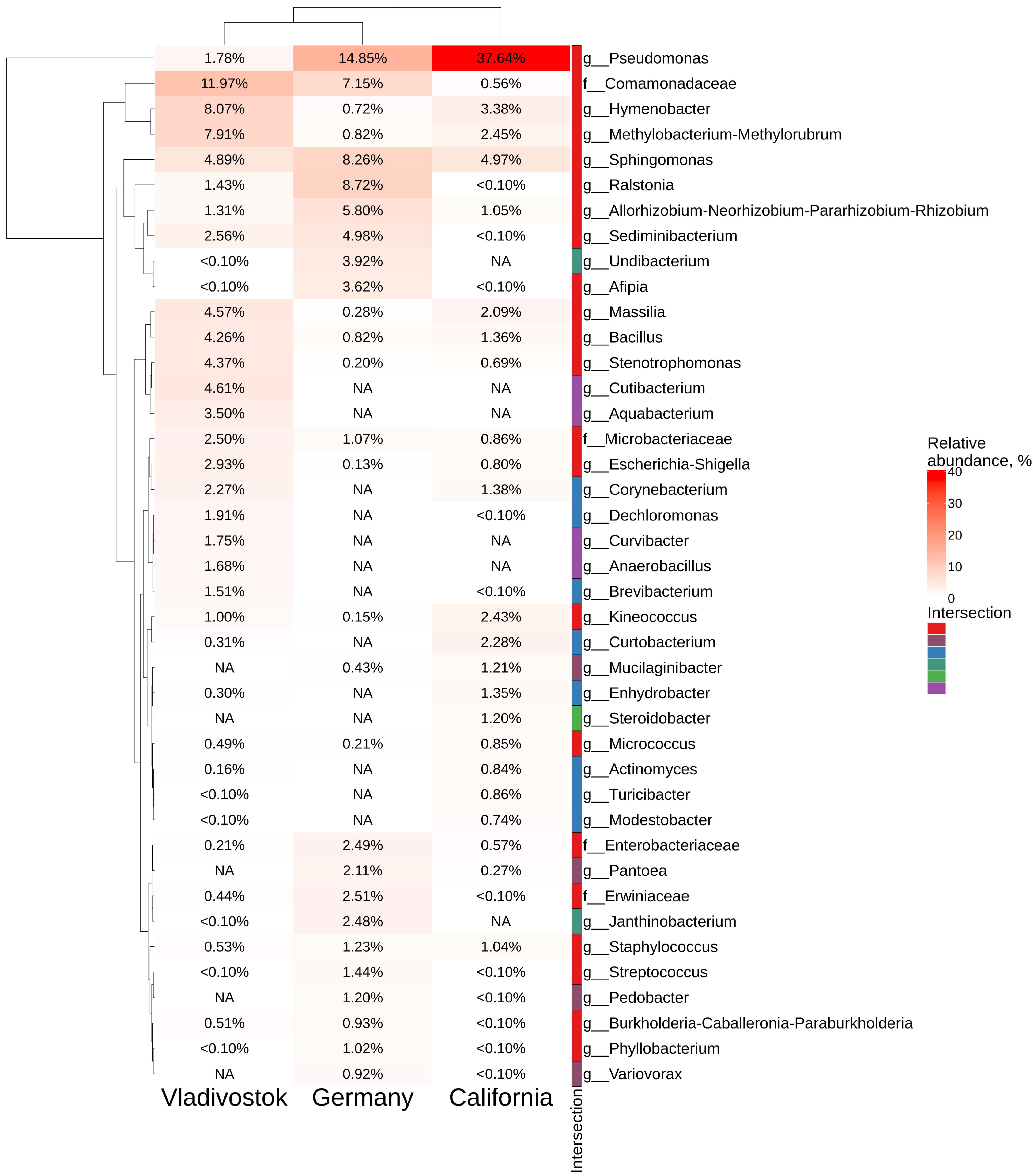
2.5. Comparative Analysis of Endophytic Bacteria Biodiversity in V. amurensis and V. vinifera

We conducted a comparative analysis of the bacteriomes of V. amurensis obtained in this study with the previously studied bacteriomes of V. vinifera grapevines growing in Germany [26] and California, USA [12] (Supporting Information S8). We analyzed the amplicon data of each sample site with respect to the location. The results for alpha and beta diversity analysis are shown in Figure 7a,b, respectively. The grape samples collected in Russia (Vladivostok) and USA (California) were similar in alpha diversity. The Germany samples exhibit reduced alpha diversity compared to Russia (Vladivostok) (p < 0.001) and to USA (California) (p < 0.001). (Figure 7a and Supporting Information S9). The beta diversity results are presented in the nonmetric multidimensional scaling (NMDS) ordination (Figure 7b). NMDS ordination showed that samples from Russia (Vladivostok), USA (California) and Germany were located in separate clusters (Figure 7b). The PERMANOVA test demonstrated that the samples collected in Vladivostok and California were more similar (R2: 1,4%, p = 0.473) based on beta diversity than samples from California and Germany (R2: 4,9%, p = 0.002), while the samples from Russia and Germany were most diverse (R2: 11,8%, p = 0.001) (Supporting Information S10). These results were also supplemented by UpSet intersection diagrams. Samples collected in Vladivostok and California displayed 47 intersection genera, California and Germany had 21, and Vladivostok and Germany had only 5 (Figure 7d and Supporting Information S11).
According to the analysis, the grape bacteriome is represented by 23 main classes of bacteria (Figure 7c). The classes Gammaproteobacteria, Alphaproteobacteria, Actinobacteria, Bacteroidia and Bacilli dominated over other classes in all bacteriomes included in the analysis. Gammaproteobacteria was the dominant class in all grapevine samples (40–50%), with Pseudomonas as the most represented genus in California and Germany samples (Figure 8). The most abundant taxa in the samples collected in Russia (Vladivostok) was Comamonadaceae (Betaproteobacteria). There were 67 genera common to all regions of the material collection. The analysis also revealed 22 unique genera for USA (California), 13—for Russia (Vladivostok) and 5—for Germany (Figure 7d and Supporting Information S11). For the wild grape V. amurensis, the unique genera were Alcaligenes, Ampullimonas, Anaerobacillus, Aquabacterium, Curvibacter, Cutibacterium, Dermacoccus, Erwinia, Fibrella, Frondihabitans, Heliimonas, Neochlamydia and Nitrospirillum (Figure 8 and Supporting Information S11).

3. Discussion

The microbiota of grapes are highly variable, mostly due to the influence of external factors, such as environmental cues, geographical location, or individual characteristics of grape varieties [8]. This study focused on the bacterial endophytes from wild V. amurensis growing in natural conditions. A comparative analysis of findings from the cultivation-dependent (bacteriological seeding in 2018–2021) and cultivation-independent (NGS) approaches revealed that Gammaproteobacteria, Alphaproteobacteria, Actinobacteria, Bacteroidia and Bacilli were the dominant classes in the endophytic bacteriome of the wild grape. However, the ratio between the genera of endophytic bacteria considerably varied according to the two methods. This can be explained by the fact that we sowed bacterial strains over the 4-year period, while NGS was performed only for probes collected in the summer and autumn of 2021. Weather conditions varied considerably each year. In addition, the lower sensitivity of the culture-dependent approach contributed to the difference between the results. In our opinion, these two conditions led to the observed differences in the percentages of endophytic bacterial genera. Therefore, it is necessary to apply both a cultivation-dependent and a cultivation-independent approach over a time period of several years in order to obtain a more comprehensive picture of endophytic bacteria assemblage in the same plant.
The cultivation-dependent method demonstrated that 10 bacterial genera prevailed in the V. amurensis endophytic community (Pantoea, Erwinia, Pseudomonas, Bacillus, Curtobacterium, Rhizobium (sphaerophysae group), Frigoribacterium, Sphingomonas, Xantomonas, and Buttiauxella) (Figure 2). The data obtained on the main genera of endophytic bacteria present in V. amurensis were similar to the previously published data on bacterial community composition in V. vinifera from Australia and Italy [6,7,8,9,27].
While studying the endophytic bacteria distribution over several years (2018, 2019, 2020 and 2021), we discovered an interesting relationship between the weather conditions and bacterial biodiversity: a minimal number of endophytic bacteria (only 155 strains) were detected under hot and dry weather conditions in the Primorsky Territory of Russia (2021 year) (Figure 4a and Table 1). On the contrary, we isolated the maximum number of endophytic bacteria (363 strains) under cold and damp conditions in 2020 using the same methods (Figure 4a and Table 1). Thus, an average temperature of 15 °C and a large amount of precipitation contribute to both quantitative and qualitative biodiversity of endophytic bacteria of V. amurensis.
Similar to the results obtained by Baldan et al. [11], the bacterial composition changed depending on the season of sampling. We showed that the composition of bacterial endophytes was richer in autumn than in summer. This was probably due to the gradual colonization of endophytic bacteria during growth and development of the aboveground grapevine organs, e.g., leaves. In addition, there were some unique genera of bacteria in wild grape V. amurensis either in the summer or in the autumn periods. This indicates a special influence of various environmental physical parameters (air temperature, precipitation, illumination, etc.), on the percentage ratio between different classes of bacteria present in the intercellular space of grapevine tissues.
The analysis of the bacteriological community in different organs of V. amurensis showed that most endophytic bacteria inhabited stems and leaves. This conclusion confirmed previously known information that the endophytes of grapes travel from the root system through conducting vessels into the stem and then move into the leaves [12]. Moreover, we noted that number of endophytic bacterial strains and the amount of ASVs were significantly lower in summer leaves than in autumn ones. The data indicate that the bacteria had not arrived in time to settle in the leaves at the beginning of the summer season compared to the autumn. A similar situation was also observed in the case of berries and seeds: the number of ASVs detected and the number of isolated strains of endophytic bacteria were 3–12 and 2.2–8.9 times lower in the berries than in the stems and leaves, respectively (Figure 1). This effect was most likely due to the fact that the berries developed later than the leaves and contained more phenolic compounds, which prevented active endophyte accumulation.
The lowest biodiversity of endophytic bacteria was found in the grape seeds, which can be explained by the better protection of seeds by the berry pulp. Interestingly, the metagenomic analysis revealed two genera of endophytic bacteria unique to the grape seeds, i.e., the genera Amycolatopsis and Pseudoalteromonas (Figure 1 and Supporting Information S2). It is known that bacteria of the genus Amycolatopsis are able to produce antibiotics [28], while bacteria of the genus Pseudoalteromonas are usually found in marine eukaryotes and are capable of producing bioactive compounds [29]. Therefore, it is possible that symbiosis of the seed-associated bacteria with the plant could contribute to seed protection.
Next, we compared the data on endophytic microbial assemblage in V. amurensis with the previously studied endophytic bacteria communities in grapevines from other regions of the world, including Germany [26] and USA (California) [12]. Nonmetric multidimensional scaling showed that samples from Russia (Vladivostok), USA (California), and Germany were located in separate clusters. The bacteriome of grapes was represented by 23 main classes of bacteria. The prevalent classes were Gammaproteobacteria, Alphaproteobacteria, Actinobacteria, Bacteroidia and Bacilli for all grapes. Gammaproteobacteria was the dominant class in all samples (40–50%), with Pseudomonas as the most represented genus in USA and Germany, and Comamonadaceae (Betaproteobacteria)–in Russia. There were 46 genera common to all regions of material collection. We also detected a number of unique genera for USA (14 genera), Russia (11 genera), and Germany (4 genera). The differences between the endophytic bacteriome compositions in the grapevines show that the place of growth also affects the percentage ratio between classes of endophytic bacteria. However, this issue needs further investigation with additional RNA-seq libraries from other cultivars. In order to present a more complete picture of grapevine microbiota, it is necessary to expand the studies on biodiversity of bacterial endophytes to a higher number of cultivars and individuals from the wild and cultivated Vitis species growing globally.
The data obtained in this investigation are interesting for practical application in agriculture. Knowledge about the biological diversity of endophytic bacteria in wild grapes, including V. amurensis, could prove useful for the development of new approaches to increase the stress resistance of cultivated grapes, yield, and fruit quality. For example, it has been shown that some endophytic bacterial isolates from domesticated and wild V. vinifera grapevines were highly active against Botrytis cinerea, Neofusicoccum parvum, Botryosphaeria dothidea, Botryosphaeria obtuse, Pochonia chlamydospora, Plasmopara viticola, and Rhizobium vitis in vitro [9,14,30]. Bacillus and Pantoea exhibited the most prominent antimicrobial activities. Moreover, bacterial endophyte Bacillus licheniformis isolated from V. vinifera has been shown to stimulate the production of secondary metabolites, such as monoterpenes, exhibiting antioxidant activity, and sesquiterpenes, exhibiting antibacterial effects [31]. Moreover, B. licheniformis produced carotenoids, which can act as antioxidants useful for plant stress protection [32]. It has been shown that bacterial endophytes B. licheniformis and Pseudomonas fluorescens can modulate ABA metabolism in inoculated grape plants, which gives them an advantage over uninfected plants in drought conditions [31,33].
Grape endophytic bacteria have previously been shown to favorably affect the quality of grape fruit. For example, grapevine inoculation with endophytic Acinetobacter lwoffii, Bacillus subtilis, and Pseudomonas fluorescens was effective against Botrytis cinerea and led to the accumulation of host-synthesized phytoalexins, especially trans-resveratrol (3,5,4′-tryhydroxystilbene) and its oligomer, trans-ε-viniferin, which could contribute to the grape fruit metabolite composition [34]. Interestingly, native endophytes and products based on endophytes from the wild grape V. amurensis can stimulate stilbene production in grape cell suspensions, which could further contribute to the development of a new stimulators of stilbene biosynthesis in grapevine or grape cell cultures [35,36].
Taken together, these obtained data can be employed to create endophyte-based preparations for plant pathogen protection. Thus, future studies on the biochemical properties (e.g., the ability to secrete phytohormones or biologically active substances) or biological functions (e.g., plant disease protection) of isolated endophytic bacteria can greatly contribute to crop protection and plant functional studies.

4. Materials and Methods

4.1. Plant Material

For both cultivation-dependent and cultivation-independent approaches, we used tissues of two healthy 10–15-year-old vines of V. amurensis located at a distance of 1 km from each other in a nonprotected natural population near Vladivostok, Russia (the southern Primorsky Territory of the Russian Far East, longitude 43.2242327 and latitude 131.99112300). Shoots, leaves (young stems 7–8 cm long with three healthy leaves), berries (green and mature), and seeds were collected at 10–11 a.m. on low-cloud days without precipitation, the air temperature was 17–20 °C. The values of the average temperatures and precipitation in 2018–2021 in Vladivostok (Primorsky Territory of Russia) are shown in the Table 1. Each plant material specimen was delivered to the laboratory within 30 min.
For the cultivation-dependent approach (bacterial sowing), plant materials were selected in July and September from 2018 to 2021. Two biological replicates (two individual vines) were collected in July, and two biological replicates–in September. Thus, there were 4 biological replicates per year. In total, 16 biological replicates were collected and analyzed by the cultivation-dependent approach from 2018 to 2021. For the cultivation-independent approach (NGS) we used grapevine material collected in July and September of 2021 (a total of 4 biological replicates) and applied 2 technical replications per biological replicate.

4.2. Isolation and Identification of the Endophytic Bacteria

The grapevine tissues (1.5 g) were washed under running water with soap and washed sequentially under sterile conditions in 75% ethanol for 2 min, 10% hydrogen peroxide for 1 min, and five times in sterile water. To check the efficacy of this method of surface sterilization, 100 µL of the last wash water was incubated on the R2A medium [37]. No microorganism growth was observed 3 days after the last portion of washing water had been plated in the Petri plates containing the growth media. This validated the quality of the performed superficial sterilization of the grape tissues.
The surface-sterilized tissue of V. amurensis was ground to a homogeneous mass in a sterile mortar; the resulting juice was squeezed, and a 100 μL aliquot was transferred to R2A media in Petri plates. After 3 days, the grown bacterial colonies were sampled and carefully transferred to a new sterile Petri plate for repeated cultivation. We isolated almost all the seeded colonies into separate strains, a total of 933 separate strains of endophytic bacteria were obtained over 4 years of biological sowing.
DNA of the 933 bacteria strains was isolated by the hexadecyltrimethylammonium bromide (CTAB) method with modifications [38]. Bacterial 16S rRNA gene sequences were amplified using universal bacterial primers for the amplification of approximately 1500 bp 16S PCR products (8F, 5′AGA GTT TGA TCM TGG CTC AG and 1522R, 5′AAG GAG GTG ATC CAR CCG CA) [39]. PCR products were sequenced using an ABI 3130 Genetic Analyzer (Applied Biosystems, Foster City, CA, USA) according to the manufacturer’s instructions as described [35,36]. The Basic Local Alignment Search Tool (BLAST) program was used for sequence analysis. Multiple sequence alignments were performed using the Clustal X program [40]. A sequence identity of ≥99% is considered as a sufficient threshold value for taxonomic identification bacteria genus.

4.3. DNA Extraction, PCR Condition, Library Preparation, and Sequencing

DNA for NGS was isolated using two approaches. The first approach used the method employed in our laboratory [38] with small modifications. Namely, 50 mg of V. amurensis tissue was taken 800 µL CTAB-buffer with 2× content of NaCl (100 mM Tris pH 7.5, 1.4 M NaCl, 40 mM EDTA pH 7.5, 1% CTAB) was added, incubated for 1 h 60 °C in a Gnom thermostat (DNA-technology, Moscow, Russia). Then 300 µL of chloroform was added, gently mixed and centrifuged for 10 min at 4 °C and 13,200 rpm (Ependorf, Germany). Further, a supernatant (420 µL) was selected in separate test tubes with 950 µL of 96% ethanol and frozen at −20 °C overnight. Then, the DNA in ethanol was centrifuged for 10 min at 4 °C and 13,200 rpm, and then the supernatant was removed. The precipitate was dried until the ethanol completely evaporated at room temperature (30 min). The precipitate was dissolved in 100 µL of distilled water and then purified on Plasmid DNA purification columns Plasmid MiniPrep kit per manufacturer’s protocol (Evrogen, Moscow, Russia). As a result, the DNAs were eluted in 50 µL of elution buffer.
According to the second approach, the DNA was extracted from 30 mg of V. amurensis tissue using the ZymoBIOMICS DNA miniprep kit per manufacturer’s protocol (Zymo Research, Irvine, CA, USA). DNA was assessed for quality and quantity using the NanoPhotometer P300 (IMPLEN, Munich, Germany).
The DNA samples were sent to Evrogene (Moscow, Russia) for high-throughput sequencing using Illumina technology. The libraries were prepared for sequencing according to the protocol described in the manual “16S Meta-genomic Sequencing Library Preparation” (Part #15,044,223 Rev. B; Illumina, San Diego, CA, USA). Bacterial 16S rRNA regions were amplified from all samples using primers 515F (5′GGTAATACGKAGGKKGCDAGC) and 806R (5′RTGGACTACCAGGGTATCTAA) modified for Vitis sp. plants. After obtaining the amplicons, the libraries were purified and mixed equimolarly using the SequalPrep™ Normalization Plate Kit (Cat #A10510-01, ThermoFisher, Waltham, MA, USA). Quality control of the obtained library pools was performed using Fragment Analyzer and quantitative analysis was performed using qPCR. The library pool was sequenced on Illumina MiSeq (2 × 250 paired end) using MiSeq Reagent Kit v2 (500 cycles). The FASTQ files were obtained using bcl2fastq v 2.17.1.14 Conversion Software (Illumina, San Diego, CA, USA). The phage PhiX library was used to control sequencing parameters. Most of the reads pertaining to phage DNA were removed during demultiplexing.
Bacterial sequences were deposited in NCBI under the accession number PRJNA813962 and in database of laboratory Biotechnology, Federal Scientific Center of the East Asia Terrestrial Biodiversity, Far Eastern Branch of the Russian Academy of Sciences, Russia (https://biosoil.ru/downloads/biotech/Vitis%20metagenom/2021-09=Vitis_amurensis_endophytes_16s), (accessed on 14 March 2022).

4.4. Computational Analysis

For the comparative analysis of endophytic bacteriomes from V. amurensis and V. vinifera, we selected two studies from California (cv. ‘Syrah’) and Germany (cv. ‘Cabernet Dorsa’) where the biodiversity of endophytic bacteria was evaluated [12,26]. The main criteria for the selection of metagenomic studies were the presence of a bacterial film elimination step in the “Materials and Methods” section of research, different geographical location and the same 16s rRNA sequencing region (515F-806R). From the selected studies, we selected samples belonging to the above-ground parts of the plant and not subjected to any treatments or infections. The samples used in the comparative analysis are presented in Supporting Information S8. Raw readings of all samples were subjected to further bioinformatic analysis.
NGS reads were preprocessed using QIIME 2 [41] and DADA2 programs [42]. The primers, remaining PhiX reads, and chimeric sequences were removed, and paired-end reads were merged and sorted. The authors of the DADA2 algorithm refer to the sequences as amplicon sequence variants (ASVs). This algorithm has the capacity to resolve sequences differences to a single nucleotide, allowing for more robust identification. Taxonomic identification of ASVs was performed using the QIIME 2 Scikit-learn algorithm [43] using the Silva 138 pre-trained classifier (99% OTUs from 515F/806R region of sequences) [44].
The obtained data were processed using the R language. The phyloseq library [45] and tidyverse package [46] were used in pre-filtering and data preparation. Taxa for bar plot, heatmap and UpSet diagram visualizations were filtered based on relative abundance of >0.1% for each biocompartment. We merged the taxonomic ranks in bar plots that were relative abundant <0.1% in each factor to one group called “other”. Shannon alpha diversity and Bray–Curtis beta diversity data were obtained using the Vegan package [47]. Bray–Curtis dissimilarity data were transformed to even sampling depth and converted to nonmetric multidimensional scaling (NMDS). A pairwise Wilcoxon rank sum test with Bonferroni correction was performed to analyze the alpha diversity data between groups. Statistical validation of beta diversity data was performed using the PERMANOVA test with 999 permutations [47]. The ggplot2 [46] and ComplexHeatmap [48] R libraries were used in the graphical representation of the results.

5. Conclusions

In this study, we firstly described the biodiversity of endophytic bacteria in wild-growing grapevine V. amurensis. According to both the cultivation-dependent and cultivation-independent approaches, Gammaproteobacteria, Actinobacteria, Alphaproteobacteria, Bacilli and Bacteroidia represented the dominant classes of bacteria. The data indicated that the weather conditions significantly affected the number and biodiversity of endophytic bacteria in V. amurensis. Namely, lower temperatures (15–16 °C) and increased precipitation (140–280 mm in month) favored both the quantitative and qualitative diversity of endophytic bacteria inhabiting the intercellular space of V. amurensis tissues. The obtained data also show that the composition of bacterial endophytes was richer in autumn than in summer. Among the aboveground organs of grapes, the stems and leaves were the leaders in quantitative composition and biodiversity of endophytic community. In addition, this study showed that the community of endophytic bacteria of V. amurensis was closer to the endophytic community of V. vinifera growing in Germany. Thus, this paper presented and discussed various factors that affect the biodiversity of endophytic bacteria in wild grapevine V. amurensis. Considering that a high number of factors affects the biodiversity of endophytic bacteria in V. amurensis, this study should be expanded to include more individuals and geographic areas where this grapevine species is present. The knowledge about the biological diversity of endophytic bacteria in wild grapes V. amurensis will development of new approaches to increase grapevine stress resistance as well as the yield and quality of fruits.

Supplementary Materials

The following are available online at https://www.mdpi.com/article/10.3390/plants11091128/s1, Supporting Information S1. Intersections in our data (Data type); Supporting Information S2. Intersections in our metagenome data (Organ material)—Figure 1d; Supporting Information S3. NMDS ordination plots and PERMANOVA results in our data; Supporting Information S4. Intersections in data of cultivate-dependent method (Date)—Figure 4b; Supporting Information S5. Intersections in data of cultivate-dependent method (Season)—Figure 5c; Supporting Information S6. Intersections in our metagenome data (Season)—Figure 5d; Supporting Information S7. Intersections in our metagenome data (Plants)—Figure 6b; Supporting Information S8. Samples used in comparative analysis of the grape bacteriomes; Supporting Information S9. Alpha diversity results; Supporting Information S10. PERMANOVA results in comparative analysis of the bacterial biodiversity between grapes; Supporting Information S11; Intersections between Vitis metagenome data—Figure 7d.

Author Contributions

O.A.A. and K.V.K. performed research design, data analysis, paper preparation, and the experimental process. O.A.A. and N.N.N. performed DNA isolation of microorganisms, PCR, and sequencing analysis. O.A.A. performed DNA isolation for NGS. N.N.N. performed bioinformatic analysis. A.S.D. performed paper preparation. All authors have read and agreed to the published version of the manuscript.

Funding

This work was supported by a grant from the Russian Science Foundation (grant number 20–74–00002).

Data Availability Statement

The data presented in this study are available within the article and supplementary material.

Conflicts of Interest

The authors declare no conflict of interest.

References

  1. Wen, J.; Nie, Z.-L.; Soejima, A.; Meng, Y. Phylogeny of Vitaceae based on the nuclear GAI1 gene sequences. Can. J. Bot. 2007, 85, 731–745. [Google Scholar] [CrossRef] [Green Version]
  2. Liu, X.-Q.; Ickert-Bond, S.M.; Nie, Z.-L.; Zhou, Z.; Chen, L.-Q.; Wen, J. Phylogeny of the Ampelocissus–Vitis clade in Vitaceae supports the New World origin of the grape genus. Mol. Phylogenetics Evol. 2016, 95, 217–228. [Google Scholar] [CrossRef] [PubMed] [Green Version]
  3. Stranska, M.; Lovecka, P.; Vrchotova, B.; Uttl, L.; Bechynska, K.; Behner, A.; Hajslova, J. Bacterial Endophytes from Vitis vinifera L.—Metabolomics Characterization of Plant-Endophyte Crosstalk. Chem. Biodivers. 2021, 18, e2100516. [Google Scholar] [CrossRef] [PubMed]
  4. Hardoim, P.R.; van Overbeek, L.S.; Berg, G.; Pirttilä, A.M.; Compant, S.; Campisano, A.; Döring, M.; Sessitsch, A. The Hidden World within Plants: Ecological and Evolutionary Considerations for Defining Functioning of Microbial Endophytes. Microbiol. Mol. Biol. Rev. 2015, 79, 293–320. [Google Scholar] [CrossRef] [Green Version]
  5. Kandel, S.L.; Joubert, P.M.; Doty, S.L. Bacterial Endophyte Colonization and Distribution within Plants. Microorganisms 2017, 5, 77. [Google Scholar] [CrossRef] [Green Version]
  6. West, E.R.; Cother, E.J.; Steel, C.C.; Ash, G.J. Thse characterization and diversity of bacterial endophytes of grapevine. Can. J. Microbiol. 2010, 56, 209–216. [Google Scholar] [CrossRef]
  7. Compant, S.; Mitter, B.; Colli-Mull, J.G.; Gangl, H.; Sessitsch, A. Endophytes of Grapevine Flowers, Berries, and Seeds: Identification of Cultivable Bacteria, Comparison with Other Plant Parts, and Visualization of Niches of Colonization. Microb. Ecol. 2011, 62, 188–197. [Google Scholar] [CrossRef]
  8. Salvetti, E.; Campanaro, S.; Campedelli, I.; Fracchetti, F.; Gobbi, A.; Tornielli, G.B.; Torriani, S.; Felis, G.E. Whole-Metagenome-Sequencing-Based Community Profiles of Vitis vinifera L. cv. Corvina Berries Withered in Two Post-harvest Conditions. Front. Microbiol. 2016, 7, 973. [Google Scholar] [CrossRef] [Green Version]
  9. Andreolli, M.; Lampis, S.; Zapparoli, G.; Angelini, E.; Vallini, G. Diversity of bacterial endophytes in 3 and 15 year-old grapevines of Vitis vinifera cv. Corvina and their potential for plant growth promotion and phytopathogen control. Microbiol. Res. 2016, 183, 42–52. [Google Scholar] [CrossRef]
  10. Gamalero, E.; Bona, E.; Novello, G.; Boatti, L.; Mignone, F.; Massa, N.; Cesaro, P.; Berta, G.; Lingua, G. Discovering the bacteriome of Vitis vinifera cv. Pinot Noir in a conventionally managed vineyard. Sci Rep 2020, 10, 6453. [Google Scholar] [CrossRef] [PubMed] [Green Version]
  11. Baldan, E.; Nigris, S.; Populin, F.; Zottini, M.; Squartini, A.; Baldan, B. Identification of culturable bacterial endophyte community isolated from tissues of Vitisvinifera “Glera”. Plant Biosyst. Int. J. Deal. All Asp. Plant Biol. 2014, 148, 508–516. [Google Scholar] [CrossRef]
  12. Deyett, E.; Rolshausen, P.E. Endophytic microbial assemblage in grapevine. FEMS Microbiol. Ecol. 2020, 96, fiaa053. [Google Scholar] [CrossRef] [PubMed]
  13. Deyett, E.; Rolshausen, P.E. Temporal Dynamics of the Sap Microbiome of Grapevine Under High Pierce’s Disease Pressure. Front. Plant Sci. 2019, 10, 1246. [Google Scholar] [CrossRef] [PubMed]
  14. Campisano, A.; Pancher, M.; Puopolo, G.; Puddu, A.; Lòpez-Fernàndez, S.; Biagini, B.; Yousaf, S.; Pertot, I. Diversity in Endophyte Populations Reveals Functional and Taxonomic Diversity between Wild and Domesticated Grapevines. Am. J. Enol. Vitic. 2015, 66, 12–21. [Google Scholar] [CrossRef]
  15. Chen, Q.; Diao, L.; Song, H.; Zhu, X. Vitis amurensis Rupr: A review of chemistry and pharmacology. Phytomedicine 2018, 49, 111–122. [Google Scholar] [CrossRef]
  16. Liu, L.; Li, H. Review: Research progress in amur grape, Vitis amurensis Rupr. Can. J. Plant Sci. 2013, 93, 565–575. [Google Scholar] [CrossRef] [Green Version]
  17. Wu, J.; Zhang, Y.; Zhang, H.; Huang, H.; Folta, K.M.; Lu, J. Whole genome wide expression profiles of Vitis amurensisgrape responding to downy mildew by using Solexa sequencing technology. BMC Plant Biol. 2010, 10, 234. [Google Scholar] [CrossRef] [Green Version]
  18. Dubrovina, A.S.; Kiselev, K.V. Regulation of stilbene biosynthesis in plants. Planta 2017, 246, 597–623. [Google Scholar] [CrossRef]
  19. Jenifer Selvarani, A.; Nishanthini, P.; Raji, P.; Sree Samanvitha, K.; Paulraj, P.; Iyappan, P.; Chandramohan, M.; Samrot, A.V. Antioxidant and Quorum Quenching Activity against Pseudomonas aeruginosa SU-18 of some Edible Fruit Juices. J. Pure Appl. Microbiol. 2019, 13, 1863–1876. [Google Scholar] [CrossRef] [Green Version]
  20. Islam, M.A.; Mazumdar, R.M.; Islam, S.; Alam, M.J.; Urmee, S.A. Isolation, Identification and In Vitro Antibiotic Sensitivity Pattern of Citrus Canker Causing Organism Xanthomonas axonopodis. Adv. Life Sci. 2014, 1, 215–222. [Google Scholar]
  21. Yim, N.; Ha, D.T.; Trung, T.N.; Kim, J.P.; Lee, S.; Na, M.; Jung, H.; Kim, H.S.; Kim, Y.H.; Bae, K. The antimicrobial activity of compounds from the leaf and stem of Vitis amurensis against two oral pathogens. Bioorganic Med. Chem. Lett. 2010, 20, 1165–1168. [Google Scholar] [CrossRef] [PubMed]
  22. Mousa, W.K.; Shearer, C.R.; Limay-Rios, V.; Zhou, T.; Raizada, M.N. Bacterial endophytes from wild maize suppress Fusarium graminearum in modern maize and inhibit mycotoxin accumulation. Front. Plant Sci. 2015, 6, 805. [Google Scholar] [CrossRef] [PubMed] [Green Version]
  23. Shehata, H.R.; Lyons, E.M.; Jordan, K.S.; Raizada, M.N. Bacterial endophytes from wild and ancient maize are able to suppress the fungal pathogen Sclerotinia homoeocarpa. J. Appl. Microbiol. 2016, 120, 756–769. [Google Scholar] [CrossRef] [PubMed] [Green Version]
  24. Zhang, Z.; Liu, T.; Zhang, X.; Xie, J.; Wang, Y.; Yan, R.; Jiang, Y.; Zhu, D. Cultivable Endophytic Bacteria in Seeds of Dongxiang Wild Rice and Their Role in Plant-Growth Promotion. Diversity 2021, 13, 665. [Google Scholar] [CrossRef]
  25. Liu, L.-H.; Yuan, T.; Zhang, J.-Y.; Tang, G.-X.; Lü, H.; Zhao, H.-M.; Li, H.; Li, Y.-W.; Mo, C.-H.; Tan, Z.-Y.; et al. Diversity of endophytic bacteria in wild rice (Oryza meridionalis) and potential for promoting plant growth and degrading phthalates. Sci. Total Environ. 2022, 806, 150310. [Google Scholar] [CrossRef]
  26. Faist, H.; Keller, A.; Hentschel, U.; Deeken, R. Grapevine (Vitis vinifera) Crown Galls Host Distinct Microbiota. Appl. Environ. Microbiol. 2016, 82, 5542–5552. [Google Scholar] [CrossRef] [Green Version]
  27. Campisano, A.; Antonielli, L.; Pancher, M.; Yousaf, S.; Pindo, M.; Pertot, I. Bacterial Endophytic Communities in the Grapevine Depend on Pest Management. PLoS ONE 2014, 9, e112763. [Google Scholar] [CrossRef]
  28. Greule, A.; Charkoudian, L.K.; Cryle, M.J. Chapter Six—Studying trans-acting enzymes that target carrier protein-bound amino acids during nonribosomal peptide synthesis. In Methods in Enzymology; Schmidt-Dannert, C., Quin, M.B., Eds.; Metabolons and Supramolecular Enzyme Assemblies; Academic Press: Cambridge, MA, USA, 2019; Volume 617, pp. 113–154. [Google Scholar]
  29. Holmström, C.; Kjelleberg, S. Marine Pseudoalteromonas species are associated with higher organisms and produce biologically active extracellular agents. FEMS Microbiol. Ecol. 1999, 30, 285–293. [Google Scholar] [CrossRef]
  30. Bell, C.R.; Dickie, G.A.; Harvey, W.L.G.; Chan, J.W.Y.F. Endophytic bacteria in grapevine. Can. J. Microbiol. 1995, 41, 46–53. [Google Scholar] [CrossRef]
  31. Salomon, M.V.; Bottini, R.; de Souza Filho, G.A.; Cohen, A.C.; Moreno, D.; Gil, M.; Piccoli, P. Bacteria isolated from roots and rhizosphere of Vitis vinifera retard water losses, induce abscisic acid accumulation and synthesis of defense-related terpenes in in vitro cultured grapevine. Physiol. Plant. 2014, 151, 359–374. [Google Scholar] [CrossRef]
  32. Cohen, A.C.; Dichiara, E.; Jofré, V.; Antoniolli, A.; Bottini, R.; Piccoli, P. Carotenoid profile produced by Bacillus licheniformis Rt4M10 isolated from grapevines grown in high altitude and their antioxidant activity. Int. J. Food Sci. Technol. 2018, 53, 2697–2705. [Google Scholar] [CrossRef]
  33. Torres, N.; Goicoechea, N.; Zamarreño, A.M.; Carmen Antolín, M. Mycorrhizal symbiosis affects ABA metabolism during berry ripening in Vitis vinifera L. cv. Tempranillo grown under climate change scenarios. Plant Sci. 2018, 274, 383–393. [Google Scholar] [CrossRef]
  34. Verhagen, B.; Trotel-Aziz, P.; Jeandet, P.; Baillieul, F.; Aziz, A. Improved Resistance Against Botrytis cinerea by Grapevine-Associated Bacteria that Induce a Prime Oxidative Burst and Phytoalexin Production. Phytopathology 2011, 101, 768–777. [Google Scholar] [CrossRef] [PubMed] [Green Version]
  35. Aleynova, O.A.; Nityagovsky, N.N.; Suprun, A.R.; Kiselev, K.V. Enhancement of Stilbene Biosynthesis in Grape-Cell Cultures by Natural Products Based on Endophytes of the Wild Grape Species Vitis amurensis RUPR. Appl. Biochem. Microbiol. 2022, 58, 45–56. [Google Scholar] [CrossRef]
  36. Aleynova, O.A.; Suprun, A.R.; Nityagovsky, N.N.; Dubrovina, A.S.; Kiselev, K.V. The Influence of the Grapevine Bacterial and Fungal Endophytes on Biomass Accumulation and Stilbene Production by the In Vitro Cultivated Cells of Vitis amurensis Rupr. Plants 2021, 10, 1276. [Google Scholar] [CrossRef] [PubMed]
  37. Reasoner, D.J.; Geldreich, E.E. A new medium for the enumeration and subculture of bacteria from potable water. Appl. Environ. Microbiol. 1985, 49, 1–7. [Google Scholar] [CrossRef] [Green Version]
  38. Kiselev, K.V.; Dubrovina, A.S.; Tyunin, A.P. The methylation status of plant genomic DNA influences PCR efficiency. J. Plant Physiol. 2015, 175, 59–67. [Google Scholar] [CrossRef]
  39. Lane, D. 1 16S/23S rRNA sequencing. Nucleic Acid Tech. Bact. Syst. 1991, 115–175. [Google Scholar]
  40. Altschul, S.F.; Gish, W.; Miller, W.; Myers, E.W.; Lipman, D.J. Basic local alignment search tool. J. Mol. Biol. 1990, 215, 403–410. [Google Scholar] [CrossRef]
  41. Bolyen, E.; Rideout, J.R.; Dillon, M.R.; Bokulich, N.A.; Abnet, C.C.; Al-Ghalith, G.A.; Alexander, H.; Alm, E.J.; Arumugam, M.; Asnicar, F.; et al. Reproducible, interactive, scalable and extensible microbiome data science using QIIME 2. Nat. Biotechnol. 2019, 37, 852–857. [Google Scholar] [CrossRef]
  42. Callahan, B.J.; McMurdie, P.J.; Rosen, M.J.; Han, A.W.; Johnson, A.J.A.; Holmes, S.P. DADA2: High-resolution sample inference from Illumina amplicon data. Nat. Methods 2016, 13, 581–583. [Google Scholar] [CrossRef] [PubMed] [Green Version]
  43. Pedregosa, F.; Varoquaux, G.; Gramfort, A.; Michel, V.; Thirion, B.; Grisel, O.; Blondel, M.; Prettenhofer, P.; Weiss, R.; Dubourg, V.; et al. Scikit-learn: Machine Learning in Python. J. Mach. Learn. Res. 2011, 12, 2825–2830. [Google Scholar]
  44. Bokulich, N.A.; Kaehler, B.D.; Rideout, J.R.; Dillon, M.; Bolyen, E.; Knight, R.; Huttley, G.A.; Gregory Caporaso, J. Optimizing taxonomic classification of marker-gene amplicon sequences with QIIME 2’s q2-feature-classifier plugin. Microbiome 2018, 6, 90. [Google Scholar] [CrossRef] [PubMed]
  45. McMurdie, P.J.; Holmes, S. phyloseq: An R Package for Reproducible Interactive Analysis and Graphics of Microbiome Census Data. PLoS ONE 2013, 8, e61217. [Google Scholar] [CrossRef] [Green Version]
  46. Wickham, H.; Averick, M.; Bryan, J.; Chang, W.; McGowan, L.D.; François, R.; Grolemund, G.; Hayes, A.; Henry, L.; Hester, J.; et al. Welcome to the Tidyverse. J. Open Source Softw. 2019, 4, 1686. [Google Scholar] [CrossRef]
  47. Oksanen, J.; Blanchet, F.G.; Friendly, M.; Kindt, R.; Legendre, P.; McGlinn, D.; Minchin, P.R.; O’Hara, R.B.; Simpson, G.L.; Solymos, P.; et al. Vegan: Community Ecology Package, R Package Version 2.5-7. 2020. Available online: https://cran.r-project.org/web/packages/vegan/vegan.pdf (accessed on 28 November 2020).
  48. Gu, Z.; Eils, R.; Schlesner, M. Complex heatmaps reveal patterns and correlations in multidimensional genomic data. Bioinformatics 2016, 32, 2847–2849. [Google Scholar] [CrossRef] [Green Version]
Figure 1. Comparative analysis of the endophytic community composition in different organs of wild grape Vitis amurensis obtained by the cultivation-dependent (bacteriological sowing (Sow)) and cultivation-independent approach (the next generation of sequencing (NGS)). The composition of endophytic bacteria of grape V. amurensis depends on the plant organ: (a) Class-level taxonomical bar plots for the bacterial community of bacteriological sow in berry, leaf, seed, stem and the sum of data for all organs; (b) Class-level taxonomical bar plots for the bacterial community as a result of the next generation of sequencing (NGS) in berry, leaf, seed, stem and the sum of data for all organs; (c) Genus-level UpSet diagrams depicting overlapping taxa of bacteriological sow in berry, leaf, seed, stem and the sum of data for all organs; (d) Genus-level UpSet diagrams depicting overlapping taxa of NGS in berry, leaf, seed and stem. Taxa were filtered based on relative abundance of >0.1% for each biocompartment. Filtered taxa in bar plot placed in “other” category and removed from UpSet diagram. Number of colonies (for sow) or amplicon sequence variants (ASVs) located above taxonomical bar plots.
Plants 11 01128 g001
Figure 2. Genus-level relative abundance heat maps of significant taxa obtained through the cultivation-independent method (next generation of sequencing (NGS)) and cultivation-dependent method (bacteriological sowing (Sow)). The top 10 most abundant taxa from each factor are displayed. White squares (NA) represent absence of taxa.
Plants 11 01128 g002
Figure 3. Genus-level relative endophytic bacteria abundance heat maps of significant taxa the next generation of sequencing (NGS) in different grape organs (stem, leaf, berry and seed). The top 40 most abundant taxa from each factor are displayed. White squares (NA) represent absence of taxa. The intersection selection is made based on the Figure 1d.
Plants 11 01128 g003
Figure 4. Composition of endophytic bacterial community in wild grape Vitis amurensis depending on the year of material collection. (a) Class-level taxonomical bar plots for the bacterial community composition in 2018–2021; (b) Genus-level UpSet diagrams depicting overlapping taxa of bacteriological sow and next-generation sequencing. Taxa were filtered based on relative abundance of >0.1% for each biocompartment. Number of colonies located above taxonomical bar plots.
Plants 11 01128 g004
Figure 5. Composition of endophytic bacterial community in wild grape Vitis amurensis depends on the season of material collection: (a) Class-level taxonomical bar plots for the bacterial community of cultivation-dependent approach in summer and autumn; (b) Class-level taxonomical bar plots for the bacterial community as a result of cultivation-independent approach (next generation of sequencing (NGS)) in summer and autumn; (c) Genus-level UpSet diagrams depicting overlapping taxa of bacteriological sow in summer and fall; (d) Genus-level UpSet diagrams depicting overlapping taxa of NGS in autumn and summer. Taxa were filtered based on relative abundance of >0.1% for each biocompartment. Filtered taxa in bar plots placed in “other” category and removed from UpSet diagram. Number of colonies or amplicon sequence variants (ASVs) located above taxonomical bar plots.
Plants 11 01128 g005
Figure 6. Composition of endophytic bacterial community in two different Vitis amurensis plants: (a) Class-level taxonomical bar plots for the bacterial community as a result cultivation-independent approach (the next generation of sequencing (NGS)) in Plant A and Plant B; (b) Genus-level UpSet diagrams depicting overlapping taxa of NGS in Plant A and Plant B. Taxa were filtered based on relative abundance of >0.1% for each biocompartment. Filtered taxa in bar plots placed in “other” category and removed from UpSet diagram. Number of amplicon sequence variants (ASVs) located above taxonomical bar plots.
Plants 11 01128 g006
Figure 7. A comparison of endophytic bacterial communities in cultivated Vitis vinifera from USA (California) and Germany with the endophytic community in Vitis amurensis from the Russian Far East (Vladivostok) (a) Shannon’s alpha diversity boxplot; (b) Bray–Curtis beta diversity NMDS plot; (c) Class-level taxonomical bar plots for the bacterial community composition V. vinifera from USA (California), Germany, and V. amurensis from Russia (Vladivostok); (d) Genus-level UpSet diagrams depicting overlapping taxa of bacteriological sow and next-generation sequencing. Taxa were filtered based on relative abundance of >0.1% for each biocompartment. Filtered taxa in bar plot placed in “other” category and removed from UpSet diagram. Number of amplicon sequence variants (ASVs) located above taxonomical bar plots.
Plants 11 01128 g007
Figure 8. Genus-level relative endophytic bacteria abundance heat maps of significant taxa of Vitis vinifera from USA (California), Germany, and Vitis amurensis from Russian Far East (Vladivostok). The top 20 most abundant taxa from each factor are displayed. White squares (NA) represent absence of taxa. The intersection selection is made based on the Figure 7d.
Plants 11 01128 g008
Table 1. The values of the average temperature and amount of precipitation from 2018–2021 in Vladivostok, Primorsky Territory of Russia.
SummerAverage t, °CPrecipitation, mmAutumnAverage t, °CPrecipitation, mm
The norm18.1159The norm16103
July 201818.5124September 201816.4163
July 201917.1131September 201917.344
July 202014.7281September 202016.3138
July 202121.324September 202117.7120
http://www.pogodaiklimat.ru/ (accessed on 20 January 2022)
Publisher’s Note: MDPI stays neutral with regard to jurisdictional claims in published maps and institutional affiliations.

