Article

Chemical Composition and In Vitro Anti-Helicobacter pylori Activity of Campomanesia lineatifolia Ruiz & Pavón (Myrtaceae) Essential Oil

by
Nívea Cristina Vieira Neves
1,2,3,*,
Morgana Pinheiro de Mello
1,
Sinéad Marian Smith
4,
Fabio Boylan
2,
Marcelo Vidigal Caliari
5 and
Rachel Oliveira Castilho
1,6,*
1
GnosiaH, Laboratório de Farmacognosia, Faculdade de Farmácia, Universidade Federal de Minas Gerais, Belo Horizonte 31270-901, Brazil
2
School of Pharmacy and Pharmaceutical Sciences, Trinity Biomedical Institute, Trinity College Dublin, Dublin 2, Ireland
3
Departamento de Farmácia, Centro Universitário Santa Rita, Conselheiro Lafaiete 36408-899, Brazil
4
Department of Clinical Medicine, School of Medicine, Trinity College Dublin, Trinity Centre, Tallaght University Hospital, Dublin 24, Ireland
5
Departamento de Patologia Geral, Instituto de Ciências Biológicas, Universidade Federal de Minas Gerais, Belo Horizonte 31270-901, Brazil
6
Consórcio Acadêmico Brasileiro de Saúde Integrativa, CABSIN, São Paulo 05449-070, Brazil
*
Authors to whom correspondence should be addressed.
Plants 2022, 11(15), 1945; https://doi.org/10.3390/plants11151945
Submission received: 9 June 2022 / Revised: 13 July 2022 / Accepted: 18 July 2022 / Published: 27 July 2022
(This article belongs to the Special Issue Medicinal Plants: Advances in Phytochemistry and Ethnobotany)

Abstract

:
Helicobacter pylori is the most common cause of gastritis and peptic ulcers, and the number of resistant strains to multiple conventional antimicrobial agents has been increasing in different parts of the world. Several studies have shown that some essential oils (EO) have bioactive compounds, which can be attributed to antimicrobial activity. Therefore, EOs have been proposed as a natural alternative to antibiotics, or for use in combination with conventional treatment for H. pylori infection. Campomanesia lineatifolia is an edible species found in the Brazilian forests, and their leaves are traditionally used for the treatment of gastrointestinal disorders. Anti-inflammatory, gastroprotective, and antioxidant properties are attributed to C. lineatifolia leaf extracts; however, studies related to the chemical constituents of the essential oil and anti-H. pylori activity is not described. This work aims to identify the chemical composition of the EO from C. lineatifolia leaves and evaluate the anti-H. pylori activity. The EO was obtained by hydrodistillation from C. lineatifolia leaves and characterized by gas chromatography–mass spectrometry analyses. To assess the in vitro anti-H. pylori activity of the C. lineatifolia leaf’s EO (6 μL/mL–25 μL/mL), we performed broth microdilution assays by using type cultures (ATCC 49503, NCTC 11638, both clarithromycin-sensitive) and clinical isolate strains (SSR359, clarithromycin-sensitive, and SSR366, clarithromycin-resistant). A total of eight new compounds were identified from the EO (3-hexen-1-ol (46.15%), α-cadinol (20.35%), 1,1-diethoxyethane (13.08%), 2,3-dicyano-7,7-dimethyl-5,6-benzonorbornadiene (10.78%), aromadendrene 2 (3.0%), [3-S-(3α, 3aα, 6α, 8aα)]-4,5,6,7,8,8a-hexahydro-3,7,7-trimethyl-8-methylene-3H-3a,6-methanoazulene (2.99%), α-bisabolol (0.94%), and β-curcumene (0.8%)), corresponding to 98.09% of the total oil composition. The EO inhibited the growth of all H. pylori strains tested (MIC 6 μL/mL). To our knowledge, the current study investigates the relation between the chemical composition and the anti-H. pylori activity of the C. lineatifolia EO for the first time. Our findings show the potential use of the C. lineatifolia leaf EO against sensitive and resistant clarithromycin H. pylori strains and suggest that this antimicrobial activity could be related to its ethnopharmacological use.

Graphical Abstract

1. Introduction

Helicobacter pylori, discovered by Marshall and Warren [1], is a Gram-negative spiral flagellated bacterium found in the stomachs of patients with chronic gastritis and peptic ulcers, and infects the stomachs of approximately half the global population [2]. The current treatment of H. pylori infection involves a combination of an antisecretory drug, such as proton pump inhibitors (PPI) or H2-blockers, together with two or three antimicrobials [3,4,5,6,7]. As expected for a multidrug therapeutic regimen, several side effects may be associated. In these cases, the patient may abandon treatment, contributing to the emergence of drug-resistant strains of H. pylori [8]. Bi et al. [9] demonstrated that herbal medicines are effective in treating gastric ulcers and have fewer side effects and lower recurrence rates. Moreover, the combination of herbal medicines and conventional anti-gastric ulcer drugs displays a synergistic effect against gastric ulcers [10,11]
In 2017, the World Health Organization listed clarithromycin-resistant H. pylori in the high priority category, in which prescribed treatment requires attention. The main point of this problem is that drug resistance has been markedly increasing and, consequently, the success of therapeutic regimens that include clarithromycin for eradicating H. pylori infection has been declining [12].
The genus Campomanesia is a member of the Myrtaceae family that comprises more than 100 genera and 4000 species around the world [13]. In Brazil, 41 species are known, of which 32 are endemic. These plants are represented by shrubs and trees found in the Brazilian Amazon rainforest, Atlantic forest, Cerrado and Caatinga, occurring in several regions of the country [14]. The Campomanesia species are popularly known as “guavira”, “guabirobeira”, or “gabiroba”, characterized by fruits with a citrus aroma and flavor that are used to prepare liqueurs, juices and sweets [15,16]. In addition, several species of Campomanesia are traditionally used for medicinal purposes, such as dysentery, stomach problems, diarrhea [17], cystitis and urethritis [18], as well as hepatic disorders [19]. Due to empirical knowledge of the health benefits associated with these species, the biological and pharmaceutical effects associated with Campomanesia have been the object of studies by different research groups [20,21,22,23].
When considering the antimicrobial activity, the essential oil obtained from C. guazumifolia showed strong inhibition for Staphylococcus aureus, Escherichia coli and Candida albicans, and demonstrated significant antioxidant activity [21]. In a study on the antimicrobial potential of essential oils from Cerrado plants against multidrug-resistant foodborne microorganisms, the essential oil from C. sessiliflora showed strong inhibition in clinical Staphylococcus strains, which cause bovine mastitis and are related to milk-borne diseases [22]. Furthermore, the antimicrobial and antibiofilm activities of the EO of the C. aurea showed sufficient capability against Listeria monocytogenes [23]. These results reveal the antimicrobial potential of the essential oils obtained from the Campomanesia species.
Although several studies showed that the plants and plant extracts/constituents exhibit anti-H. pylori activity and gastroprotective action, in recent years, few studies have described the effects of specific essential oils on H. pylori growth and viability [24,25,26].
Previous studies conducted by our research group demonstrated that C. lineatifolia Ruiz & Pavón leaf extracts protect the gastric mucosa against experimental ethanol- and indomethacin-induced gastric lesions [27]. Furthermore, the gastroprotective effects appear to be related to their antioxidant properties and polyphenolic contents. In a study of Brazilian medicinal plants in lipopolysaccharide (LPS)-stimulated THP-1 cells, Henriques et al. [28] demonstrated the in vitro tumor necrosis factor α (TNF- α) inhibitory activity of C. lineatifolia.
By considering a causal relationship between inflammation, gastric ulcers and H. pylori infection, we proposed a study to determine whether C. lineatifolia essential oil (EO) has anti-H. pylori activity. In the present study, we also aimed to identify the chemical composition of the EO that could be related to anti-H. pylori activity so that it can be used as an alternative or adjunctive agent to the current therapy for H. pylori infection. As far as we know, our study investigates, for the first time, the chemical composition and the anti-H. pylori activity from the essential oil of C. lineatifolia.

2. Results

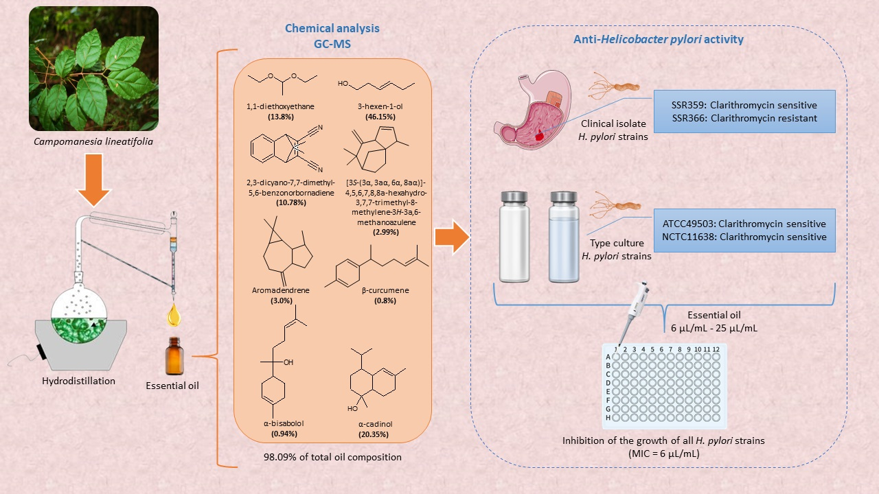
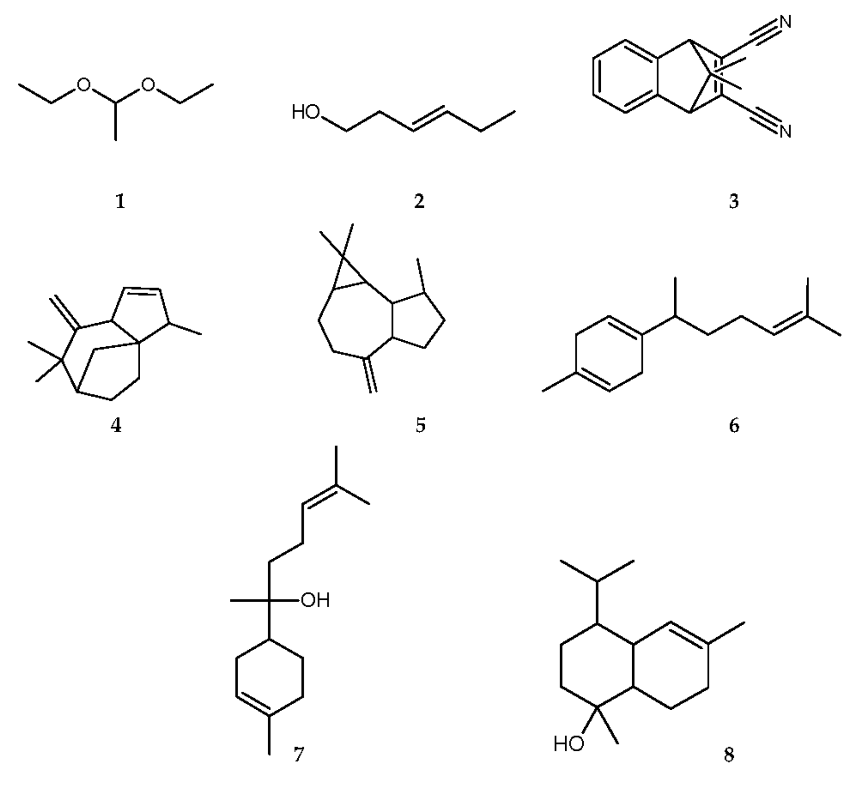
2.1. Chemical Analysis of the Essential Oil

The GC-MS chemical analysis of C. lineatifolia essential oil showed the presence of eight components that represent 98.09% of the total oil composition (Table 1). The main components of the oil were 3-hexen-1-ol (46.15%), α-cadinol (20.35%), 1,1-diethoxyethane (13.08%), and 2,3-dicyano-7,7-dimethyl-5,6-benzonorbornadiene (10.78%), followed by aromadendrene 2 (3.0%), and [3-S-(3α, 3aα, 6α, 8aα)]-4,5,6,7,8,8a-hexahydro-3,7,7-trimethyl-8-methylene-3H-3a,6-methanoazulene (2.99%), respectively. The other components, such as α-bisabolol (0.94%) and β-curcumene (0.8%), were traced in the C. lineatifolia oil. The structures of the identified compounds are presented in Figure 1.

2.2. In Vitro Anti-Helicobacter pylori Activity Evaluation

The antimicrobial activity of the C. lineatifolia EO at 6 μL/mL–25 μL/mL concentrations was evaluated against ATCC 49503; NCTC116328 (both clarithromycin-sensitive strains), and SSR359; SSR366, clarithromycin-sensitive clinical isolate, clarithromycin-resistant clinical isolate, respectively. Clarithromycin was used as a positive control with inhibitory activity for the H. pylori growth in a concentration range of 0.03 μg/mL–256 μg/mL. It was shown that the EO was able to inhibit the growth of all the H. pylori strains at the lowest concentration tested, with a calculated MIC of 6 μL/mL. Clarithromycin inhibited the H. pylori growth at 0.03 μg/mL in the sensitive strains (ATCC 49503, NCTC11638, and SSR359) and at 8 μg/mL in the clarithromycin-resistant clinical isolate (SSR366).

3. Discussion

Essential oils are complex mixtures of volatile compounds belonging to various chemical classes, such as alcohol, ethers or oxides, aldehydes, ketones, esters, amines, amides, phenols, heterocycles, and mainly the terpenoids, especially monoterpenes and sesquiterpenes [36]. The chemical analysis of the EO composition from the C. lineatifolia leaves consisted mainly of alcohol (2) (46.15%), ether (1) (13.08%), and sesquiterpenes (38) (38.86%) as the majority constituents.
Osorio et al. [37] demonstrated that the presence of alcohols was predominantly characterized in the volatile extract of C. lineatifolia leaves, and among them, (Z)-3-hexenol was one of the majority constituents. In addition, cadinanes sesquiterpenes, β-cadinol, γ-cadinol, and δ-cadinol were identified. Similarly, in this study, the alcohol 3-hexen-1-ol (2) corresponded to the majority compound in the essential oil, and the cadinane sesquiterpene, α-cadinol (8), was also identified in C. lineatifolia.
Moreover, the compounds ether 1,1-diethoxyethane (1), 2,3-dicyano-7,7-dimethyl-5,6-benzonorbornadiene (3), [3S-(3α, 3aα, 6α, 8aα)]-4,5,6,7,8,8a-hexahydro-3,7,7-trimethyl-8-methylene-3H-3a,6-methanoazulene (4), aromadendrene 2 (5), β-curcumene (6), and α-bisabolol (7) were identified for the first time in C. lineatifolia.
The phytochemical analysis of the Psidium guajava leaves, Myrtaceae family, showed the presence of the ether 1,1-diethoxyethane (1) as one of the constituents that confer fruit aroma [38].
Compound 3, 2,3-dicyano-7,7-dimethyl-5,6-benzonorbornadiene, was described in a study of the chemical composition of the essential oils isolated from the Mentha microphylla (Labiatae) and Lantana camara (Verbenaceae) species [39], with related insecticidal and fungicidal activities. This sesquiterpene was also identified in the dichloromethane-extracted rhizome essential oil from Aframomum atewae (Zingiberaceae), and the EO exhibited fungicidal activity against Candida albicans [40].
Compound 4, [3S-(3α, 3aα, 6α, 8aα)]-4,5,6,7,8,8a-hexahydro-3,7,7-trimethyl-8-methylene-3H-3a,6-methanoazulene, showed an identical fragmentation profile to the diene derivative of the sesquiterpene zizanol, found in Vetiveria zizanioides (L.) Nash, sin. Chrysopogon zizanioides (L.) Roberty (Poaceae) [31]. Vetiver oil is commonly used in the perfume and food industry as a flavoring agent [41]. Aside from its special aroma, its antioxidant [42], antibacterial [43] and anti-inflammatory [44] activities are described in the literature.
Aromadendrene 2 (5) was illustrated in several species of the Myrtaceae family, such as Eucalyptus sp. [32,45], Psidium guajava [46], and Lophomyrtus sp. [47].
Compound 6, β-curcumene, was identified as one of the major constituents in the essential oil from the rhizomes of Curcuma amada and C. longa (Zingiberaceae) [48,49]. Curcuma sp. is traditionally used to treat various intestinal diseases, fever and jaundice [50] and its anti-H. pylori activity has been described [51,52]. In the Myrtaceae family, γ-curcumene is found in the essential oil of Eucalyptus microtheca leaves [32] and β-curcumene in Myrcia sylvatica [53].
Compound 7, α-bisabolol, was identified in the essential oils of some species from the Myrtaceae family, such as Plinia cerrocampanensis [54] and Psidium myrtoides [55].
The treatment for H. pylori infection commonly involves first-line triple therapy with clarithromycin (a combination of second-generation PPI, amoxicillin and clarithromycin for 14 days) [7]. In areas of high clarithromycin resistance (> 15%), a quadruple bismuth therapy (PPI, bismuth salt, tetracycline, nitroimidazole) or a concomitant therapy (PPI, clarithromycin, metronidazole, amoxicillin) is recommended [3,4,5]. However, these regimens may increase the risk of side effects and the cost of treatment, which brings difficulties for their implementation in clinical practice and often causes treatment abandonment by the patient. In addition, the use of multiple antimicrobial agents for H. pylori infection may increase the risk of future microbial resistance [56]. In this scenario, new regimens and approaches that allow the minimal use of antimicrobials for a shorter treatment period are needed.
Plant-based medicines are effective in treating gastric ulcers and have fewer side effects and lower recurrence rates, and when used in monotherapy or in combination with conventional drugs, they can become an alternative to treat certain gastric ulcers and prevent their recurrence [9]. Clinical studies have already demonstrated the efficacy of some natural products for the treatment of gastrointestinal diseases. For example, capsaicin (derived from the peppers of the Capsicum genus) protected the gastric mucosa in healthy volunteers who took acetylsalicylic acid [57] and decreased the intensity of dyspeptic symptoms in patients with functional dyspepsia [58]. Curcumin, from Curcuma longa (turmeric), improved dyspeptic symptoms, improved quality of life, and provided a satisfactory equivalent to omeprazole in a double-blind, placebo-controlled trial in patients diagnosed with functional dyspepsia [59]. Patients treated with a Maytenus ilicifolia (espinheira-santa) extract for 28 days showed a significant improvement compared to the placebo group, with regard to the overall dyspeptic symptoms, and especially for symptoms of heartburn and gastralgia [60].
Regarding the essential oils, Korona-Glowniak et al. [24] evaluated the in vitro activity of the five essential oils, silver fir, pine needle, tea tree, lemongrass and cedarwood oils, against H. pylori. The most active against the clinical strains of H. pylori were cedarwood oil and oregano oil. Moreover, the cedarwood oil inhibited the urease activity at sub-inhibitory concentrations, suggesting that this essential oil can be a useful component of the plant preparations supporting the eradication of H. pylori therapy. The Pelargonium graveolens oil exhibited good activity against H. pylori at a MIC of 15.63 µg/mL, and once combined with clarithromycin, a significant synergistic effect appeared at a fractional inhibitory concentration index of 0.38 µg/mL [25]. Thus, the combination of herbal medicines with anti-H. pylori activity could improve the outcome for gastric ulcer patients.
The alcohol, 3-hexen-1-ol, is described as one of the majority constituents of the EO of Fagopyrum esculentum (Polygonaceae) flowers and is related to the antimicrobial activity against Gram-positive bacteria Bacillus subtilis and Staphylococcus aureus, and Gram-negative bacteria, Escherichia coli and Pseudomonas lachrymans [61]. Pino et al. [62] also described the presence of 3-hexen-1-ol as the majority constituent of the EO from Galinsoga parviflora (Asteraceae) leaves and demonstrated that this EO exhibited antimicrobial activity against Gram-positive bacteria S. aureus and Bacillus cereus. Similarly, this alcohol corresponded with the majority constituent in the C. lineatifolia EO, and together with the data described in the literature, one can suggest its potential anti-H. pylori activity, observed in this study.
David et al. [63] demonstrated that the EO obtained by the hydrodistillation of Vetiveria zizanioides leaves showed potent antimicrobial activity against the Gram-positive bacterium S. aureus and moderate activity for the Gram-negative bacteria P. aeruginosa and E. coli. It is important to consider that V. zizanioides contains a diene derivative of the sesquiterpene zizanol, [3S-(3α, 3aα,6α,8aα)]-4,5,6,7,8,8a-hexahydro-3,7,7-trimethyl-8-methylene-3H-3a,6-methanoazulene [27], also identified in the current study for C. lineatifolia EO. Mulyaningsih et al. [64] identified that aromadendrene contributes to the antimicrobial activity of Eucalyptus globulus essential oil, markedly on multidrug-resistant bacteria, such as methicillin-resistant S. aureus (MRSA) and vancomycin-resistant Enterococcus faecalis (VRE), in addition to activity against the Gram-negative bacterium Acinetobacter baumanii. Furthermore, related to Campomanesia adamantium and C. pubescens species, in which aromadendrene and α-cadinol were identified, the hexanic extract of the fruits inhibited the growth of all bacteria tested, i.e., S. aureus, P. aeruginosa, and E. coli, demonstrating that the volatile compounds present showed good activity against Gram-positive and Gram-negative bacteria [65].
It has been shown that β-curcumene and α-bisabolol, the majority constituents of the EOs of Curcuma sp. (Zingiberaceae) and Plinia cerrocampanensis (Myrtaceae), respectively, have anti-H. pylori activities [46,47,49]. In addition, Brehm-Stecher & Johnson [66] described that bisabolol is able to increase bacterial wall permeability and, consequently, susceptibility to antibiotics of clinical importance, suggesting a possible mechanism for its activity.
Studies indicate that plant essential oils have bactericidal effects against H. pylori, and this activity could be related mainly to the terpenoid content, followed by other compounds, such as phenols, alcohols, aldehydes and ketones [24,26,36,67]. Additionally, the EOs have equivalent bactericidal effects against both the antibiotic-susceptible strains and other H. pylori strains that resist antibiotics, suggesting that EOs may be promising candidates to treat H. pylori infection [68].
All these findings related to the compounds identified in the EO of C. lineatifolia may suggest that the anti-H. pylori activity of the EO may originate from one of its main constituents or even synergism between them. However, it is important to note that the antibacterial assay was performed as an initial screen to determine whether the C. lineatifolia EO demonstrated anti-H. pylori activity. In the current study, the microbroth dilution assay data presented demonstrates the bacteriostatic effect of the EO at a MIC of 6 μL/mL, as H. pylori growth was inhibited at all concentrations of EOs tested (6 μL/mL–25 μL/mL). Furthermore, lower concentrations of EO (i.e., < 6 μL/mL) could possibly demonstrate anti-H. pylori activity, and will be the subject of our future MIC and minimum bactericidal concentration (MBC) studies after purifying additional EO from fresh C. lineatifolia leaves to determine whether the EO also possesses bactericidal activity.
These results showed the potential of the C. lineatifolia EO against clarithromycin-sensitive and resistant H. pylori-type cultures and clinical isolate strains, making the species promising in the treatment of gastric ulcers associated with H. pylori infection.

4. Material and Methods

4.1. Plant Material

Campomanesia lineatifolia leaves were collected in February 2020 from Minas Gerais, Brazil (19°52′9.87″ S; 43°58′12.04″ W). The species was identified by Dr Marcos Sobral from the Botany Department of Biological Sciences, the Institute at the Federal University of Minas Gerais (UFMG), Belo Horizonte. A voucher specimen (BHCB 150.606) was deposited at the UFMG Herbarium. The registration in the National System of Genetic Heritage and Associated Traditional Knowledge Management (SisGen) was carried out and given the code A216C7C.

4.2. Isolation of the Essential Oil

The EO was extracted from the fresh leaves of C. lineatifolia by hydrodistillation using a Clevenger-type apparatus. The extraction was carried out for 3 h by mixing 143 g of plants in 1000 mL of distilled water. The EO were dried with sodium sulphate anhydrous (Dinâmica Química Contemporânea Ltda), and concentrated under reduced pressure by a rotatory evaporator to evaporate water. The pure oil was stored at −25 °C until further analysed. The essential oils’ yield was 0.5 mL.

4.3. Analysis of the Essential Oil

The essential oil chemical composition assessments and the identification of the main constituents were conducted by GC-MS analyses. The GC-MS analysis was performed in triplicate using a Hewlett-Packard 6890N gas chromatograph, coupled with a 5975B mass selective detector (MSD; Agilent Technologies, Inc., Santa Clara, CA, USA) operating at 70 eV over a mass range of 35–500 amu and a scanning speed of 0.34 and equipped with a DB-5MS fused-silica capillary column (5% phenylmethylsiloxane; length—30 m, internal diameter—0.25 mm, film thickness—0.25 μm). The oven temperature was raised from 70 °C to 290 °C at a heating rate of 5 °C/min and held isothermal for 10 min; injector temperature, 250 °C; interface temperature, 300 °C; carrier gas, helium at 1.0 mL/min. The sample oil was dissolved in Et2O at the ratio of 1:1000 and injected into a pulsed split mode. The flow rate was 1.5 mL/min for the first thirty seconds (0.50 min), then modified to 1.0mL/min for the remainder of the run; the split ratio was 40:1. The identification of the constituents was based on the comparison of their linear retention indices relative to the series of n-hydrocarbons (C6–C40), and on computer matching against commercial (NIST or Wiley library) and home-made library mass spectra that were built up from pure substances and components of known oils, and MS literature data.

4.4. Anti-Helicobacter pylori Activity

4.4.1. Bacterial Strains and Culture Conditions

The H. pylori strains were obtained from the American Type Culture Collection (ATCC 49503, clarithromycin-sensitive), National Collection of Type Cultures (NCTC11638, clarithromycin-sensitive), and from Tallaght University Hospital, Dublin 24, Ireland (SSR359, clarithromycin-sensitive clinical isolate, and SSR366, clarithromycin-resistant clinical isolate). All strains were maintained on Columbia blood agar (CBA) plates containing 5% sheep’s blood (VWR) at 37 °C under microaerobic conditions (CampyGen, Oxoid). Bacteria were inoculated into a brain heart infusion (BHI) broth (Sigma) containing 10% foetal bovine serum (Gibco) and incubated under microaerobic conditions at 37 °C with shaking (120 rpm) for 24h to prepare the bacterial suspensions for the broth microdilution assays.

4.4.2. Broth Microdilution Assays

The determination of the minimum inhibitory concentration (MIC) of EO against H. pylori was evaluated using the broth microdilution assays in 96-well plates, according to the Standards for Antimicrobial Susceptibility Testing protocol [69], with adaptations [70]. The EO was diluted with the BHI broth supplemented with 10% foetal bovine serum and dimethylsulfoxide 2% (DMSO, Sigma) at 47 °C to prepare the concentrations at 6 μL/mL–25 μL/mL (0.6–2.5% v/v).
A total of 100 µL aliquots of overnight bacterial suspension (approximately 106 CFU/mL; 1:20 0.5 McFarland Standard) [70] was added to each well of a 96-well plate. The cultures were incubated with 100 µL aliquots of EO for 72 h at 37 °C under microaerobic conditions (CampyGen) with shaking (120 rpm). The absorbances at a wavelength of 600 nm were read using a Varioskan LUX Microplate Reader (Thermo Fisher Scientific). The MIC values were determined as the lowest concentration at which no bacterial growth occurred. Clarithromycin was used as the positive control at 0.03 μg/mL–256 μg/mL (0.003–25.6% v/v).

4.5. Statistical Analysis

The experiments were performed in triplicate, and the results were expressed as mean ± SEM. A one-way ANOVA using GraphPad Prism 6.0, followed by a Bonferroni post-test, was performed, considering values of a p ≤ 0.05 as statistically significant. The MIC values were calculated considering the concentrations that did not differ statistically from the BHI broth control.

5. Conclusions

As far as we know, our study investigates, for the first time, the chemical composition of the essential oil from C. lineatifolia fresh leaves and its anti-H. pylori activity. Using the GC-MS technique, it was possible to identify eight new compounds for the species, representing 98.09% of the total oil composition, the major components of the alcohol 3-hexen-1-ol and the sesquiterpene α-cadinol.
Our findings demonstrate that the C. lineatifolia EO has anti-H. pylori activity at the lower concentration tested (MIC 6 μL/mL), suggesting a bacteriostatic property and a relation with its ethnopharmacological use as a medicinal plant for the treatment of gastric ulcers. Furthermore, the antibacterial activity against clarithromycin-resistant clinical isolates indicates that essential oils could be used in combination, as even antibiotics with high activity are not effective when used singly in therapeutic regimens involving patients with resistant H. pylori infection. Evidence for the use of medicinal plants in therapy shows that they have fewer adverse effects and lower rates of infection recurrence, as well as reducing time and improving the patient’s compliance with treatment.
For the follow-on work to this study, additional EO from fresh C. lineatifolia leaves will be purified to determine whether the EO also possesses bactericidal activity by performing MBC assays. In addition, it will be necessary to examine each antibacterial component of essential oil separately and in combination to ascertain whether they act alone or synergistically. Furthermore, studies involving in vivo models of gastric ulcers are proposed to determine the mechanisms by which the chemical constituents described here exert their anti-H. pylori activity.

Author Contributions

Conceptualization, N.C.V.N., F.B. and R.O.C.; methodology, R.O.C., F.B. and S.M.S.; validation, N.C.V.N., F.B., R.O.C. and S.M.S.; formal analysis, N.C.V.N., F.B., R.O.C. and S.M.S.; investigation, N.C.V.N., M.P.d.M. and R.O.C.; resources, R.O.C., F.B. and S.M.S.; data curation, N.C.V.N.; writing—original draft preparation, N.C.V.N.; writing—review and editing, R.O.C., F.B. and S.M.S.; supervision, R.O.C., M.V.C., F.B. and S.M.S.; project administration, R.O.C.; funding acquisition, R.O.C. All authors have read and agreed to the published version of the manuscript.

Funding

This research was funded by Coordenação de Aperfeiçoamento de Pessoal de Nível Superior (CAPES) through the Programa Institucional de Internalização (PRINT), grant number CAPES-PRINT 88887.370558/2019-00; Fundação de Amparo à Pesquisa do Estado de Minas Gerais (FAPEMIG), grant number APQ-00901-21; Conselho Nacional de Desenvolvimento Científico e Tecnológico (CNPq), grant number 303757/2019-1, and Pró-Reitoria de Pesquisa (PRPq), Universidade Federal de Minas Gerais (UFMG).

Data Availability Statement

All data generated or analyzed during this study are included in this article.

Acknowledgments

N.C.V.N and F.B. thank Niko S. Radulović, Department of Chemistry, Faculty of Sciences and Mathematics, University of Niš, Višegradska 33, Niš 18000, Serbia, for the essential oil chemical analysis support.

Conflicts of Interest

The authors declare no conflict of interest.

References

  1. Marshall, B.; Warren, J.R. Unidentified curved bacilli in the stomach of patients with gastritis and peptic and peptic ulceration. Lancet 1984, 323, 1311–1315. [Google Scholar] [CrossRef]
  2. Smith, S.M. An update on the treatment of Helicobacter pylori infection. EMJ Gastroenterol. 2015, 4, 101–107. [Google Scholar]
  3. Fallone, C.A.; Chiba, N.; van Zanten, S.V.; Fischbach, L.; Gisbert, J.P.; Hunt, R.H.; Jones, N.L.; Render, C.; Leontiadis, G.I.; Moayyedi, P.; et al. The Toronto consensus for the treatment of Helicobacter pylori infection in adults. Gastroenterology 2016, 151, 51–69.e14. [Google Scholar] [CrossRef] [PubMed] [Green Version]
  4. Chey, W.D.; Leontiadis, G.I.; Howden, C.W.; Moss, S.F. ACG clinical guideline: Treatment of Helicobacter pylori infection. Am. J. Gastroenterol. 2017, 112, 212–239. [Google Scholar] [CrossRef] [PubMed]
  5. Malfertheiner, P.; Megraud, F.; O’Morain, C.A.; Gisbert, J.P.; Kuipers, E.J.; Axon, A.T.; Bazzoli, F.; Gasbarrini, A.; Atherton, J.; Graham, D.Y.; et al. Management of Helicobacter pylori infection—the Maastricht V/Florence consensus report. Gut 2017, 66, 6–30. [Google Scholar] [CrossRef] [Green Version]
  6. Liu, W.Z.; Xie, Y.; Lu, H.; Cheng, H.; Zeng, Z.R.; Zhou, L.Y.; Chen, Y.; Wang, J.B.; Du, Y.Q.; Lu, N.H.; et al. Fifth Chinese National Consensus Report on the management of Helicobacter pylori infection. Helicobacter 2018, 23, e12475. [Google Scholar] [CrossRef]
  7. Coelho, L.G.V.; Marinho, J.R.; Genta, R.; Ribeiro, L.T.; Passos, M.D.C.F.; Zaterka, S.; Assumpção, P.P.; Barbosa, A.J.A.; Barbuti, R.; Braga, L.L.; et al. IVth Brazilian Consensus Conference on Helicobacter pylori infection. Arq. Gastroenterol. 2018, 16, 97–121. [Google Scholar] [CrossRef]
  8. Ustün, O.; Ozçelik, B.; Akyön, Y.; Abbasoglu, U.; Yesilada, E. Flavonoids with anti-Helicobacter pylori activity from Cistus laurifolius leaves. J. Ethnopharmacol. 2006, 108, 457–461. [Google Scholar] [CrossRef]
  9. Bi, W.P. Efficacy and safety of herbal medicines in treating gastric ulcer: A review. World J. Gastroenterol. 2014, 20, 17020–17028. [Google Scholar] [CrossRef]
  10. Krzyżek, P.; Junka, A.; Słupski, W.; Dołowacka-Jóźwiak, A.; Płachno, B.J.; Sobiecka, A.; Matkowski, A.; Chodaczek, G.; Płusa, T.; Gościniak, G.; et al. Antibiofilm and antimicrobial-enhancing activity of Chelidonium majus and Corydalis cheilanthifolia extracts against multidrug-resistant Helicobacter pylori. Pathogens 2021, 10, 1033. [Google Scholar] [CrossRef]
  11. Hassan, S.T.S.; Berchová, K.; Majerová, M.; Pokorná, M.; Švajdlenka, E. In vitro synergistic effect of Hibiscus sabdariffa aqueous extract in combination with standard antibiotics against Helicobacter pylori clinical isolates. Pharm. Biol. 2016, 54, 1736–1740. [Google Scholar] [CrossRef] [PubMed] [Green Version]
  12. Abadi, A.T.B. Resistance to clarithromycin and gastroenterologist’s persistence roles in nomination for Helicobacter pylori as high priority pathogen by World Health Organization. World J. Gastroenterol. 2017, 23, 6379–6384. [Google Scholar] [CrossRef] [PubMed]
  13. Wilson, P.G.; O’Brien, M.M.; Gadek, P.A.; Quinn, C.J. Myrtaceae revisited: A reassessment of infrafamilial groups. Am. J. Bot. 2001, 88, 2013–2025. [Google Scholar] [CrossRef] [PubMed]
  14. Sobral, M.; Proença, C.; Souza, M.; Mazine, F.; Lucas, E. Myrtaceae. In Lista de Espécies da Flora do Brasil; Jardim Botânico do Rio de Janeiro: Rio de Janeiro, Brazil, 2015. Available online: http://floradobrasil2015.jbrj.gov.br/FB171 (accessed on 8 June 2022).
  15. Villachica, H.; Carvalho, J.F.U.; Muller, C.H.; Diaz, C.S.; Almanza, M. Frutales y Hortializas Promisorios de la Amazonia. Tratado de Cooperacion Amazonica; Secretaria Pro-Tempore Publicaciones: Lima, Peru, 1996; Volume 44, pp. 212–213. [Google Scholar]
  16. Lorenzi, H.; Bacher, L.; Lacerda, M.; Sartori, S. Frutas Brasileiras e Exóticas Cultivadas: (de Consumo in Natura), 1st ed.; Instituto Plantarum: São Paulo, Brazil, 2006. [Google Scholar]
  17. Pio Corrêa, M. Dicionário das Plantas Úteis do Brasil e das Exóticas Cultivadas; Imprensa Nacional: Rio de Janeiro, Brazil, 1952.
  18. D’Ávilla, M.C. Da Flora Medicinal do Rio Grande do Sul; Faculdade Livre de Medicina e Pharmacia de Porto Alegre: Porto Alegre, Brazil, 1910; p. 155. [Google Scholar]
  19. Moraes, M. Botânica Brasileira: Applicada à Medicina às Artes e à Indústria (Seguida de um Suplemento de Matéria Médica, Inclusive as Plantas Conhecidas e Applicadas Pelos Índios em Suas Enfermidades); Garnier: Rio de Janeiro, Brazil, 1881. [Google Scholar]
  20. Lescano, C.H.; Freitas de Lima, F.; Caires, A.R.L.; de Oliveira, I.P. Polyphenols present in Campomanesia Genus: Pharmacological and nutraceutical approach. In Polyphenols in Plants, 2nd ed.; Watson, R.R., Ed.; Academic Press: Cambridge, MA, USA, 2019; Chapter 25; pp. 407–420. [Google Scholar]
  21. Dos Santos, A.L.; Polidoro, A.D.S.; Cardoso, C.A.L.; Batistote, M.; do Carmo Vieira, M.; Jacques, R.A.; Caramão, E.B. GC×GC/qMS analyses of Campomanesia guazumifolia (Cambess.) O. Berg essential oils and their antioxidant and antimicrobial activity. Nat. Prod. Res. 2019, 16, 593–597. [Google Scholar] [CrossRef]
  22. De Jesus, G.S.; Micheletti, A.C.; Padilha, R.G.; de Souza de Paula, J.; Alves, F.M.; Leal, C.R.B.; Garcez, F.R.; Garcez, W.S.; Yoshida, N.C. Antimicrobial potential of essential oils from cerrado plants against multidrug–resistant foodborne microorganisms. Molecules 2020, 25, 3296. [Google Scholar] [CrossRef]
  23. Pacheco, L.A.; Ethur, E.M.; Sheibel, T.; Buhl, B.; Weber, A.C.; Kauffmann, C.; Marchi, M.I.; Freitas, E.M.; Hoehne, L. Chemical characterization and antimicrobial activity of Campomanesia aurea against three strains of Listeria monocytogenes. Braz. J. Biol. 2021, 81, 69–76. [Google Scholar] [CrossRef] [Green Version]
  24. Korona-Glowniak, I.; Glowniak-Lipa, A.; Ludwiczuk, A.; Baj, T.; Malm, A. The in vitro activity of essential oils against Helicobacter pylori growth and urease activity. Molecules 2020, 25, 586. [Google Scholar] [CrossRef] [Green Version]
  25. Ibrahim, M.A.; Sallem, O.W.; Abdelhassib, M.R.; Eldahshan, O.A. Potentiation of anti-Helicobacter pylori activity of clarithromycin by Pelargonium graveolens oil. Arab J. Gastroenterol. 2021, 22, 224–228. [Google Scholar] [CrossRef]
  26. Spósito, L.; Oda, F.B.; Vieira, J.H.; Carvalho, F.A.; dos Santos Ramos, M.A.; de Castro, R.C.; Crevelin, E.J.; Crotti, A.E.M.; Santos, A.G.; da Silva, P.B.; et al. In vitro and in vivo anti-Helicobacter pylori activity of Casearia sylvestris leaf derivatives. J. Ethnopharmacol. 2019, 233, 1–12. [Google Scholar] [CrossRef]
  27. Madalosso, R.C.; Oliveira, G.C.; Martins, M.T.; Vieira, A.E.D.; Barbosa, J.; Caliari, M.V.; Castilho, R.O.; Tagliati, C.A. Campomanesia lineatifolia Ruiz & Pav. as a gastroprotective agent. J. Ethnopharmacol. 2012, 139, 772–779. [Google Scholar]
  28. Henriques, B.O.; Corrêa, O.; Azevedo, E.P.C.; Pádua, R.M.; Oliveira, V.L.S.D.; Oliveira, T.H.C.; Boff, D.; Dias, A.C.F.; Souza, D.G.D.; Amaral, F.A.; et al. In vitro TNF-α inhibitory activity of brazilian plants and anti-inflammatory effect of Stryphnodendron adstringens in an acute arthritis model. Evid. Based Complement. Altern. Med. 2016, 2016, 9872598. [Google Scholar] [CrossRef] [PubMed] [Green Version]
  29. Willner, B.; Granvogl, M.; Schieberle, P. Characterization of the key aroma compounds in bartlett pear brandies by means of the sensomics concept. J. Agric. Food Chem. 2013, 61, 9583–9593. [Google Scholar] [CrossRef] [PubMed]
  30. Marongiu, B.; Porcedda, S.; Porta, G.D.; Reverchon, E. Extraction and isolation of Salvia desoleana and Mentha spicata subsp. insularis essential oils by supercritical CO2. Flavour Fragr. J. 2001, 16, 384–388. [Google Scholar] [CrossRef]
  31. Kang, S.H.; Monti, S.A. Revised structure of zizanol. J. Org. Chem. 1984, 49, 3830–3832. [Google Scholar] [CrossRef]
  32. Maghsoodlou, M.T.; Kazemipoor, N.; Valizadeh, J.; Falak Nezhad Seifi, M.; Rahneshan, N. Essential oil composition of Eucalyptus microtheca and Eucalyptus viminalis. Avicenna J. Phytomed. 2015, 5, 540–552. [Google Scholar]
  33. Lucero, M.E.; Estell, R.E.; Fredrickson, E.L. The essential oil composition of Psorothamnus scoparius (A. Gray) RybB. J. Essent. Oil Res. 2003, 15, 108–111. [Google Scholar] [CrossRef]
  34. Zheng, C.H.; Kim, T.H.; Kim, K.H.; Leem, Y.H.; Lee, H.J. Characterization of potent aroma compounds in Chrysanthemum coronarium L. (Garland) using aroma extract dilution analysis. Flavour Fragr. J. 2004, 19, 401–405. [Google Scholar] [CrossRef]
  35. Yu, E.J.; Kim, T.H.; Kim, K.H.; Lee, H.J. Aroma-active compounds of Pinus densiflora (red pine) needles. Flavour Fragr. J. 2004, 19, 532–537. [Google Scholar] [CrossRef]
  36. Dhifi, W.; Bellili, S.; Jazi, S.; Bahloul, N.; Mnif, W. Essential oils’ chemical characterization and investigation of some biological activities: A critical review. Medicines 2016, 3, 25. [Google Scholar] [CrossRef] [Green Version]
  37. Osorio, C.; Alarcon, M.; Moreno, C.; Bonilla, A.; Barrios, J.; Garzon, C.; Duque, C. Characterization of odor-active volatiles in champa (Campomanesia lineatifolia R. & P.). J. Agric. Food Chem. 2006, 54, 509–516. [Google Scholar]
  38. Craveiro, A.A.; Fernandes, G.F.; Andrade, C.H.S. Óleos Essenciais de Plantas do Nordeste; Edições UFC: Fortaleza, Brazil, 1981. [Google Scholar]
  39. Abdelgaleil, S.A.M. Chemical composition, insecticidal and fungicidal activities of essential oils isolated from Mentha microphylla and Lantana camara growing in Egypt. Alex. Sci. Exch. J. 2006, 27, 18–28. [Google Scholar]
  40. Coffie, E. Essential Oils and Compounds Isolated from the Leaves and Rhizomes of Aframomum atewae. Ph.D. Thesis, University of Ghana, Accra, Ghana, July 2019. [Google Scholar]
  41. Martinez, J.; Rosa, P.T.V.; Menut, C.; Leydet, A.; Brat, P.; Pallet, D.; Meireles, M.A.A. Valorization of brazilian vetiver (Vetiveria zizanioides (L.) nash ex small) oil. J. Agric. Food Chem. 2004, 52, 6578–6584. [Google Scholar] [CrossRef] [PubMed]
  42. Luqman, S.; Kumar, R.; Kaushik, S.; Srivastava, S.; Darokar, M.P.; Khanuja, S.P.S. Antioxidant potential of the root of Vetiveria zizanioides (L.) nash. Indian J. Biochem. Biophys. 2009, 46, 122–125. [Google Scholar] [PubMed]
  43. Luqman, S.; Srivastava, S.; Darokar, M.P.; Khanuja, S.P.S. Detection of antibacterial activity in spent roots of two genotypes of aromatic grass Vetiveria zizanioides. Pharm. Biol. 2005, 43, 732–736. [Google Scholar] [CrossRef]
  44. Balasankar, D.; Vanilarasu, K.; Selva Preetha, P.; Rajeswari, M.; Umadevi, S.; Bhowmik, D. Traditional and medicinal uses of vetiver. J. Med. Plants Stud. 2013, 1, 191–200. [Google Scholar]
  45. Yangui, I.; Zouaoui Boutiti, M.; Boussaid, M.; Messaoud, C. Essential oils of myrtaceae species growing wild in tunisia: Chemical variability and antifungal activity against Biscogniauxia mediterranea, the causative agent of charcoal canker. Chem. Biodivers. 2017, 14, e1700058. [Google Scholar] [CrossRef]
  46. García-Díaz, E.; Trejo, R.; Tafoya, F.; Aragón-García, A.; Elizalde-González, M.P. Profile of terpenoid compounds mediating a plant-herbivore interaction: Screening by static headspace solid-phase microextraction-gas chromatography/Q-ToF mass spectrometry. Chem. Biodivers. 2020, 17, e2000564. [Google Scholar] [CrossRef]
  47. Woollard, J.M.R.; Perry, N.B.; Weavers, R.T.; van Klink, J.W. Bullatenone, 1,3-dione and sesquiterpene chemotypes of Lophomyrtus species. Phytochemistry 2008, 69, 1313–1318. [Google Scholar] [CrossRef]
  48. Srivastava, A.K.; Srivastava, S.K.; Shah, N.C. Constituents of the rhizome essential oil of Curcuma amada Roxb. from India. J. Essent. Oil Res. 2001, 13, 63–64. [Google Scholar] [CrossRef]
  49. Ministério da Saúde. Monografia da Espécie Curcuma longa L. (curcuma); Agência Nacional de Vigilância Sanitária: Brasília, Brazil, 2015.
  50. Jain, S.K. Ethnobotanical diversity in zingibers of India. Ethnobotany 1995, 7, 83–88. [Google Scholar]
  51. Zaidi, S.F.H.; Yamada, K.; Kadowaki, M.; Usmanghani, K.; Sugiyama, T. Bactericidal activity of medicinal plants, employed for the treatment of gastrointestinal ailments, against Helicobacter pylori. J. Ethnopharmacol. 2009, 121, 286–291. [Google Scholar] [CrossRef] [PubMed]
  52. Koosirirat, C.; Linpisarn, S.; Changsom, D.; Chawansuntati, K.; Wipasa, J. Investigation of the anti-inflammatory effect of Curcuma longa in Helicobacter pylori-infected patients. Int. Immunopharmacol. 2010, 10, 815–818. [Google Scholar] [CrossRef] [PubMed]
  53. Silva, L.; Sarrazin, S.; Oliveira, R.; Suemitsu, C.; Maia, J.; Mourão, R. Composition and antimicrobial activity of leaf essential oils of Myrcia sylvatica (G. Mey.) DC. Eur. J. Med. Plants 2016, 13, 1–9. [Google Scholar] [CrossRef]
  54. Vila, R.; Santana, A.I.; Pérez-Rosés, R.; Valderrama, A.; Castelli, M.V.; Mendonca, S.; Zacchino, S.; Gupta, M.P.; Cañigueral, S. Composition and biological activity of the essential oil from leaves of Plinia cerrocampanensis, a new source of α-bisabolol. Bioresour. Technol. 2010, 101, 2510–2514. [Google Scholar] [CrossRef] [PubMed]
  55. Dias, A.L.B.; Batista, H.R.F.; Estevam, E.B.B.; Alves, C.C.F.; Forim, M.R.; Nicolella, H.D.; Furtado, R.A.; Tavares, D.C.; Silva, T.S.; Martins, C.H.G.; et al. Chemical composition and in vitro antibacterial and antiproliferative activities of the essential oil from the leaves of Psidium myrtoides O. Berg (Myrtaceae). Nat. Prod. Res. 2019, 33, 2566–2570. [Google Scholar] [CrossRef]
  56. Suzuki, S.; Gotoda, T.; Kusano, C.; Ikehara, H.; Ichijima, R.; Ohyauchi, M.; Ito, H.; Kawamura, M.; Ogata, Y.; Ohtaka, M.; et al. Seven-day vonoprazan and low-dose amoxicillin dual therapy as first-line Helicobacter pylori treatment: A multicentre randomised trial in Japan. Gut 2020, 69, 1019–1026. [Google Scholar] [CrossRef] [Green Version]
  57. Yeoh, K.G.; Kang, J.Y.; Yap, I.; Guan, R.; Tan, C.C.; Wee, A.; Teng, C.H. Chili protects against aspirin-induced gastroduodenal mucosal injury in humans. Dig. Dis. Sci. 1995, 40, 580–583. [Google Scholar] [CrossRef]
  58. Bortolotti, M.; Coccia, G.; Grossi, G.; Miglioli, M. The treatment of functional dyspepsia with red pepper. Aliment. Pharmacol. Ther. 2002, 16, 1075–1082. [Google Scholar] [CrossRef]
  59. Yongwatana, K.; Harinwan, K.; Chirapongsathorn, S.; Opuchar, K.; Sanpajit, T.; Piyanirun, W.; Puttapitakpong, C. Curcuma longa Linn versus omeprazole in treatment of functional dyspepsia: A randomized, double-blind, placebo-controlled trial. J. Gastroenterol. Hepatol. 2022, 37, 335–341. [Google Scholar] [CrossRef]
  60. Santos-Oliveira, R.; Coulaud-Cunha, S.; Colaço, W. Revisão da Maytenus ilicifolia Mart. ex Reissek, Celastraceae. Contribuição ao estudo das propriedades farmacológicas. Rev. Bras. Farmacogn. 2009, 19, 650–659. [Google Scholar] [CrossRef]
  61. Zhao, J.; Jiang, L.; Tang, X.; Peng, L.; Li, X.; Zhao, G.; Zhong, L. Chemical composition, antimicrobial and antioxidant activities of the flower volatile oils of Fagopyrum esculentum, Fagopyrum tataricum and Fagopyrum Cymosum. Molecules 2018, 23, 182. [Google Scholar] [CrossRef] [PubMed] [Green Version]
  62. Pino, J.A.; Gaviria, M.; Quevedo-Vega, J.; García-Lesmes, L.; Quijano-Celis, C.E. Essential oil of Galinsoga parviflora leaves from Colombia. Nat. Prod. Commun. 2010, 5, 1831–1832. [Google Scholar] [CrossRef] [PubMed] [Green Version]
  63. David, A.; Wang, F.; Sun, X.; Li, H.; Lin, J.; Li, P.; Deng, G. Chemical composition, antioxidant, and antimicrobial activities of Vetiveria zizanioides (L.) nash essential oil extracted by carbon dioxide expanded ethanol. Molecules 2019, 24, 1897. [Google Scholar] [CrossRef] [PubMed] [Green Version]
  64. Mulyaningsih, S.; Sporer, F.; Zimmermann, S.; Reichling, J.; Wink, M. Synergistic properties of the terpenoids aromadendrene and 1,8-cineole from the essential oil of Eucalyptus globulus against antibiotic-susceptible and antibiotic-resistant pathogens. Phytomedicine 2010, 17, 1061–1066. [Google Scholar] [CrossRef]
  65. Cardoso, C.A.L.; Salmazzo, G.R.; Honda, N.K.; Prates, C.B.; Vieira, M.D.C.; Coelho, R.G. Antimicrobial activity of the extracts and fractions of hexanic fruits of Campomanesia species (Myrtaceae). J. Med. Food. 2010, 13, 1273–1276. [Google Scholar] [CrossRef]
  66. Brehm-Stecher, B.F.; Johnson, E.A. Sensitization of Staphylococcus aureus and Escherichia coli to antibiotics by the sesquiterpenoids nerolidol, farnesol, bisabolol, and apritone. Antimicrob. Agents Chemother. 2003, 47, 3357–3360. [Google Scholar] [CrossRef] [Green Version]
  67. Bergonzelli, G.E.; Donnicola, D.; Porta, N.; Corthésy-Theulaz, I.E. Essential oils as components of a diet-based approach to management of Helicobacter infection. Antimicrob. Agents Chemother. 2003, 47, 3240–3246. [Google Scholar] [CrossRef] [Green Version]
  68. Ohno, T.; Kita, M.; Yamaoka, Y.; Imamura, S.; Yamamoto, T.; Mitsufuji, S.; Kodama, T.; Kashima, K.; Imanishi, J. Antimicrobial activity of essential oils against Helicobacter pylori. Helicobacter 2003, 8, 207–215. [Google Scholar] [CrossRef]
  69. Patel, J.B.; Cockerill, F.R.; Bradford, P.A. M100-S25 performance standards for antimicrobial susceptibility testing; twenty-fifth informational supplement. Clin. Lab. Stand. Inst. 2015, 35, 29–50. [Google Scholar]
  70. Borges, A.S.; Minozzo, B.R.; Santos, H.; Ardisson, J.S.; Rodrigues, R.P.; Romão, W.; de Souza Borges, W.; Gonçalves, R.D.C.R.; Beltrame, F.L.; Kitagawa, R.R. Plectranthus barbatus Andrews as anti-Helicobacter pylori agent with activity against adenocarcinoma gastric cells. Ind. Crops Prod. 2020, 146, 112207. [Google Scholar] [CrossRef]
Figure 1. Structures of identified compounds (1–8) from Campomanesia lineatifolia fresh leaves essential oil.
Plants 11 01945 g001
Table 1. Chemical compounds identified in essential oil from Campomanesia lineatifolia fresh leaves by gas chromatography–mass spectrometry. For methodology, see Material and Methods.
CompoundRT (min)RIMolecular WeightMolecular Formula%MSID
1,1-diethoxyethane (1)2.483718118C6H14O213.08103, 75, 73, 61, 47, 45, 43, 31, 29NIST; Willner, Granvogl & Schieberle (2013) [29]
3-hexen-1-ol (2)3.523850100C6H12O46.1582, 79, 72, 69, 67, 65, 57, 55, 53, 51, 41, 39NIST; Marongiu et al. (2001) [30]
2,3-dicyano-7,7-dimethyl-5,6-benzonorbornadiene (3)18.1581506220C15H12N210.78223, 221, 207, 206, 205Wiley
[3-S-(3α, 3aα, 6α, 8aα)]-4,5,6,7,8,8a-hexahydro-3,7,7-trimethyl-8-methylene-3H-3a,6-methanoazulene (4)20.0441586202C15H222.99187, 159, 145, 131, 119, 105, 91, 77Kang & Monti (1984) [31]
Aromadendrene 2 (5)20.2751596204C15H243.0161, 133, 119, 105, 93, 91, 81, 79NIST; Maghsoodlou et al. (2015) [32]
β-curcumene (6)21.5471652204C15H240.8161, 119, 105, 93, 81, 55, 41Wiley; Lucero, Estell & Fredrickson (2003) [33]
α-bisabolol (7)21.5551652222C15H26O0.94204, 161, 119, 105, 93, 69, 41NIST; Zheng et al. (2004) [34]
α-cadinol (8)21.8091664222C15H26O20.35204, 189, 161, 134, 121, 109, 105, 95, 81, 71NIST; Yu et al. (2004) [35]
RT: retention time; RI: retention index, determined relative to n-alkanes (C6–C40) on the DB-5MS column; %: relative percentage, obtained from peak area; MS: mass spectrum fragmentation; ID: proposed identification according to mass spectrum compared to NIST or Wiley library/literature and retention index, according to literature.
Publisher’s Note: MDPI stays neutral with regard to jurisdictional claims in published maps and institutional affiliations.

