Review

Highly Specialized Mechanisms for Mitochondrial Transport in Neurons: From Intracellular Mobility to Intercellular Transfer of Mitochondria

1
Institute for Genetics, University of Cologne, 50931 Cologne, Germany
2
Cologne Excellence Cluster on Cellular Stress Responses in Aging-Associated Diseases (CECAD), 50931 Cologne, Germany
3
Department of Medicine, University of Udine, 33100 Udine, Italy
*
Authors to whom correspondence should be addressed.
Biomolecules 2023, 13(6), 938; https://doi.org/10.3390/biom13060938
Submission received: 12 May 2023 / Revised: 26 May 2023 / Accepted: 1 June 2023 / Published: 3 June 2023
(This article belongs to the Special Issue Molecular Functions of Microtubules)

Abstract

:
The highly specialized structure and function of neurons depend on a sophisticated organization of the cytoskeleton, which supports a similarly sophisticated system to traffic organelles and cargo vesicles. Mitochondria sustain crucial functions by providing energy and buffering calcium where it is needed. Accordingly, the distribution of mitochondria is not even in neurons and is regulated by a dynamic balance between active transport and stable docking events. This system is finely tuned to respond to changes in environmental conditions and neuronal activity. In this review, we summarize the mechanisms by which mitochondria are selectively transported in different compartments, taking into account the structure of the cytoskeleton, the molecular motors and the metabolism of neurons. Remarkably, the motor proteins driving the mitochondrial transport in axons have been shown to also mediate their transfer between cells. This so-named intercellular transport of mitochondria is opening new exciting perspectives in the treatment of multiple diseases.

1. Introduction

Neurons are polarized structures divided into compartments, which are functionally distinct units. Organelles, proteins and RNA are transported along neuronal processes—named axons and dendrites—to distal areas such as synapses, growth cones and branching points where mitochondria play different roles. Mitochondria are abundant in presynapses, while roughly 10% of dendritic spines contain mitochondria in basal conditions [1]. Neuronal activity increases the transport of mitochondria in synapses [1]. At a wider level, the somatodendritic and the axonal compartments have different distribution and trafficking properties in terms of cargo vesicles and organelles, which do not diffuse but are actively selected at the pre-axonal exclusion zone [2]. The majority of mitochondria are stationary in axons across different species [3]. The other motile mitochondria travel long distances and the direction of the transport is named anterograde or retrograde if it is from the cell body to the distal area of axons or vice versa. Motile mitochondria show complex trajectories, including linear and oscillatory, with pauses and changes in direction [3]. The fraction of mitochondria in a moving or a stationary state is associated with axonal growth: in the region of active growth cones, there is a motile-to-stationary shift of mitochondria that is reversed when axonal growth is blocked. The consequence of this dynamic balance is that the net transport is anterograde in growing axons and retrograde in blocked axons [4]. In this review, we focus on the mechanisms regulating the complex transport of mitochondria along the extraordinary distances over which neurons can extend.
Another type of long-distance transport contributing to the maintenance of neural homeostasis is represented by the transfer of mitochondria between adjacent cells. Different processes drive this transcellular communication, including the formation of tunneling nanotubes (TNTs). These structures, described for the first time twenty years ago by Rustom and colleagues [5], seem to rely on the same molecular system used for mitochondrial transport and docking in axons. The intracellular transmission of mitochondria observed in astrocytes, microglia, and neurons, is important for the recovery of neural functions supporting both their viability and post-injury recovery [6,7,8,9]. Similarly to spontaneous mitochondrial transfer between cells, stem cell-derived mitochondrial transplantation can provide an exogenous mitochondrial source thus restoring the mitochondrial functions in recipient cells [10,11]. As the primary hallmark in a wide range of brain states and pathologies is mitochondrial dysfunction, mitochondrial delivery into injured cells is opening a novel horizon for treating many diseases. Here, we summarize the clinical applications highlighting the opportunities and challenges of mitochondrial transfer/transplantation, especially in brain disorders.

2. Mitochondria Move along Microtubules and Actin Filaments
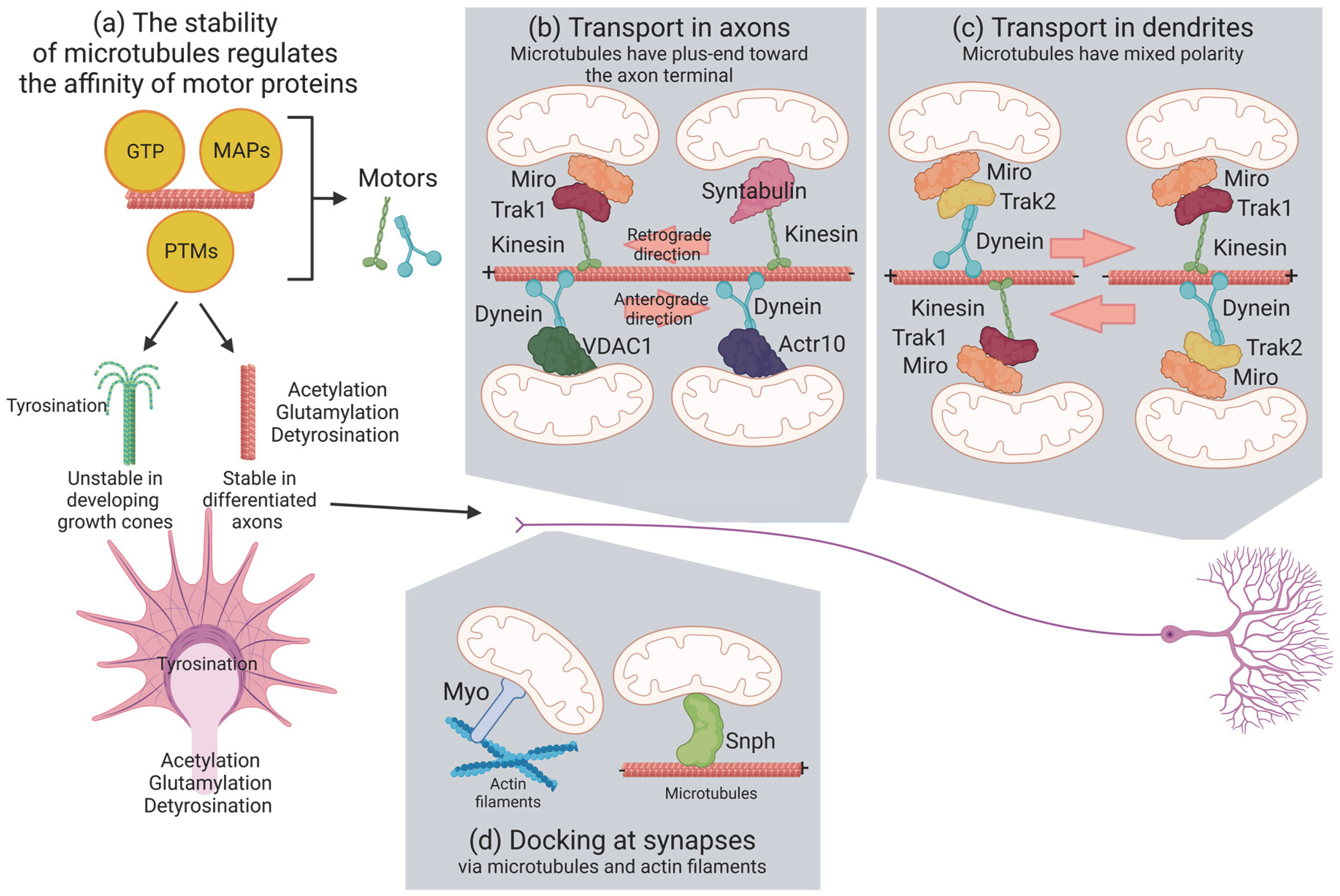
The cytoskeleton in neurons is composed of microtubules, actin filaments and neurofilaments. Neurofilaments are enriched in axons where they determine the diameter and the conductance. Microtubules and actin filaments regulate axonal maturation and growth and build the support for the transport of organelles, including mitochondria [12] (Figure 1).

2.1. Microtubules

Microtubules are tubes assembled by dimers of α- and β-tubulin oriented head-to-tail in rapid phases of growth and collapse named “dynamic instability” [13]. These structures confer the distinct polarity of microtubules, where α- and β-tubulins are exposed at the minus- and plus-end of microtubules, respectively. Microtubules have a peculiar organization and dynamics in neurons: they present a plus-end toward the distal part of axons and have a mixed polarity in dendrites [14]. Microtubules polymerize at the plus-end via the incorporation of fresh guanosine triphosphate (GTP) to β-tubulin, which is hydrolyzed to guanosine diphosphate (GDP) in already incorporated tubulin dimers. However, GTP-bound tubulin dimers have also been described in the stable microtubule lattice [15,16,17] and are more enriched in axons than dendrites [17,18]. These so-named GTP islands protect microtubule depolymerization and promote self-repair [19,20] but also regulate the local conformation of tubulin to modulate the transport of mitochondria [18]. A recent study showed that anterograde mitochondria halt along GTP-bound elongated dimers within the microtubule bundle but they remain motile at the rim of the microtubule bundle [18]. Furthermore, the affinity of the motor proteins kinesins linking organelles to microtubules depends on the different conformation of GDP- or GTP-bound dimers [17,21,22]. For mitochondria, these elongated GTP islands increase the velocity of the Kinesin motor for mitochondria KIF5B [18].
Post-translational modifications of tubulin appear after the polymerization of microtubules and include acetylation, detyrosination, glycylation and glutamylation (Figure 1a). Developing axons grow in response to attractive and repulsive chemical guidance clues. This process is highly dynamic and involves cycles of the de- and re-polymerization of actin and microtubules in the terminal part of axons, named the growth cone. In an initial phase, actin extends in protrusions, which are later invaded by microtubules and organelles as mitochondria and endoplasmic reticulum (engorgement). No acetylation is detected in growth cones, consistent with the presence of highly dynamic microtubules [23]. Finally, the new formed structure is consolidated by the depolymerization of actin and stabilization of microtubules [24]. This process is regulated by kinases activated by growth factors such as Slit, Wnt or nerve growth factor (NGF) bound to the receptors roundabout (Robo), trafficking kinesin protein (Trak) A and frizzled (Frz)/low-density lipoprotein receptor-related protein (LRP), respectively. These glycogen synthase kinase 3β (GSK3β) and Abl kinases regulate the localization of plus-end tracking proteins such as cytoplasmic linker-associated protein 2 (Clasp2), adenomatous polyposis coli (APC) and microtubule-associated protein 1B (Map1B), which alter the stability of microtubules [25]. Differentiated axons exhibit stable microtubules with high acetylation, glutamylation and detyrosination [23]. In addition to their role in regulating the stability of microtubules, post-translational modifications of tubulin also regulate the affinity of motor adaptors to microtubules and alter the general transport of organelles rather than being specific to mitochondria [26,27]. Accessory proteins of microtubules regulate the transport of mitochondria in a similar way. For instance, Map1B knockout neurons increase the retrograde transport [28] and mitochondria of N2a cells, or primary neurons overexpressing Tau do not travel in the anterograde direction [29,30,31]. This is probably due to a combination of Tau that can generally destabilize microtubules [32] and inhibit the kinesin-dependent transport of vesicles and organelles [31]. Taken together, these studies seem to indicate that the transport of mitochondria is not specifically regulated at the level of microtubule organization. However, it is possible that the studies conducted so far analyzed the general movement of mitochondria (stationary over motile mitochondria) and that broader methods to dissect the complex motility of mitochondria could show a subtle but specific role of accessory proteins and post-translational modifications of tubulin [33].

2.2. Actin

Actin filaments are formed by polarized globular monomers bound via weak interactions. Thus, actin polymers are intrinsically unstable and difficult to visualize in neurons. Electron microscopy allows the visualization of patches of actin along axons, synapses and growth cones [12]. Very little is known about the role of actin in the long-range transport of mitochondria. It appears that microtubules do not have an exclusive role in this transport because the depolymerization of microtubules using nocodazole or vinblastine does not completely stop mitochondria in axons and dendrites [34,35]. In addition, the disruption of the actin cytoskeleton using cytochalasin-D or lantruculin B has no effect [34]. A more recent study shows that cytochalasin-D stabilizes axonal mitochondria and destabilizes dendritic mitochondria [36], suggesting that actin organization modulates the transport of mitochondria via different mechanisms in the two compartments. As we mentioned previously, actin is also present in synapses, where it mediates the docking of mitochondria after the administration of NGF [37], regulating the short-range transport of mitochondria.

3. Molecular Motors Transport Mitochondria via Microtubules

Mitochondria associate with the microtubule network through kinesin and dynein, the molecular motors that drive mitochondria in the anterograde and retrograde direction, respectively (Figure 1b,c). Therefore, kinesins move mitochondria towards the plus-ends of microtubules and dynein mediates minus-end-directed mitochondrial transport. Among the kinesin superfamily proteins (also known as KIFs) encoded in humans and mice by 45 different genes [38], KIF5/Kinesin-1, KIF1B/Kinesin-3 and Kinesin-Like Protein 6 (KLP6) are the main kinesins that mediate mitochondrial transport in neurons [39,40,41]. Two kinesin heavy chains (KHCs) and two kinesin light chains (KLCs) form a 380 kDa heterotetramer complex. This complex is composed of a conserved globular motor domain (or head) consisting of an adenosine triphosphate (ATP)-binding motif and a microtubule-binding domain, attached to a stalk domain for dimerization and to a tail domain with binding and regulatory functions. While the motor domains are highly conserved, the remaining sequences are unique for each kinesin, determining the cargo specificity and the direction of the transport. In contrast to the large specialized kinesin superfamily, only the cytoplasmatic Dynein 1 drives the minus-end-directed microtubule transport. Then, different mechanisms must explain how dynein specifically transports the numerous different cargoes. Dynein is a very large protein complex (1.2 MDa) composed of distinct polypeptides, all of which are present in two copies. The dynein heavy chain (DHC) contains the motor domain with six distinct AAA domains folded into a ring-shaped structure, together with a microtubule-binding domain and a tail for the assembly of the other components: the intermediate chains (DIC), the light intermediate chains (DLIC), and three different light chains (DLC) [42,43]. To be fully active, dynein requires interaction with the dynactin complex and a coiled-coil cargo adaptor [44]. Dynactin anchors dynein to microtubules through its larger subunit, p150Glued. In addition, dynactin contains two actin-related proteins, Arp1 and Actr10/Arp11, and the subunits p62, p25, and p27 [45]. The loss of Actr10 selectively reduces the mitochondrial retrograde transport, leading to the accumulation of mitochondria in axon terminals [45]. Specific adaptor proteins link kinesin and dynein–dynactin to each cargo for its transport along microtubules. On the outer mitochondrial membrane, the Mitochondrial Rho GTPase protein (Miro 1 and Miro 2; in humans, RHOT1 and RHOT2) recruits motor proteins by Milton/Trak adaptors and Metaxin (MTX) proteins. Miro contains a transmembrane domain in its C-terminus and two GTPase domains, one at the N-terminal and one near the C-terminal, flanking two calcium-binding EF hand motifs which regulate the motility of mitochondria depending on calcium. The GTPase domains are crucial for regulating mitochondrial distribution: when GDP is bound, Miro does not recruit adaptor and motor proteins [46]. In mammals, the different binding specificity of the two Trak proteins targets mitochondria to dendrites and axons; while Trak1 binds to both kinesin and dynein components and is responsible for the axonal localization of mitochondria, Trak2 primarily interacts with dynein and plays a critical role in targeting mitochondria to dendrites [47] (Figure 1b,c).
Interestingly, as in Miro1/2 double-knockout cells Traks are still recruited to the outer mitochondrial membrane to drive mitochondrial trafficking, Miro cannot be the only outer-membrane protein that links mitochondria to motor proteins [48]. In fact, the loss of Drosophila Miro (dMiro) cannot fully block mitochondrial movement [49]. Thus, several other kinesin-containing complexes have been discovered in neurons, including syntabulin (SYBU), fasciculation and elongation protein zeta 1 (FEZ1), or Ran-binding protein 2 (RanBP2) [50,51,52]. Interestingly all of these adaptors contribute to the maintenance and remodeling of synapses [53,54]. Another interactor of the Miro/Milton complex is Mitofusin 2 (Mfn2), the activity of which is disrupted in the presence of pathogenic mutants, which arrest mitochondria independently from Mfn2 profusion activity [55]. In contrast to the anterograde transport machineries, the players that link dynein to mitochondria are not well characterized. One suggested system is represented by the direct interaction of the outer mitochondrial membrane (OMM) protein voltage-dependent anion-selective channel (VDAC) with the dynein motor protein [56]. In conclusion, the main molecular actors involved in anterograde mitochondrial transport form a complex with Miro (receptor), KIF5 (motor) and Milton/Traks (adaptors). In contrast, dynein in complex with dynactin mediates retrograde mitochondrial transport by interacting with Milton/Trak2 and Miro.

4. Mitochondrial Docking and Anchoring Machineries in Neurons

Mitochondria localize in the specific regions of the neuron that need the most energy and a high ion flux, such as the synapses or the distal regions of actively growing axons [57]. While a large proportion of mitochondria move during neuronal development, in mature neurons the stationary pool of mitochondria represents more than two-thirds. Remarkably, more than 30% of the synapse is occupied by anchored mitochondria serving as ‘power stations’ [58]. In addition, as mitochondria can temporarily stop and start moving again at various subcellular localizations, specific docking mechanisms are needed. The mitochondria-associated protein Syntaphilin (SNPH) is a central microtubule calcium-dependent docking system (Figure 1d). SNPH directly interacts with microtubules through its N-terminal microtubule-binding domain while its C-terminal tail inserts into the OMM [59]. The dynein light chain LC8 binds to SNPH, thus enhancing the SNPH-based docking of mitochondria to microtubules [60]. SNPH can also interact with myosin VI (Myo6, Jaguar in Drosophila) and anchor mitochondria on presynaptic filamentous (F)-actin. The AMP-activated protein kinase (AMPK)-dependent phosphorylation of Myo6 drives the capture of mobile axonal mitochondria at presynaptic terminals, switching them from microtubule-dependent transport to actin-mediated tethering [61]. Other myosin motor proteins in association with the actin cytoskeleton have been shown to interrupt mitochondrial transport on the microtubule tracks, facilitating their positioning. For example, Myo19 stops mitochondria in actin filaments through Miro [62] (Figure 1d). In general, where mitochondria are more urgently needed, actin is enriched and docks mitochondria away from microtubules.

5. Metabolic Control of Mitochondrial Transport
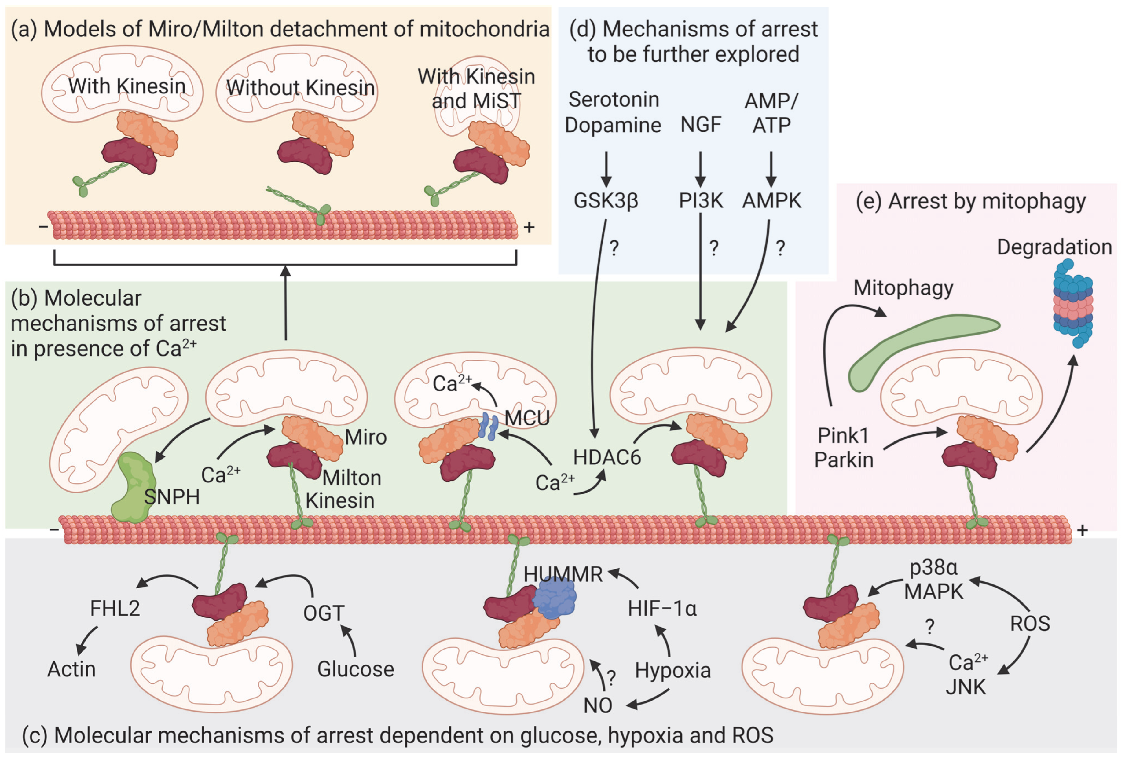
Mitochondria produce ATP and buffer calcium for the functioning and survival of neurons. Thus, it is not surprising that mitochondria are more abundant in neurons than in other cell types and that calcium and adenosine diphosphate (ADP)/ATP are prime signaling molecules to regulate the distribution of mitochondria. Glutamate increases the amount of calcium and immobilizes mitochondria in synapses. A similar effect is obtained after injecting ADP or in hypoxia [63]. It seems that the localization of mitochondria is largely regulated by stationarity rather than transport, as the studies of signaling molecules have pointed out so far.

5.1. Calcium

As mentioned above, mitochondria rely on the adaptor proteins Milton in Drosophila and Traks in mammals for anterograde movement along axonal microtubules. These adaptors bridge the receptor proteins Miro1 and 2 located in the outer mitochondrial membrane to Kinesin-1 [62]. Miro contains two EF hands that bind calcium. In the absence of these domains, mitochondria fail to stop in the presence of calcium in axons and dendrites [62,64], placing Miro as a regulator of mitochondrial arrest in sites where cytoplasmic calcium concentration is high. Two models explain a conformational change in Miro in the presence of calcium, but the mechanism is the opposite (Figure 2a). According to Wang and Schwarz, Kinesin-1 binds microtubules with its N-terminal domain and the Miro/Milton complex via its C-terminal region [64]. This complex transports mitochondria, but calcium induces the sequestration of the whole complex from microtubules. In MacAskill et al., Miro directly binds Kinesin-1 and calcium detaches Kinesin-1 from mitochondria but not from microtubules [62]. In contrast, another study shows that Miro1 binds the mitochondrial calcium uniporter (MCU) through its N-terminal domain on the outer mitochondrial membrane [65] (Figure 2b). This interaction is required for the transport of mitochondria and depends on the MCU-dependent calcium influx in the mitochondrial matrix rather than cytoplasmic calcium [66]. E208K/E328K mutations in the EF hands of Miro abolish calcium entry in the matrix [66]. Curiously, a different mutation (R272Q) in the EF hands of Miro has no effect on mitochondrial movement, although it disrupts calcium handling in the mitochondria [67]. The discrepancies in the role of Miro could be explained by the use of different mutants and raise the possibility that Miro is regulated by accessory proteins bound to the EF hands, or that the regulation of the transport complex is yet to be fully addressed [68]. Indeed, the EF1 domain of Miro1 senses cytosolic calcium and changes the shape of mitochondria independently of MCU-dependent calcium uptake and fusion/fission proteins in fibroblasts [69]. Furthermore, the analysis performed in neurites of iPSCs and in axons and dendrites of primary neurons could also indicate different functions of Miro, depending on its location in neurons. For example, histone deacetylase 6 (HDAC6) deacetylates Miro and stops mitochondria in calcium-rich axons [70] (Figure 2b). Although intriguing, given the broad functions of Mfn2 it is difficult to directly link mitochondrial transport to the levels of mitochondrial calcium [71]. SNPH is another calcium sensor that arrests mitochondria in axons [59,60]. In the presence of elevated calcium, SNPH dissociates Kinesin-1 from mitochondria to enhance their docking at presynapses. High calcium levels disrupt the Miro/Trak/Kinesin complex, favoring the anchoring of mitochondria by SNPH, which inhibits the activity of kinesin [72]. The anchoring of mitochondria to microtubules by SNPH is reversible since in the absence of calcium Miro can rapidly resume its calcium-free conformation and form the Miro/Trak/Kinesin complex to drive mitochondrial motility [62] (Figure 2b).

5.2. Glucose

Elevated electric activity mobilizes the Glut3 and Glut4 transporters at presynapses to increase the availability of glucose to boost local metabolism [73,74,75]. Thus, it is not surprising that glucose additionally halts mitochondria via an active mechanism [76]. O-GlcNAcylation is a post-translational modification important for proteins involved in neuronal signaling and synaptic plasticity [77,78]. Glucose activates O-GlcNAc transferase and increases the O-GlcNAcylation of Milton, which inhibits the transport of mitochondria [76]. The four and a half LIM domain protein 2 (FHL2) associates with O-GlcNAcylated Milton and favors the docking of mitochondria to actin [79] (Figure 2c).

5.3. ATP

The ratio of ADP and ATP regulates the positioning of mitochondria in primary neurons [63,80], possibly involving mechanisms to sense and drive mitochondria to sites of high energy consumption. Since motor proteins require ATP to transport mitochondria, it is possible that in sites of high ATP consumption, mitochondria halt because kinesin and dynein are not functional. The contributions of ATP and calcium signals to mitochondrial motility are difficult to disentangle because mitochondria are major regulators of the concentration of these two molecules. A study shows that neuronal depolarization decreases ATP levels and activates AMPK, increasing the anterograde transport of mitochondria into axons [80]. Similar results were obtained when lactate uptake was inhibited locally [81]. These studies suggest that the motility of mitochondria can be regulated by ATP independently of calcium. However, it is difficult to understand if this transport is directly regulated by AMPK or if it is the consequence of the broad mechanisms downstream of AMPK (Figure 2c).

5.4. Hypoxia

Hypoxia is an important factor during brain ischemia, and it has been shown to regulate mitochondrial motility in neurons [82,83] (Figure 2c). Decreased oxygen induces hypoxia-inducible factor 1α (HIF-1α) which, among its other targets, increases the expression of hypoxia up-regulated mitochondrial movement regulator (HUMMR). Reduced HUMMR in normoxia does not change the motility of mitochondria but diminishes the amount of motile mitochondria with anterograde movement in hypoxia, consistent with a defect in Kinesin-1 transport [82]. Indeed, HUMMR coimmunoprecipitates with Miro and Milton in normoxia [82]. Consistently, the upregulation of HUMMR recovers the average number of axonal mitochondria that are normally reduced by hypoxia [83]. It would be interesting to understand if HUMMR is also a sensor of hypoxia and if it increases its binding to the Miro/Milton complex. Interestingly, nitric oxide (NO) inhibitors recovered the motility of mitochondria during hypoxia and NO administration halted mitochondria [83,84]. It is not clear if this is a direct or indirect effect, nor if HUMMR is involved.

5.5. Reactive Oxygen Species (ROS)

Sources of ROS in the CNS are mitochondria and reactive microglia during inflammation. Intracellular and extracellular ROS equally block mitochondria without affecting other organelles [85,86,87]. In these experiments, axons are more vulnerable than dendrites [86]. Two opposing studies show that ROS inhibit the Miro/Milton complex by activating p38α mitogen-activated protein kinase (MAPK) independently from calcium [87] and that ROS increase calcium and the activity of the c-Jun N-terminal Kinase [85] (Figure 2c).

5.6. Growth Factors and Neurotransmitters

In addition, molecules for neuronal communication and growth seem to modulate the transport of mitochondria (Figure 2d). In developing neurons, the administration of NGF accumulates mitochondria close to the site of treatment [37]. Although the mechanism is not clear, it seems to involve the phosphoinositide 3-kinase (PI3K) pathway [88]. Moreover, serotonin and dopamine act via their respective receptors on the AKT-GSK3β pathway with opposite effects on mitochondria: dopamine halts and serotonin mobilizes mitochondria [89,90]. Interestingly, GSK3β was found to be localized to HDAC6 and HDAC6 activity and phosphorylation was regulated by GSK3β in primary neurons [91].

6. Mitophagy

Mitochondria can also be arrested when they are sequestered and degraded by autophagic engulfment, a process known as mitophagy. Damaged and depolarized mitochondria are cleared by the Pink1/Parkin pathway (Figure 2e). In these conditions, Pink1 is recruited and stabilized by Parkin on the outer mitochondrial membrane. This complex interacts with the Miro/Milton complex and induces the proteasomal degradation of Miro, thus releasing kinesin and arresting mitochondria [92,93]. It is under debate where autophagy occurs, if it involves the transport of mitochondria to the soma or if it happens in distal axons [94,95]. Nevertheless, it is clear that the transport and degradation of mitochondria are intertwined processes regulated by metabolism [96,97].

7. Specialized Cytoskeleton Structures Allow Mitochondria to Cross Cell Boundaries

Proper neuronal homeostasis is maintained not only by the intracellular trafficking of mitochondria but also by the intercellular exchange of mitochondria. For example, astrocytes can transfer healthy mitochondria to damaged neurons and provide neuroprotection and neurorepair [7,8,9,98]. Vice versa, astrocytes may internalize damaged mitochondria. Davis and colleagues described this process, named transmitophagy, at the optic nerve head where retinal ganglion cell axons shed the damaged mitochondria to be degraded by astrocytes [99]. Transmitophagy is restricted to a site of high energy demand with limited space, far from the cell body of retinal ganglion cells, suggesting that the cooperation of nearby cells is more advantageous than using resources within the cell to degrade damaged mitochondria. Axonal protrusions in contact with astrocytes contain mitochondria, and microtubules are found proximal to these mitochondria in electron microscopy [99]. Although lacking temporal resolution, this observation leads us to hypothesize the existence of an active mechanism to transfer mitochondria between neural cells. Additional publications show that this mechanism is more widespread in other cells and allow it to be studied in models simpler than the retina. Indeed, stem cells transplanted in vivo have been shown to perform a similar bidirectional mitochondrial exchange [100,101,102]. Therefore, stem cells can both donate their healthy mitochondria and take up damaged mitochondria from stressed somatic cells for degradation [103,104].

7.1. Structure of TNTs

While different mechanisms for the transcellular transfer of mitochondria have been identified, including extracellular vesicles and gap junction channels [105], TNTs represent unique actin-rich structures that provide cytoplasmatic continuity between cells, enabling the bidirectional transport of cargoes. TNTs are membrane protrusions linking two or more cells and, as such, they have been described in multiple cell types [106]. They form transient cytoplasmic bridges containing a skeleton mainly composed of F-actin, microtubules or both. Thinner TNTs (<100 nm in diameter) only contain F-actin, whereas thicker TNTs (>100 nm in diameter) are composed of both F-actin and microtubules [107]. Electron and fluorescent microscopy studies performed so far have lacked the spatial resolution to fully investigate the structure and dynamics of TNTs. A recent study using single-molecule-localization-based stochastic optical reconstruction microscopy (STORM) revealed in more detail the organization of TNTs [106]. Interestingly, mitochondria appear to be more abundant in areas closer to cells but also in the bud-shaped sides of TNTs, which are enriched in actin filaments [106]. This observation probably suggests that the movement of mitochondria in TNTs could be regulated in an actin-mediated docking system, as Qin and colleagues speculated in their review [108]. Furthermore, STORM images also reveal that the organization of TNT microtubules differs between cell lines, opening up intriguing scenarios of different mechanisms to traffic mitochondria depending on the type of cell. However, it has to be pointed out that only cell lines have been used in this study and that cells were permeabilized before imaging. Therefore, this method is not suitable to study the motility of mitochondria. So far, it has been shown that TNTs may polymerize and depolymerize rapidly in 30–60 s, spanning distances of up to 300 µm [107], but the use of a higher resolution for a deeper analysis may answer the questions we previously raised.
Prior to the evidence of mitochondrial exchange through membranous channels in 2004 [5,109], Ramírez-Weber and Kornberg provided the first observation of thin actin-based extensions (cytonemes) in disc cells in Drosophila [110]. At that time, the authors suggested that cytonemes might be responsible for some forms of long-range cell–cell communication. To date, microscopy imaging of both live and fixed cells has provided direct evidence of the horizontal transfer of mitochondria through the formation of TNTs under (patho)physiological conditions. Moreover, by combining live imaging and correlative light- and cryo-electron tomography approaches, Sartori-Rupp et al. revealed the ultrastructural features of TNTs. In human and mouse neuronal cells, they proved the different nature of TNTs with respect to other cell protrusions such as filopodia, showing that TNTs form bundles of parallel tubes (iTNTs) braided together by linkers with N-Cadherin, which contain vesicles and mitochondria [111]. While TNTs have been described in many different cell types, no TNT-specific marker has been identified, and they can be recognized based only on morphological characteristics, thus limiting the selective molecular approaches to study TNT formation and the movement of mitochondria [112,113].

7.2. Transfer of Mitochondria to Neuronal Cells

It is known that the movement of mitochondria along TNTs is mediated by transport complexes [114]. As for intracellular trafficking, the key protein that modulates intercellular mitochondria transfer is Miro. It interacts directly with the motor protein KIF5, or through adaptor proteins, including Trak1 and Trak2 and Myo10 and Myo19. The knocking down or overexpression of Miro affects the efficiency of the transfer of mitochondria from mesenchymal stem cells (MSCs) to nerve cells in order to repair injury in vitro [102,115]. One fundamental caveat of these experiments is that Miro regulates the mobility of mitochondria within cells and the decreased transfer might be a consequence of a general dysfunction of mitochondrial availability. Furthermore, the downregulation of Miro1 or Miro2 does not completely abrogate the transfer of mitochondria [115]. This observation could be explained by a redundancy of the two proteins, which should be tested by downregulating Miro1 and Miro2 simultaneously in neural cells. Given that Miro mediates the long-range transport of mitochondria using microtubules, it is possible that other mechanisms in the transfer of mitochondria exist, but studies on this matter are still missing. We may also speculate that other components of the well-known system to traffic mitochondria in neurons might be involved and that actin or the orientation of microtubules may provide additional lines of study for mitochondrial transfer between neural cells. Surprisingly, the injection of multipotent mesenchymal stem cells overexpressing Miro1 is able to restore the neurological status of ischemic rats without recovering the ischemic damage [102]. However, this study does not provide evidence of an active transfer of mitochondria in vivo and thus requires further validation. Furthermore, the rescue of neurological functions was measured for 14 days and the efficacy of transplantation in the long term was not evaluated.

7.3. The Heterogeneous Nature of TNTs

Although the mechanism regulating the transition of mitochondria entering the TNTs from the cytoplasm is not known, a speculative mechanism has been proposed to be based on an actin docking system [108]. In general, actin has a dominant role in TNTs formation since low doses of the actin-specific inhibitor cytochalasin B are able to dramatically affect the formation of both F-actin and microtubule-based TNTs. Oppositely, microtubule-specific inhibitors do not reduce the formation of TNTs [116,117]. Consistently, regulators of the actin cytoskeleton were identified as key drivers of the membrane nanotube’s formation. For instance, M-Sec (TNFAIP2) in association with Lst1, RelA and other components forms a complex of small GTPases, the exocyst complex, which promotes the formation of TNTs in stressed cells [118,119]. In addition to M-Sec, its interacting protein nucleolin [120], an RNA-binding protein, can promote TNTs formation by binding to and stabilizing the 14–3–3ζ mRNA, thus regulating cortical actin dynamics between primary cortical neurons and astrocytes. Among other proteins known to participate in actin cytoskeleton remodeling, some Rab GTPases have been found to play a role in TNTs formation. Bhat et al. showed that similar to neurite elongation, TNTs formation in neuronal cells is positively regulated by Rab35 through ACAP2 and ARF6-GDP [121]. Ljubojevic et al. recently provided an overview on the actin-related proteins that play a role in TNTs formation [122].
Horizontal mitochondrial transfer has been referred to by Liu et al. as “find me” and “save me” intercellular communication, since damaged cells take up functional mitochondria from healthy donor cells [123]. Cells exposed to ethidium bromide to induce mitochondrial DNA (mtDNA) deletion have been used to prove the intercellular movement of mitochondria from healthy donor cells. Spees et al. demonstrated that these cells that are incapable of aerobic respiration and growth can acquire mtDNA and mitochondria from healthy donor cells and finally regain their oxidative capacity [124]. Consistent with this finding, Lin et al. showed that mtDNA-deficient ρ0 tumor cells co-cultured with mesenchymal cells rescue mitochondrial bioenergetics and oxidative-phosphorylation-dependent cellular growth and motility by acquiring mtDNA [125]. Multiple studies show that the intercellular mitochondrial transfer from healthy donor cells rescues the damage in injured cells [105]. MSCs are the most popular donor cells, indicating that the reparative function in stem cell therapy is partially mediated by mitochondrial transfer [126]. Although in the last decade the number of studies describing mitochondrial transfer has greatly increased, the signaling mechanisms that initiate transcellular mitochondrial trafficking remain largely unknown. For example, it is not clear whether the route is dependent on the recipient damaged or on the healthy donor cell. Several signaling mechanisms of TNTs initiation have been identified. In particular, M-Sec was found to be essential for the formation of TNTs from recipient cells in association with other components forming a complex of small GTPases, the exocyst complex, comprising Cdc42, which is required for the extension of TNTs [118].
In general, the mechanisms of TNTs formation and the initiation factors that drive mitochondrial transfer are still poorly understood and need to be investigated in future studies. Since the intercellular mitochondrial transfer represents what is probably the most exciting therapeutic option for multiple diseases, it will be important to decipher in detail the mechanisms regulating mitochondrial entry into damaged cells, including TNTs formation. Many studies show that it is feasible to treat many diseases in the brain associated with mitochondrial dysfunctions by the described mitochondrial transfer process between cells, or even by the direct transplantation of isolated mitochondria (Table 1).

8. Conclusions

In this review, we summarized the complexity of the transport of mitochondria in neurons and the similarity of this system to the intracellular transport of mitochondria. Although the first has been broadly characterized, it could provide a basis for understanding how the latter happens, and ameliorate its potential benefits in therapeutic approaches. The current understanding of this mode of transport is quite extensive, but it is clear that some of the conclusions established in the past were oversimplified. Recent advances in technology and analysis will help to investigate the fine regulation of mitochondrial motility, especially regarding poorly studied movements such as the duration of pauses and oscillatory movements. These mechanisms will possibly link microtubule dynamics to motor proteins at a more refined resolution, scaling down to micron-size compartments such as spines. It will be intriguing to understand how metabolism regulates such movement at a single-mitochondrion scale.

Author Contributions

Conceptualization, C.B. and M.Z.; writing—original draft preparation, C.B. and M.Z.; writing—review and editing, C.B. and M.Z.; supervision, C.B. and M.Z. All authors have read and agreed to the published version of the manuscript.

Funding

M.Z. was supported by the Deutsche Forschungsgemeinschaft (project number 269925409).

Institutional Review Board Statement

Not applicable.

Informed Consent Statement

Not applicable.

Data Availability Statement

No new data were created or analyzed in this study. Data sharing is not applicable to this article.

Acknowledgments

We gratefully acknowledge the substantial contributions of all the investigators in the field that we could not include in this review due to space limitations.

Conflicts of Interest

The authors declare no conflict of interest.

Abbreviations

ACRT10Actin-related protein 10
ADPAdenosine diphosphate
AMPAdenosine monophosphate
AMPKAMP-activated protein kinase
APCAdenomatous polyposis coli
ATPAdenosine triphosphate
CLASP2Cytoplasmic linker-associated protein 2
DHCDynein heavy chain
DICDynein intermediate chain
DLCDynein light chain
DLICDynein light intermediate chain
FEZ1Fasciculation and elongation protein zeta 1
FHL2Four and a half LIM domain protein 2
FRZFrizzled
GDPGuanosine diphosphate
GSK3βGlycogen synthase kinase 3β
GTPGuanosine triphosphate
HDAC6Histone deacetylate 6
HIF-1αHypoxia-inducible factor 1α
HUMMRHypoxia up-regulated mitochondrial movement regulator
JNKc-Jun N-terminal Kinase
KHCKinesin heavy chain
KLCKinesin light chain
KLP6Kinesin-Like Protein 6
LRPLow-density lipoprotein receptor-related protein
MAPMicrotubule-associated protein
MAP1BMicro+A1:B48tubule-associated protein 1B
MAPKMitogen-activated protein kinase
MCAOMiddle cerebral artery occlusion
MCUMitochondrial calcium uniporter
MFN2Mitofusin 2
MiSTMitochondrial shape transition
MSCMesenchymal stem cell
mtDNAMitochondrial DNA
MTXMetaxins
MYOMyosin
NGFNerve growth factor
NONitric oxide
OMMOuter mitochondrial membrane
PI3KPhosphoinositide 3-kinase
PTMPost-translational modification
RANBP2Ran-binding protein 2
ROBORoundabout
SNPHSyntaphilin
STORMStochastic optical reconstruction microscopy
SYBUSyntabulin
TNTTunneling nanotube
TRAKTrafficking kinesin protein
VDACVoltage-dependent anion-selective channel

References

  1. Li, Z.; Okamoto, K.-I.; Hayashi, Y.; Sheng, M. The Importance of Dendritic Mitochondria in the Morphogenesis and Plasticity of Spines and Synapses. Cell 2004, 119, 873–887. [Google Scholar] [CrossRef] [PubMed] [Green Version]
  2. Farías, G.G.; Guardia, C.M.; Britt, D.J.; Guo, X.; Bonifacino, J.S. Sorting of Dendritic and Axonal Vesicles at the Pre-axonal Exclusion Zone. Cell Rep. 2015, 13, 1221–1232. [Google Scholar] [CrossRef] [PubMed] [Green Version]
  3. Misgeld, T.; Schwarz, T.L. Mitostasis in Neurons: Maintaining Mitochondria in an Extended Cellular Architecture. Neuron 2017, 96, 651–666. [Google Scholar] [CrossRef] [Green Version]
  4. Morris, R.; Hollenbeck, P. The regulation of bidirectional mitochondrial transport is coordinated with axonal outgrowth. J. Cell Sci. 1993, 104, 917–927. [Google Scholar] [CrossRef]
  5. Rustom, A.; Saffrich, R.; Markovic, I.; Walther, P.; Gerdes, H.-H. Nanotubular Highways for Intercellular Organelle Transport. Science 2004, 303, 1007–1010. [Google Scholar] [CrossRef] [Green Version]
  6. Wang, X.; Bukoreshtliev, N.V.; Gerdes, H.-H. Developing Neurons Form Transient Nanotubes Facilitating Electrical Coupling and Calcium Signaling with Distant Astrocytes. PLoS ONE 2012, 7, e47429. [Google Scholar] [CrossRef] [Green Version]
  7. Hayakawa, K.; Esposito, E.; Wang, X.; Terasaki, Y.; Liu, Y.; Xing, C.; Ji, X.; Lo, E.H. Transfer of mitochondria from astrocytes to neurons after stroke. Nature 2016, 535, 551–555. [Google Scholar] [CrossRef] [PubMed] [Green Version]
  8. Huang, L.; Nakamura, Y.; Lo, E.H.; Hayakawa, K. Astrocyte Signaling in the Neurovascular Unit After Central Nervous System Injury. Int. J. Mol. Sci. 2019, 20, 282. [Google Scholar] [CrossRef] [Green Version]
  9. Lippert, T.; Borlongan, C.V. Prophylactic treatment of hyperbaric oxygen treatment mitigates inflammatory response via mitochondria transfer. CNS Neurosci. Ther. 2019, 25, 815–823. [Google Scholar] [CrossRef]
  10. Rocca, C.J.; Goodman, S.M.; Dulin, J.N.; Haquang, J.H.; Gertsman, I.; Blondelle, J.; Smith, J.L.M.; Heyser, C.J.; Cherqui, S. Transplantation of wild-type mouse hematopoietic stem and progenitor cells ameliorates deficits in a mouse model of Friedreich’s ataxia. Sci. Transl. Med. 2017, 9, eaaj2347. [Google Scholar] [CrossRef] [Green Version]
  11. Liu, K.; Guo, L.; Zhou, Z.; Pan, M.; Yan, C. Mesenchymal stem cells transfer mitochondria into cerebral microvasculature and promote recovery from ischemic stroke. Microvasc. Res. 2019, 123, 74–80. [Google Scholar] [CrossRef] [PubMed]
  12. Kevenaar, J.T.; Hoogenraad, C.C. The axonal cytoskeleton: From organization to function. Front. Mol. Neurosci. 2015, 8, 44. [Google Scholar] [CrossRef] [Green Version]
  13. Desai, A.; Mitchison, T.J. Microtubule polymerization dynamics. Annu. Rev. Cell Dev. Biol. 1997, 13, 83–117. [Google Scholar] [CrossRef] [PubMed] [Green Version]
  14. Baas, P.W.; Black, M.M.; A Banker, G. Changes in microtubule polarity orientation during the development of hippocampal neurons in culture. J. Cell Biol. 1989, 109, 3085–3094. [Google Scholar] [CrossRef] [PubMed] [Green Version]
  15. Dimitrov, A.; Quesnoit, M.; Moutel, S.; Cantaloube, I.; Poüs, C.; Perez, F. Detection of GTP-Tubulin Conformation in Vivo Reveals a Role for GTP Remnants in Microtubule Rescues. Science 2008, 322, 1353–1356. [Google Scholar] [CrossRef]
  16. Aumeier, C.; Schaedel, L.; Gaillard, J.; John, K.; Blanchoin, L.; Théry, M. Self-repair promotes microtubule rescue. Nature 2016, 18, 1054–1064. [Google Scholar] [CrossRef] [Green Version]
  17. Nakata, T.; Niwa, S.; Okada, Y.; Perez, F.; Hirokawa, N. Preferential binding of a kinesin-1 motor to GTP-tubulin–rich microtubules underlies polarized vesicle transport. J. Cell Biol. 2011, 194, 245–255. [Google Scholar] [CrossRef] [Green Version]
  18. Van Steenbergen, V.; Lavoie-Cardinal, F.; Kazwiny, Y.; Decet, M.; Martens, T.; Verstreken, P.; Boesmans, W.; De Koninck, P.; Berghe, P.V. Nano-positioning and tubulin conformation contribute to axonal transport regulation of mitochondria along microtubules. Proc. Natl. Acad. Sci. USA 2022, 119, e2203499119. [Google Scholar] [CrossRef]
  19. Tropini, C.; Roth, E.A.; Zanic, M.; Gardner, M.K.; Howard, J. Islands Containing Slowly Hydrolyzable GTP Analogs Promote Microtubule Rescues. PLoS ONE 2012, 7, e30103. [Google Scholar] [CrossRef] [Green Version]
  20. Vemu, A.; Szczesna, E.; Zehr, E.A.; Spector, J.O.; Grigorieff, N.; Deaconescu, A.M.; Roll-Mecak, A. Severing enzymes amplify microtubule arrays through lattice GTP-tubulin incorporation. Science 2018, 361, eaau1504. [Google Scholar] [CrossRef]
  21. Guedes-Dias, P.; Nirschl, J.; Abreu, N.; Tokito, M.K.; Janke, C.; Magiera, M.M.; Holzbaur, E.L. Kinesin-3 Responds to Local Microtubule Dynamics to Target Synaptic Cargo Delivery to the Presynapse. Curr. Biol. 2019, 29, 268–282.e8. [Google Scholar] [CrossRef] [PubMed] [Green Version]
  22. Shima, T.; Morikawa, M.; Kaneshiro, J.; Kambara, T.; Kamimura, S.; Yagi, T.; Iwamoto, H.; Uemura, S.; Shigematsu, H.; Shirouzu, M.; et al. Kinesin-binding–triggered conformation switching of microtubules contributes to polarized transport. J. Cell Biol. 2018, 217, 4164–4183. [Google Scholar] [CrossRef] [PubMed] [Green Version]
  23. Song, Y.; Brady, S.T. Post-translational modifications of tubulin: Pathways to functional diversity of microtubules. Trends Cell Biol. 2014, 25, 125–136. [Google Scholar] [CrossRef] [PubMed] [Green Version]
  24. Dent, E.W.; Gertler, F.B. Cytoskeletal Dynamics and Transport in Growth Cone Motility and Axon Guidance. Neuron 2003, 40, 209–227. [Google Scholar] [CrossRef] [PubMed] [Green Version]
  25. Geraldo, S.; Gordon-Weeks, P.R. Cytoskeletal dynamics in growth-cone steering. J. Cell Sci. 2009, 122, 3595–3604. [Google Scholar] [CrossRef] [Green Version]
  26. Hammond, J.; Huang, C.-F.; Kaech, S.; Jacobson, C.; Banker, G.; Verhey, K.J. Posttranslational Modifications of Tubulin and the Polarized Transport of Kinesin-1 in Neurons. Mol. Biol. Cell 2010, 21, 572–583. [Google Scholar] [CrossRef] [Green Version]
  27. Nirschl, J.J.; Magiera, M.M.; Lazarus, J.E.; Janke, C.; Holzbaur, E.L. α-Tubulin Tyrosination and CLIP-170 Phosphorylation Regulate the Initiation of Dynein-Driven Transport in Neurons. Cell Rep. 2016, 14, 2637–2652. [Google Scholar] [CrossRef] [Green Version]
  28. Jiménez-Mateos, E.-M.; Gonzalez-Billault, C.; Dawson, H.N.; Vitek, M.P.; Avila, J. Role of MAP1B in axonal retrograde transport of mitochondria. Biochem. J. 2006, 397, 53–59. [Google Scholar] [CrossRef] [Green Version]
  29. Ebneth, A.; Godemann, R.; Stamer, K.; Illenberger, S.; Trinczek, B.; Mandelkow, E.-M. Overexpression of Tau Protein Inhibits Kinesin-dependent Trafficking of Vesicles, Mitochondria, and Endoplasmic Reticulum: Implications for Alzheimer’s Disease. J. Cell Biol. 1998, 143, 777–794. [Google Scholar] [CrossRef] [Green Version]
  30. Shahpasand, K.; Uemura, I.; Saito, T.; Asano, T.; Hata, K.; Shibata, K.; Toyoshima, Y.; Hasegawa, M.; Hisanaga, S.-I. Regulation of Mitochondrial Transport and Inter-Microtubule Spacing by Tau Phosphorylation at the Sites Hyperphosphorylated in Alzheimer’s Disease. J. Neurosci. 2012, 32, 2430–2441. [Google Scholar] [CrossRef] [Green Version]
  31. Stamer, K.; Vogel, R.; Thies, E.; Mandelkow, E.; Mandelkow, E.-M. Tau blocks traffic of organelles, neurofilaments, and APP vesicles in neurons and enhances oxidative stress. J. Cell Biol. 2002, 156, 1051–1063. [Google Scholar] [CrossRef] [PubMed]
  32. Alonso, A.C.; Zaidi, T.; Grundke-Iqbal, I.; Iqbal, K. Role of abnormally phosphorylated tau in the breakdown of microtubules in Alzheimer disease. Proc. Natl. Acad. Sci. 1994, 91, 5562–5566. [Google Scholar] [CrossRef] [PubMed] [Green Version]
  33. Bodakuntla, S.; Magiera, M.M.; Janke, C. Measuring the Impact of Tubulin Posttranslational Modifications on Axonal Transport. Methods Mol. Biol. 2019, 2101, 353–370. [Google Scholar] [CrossRef]
  34. Ligon, L.A.; Steward, O. Role of microtubules and actin filaments in the movement of mitochondria in the axons and dendrites of cultured hippocampal neurons. J. Comp. Neurol. 2000, 427, 351–361. [Google Scholar] [CrossRef]
  35. Morris, R.L.; Hollenbeck, P.J. Axonal transport of mitochondria along microtubules and F-actin in living vertebrate neurons. J. Cell Biol. 1995, 131, 1315–1326. [Google Scholar] [CrossRef] [Green Version]
  36. Rangaraju, V.; Lauterbach, M.; Schuman, E.M. Spatially Stable Mitochondrial Compartments Fuel Local Translation during Plasticity. Cell 2019, 176, 73–84.e15. [Google Scholar] [CrossRef] [PubMed] [Green Version]
  37. Chada, S.R.; Hollenbeck, P.J. Nerve Growth Factor Signaling Regulates Motility and Docking of Axonal Mitochondria. Curr. Biol. 2004, 14, 1272–1276. [Google Scholar] [CrossRef] [PubMed] [Green Version]
  38. Hirokawa, N.; Noda, Y.; Tanaka, Y.; Niwa, S. Kinesin superfamily motor proteins and intracellular transport. Nat. Rev. Mol. Cell Biol. 2009, 10, 682–696. [Google Scholar] [CrossRef]
  39. Lin, M.-Y.; Sheng, Z.-H. Regulation of mitochondrial transport in neurons. Exp. Cell Res. 2015, 334, 35–44. [Google Scholar] [CrossRef] [Green Version]
  40. Nangaku, M.; Sato-Yoshitake, R.; Okada, Y.; Noda, Y.; Takemura, R.; Yamazaki, H.; Hirokawa, N. KIF1B, a novel microtubule plus end-directed monomeric motor protein for transport of mitochondria. Cell 1994, 79, 1209–1220. [Google Scholar] [CrossRef]
  41. Wozniak, M.J.; Melzer, M.; Dorner, C.; Haring, H.-U.; Lammers, R. The novel protein KBP regulates mitochondria localization by interaction with a kinesin-like protein. BMC Cell Biol. 2005, 6, 35. [Google Scholar] [CrossRef] [PubMed] [Green Version]
  42. Neuwald, A.F.; Aravind, L.; Spouge, J.L.; Koonin, E.V. AAA+: A class of chaperone-like ATPases associated with the assembly, operation, and disassembly of protein complexes. Genome Res. 1999, 9, 27–43. [Google Scholar] [CrossRef] [PubMed]
  43. Canty, J.T.; Tan, R.; Kusakci, E.; Fernandes, J.; Yildiz, A. Structure and Mechanics of Dynein Motors. Annu. Rev. Biophys. 2021, 50, 549–574. [Google Scholar] [CrossRef] [PubMed]
  44. McKenney, R.J.; Huynh, W.; Tanenbaum, M.E.; Bhabha, G.; Vale, R.D. Activation of cytoplasmic dynein motility by dynactin-cargo adapter complexes. Science 2014, 345, 337–341. [Google Scholar] [CrossRef] [Green Version]
  45. Drerup, C.M.; Herbert, A.L.; Monk, K.R.; Nechiporuk, A.V. Regulation of mitochondria-dynactin interaction and mitochondrial retrograde transport in axons. Elife 2017, 6, e22234. [Google Scholar] [CrossRef]
  46. Davis, K.; Basu, H.; Izquierdo-Villalba, I.; Shurberg, E.; Schwarz, T.L. Miro GTPase domains regulate the assembly of the mitochondrial motor–adaptor complex. Life Sci. Alliance 2022, 6, e202201406. [Google Scholar] [CrossRef]
  47. van Spronsen, M.; Mikhaylova, M.; Lipka, J.; Schlager, M.; van den Heuvel, D.J.; Kuijpers, M.; Wulf, P.S.; Keijzer, N.; Demmers, J.; Kapitein, L.C.; et al. TRAK/Milton Motor-Adaptor Proteins Steer Mitochondrial Trafficking to Axons and Dendrites. Neuron 2013, 77, 485–502. [Google Scholar] [CrossRef] [Green Version]
  48. López-Doménech, G.; Covill-Cooke, C.; Ivankovic, D.; Halff, E.F.; Sheehan, D.F.; Norkett, R.; Birsa, N.; Kittler, J.T. Miro proteins coordinate microtubule-and actin-dependent mitochondrial transport and distribution. EMBO J. 2018, 37, 321–336. [Google Scholar] [CrossRef]
  49. Guo, X.; Macleod, G.T.; Wellington, A.; Hu, F.; Panchumarthi, S.; Schoenfield, M.; Marin, L.; Charlton, M.P.; Atwood, H.L.; Zinsmaier, K.E. The GTPase dMiro Is Required for Axonal Transport of Mitochondria to Drosophila Synapses. Neuron 2005, 47, 379–393. [Google Scholar] [CrossRef] [Green Version]
  50. Cai, Q.; Gerwin, C.; Sheng, Z.-H. Syntabulin-mediated anterograde transport of mitochondria along neuronal processes. J. Cell Biol. 2005, 170, 959–969. [Google Scholar] [CrossRef] [Green Version]
  51. Cho, K.; Cai, Y.; Yi, H.; Yeh, A.; Aslanukov, A.; Ferreira, P.A. Association of the Kinesin-Binding Domain of RanBP2 to KIF5B and KIF5C Determines Mitochondria Localization and Function. Traffic 2007, 8, 1722–1735. [Google Scholar] [CrossRef]
  52. Ikuta, J.; Maturana, A.; Fujita, T.; Okajima, T.; Tatematsu, K.; Tanizawa, K.; Kuroda, S. Fasciculation and elongation protein zeta-1 (FEZ1) participates in the polarization of hippocampal neuron by controlling the mitochondrial motility. Biochem. Biophys. Res. Commun. 2007, 353, 127–132. [Google Scholar] [CrossRef]
  53. Xiong, G.-J.; Cheng, X.-T.; Sun, T.; Xie, Y.; Huang, N.; Li, S.; Lin, M.-Y.; Sheng, Z.-H. Defects in syntabulin-mediated synaptic cargo transport associate with autism-like synaptic dysfunction and social behavioral traits. Mol. Psychiatry 2020, 26, 1472–1490. [Google Scholar] [CrossRef]
  54. Zhao, J.; Fok, A.H.K.; Fan, R.; Kwan, P.-Y.; Chan, H.-L.; Lo, L.H.-Y.; Chan, Y.-S.; Yung, W.-H.; Huang, J.; Lai, C.S.W.; et al. Specific depletion of the motor protein KIF5B leads to deficits in dendritic transport, synaptic plasticity and memory. Elife 2020, 9, e53456. [Google Scholar] [CrossRef] [PubMed]
  55. Misko, A.; Jiang, S.; Wegorzewska, I.; Milbrandt, J.; Baloh, R.H. Mitofusin 2 Is Necessary for Transport of Axonal Mitochondria and Interacts with the Miro/Milton Complex. J. Neurosci. 2010, 30, 4232–4240. [Google Scholar] [CrossRef] [PubMed] [Green Version]
  56. Schwarzer, C.; Barnikol-Watanabe, S.; Thinnes, F.P.; Hilschmann, N. Voltage-dependent anion-selective channel (VDAC) interacts with the dynein light chain Tctex1 and the heat-shock protein PBP74. Int. J. Biochem. Cell Biol. 2002, 34, 1059–1070. [Google Scholar] [CrossRef] [PubMed]
  57. Schwarz, T.L. Mitochondrial Trafficking in Neurons. Cold Spring Harb. Perspect. Biol. 2013, 5, a011304. [Google Scholar] [CrossRef] [Green Version]
  58. Wilhelm, B.G.; Mandad, S.; Truckenbrodt, S.; Kröhnert, K.; Schäfer, C.; Rammner, B.; Koo, S.J.; Claßen, G.A.; Krauss, M.; Haucke, V.; et al. Composition of isolated synaptic boutons reveals the amounts of vesicle trafficking proteins. Science 2014, 344, 1023–1028. [Google Scholar] [CrossRef] [Green Version]
  59. Kang, J.-S.; Tian, J.-H.; Pan, P.-Y.; Zald, P.; Li, C.; Deng, C.; Sheng, Z.-H. Docking of Axonal Mitochondria by Syntaphilin Controls Their Mobility and Affects Short-Term Facilitation. Cell 2008, 132, 137–148. [Google Scholar] [CrossRef] [PubMed] [Green Version]
  60. Chen, Y.-M.; Gerwin, C.; Sheng, Z.-H. Dynein Light Chain LC8 Regulates Syntaphilin-Mediated Mitochondrial Docking in Axons. J. Neurosci. 2009, 29, 9429–9438. [Google Scholar] [CrossRef] [Green Version]
  61. Li, S.; Xiong, G.-J.; Huang, N.; Sheng, Z.-H. The cross-talk of energy sensing and mitochondrial anchoring sustains synaptic efficacy by maintaining presynaptic metabolism. Nat. Metab. 2020, 2, 1077–1095. [Google Scholar] [CrossRef] [PubMed]
  62. MacAskill, A.F.; Rinholm, J.E.; Twelvetrees, A.E.; Arancibia-Carcamo, I.L.; Muir, J.; Fransson, A.; Aspenstrom, P.; Attwell, D.; Kittler, J.T. Miro1 Is a Calcium Sensor for Glutamate Receptor-Dependent Localization of Mitochondria at Synapses. Neuron 2009, 61, 541–555. [Google Scholar] [CrossRef] [PubMed] [Green Version]
  63. Mironov, S.L. ADP Regulates Movements of Mitochondria in Neurons. Biophys. J. 2007, 92, 2944–2952. [Google Scholar] [CrossRef] [PubMed] [Green Version]
  64. Wang, X.; Schwarz, T.L. The Mechanism of Ca2+-Dependent Regulation of Kinesin-Mediated Mitochondrial Motility. Cell 2009, 136, 163–174. [Google Scholar] [CrossRef] [Green Version]
  65. Niescier, R.F.; Hong, K.; Park, D.; Min, K.-T. MCU Interacts with Miro1 to Modulate Mitochondrial Functions in Neurons. J. Neurosci. 2018, 38, 4666–4677. [Google Scholar] [CrossRef] [Green Version]
  66. Chang, K.T.; Niescier, R.F.; Min, K.-T. Mitochondrial matrix Ca 2 + as an intrinsic signal regulating mitochondrial motility in axons. Proc. Natl. Acad. Sci. 2011, 108, 15456–15461. [Google Scholar] [CrossRef] [Green Version]
  67. Schwarz, L.; Sharma, K.; Dodi, L.D.; Rieder, L.-S.; Fallier-Becker, P.; Casadei, N.; Fitzgerald, J.C. Miro1 R272Q disrupts mitochondrial calcium handling and neurotransmitter uptake in dopaminergic neurons. Front. Mol. Neurosci. 2022, 15, 966209. [Google Scholar] [CrossRef]
  68. Zinsmaier, K.E. Mitochondrial Miro GTPases coordinate mitochondrial and peroxisomal dynamics. Small GTPases 2020, 12, 372–398. [Google Scholar] [CrossRef]
  69. Nemani, N.; Carvalho, E.; Tomar, D.; Dong, Z.; Ketschek, A.; Breves, S.L.; Jaña, F.; Worth, A.M.; Heffler, J.; Palaniappan, P.; et al. MIRO-1 Determines Mitochondrial Shape Transition upon GPCR Activation and Ca2+ Stress. Cell Rep. 2018, 23, 1005–1019. [Google Scholar] [CrossRef] [Green Version]
  70. Kalinski, A.L.; Kar, A.N.; Craver, J.; Tosolini, A.P.; Sleigh, J.N.; Lee, S.J.; Hawthorne, A.; Brito-Vargas, P.; Miller-Randolph, S.; Passino, R.; et al. Deacetylation of Miro1 by HDAC6 blocks mitochondrial transport and mediates axon growth inhibition. J. Cell Biol. 2019, 218, 1871–1890. [Google Scholar] [CrossRef] [Green Version]
  71. Zaman, M.; Shutt, T.E. The Role of Impaired Mitochondrial Dynamics in MFN2-Mediated Pathology. Front. Cell Dev. Biol. 2022, 10, 858286. [Google Scholar] [CrossRef] [PubMed]
  72. Chen, Y.; Sheng, Z.-H. Kinesin-1–syntaphilin coupling mediates activity-dependent regulation of axonal mitochondrial transport. J. Cell Biol. 2013, 202, 351–364. [Google Scholar] [CrossRef] [PubMed]
  73. Ferreira, J.M.; Burnett, A.L.; Rameau, G.A. Activity-Dependent Regulation of Surface Glucose Transporter-3. J. Neurosci. 2011, 31, 1991–1999. [Google Scholar] [CrossRef] [Green Version]
  74. Weisová, P.; Concannon, C.G.; Devocelle, M.; Prehn, J.H.M.; Ward, M.W. Regulation of Glucose Transporter 3 Surface Expression by the AMP-Activated Protein Kinase Mediates Tolerance to Glutamate Excitation in Neurons. J. Neurosci. 2009, 29, 2997–3008. [Google Scholar] [CrossRef] [PubMed] [Green Version]
  75. Ashrafi, G.; Wu, Z.; Farrell, R.J.; Ryan, T.A. GLUT4 Mobilization Supports Energetic Demands of Active Synapses. Neuron 2017, 93, 606–615.e3. [Google Scholar] [CrossRef] [PubMed] [Green Version]
  76. Pekkurnaz, G.; Trinidad, J.C.; Wang, X.; Kong, D.; Schwarz, T.L. Glucose Regulates Mitochondrial Motility via Milton Modification by O-GlcNAc Transferase. Cell 2014, 158, 54–68. [Google Scholar] [CrossRef] [Green Version]
  77. Lagerlöf, O.; Hart, G.W.; Huganir, R.L. O-GlcNAc transferase regulates excitatory synapse maturity. Proc. Natl. Acad. Sci. USA 2017, 114, 1684–1689. [Google Scholar] [CrossRef] [Green Version]
  78. Yang, Y.R.; Song, S.; Hwang, H.; Jung, J.H.; Kim, S.-J.; Yoon, S.; Hur, J.-H.; Park, J.-I.; Lee, C.; Nam, D.; et al. Memory and synaptic plasticity are impaired by dysregulated hippocampal O-GlcNAcylation. Sci. Rep. 2017, 7, 44921. [Google Scholar] [CrossRef] [Green Version]
  79. Basu, H.; Pekkurnaz, G.; Falk, J.; Wei, W.; Chin, M.; Steen, J.; Schwarz, T.L. FHL2 anchors mitochondria to actin and adapts mitochondrial dynamics to glucose supply. J. Cell Biol. 2021, 220, e201912077. [Google Scholar] [CrossRef]
  80. Tao, K.; Matsuki, N.; Koyama, R. AMP-activated protein kinase mediates activity-dependent axon branching by recruiting mitochondria to axon. Dev. Neurobiol. 2013, 74, 557–573. [Google Scholar] [CrossRef]
  81. Watters, O.; Connolly, N.M.C.; König, H.-G.; Düssmann, H.; Prehn, J.H.M. AMPK Preferentially Depresses Retrograde Transport of Axonal Mitochondria during Localized Nutrient Deprivation. J. Neurosci. 2020, 40, 4798–4812. [Google Scholar] [CrossRef]
  82. Li, Y.; Lim, S.; Hoffman, D.; Aspenstrom, P.; Federoff, H.J.; Rempe, D.A. HUMMR, a hypoxia- and HIF-1α–inducible protein, alters mitochondrial distribution and transport. J. Cell Biol. 2009, 185, 1065–1081. [Google Scholar] [CrossRef] [Green Version]
  83. Zanelli, S.A.; Trimmer, P.A.; Solenski, N.J. Nitric oxide impairs mitochondrial movement in cortical neurons during hypoxia. J. Neurochem. 2006, 97, 724–736. [Google Scholar] [CrossRef]
  84. Rintoul, G.L.; Bennett, V.J.; Papaconstandinou, N.A.; Reynolds, I.J. Nitric oxide inhibits mitochondrial movement in forebrain neurons associated with disruption of mitochondrial membrane potential. J. Neurochem. 2006, 97, 800–806. [Google Scholar] [CrossRef] [PubMed]
  85. Liao, P.-C.; Tandarich, L.C.; Hollenbeck, P.J. ROS regulation of axonal mitochondrial transport is mediated by Ca2+ and JNK in Drosophila. PLoS ONE 2017, 12, e0178105. [Google Scholar] [CrossRef] [PubMed] [Green Version]
  86. Fang, C.; Bourdette, D.; Banker, G. Oxidative stress inhibits axonal transport: Implications for neurodegenerative diseases. Mol. Neurodegener. 2012, 7, 29. [Google Scholar] [CrossRef] [Green Version]
  87. Debattisti, V.; Gerencser, A.A.; Saotome, M.; Das, S.; Hajnóczky, G. ROS Control Mitochondrial Motility through p38 and the Motor Adaptor Miro/Trak. Cell Rep. 2017, 21, 1667–1680. [Google Scholar] [CrossRef] [PubMed] [Green Version]
  88. Chada, S.R.; Hollenbeck, P.J. Mitochondrial movement and positioning in axons: The role of growth factor signaling. J. Exp. Biol. 2003, 206, 1985–1992. [Google Scholar] [CrossRef] [Green Version]
  89. Chen, S.; Owens, G.C.; Crossin, K.L.; Edelman, D.B. Serotonin stimulates mitochondrial transport in hippocampal neurons. Mol. Cell. Neurosci. 2007, 36, 472–483. [Google Scholar] [CrossRef]
  90. Chen, S.; Owens, G.C.; Edelman, D.B. Dopamine Inhibits Mitochondrial Motility in Hippocampal Neurons. PLoS ONE 2008, 3, e2804. [Google Scholar] [CrossRef]
  91. Chen, S.; Owens, G.C.; Makarenkova, H.; Edelman, D.B. HDAC6 Regulates Mitochondrial Transport in Hippocampal Neurons. PLoS ONE 2010, 5, e10848. [Google Scholar] [CrossRef] [PubMed] [Green Version]
  92. Wang, X.; Winter, D.; Ashrafi, G.; Schlehe, J.; Wong, Y.L.; Selkoe, D.; Rice, S.; Steen, J.; LaVoie, M.J.; Schwarz, T.L. PINK1 and Parkin Target Miro for Phosphorylation and Degradation to Arrest Mitochondrial Motility. Cell 2011, 147, 893–906. [Google Scholar] [CrossRef] [PubMed] [Green Version]
  93. Weihofen, A.; Thomas, K.J.; Ostaszewski, B.L.; Cookson, M.R.; Selkoe, D.J. Pink1 Forms a Multiprotein Complex with Miro and Milton, Linking Pink1 Function to Mitochondrial Trafficking. Biochemistry 2009, 48, 2045–2052. [Google Scholar] [CrossRef] [Green Version]
  94. Cai, Q.; Zakaria, H.M.; Simone, A.; Sheng, Z.-H. Spatial Parkin Translocation and Degradation of Damaged Mitochondria via Mitophagy in Live Cortical Neurons. Curr. Biol. 2012, 22, 545–552. [Google Scholar] [CrossRef] [PubMed] [Green Version]
  95. Maday, S.; Wallace, K.E.; Holzbaur, E.L. Autophagosomes initiate distally and mature during transport toward the cell soma in primary neurons. J. Cell Biol. 2012, 196, 407–417. [Google Scholar] [CrossRef] [PubMed]
  96. Zheng, Y.; Zhang, X.; Wu, X.; Jiang, L.; Ahsan, A.; Ma, S.; Xiao, Z.; Han, F.; Qin, Z.-H.; Hu, W.; et al. Somatic autophagy of axonal mitochondria in ischemic neurons. J. Cell Biol. 2019, 218, 1891–1907. [Google Scholar] [CrossRef] [Green Version]
  97. Trigo, D.; Avelar, C.; Fernandes, M.; Sá, J.; e Silva, O.C. Mitochondria, energy, and metabolism in neuronal health and disease. FEBS Lett. 2022, 596, 1095–1110. [Google Scholar] [CrossRef]
  98. Wang, Y.; Cui, J.; Sun, X.; Zhang, Y. Tunneling-nanotube development in astrocytes depends on p53 activation. Cell Death Differ. 2010, 18, 732–742. [Google Scholar] [CrossRef] [Green Version]
  99. Davis, C.-H.O.; Kim, K.-Y.; Bushong, E.A.; Mills, E.A.; Boassa, D.; Shih, T.; Kinebuchi, M.; Phan, S.; Zhou, Y.; Bihlmeyer, N.A.; et al. Transcellular degradation of axonal mitochondria. Proc. Natl. Acad. Sci. USA 2014, 111, 9633–9638. [Google Scholar] [CrossRef] [Green Version]
  100. Boukelmoune, N.; Chiu, G.S.; Kavelaars, A.; Heijnen, C.J. Mitochondrial transfer from mesenchymal stem cells to neural stem cells protects against the neurotoxic effects of cisplatin. Acta Neuropathol. Commun. 2018, 6, 139. [Google Scholar] [CrossRef]
  101. Caicedo, A.; Fritz, V.; Brondello, J.-M.; Ayala, M.; Dennemont, I.; Abdellaoui, N.; de Fraipont, F.; Moisan, A.; Prouteau, C.A.; Boukhaddaoui, H.; et al. MitoCeption as a new tool to assess the effects of mesenchymal stem/stromal cell mitochondria on cancer cell metabolism and function. Sci. Rep. 2015, 5, srep09073. [Google Scholar] [CrossRef] [PubMed] [Green Version]
  102. Babenko, V.A.; Silachev, D.N.; Popkov, V.A.; Zorova, L.D.; Pevzner, I.B.; Plotnikov, E.Y.; Sukhikh, G.T.; Zorov, D.B. Miro1 Enhances Mitochondria Transfer from Multipotent Mesenchymal Stem Cells (MMSC) to Neural Cells and Improves the Efficacy of Cell Recovery. Molecules 2018, 23, 687. [Google Scholar] [CrossRef] [PubMed] [Green Version]
  103. Maeda, H.; Kami, D.; Maeda, R.; Murata, Y.; Jo, J.; Kitani, T.; Tabata, Y.; Matoba, S.; Gojo, S. TAT-dextran–mediated mitochondrial transfer enhances recovery from models of reperfusion injury in cultured cardiomyocytes. J. Cell. Mol. Med. 2020, 24, 5007–5020. [Google Scholar] [CrossRef] [PubMed] [Green Version]
  104. Mahrouf-Yorgov, M.; Augeul, L.; Da Silva, C.C.; Jourdan, M.; Rigolet, M.; Manin, S.; Ferrera, R.; Ovize, M.; Henry, A.; Guguin, A.; et al. Mesenchymal stem cells sense mitochondria released from damaged cells as danger signals to activate their rescue properties. Cell Death Differ. 2017, 24, 1224–1238. [Google Scholar] [CrossRef] [Green Version]
  105. Liu, D.; Gao, Y.; Liu, J.; Huang, Y.; Yin, J.; Feng, Y.; Shi, L.; Meloni, B.P.; Zhang, C.; Zheng, M.; et al. Intercellular mitochondrial transfer as a means of tissue revitalization. Signal Transduct. Target. Ther. 2021, 6, 65. [Google Scholar] [CrossRef] [PubMed]
  106. Huang, L.; Zhang, J.; Wu, Z.; Zhou, L.; Yu, B.; Jing, Y.; Lin, D.; Qu, J. Revealing the structure and organization of intercellular tunneling nanotubes (TNTs) by STORM imaging. Nanoscale Adv. 2022, 4, 4258–4262. [Google Scholar] [CrossRef]
  107. Gerdes, H.-H.; Rustom, A.; Wang, X. Tunneling nanotubes, an emerging intercellular communication route in development. Mech. Dev. 2013, 130, 381–387. [Google Scholar] [CrossRef]
  108. Qin, Y.; Jiang, X.; Yang, Q.; Zhao, J.; Zhou, Q.; Zhou, Y. The Functions, Methods, and Mobility of Mitochondrial Transfer Between Cells. Front. Oncol. 2021, 11, 672781. [Google Scholar] [CrossRef]
  109. Onfelt, B.; Nedvetzki, S.; Yanagi, K.; Davis, D.M. Cutting Edge: Membrane Nanotubes Connect Immune Cells. J. Immunol. 2004, 173, 1511–1513. [Google Scholar] [CrossRef] [Green Version]
  110. Ramírez-Weber, F.-A.; Kornberg, T.B. Cytonemes: Cellular Processes that Project to the Principal Signaling Center in Drosophila Imaginal Discs. Cell 1999, 97, 599–607. [Google Scholar] [CrossRef] [Green Version]
  111. Sartori-Rupp, A.; Cervantes, D.C.; Pepe, A.; Gousset, K.; Delage, E.; Corroyer-Dulmont, S.; Schmitt, C.; Krijnse-Locker, J.; Zurzolo, C. Correlative cryo-electron microscopy reveals the structure of TNTs in neuronal cells. Nat. Commun. 2019, 10, 342. [Google Scholar] [CrossRef] [PubMed] [Green Version]
  112. Austefjord, M.W.; Gerdes, H.-H.; Wang, X. Tunneling nanotubes: Diversity in morphology and structure. Commun. Integr. Biol. 2014, 7, e27934. [Google Scholar] [CrossRef]
  113. Venkatesh, V.S.; Lou, E. Tunneling nanotubes: A bridge for heterogeneity in glioblastoma and a new therapeutic target? Cancer Rep. 2019, 2, e1185. [Google Scholar] [CrossRef] [PubMed] [Green Version]
  114. Ahmad, T.; Mukherjee, S.; Pattnaik, B.; Kumar, M.; Singh, S.; Rehman, R.; Tiwari, B.K.; Jha, K.A.; Barhanpurkar, A.P.; Wani, M.R.; et al. Miro1 regulates intercellular mitochondrial transport & enhances mesenchymal stem cell rescue efficacy. EMBO J. 2014, 33, 994–1010. [Google Scholar] [CrossRef] [PubMed]
  115. Gao, L.; Zhang, Z.; Lu, J.; Pei, G. Mitochondria Are Dynamically Transferring Between Human Neural Cells and Alexander Disease-Associated GFAP Mutations Impair the Astrocytic Transfer. Front. Cell. Neurosci. 2019, 13, 316. [Google Scholar] [CrossRef] [Green Version]
  116. Bukoreshtliev, N.V.; Wang, X.; Hodneland, E.; Gurke, S.; Barroso, J.F.; Gerdes, H.-H. Selective block of tunneling nanotube (TNT) formation inhibits intercellular organelle transfer between PC12 cells. FEBS Lett. 2009, 583, 1481–1488. [Google Scholar] [CrossRef] [Green Version]
  117. Hanna, S.J.; McCoy-Simandle, K.; Miskolci, V.; Guo, P.; Cammer, M.; Hodgson, L.; Cox, D. The Role of Rho-GTPases and actin polymerization during Macrophage Tunneling Nanotube Biogenesis. Sci. Rep. 2017, 7, 8547. [Google Scholar] [CrossRef]
  118. Hase, K.; Kimura, S.; Takatsu, H.; Ohmae, M.; Kawano, S.; Kitamura, H.; Ito, M.; Watarai, H.; Hazelett, C.C.; Yeaman, C.; et al. M-Sec promotes membrane nanotube formation by interacting with Ral and the exocyst complex. Nature 2009, 11, 1427–1432. [Google Scholar] [CrossRef]
  119. Kimura, S.; Yamashita, M.; Yamakami-Kimura, M.; Sato, Y.; Yamagata, A.; Kobashigawa, Y.; Inagaki, F.; Amada, T.; Hase, K.; Iwanaga, T.; et al. Distinct Roles for the N- and C-terminal Regions of M-Sec in Plasma Membrane Deformation during Tunneling Nanotube Formation. Sci. Rep. 2016, 6, 33548. [Google Scholar] [CrossRef] [Green Version]
  120. Pergu, R.; Dagar, S.; Kumar, H.; Kumar, R.; Bhattacharya, J.; Mylavarapu, S.V.S. The chaperone ERp29 is required for tunneling nanotube formation by stabilizing MSec. J. Biol. Chem. 2019, 294, 7177–7193. [Google Scholar] [CrossRef]
  121. Bhat, S.; Ljubojevic, N.; Zhu, S.; Fukuda, M.; Echard, A.; Zurzolo, C. Rab35 and its effectors promote formation of tunneling nanotubes in neuronal cells. Sci. Rep. 2020, 10, 16803. [Google Scholar] [CrossRef] [PubMed]
  122. Ljubojevic, N.; Henderson, J.M.; Zurzolo, C. The Ways of Actin: Why Tunneling Nanotubes Are Unique Cell Protrusions. Trends Cell Biol. 2020, 31, 130–142. [Google Scholar] [CrossRef] [PubMed]
  123. Liu, K.; Ji, K.; Guo, L.; Wu, W.; Lu, H.; Shan, P.; Yan, C. Mesenchymal stem cells rescue injured endothelial cells in an in vitro ischemia–reperfusion model via tunneling nanotube like structure-mediated mitochondrial transfer. Microvasc. Res. 2014, 92, 10–18. [Google Scholar] [CrossRef]
  124. Spees, J.L.; Olson, S.d.; Whitney, M.J.; Prockop, D.J. Mitochondrial transfer between cells can rescue aerobic respiration. Proc. Natl. Acad. Sci. USA 2006, 103, 1283–1288. [Google Scholar] [CrossRef] [PubMed] [Green Version]
  125. Lin, H.-Y.; Liou, C.-W.; Chen, S.-D.; Hsu, T.-Y.; Chuang, J.-H.; Wang, P.-W.; Huang, S.-T.; Tiao, M.-M.; Chen, J.-B.; Lin, T.-K. Mitochondrial transfer from Wharton’s jelly-derived mesenchymal stem cells to mitochondria-defective cells recaptures impaired mitochondrial function. Mitochondrion 2015, 22, 31–44. [Google Scholar] [CrossRef] [PubMed]
  126. Gomzikova, M.O.; James, V.; Rizvanov, A.A. Mitochondria Donation by Mesenchymal Stem Cells: Current Understanding and Mitochondria Transplantation Strategies. Front. Cell Dev. Biol. 2021, 9, 653322. [Google Scholar] [CrossRef] [PubMed]
  127. Chang, J.-C.; Wu, S.-L.; Liu, K.-H.; Chen, Y.-H.; Chuang, C.-S.; Cheng, F.-C.; Su, H.-L.; Wei, Y.-H.; Kuo, S.-J.; Liu, C.-S. Allogeneic/xenogeneic transplantation of peptide-labeled mitochondria in Parkinson’s disease: Restoration of mitochondria functions and attenuation of 6-hydroxydopamine–induced neurotoxicity. Transl. Res. 2015, 170, 40–56.e3. [Google Scholar] [CrossRef]
  128. Shi, X.; Zhao, M.; Fu, C.; Fu, A. Intravenous administration of mitochondria for treating experimental Parkinson’s disease. Mitochondrion 2017, 34, 91–100. [Google Scholar] [CrossRef]
  129. Robicsek, O.; Ene, H.M.; Karry, R.; Ytzhaki, O.; Asor, E.; McPhie, D.; Cohen, B.M.; Ben-Yehuda, R.; Weiner, I.; Ben-Shachar, D. Isolated Mitochondria Transfer Improves Neuronal Differentiation of Schizophrenia-Derived Induced Pluripotent Stem Cells and Rescues Deficits in a Rat Model of the Disorder. Schizophr. Bull. 2017, 44, 432–442. [Google Scholar] [CrossRef] [Green Version]
  130. Zhang, Z.; Ma, Z.; Yan, C.; Pu, K.; Wu, M.; Bai, J.; Li, Y.; Wang, Q. Muscle-derived autologous mitochondrial transplantation: A novel strategy for treating cerebral ischemic injury. Behav. Brain Res. 2018, 356, 322–331. [Google Scholar] [CrossRef]
  131. Babenko, V.A.; Silachev, D.N.; Zorova, L.D.; Pevzner, I.B.; Khutornenko, A.A.; Plotnikov, E.Y.; Sukhikh, G.T.; Zorov, D.B. Improving the Post-Stroke Therapeutic Potency of Mesenchymal Multipotent Stromal Cells by Cocultivation With Cortical Neurons: The Role of Crosstalk Between Cells. Stem Cells Transl. Med. 2015, 4, 1011–1020. [Google Scholar] [CrossRef] [PubMed]
  132. Huang, P.-J.; Kuo, C.-C.; Lee, H.-C.; Shen, C.-I.; Cheng, F.-C.; Wu, S.-F.; Chang, J.C.; Pan, H.-C.; Lin, S.-Z.; Liu, C.-S.; et al. Transferring Xenogenic Mitochondria Provides Neural Protection against Ischemic Stress in Ischemic Rat Brains. Cell Transplant. 2016, 25, 913–927. [Google Scholar] [CrossRef] [PubMed] [Green Version]
  133. Pourmohammadi-Bejarpasi, Z.; Roushandeh, A.M.; Saberi, A.; Rostami, M.K.; Toosi, S.M.R.; Jahanian-Najafabadi, A.; Tomita, K.; Kuwahara, Y.; Sato, T.; Roudkenar, M.H. Mesenchymal stem cells-derived mitochondria transplantation mitigates I/R-induced injury, abolishes I/R-induced apoptosis, and restores motor function in acute ischemia stroke rat model. Brain Res. Bull. 2020, 165, 70–80. [Google Scholar] [CrossRef] [PubMed]
  134. Gollihue, J.L.; Patel, S.P.; Eldahan, K.C.; Cox, D.H.; Donahue, R.R.; Taylor, B.K.; Sullivan, P.G.; Rabchevsky, A.G.; Caprelli, M.T.; Mothe, A.J.; et al. Effects of Mitochondrial Transplantation on Bioenergetics, Cellular Incorporation, and Functional Recovery after Spinal Cord Injury. J. Neurotrauma 2018, 35, 1800–1818. [Google Scholar] [CrossRef] [PubMed]
  135. Bamshad, C.; Roudkenar, M.H.; Abedinzade, M.; Chabok, S.Y.; Pourmohammadi-Bejarpasi, Z.; Najafi-Ghalehlou, N.; Sato, T.; Tomita, K.; Jahanian-Najafabadi, A.; Feizkhah, A.; et al. Human umbilical cord-derived mesenchymal stem cells-harvested mitochondrial transplantation improved motor function in TBI models through rescuing neuronal cells from apoptosis and alleviating astrogliosis and microglia activation. Int. Immunopharmacol. 2023, 118, 110106. [Google Scholar] [CrossRef] [PubMed]
  136. Kuo, C.-C.; Su, H.-L.; Chang, T.-L.; Chiang, C.-Y.; Sheu, M.-L.; Cheng, F.-C.; Chen, C.-J.; Sheehan, J.; Pan, H.-C. Prevention of Axonal Degeneration by Perineurium Injection of Mitochondria in a Sciatic Nerve Crush Injury Model. Neurosurgery 2017, 80, 475–488. [Google Scholar] [CrossRef]
Figure 1. Organization of cytoskeleton and molecular components for the transport of mitochondria in neurons. Neurons are polarized cells divided into soma, dendrites and axons. The long-range transport of organelles is mediated via motor proteins, which bind microtubules. The regulation of this transport is regulated by the stability of microtubules (a) and by specific adaptors (bd). The stability of microtubules increases the affinity of motors and enhances the transport of organelles and cargoes. Guanosine triphosphate (GTP), post-translational modifications (PTMs) and microtubule-associated proteins (MAPs) are the main factors that regulate such stability, which is key during the development of axons (a). For instance, tyrosinated microtubules are unstable at the distal tips of axons, named growth cones, which respond to guidance clues to remodel the actin and microtubule cytoskeleton to orient the growth of axons. Once the axon is extended, microtubules are acetylated, glutamylated and detyrosinated to promote stability and a fixed orientation in mature neurons. Indeed, microtubules are plus-ended, oriented toward axonal terminals and are of mixed orientation in dendrites (b,c). The orientation of microtubules in axons determines the transport of mitochondria towards the distal part (retrograde) or the soma (anterograde) (b). The motor proteins kinesin and dynein mediate the anterograde and retrograde transport of organelles and vesicle cargoes, respectively. Specific receptors link these motor proteins to mitochondria. In axons, kinesin binds to the Miro/Trak1 complex or to Syntabulin to transport mitochondria towards the axon terminal, while actin-related protein 10 (Acrt10) and voltage-dependent anion-selective channel (VDAC1) link mitochondria to the dynein/dynactin complex in the anterograde direction (b). The Miro/Traks complex regulates the transport of mitochondria in dendrites. Trak1 and Trak2 mediate the retrograde and anterograde directions, respectively (c). Mitochondria also travel along actin filaments, but the mechanisms regulating long-range transport are largely unknown. However, actin filaments regulate a third mechanism to arrest and dock mitochondria at sites with high energy demands, such as synapses, using the adaptors Myo6 or 19 and Syntaphilin (Snph) (d). Created with Biorender.com.
Biomolecules 13 00938 g001
Figure 2. Regulation of mitochondrial transport. Mitochondria are transported along microtubules with the kinesin motor and the Milton/Miro adaptors organized in a complex. Mitochondria detach from microtubules when this complex is not active. The detailed mechanism of detachment is not yet clear. Indeed, it has been proposed that mitochondria halt following the dissociation of the whole complex, or that kinesin remains attached to microtubules, possibly involving a change in the shape of mitochondria (mitochondrial shape transition, MiST) independent of fusion and fission proteins (a). Nevertheless, multiple studies show that the main regulator of the activity of the Kinesin/Milton/Miro complex is calcium (Ca2+). Additionally, in this case, studies are contradictory. It is reported that cytoplasmatic calcium binds directly to Miro EF hands and induces a conformational change in Miro, thus arresting mitochondria (b). Alternatively, mitochondria can also arrest when the amount of calcium increases in the matrix through the mitochondrial calcium uniporter (MCU). In addition, mitochondria can stop following the deacetylation of Miro by histone deacetylate 6 (HDAC6) in the presence of cytosolic calcium (b). Moreover, mitochondria can be transferred to the adaptor Syntaphilin (SNPH) after dissociation from the Kinesin/Milton/Miro complex to enhance their docking in synapses (b). Other metabolic pathways can arrest mitochondria in neurons (c): glucose induces the O-GlcNAcylation of Miro and the recruitment of four and a half LIM domain protein 2 (FHL2) to dock mitochondria to actin filaments. Hypoxia increases the levels of nitric oxide (NO) and induces the expression of hypoxia up-regulated mitochondrial movement regulator (HUMMR), which interacts with the Milton/Miro complex to regulate kinesin-mediated transport. Furthermore, reactive oxygen species (ROS) activate p38α mitogen-activated protein kinase (MAPK), which inhibits Miro and activates c-Jun N-terminal Kinase (JNK) to stop mitochondria. There are other mechanisms that regulate the motility of mitochondria that remain to be investigated in detail (d). For instance, the neurotransmitters serotonin and dopamine are opposite modulators of mitochondrial motility, possibly via glycogen synthase kinase 3β (GSK3β). Moreover, nerve growth factor (NGF) via phosphatidylinositol-3-kinase (PI3K) and the ratio of AMP/ATP levels sensed by AMP-activated protein kinase (AMPK) regulate the movement of mitochondria by unknown mechanisms. Finally, depolarized and damaged mitochondria can recruit and stabilize the Pink1/Parkin complex, which targets Miro for proteasomal degradation (e). Thus, mitochondria stop and are degraded by autophagy. Created with Biorender.com.
Biomolecules 13 00938 g002
Table 1. Mitochondrial transfer therapy for brain diseases.
DiseaseTreatmentClinical OutcomeRef.
Rat model of Parkinson’s disease Mitochondria Restored mitochondrial functions and reduced oxidative damage in dopaminergic neurons[127]
Mouse model of PDMitochondriaIncreased electron transport chain activity, reduced ROS level and prevented apoptosis and necrosis[128]
Rat model of schizophreniaMitochondriaPrevented mitochondrial dysfunction in intra-prefrontal cortex neurons and emergence of attention deficit[129]
Middle cerebral artery occlusion (MCAO) in rats MitochondriaDecreased brain infarct volume and reversed neurological deficits.[130]
MCAO in ratsMesenchymal multipotent stromal cellsReduced infarct volume in the brain and partial restoration of neurological status *[131]
Ischemic stress in ratsMitochondriaRestored motor performance, attenuated brain infarct area and neuronal cell death[132]
MCAO in ratsMSC-derived mitochondriaDeclined blood creatine phosphokinase level, abolished apoptosis, decreased astroglyosis and microglia activation, reduced infarct size and improved motor function[133]
Ischemia–reperfusion stroke injuryMSCs Rescued damaged cerebrovascular system in stroke[123]
Spinal cord injury in ratsMitochondriaMaintenance of normal bioenergetics without recovery of motor and sensory functions[134]
Traumatic brain injury in ratsMSC-derived mitochondriaImproved sensorimotor functions[135]
Nerve crush injury in ratsMitochondriaImproved neurobehaviors, electrophysiology of nerve conduction and muscle activities[136]
* More profound neuroprotective effects have been obtained when MSCs were injected after cocultivation with neurons.
Disclaimer/Publisher’s Note: The statements, opinions and data contained in all publications are solely those of the individual author(s) and contributor(s) and not of MDPI and/or the editor(s). MDPI and/or the editor(s) disclaim responsibility for any injury to people or property resulting from any ideas, methods, instructions or products referred to in the content.

