Review

Regulation Mechanisms of Meiotic Recombination Revealed from the Analysis of a Fission Yeast Recombination Hotspot ade6-M26

Department of Chemistry, Graduate School of Science, Tokyo Metropolitan University, Minamiosawa 1-1, Hachioji-shi, Tokyo 192-0397, Japan
Biomolecules 2022, 12(12), 1761; https://doi.org/10.3390/biom12121761
Submission received: 21 October 2022 / Revised: 23 November 2022 / Accepted: 24 November 2022 / Published: 26 November 2022
(This article belongs to the Special Issue Yeast Models for Gene Regulation)

Abstract

:
Meiotic recombination is a pivotal event that ensures faithful chromosome segregation and creates genetic diversity in gametes. Meiotic recombination is initiated by programmed double-strand breaks (DSBs), which are catalyzed by the conserved Spo11 protein. Spo11 is an enzyme with structural similarity to topoisomerase II and induces DSBs through the nucleophilic attack of the phosphodiester bond by the hydroxy group of its tyrosine (Tyr) catalytic residue. DSBs caused by Spo11 are repaired by homologous recombination using homologous chromosomes as donors, resulting in crossovers/chiasmata, which ensure physical contact between homologous chromosomes. Thus, the site of meiotic recombination is determined by the site of the induced DSB on the chromosome. Meiotic recombination is not uniformly induced, and sites showing high recombination rates are referred to as recombination hotspots. In fission yeast, ade6-M26, a nonsense point mutation of ade6 is a well-characterized meiotic recombination hotspot caused by the heptanucleotide sequence 5′-ATGACGT-3′ at the M26 mutation point. In this review, we summarize the meiotic recombination mechanisms revealed by the analysis of the fission ade6-M26 gene as a model system.

1. Introduction

Meiosis is a special cell division process that creates gametes by reducing the number of chromosomes by half to maintain the original chromosome number during sexual reproduction. Meiosis consists of DNA replication followed by two sequential rounds of nuclear division: MI and II. Homologous chromosomes segregate away from each other in the first nuclear division (MI), whereas the sister chromatids separate in the second meiotic division (MII) [1]. Before MI, the two homologous chromosomes are in close contact to form bivalent chromosomes, which is pivotal for the faithful segregation of homologous chromosomes [2]. This contact is mediated by crossover (CO) (cytologically seen as chiasma) formed between two homologous chromosomes due to homologous recombination [3,4,5].
Meiotic homologous recombination contributes to the proper segregation of homologous chromosomes and augmentation of genetic diversity. This is a highly regulated process initiated by DNA double-strand breaks (DSBs), which are induced by the conserved Spo11 protein and other accessory DSB proteins including Rec114 and Mer2, which assemble on chromosomes [6,7,8,9,10,11]. Spo11 is an enzyme similar to topoisomerase II and induces DSB via nucleophilic attack of the hydroxy group in its catalytic residue tyrosine (Tyr) on the phosphodiester bond [5,12] (Figure 1A,B, DSB formation). The programmed DSBs are repaired by homologous recombination using the homologous chromosome donor sequence (Figure 1B). DSB ends are processed into 3′ single-stranded overhangs via end resection by the CtIP, Exo1, and Mre11-Rad50-Nbs1 (MRN) complex [13,14,15,16] (Figure 1B, End resection). The resultant single stranded DNA (ssDNA) can invade homologous chromosomes via homologous recombination [17,18] (Figure 1B). From bacteria to humans, most types of homologous recombination rely on the RecA family of recombination proteins (eukaryotic homologs, Rad51, and Dmc1) to promote strand pairing and invasion [17,19]. This view is supported by the facts that mutations in DMC1 gene cause infertility in human and mouse [20,21]. Each of these recombination proteins forms a nucleoprotein filament by binding to 3 nucleotides of the ssDNA [22]. The resultant nucleoprotein filament helps identify a homologous genome sequence within the donor double-stranded DNA (dsDNA) sequence and then promotes an exchange of base pairs, leading to the formation of a displacement loop (D-loop) [23]. After invasion and exchange of the base pairs, a DNA polymerase extends the 3′ end of the invading strand using a homologous template and eventually repairs the DSB. If the synthesized DNA end returns and rehybridizes to the original strand, a portion of the sequence in the homologous chromosome is copied. This process is referred to as synthesis-dependent strand annealing (SDSA) and results in gene conversion (non-crossover [NCO]) (Figure 1B, SDSA). Alternatively, annealing between the displaced strand and complementary ssDNA (second-end capture) results in the formation of a double Holliday junction (dHJ), leading to CO [24,25] (Figure 1B, dHJ). This dHJ model was proposed from studies in Saccharomyces cerevisiae (budding yeast), while other model in which COs are induced from single Holliday junction (sHJ) was proposed in the highly divergent yeast Schizosaccharomyces pombe (fission yeast) [26]. In this model, the second-end capture results in the formation of sHJ, which is resolved by Mus81 resulting in CO or NCO [26] (Figure 1B, sHJ).
The COs/chiasmata and meiotic DSBs are non-randomly distributed along the chromosome and concentrated in short DNA intervals called DSB hotspots [27,28,29,30]. The global distribution of DSB has been extensively studied in budding yeast and fission yeast [31,32,33,34]. Nucleotide resolution analysis of DSB sites using next-generation sequencing in budding yeast has provided insights into DSB sites and hierarchical contexts, such as chromosome structures, chromatin, transcription factors, and local sequence composition [35]. Many of the DSB hotspots are located in promoter regions for mRNA or noncoding RNA (ncRNA) [36,37], which are characterized by transcription factor binding, histone modifications, and low nucleosome density, suggesting a close relationship between meiotic recombination and transcription. Chromatin consists of arrays of nucleosomes, which are one of the critical determinants of all bioreactions on chromosomes, such as transcription, replication, and recombination, with the following correlations: open chromatin configuration correlates with active reactions and closed (condensed) chromatin configuration correlates with inactivation [38,39]. Moreover, the involvement of the state of chromatin figurations and histone variant in the control of CO/NCO has been shown in the genome wide researches in arabidopsis (reviewed in [40]). In addition to chromatin status, the status of mRNA and ncRNA transcription may also be critical determinants of meiotic recombination [36,37]. For several decades, researchers have explored the multiple layers of meiotic recombination modulators by analyzing the fission yeast gene ade6-M26 as a model meiotic recombination hotspot. Herein, we discuss how analyses of the fission yeast gene ade6-M26 have provided knowledge on the multiple regulatory mechanisms involved in the regulation of meiotic recombination.

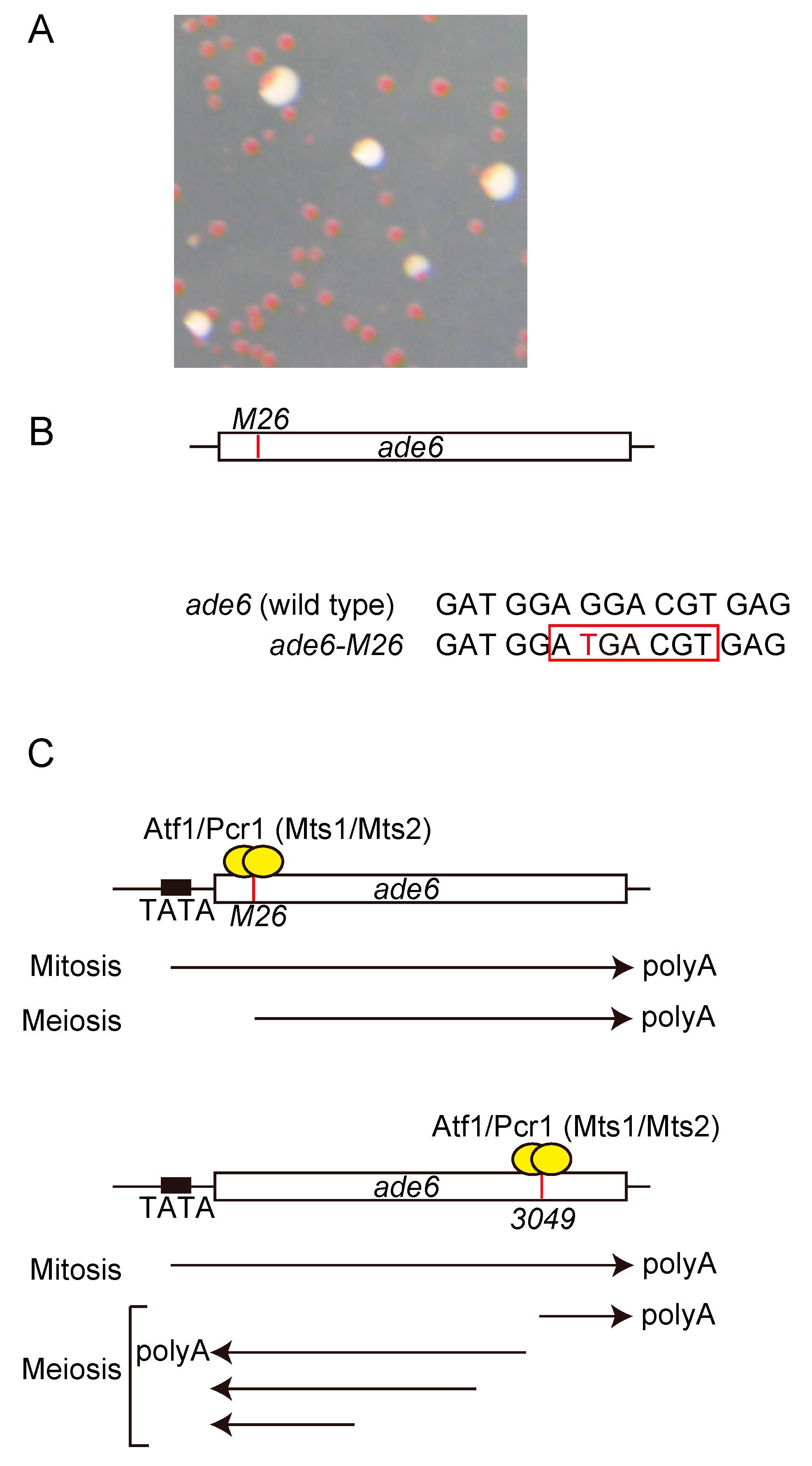
2. ade6-M26, a Meiotic Hotspot Created by a Mono-Nucleotide Substitution Mutation

The ade6 gene encodes phosphoribosylaminoimidazole carboxylase, which is required for purine biosynthesis. The adenine auxotrophic strains with a nonsense mutation in the ade6 gene form red (or pink) colonies on yeast extract medium with a limiting adenine concentration [41], providing a visual distinction between wild-type and mutant alleles (Figure 2A). Several hundred ade6 mutations induced by ultraviolet or X-ray irradiation or nitrous acid treatment were assessed for recombination to map the mutation point, and the mutation ade6-M26 was shown to create a meiotic recombination hotspot [42]. The ade6-M26 hotspot contains a G-to-T substitution in the ade6 open reading frame (ORF) [43,44] (Figure 2B, Red letter). During the early stage of genetic analysis in meiotic recombination, the rec genes required for meiotic recombination were screened using this system and several rec genes were identified [45,46]. Originally, ade6-M26 hotspot was identified as a gene conversion (NCO) hotspot [42] (Figure 1B, left SASD). Later, it was showed that this mutation hotspot also augments the meiotic CO recombination [47] (Figure 1B, left Crossover).

3. Transcription Factor Required for Meiotic Recombination at M26

The M26-mutation is a nonsense mutation that creates a cyclic adenosine monophosphate (cAMP)-responsive element (CRE)-like heptanucleotide sequence 5′-ATGACGT-3′ (Figure 2B, Red square). Later, biding factors to this CRE-like heptanucleotide sequence was identified and named as Mst1 and Mst2 and it was revealed that these bind factors activate meiotic recombination [48,49,50] (Figure 2C). After this identification, three groups almost simultaneously identified Atf1, a basic leucine zipper (bZIP) transcription factor that is activated by the mitogen-activated protein kinase (MAPK) pathway [51,52,53]. Moreover, a similar bZIP protein, Pcr1, was also identified [54], and Mts1 and Mts2 were found to be identical to Atf1 and Pcr1, respectively [48]. In fission yeast, meiosis is induced by the depletion of the nitrogen source, and the signal is mediated by PKA and stress-activated MAPK signal pathway, resulting in the activation of the genes required for meiosis [55]. In addition, during nitrogen starvation, the phosphorylation of Atf1 through a stress-activated MAPK, Spc1, is required for conjugation and meiosis [52]. Atf1 phosphorylation is also required for the activation of the fbp1 gene upon glucose starvation stress via the modulation of chromatin configuration in fbp1 upstream sequence [56,57,58,59,60,61,62,63]. Thus, Atf1 phosphorylation is involved in ade6-M26 hotspot activation, gene induction during mating and meiosis, and fbp1 transcription. Interestingly, the canonical Spc1-phospholylation sites in Atf1 are required for fbp1 transcription but not for the activation of the ade6-M26 hotspot or induction of meiotic genes, indicating a distinctly different phosphorylation mechanism required for ade6-M26 hotspot activation, gene transcriptional induction in mating and meiosis, and fbp1 transcription [64]. Similarly, a meiotic recombination hotspot, ade6-3049 allele possesses a point mutation, which concurrently creates a nonsense mutation and a CRE-like sequence at the 3′ region of ade6 gene [37]. Both ade6-M26 and ade6-3049 strains express shorter versions of mRNA initiated from M26 and 3049 point mutations, respectively [37,65] (Figure 2C), suggesting a link between transcriptional initiation and meiotic recombination. Involvement of transcriptions in the meiotic recombination activation at ade6-M26 is also highlighted by the following facts. First, hotspot activity of ade6-M26 requires ade6 promoter [66,67]. Second, insertions of other transcription factor binding sequences at the M26 mutation site in ade6 gene also created meiotic recombination hotspot [68,69]. This indicates that meiotic recombination hotspots can be created by transcription factors other than the Atf1-Pcr1 transcription factor required for the ade6-M26 hotspot [48,49,50]. This was proved to be the case when the loss of each transcription factor bound to the inserted binding site inactivated hotspot activity [70]. Moreover, the subset of naturally occurring M26 sequence sites (M26 heptanucleotide sequence occurring in wild-type cells) that facilitate meiotic recombination is implicated in approximately 20% of all natural meiotic recombination events in the fission yeast genome [71]. This might also be the case in another highly diverged yeast, Saccharomyces cerevisiae (budding yeast), since binding sites for the Atf1 ortholog Sko1 tend to be highly enriched for meiotic DSBs [35]. Furthermore, other sequence-specific binding proteins proven to activate hotspots in the ade6 gene in fission yeast (Php2, Php3, Php5, Rst2) [68,69] also have orthologs in budding yeast (Hap2, Hap3, Hap5, Adr1) in which DSBs are preferentially directed to their binding sites [35]. Such sequence-specific transcription factor-dependent hotspots created in the fission yeast ade6 gene are mediated through a common chromatin modulation system [72] (see Section 4). Taken together, these results suggest that some shared regulatory systems between recombination and transcription may play a role in a subset of meiotic recombination sites created by the binding sequence of transcription factors.

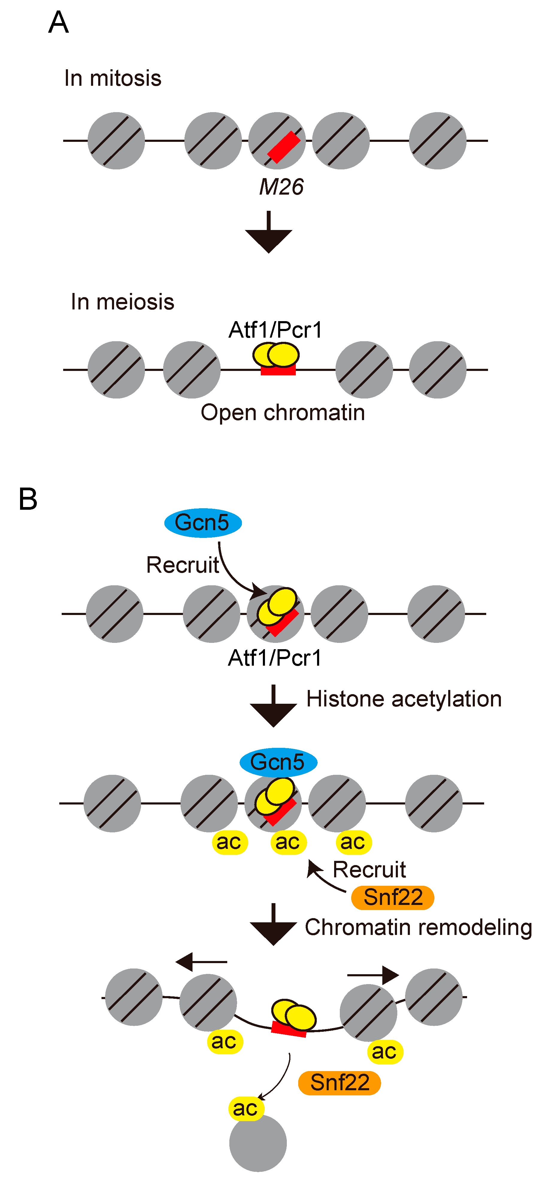
4. Chromatin Modulation Mechanisms Involved in the Regulation of ade6-M26 Meiotic Recombination Hotspot

The chromatin configurations play pivotal role in the transcriptional control (reviewed in [38,39]). This can be attributed to the fact that tightly packed chromatin limits the accessibility of trans-acting proteins such as transcription factors. Regulation of chromatin structure involves two major classes of chromatin modulation machinery. The first comprises histone modifications, such as acetylation, that are involved in chromatin modulation. For example, histone acetyltransferases add acetyl groups that are usually associated with active chromatin [73,74]. The second class comprises ATP-dependent chromatin remodelers. ATP-dependent chromatin remodelers alter chromatin configuration by sliding and disassembling nucleosomes in an ATP-dependent manner [75]. The yeast Swi2/Snf2 family ATP-dependent chromatin remodeler contains a helicase/ATPase motif as a catalytic domain and a bromodomain that recognizes acetylated histones [76,77]. Proteins carrying bromodomains, including Swi2/Snf2 family ATP-dependent chromatin remodelers, can serve as decoders for histone acetylation.
The direct involvement of histone acetylation and ATP-dependent chromatin remodeler-mediated chromatin remodeling in the regulation of meiotic recombination was first demonstrated in ade6-M26 [78]. The chromatin configurations around the M26 mutation site were assessed based on the sensitivity of the chromatin DNA to micrococcal nuclease (MNase). The chromatin configuration at the M26 mutation point was shown to convert into an open state during meiosis, which requires the involvement of Atf1, Pcr1, and CRE-like heptanucleotide sequences [79] (Figure 3A). Acetylation of the histones H3 and H4 around the M26 mutation site is augmented in an M26- and Atf1/Pcr1-dependent manner early in meiosis. Gcn5 histone acetyl transferase is involved in the histone H3 acetylation around M26 [78]. Moreover, deletion of the gcn5 gene results in a significant delay in chromatin remodeling and partial reduction in the M26 meiotic recombination frequency [78]. Deletion of the snf22 gene (which encodes Swi2/Snf2-chromatin remodeler) abolishes chromatin remodeling and results in the critical reduction of meiotic recombination at ade6-M26 site [78]. These results suggest that Gcn5 histone acetyl transferase and Snf22 Swi2/Snf2-chromatin remodeler cooperatively alter the local chromatin structure to activate meiotic recombination at M26 in a site-specific manner (Figure 3B). Later, two CHD-1 family chromatin remodelers (Hrp1 and Hrp3) involved in chromatin regulation, a Gcn5 histone acetyltransferase component (Ada2), a member of the Moz-Ybf2/Sas3-Sas2-Tip60 family histone acetyltransferase (Mst2), and a histone variant H2A.Z have been reported in the regulation of the of ade6-M26 hotspot activity [80,81]. Moreover, proteomic analysis of the ade6-M26 recombination hotspot revealed that chromatin-mediated regulators, including histone chaperones (Nap1, Hip1/Hir1), subunits of the Ino80 complex (Arp5, Arp8), a DNA helicase/E3 ubiquitin ligase (Rrp2), components of a Swi2/Snf2 family remodeling complex (Swr1, Swc2), and a nucleosome evictor (Fft3/Fun30), interact with the ade6-M26 recombination hotspot during meiosis [82]. Taken together, these studies revealed that remarkably diverse chromatin modulators and histone post-translational modifications participate in the regulation of M26 meiotic recombination.

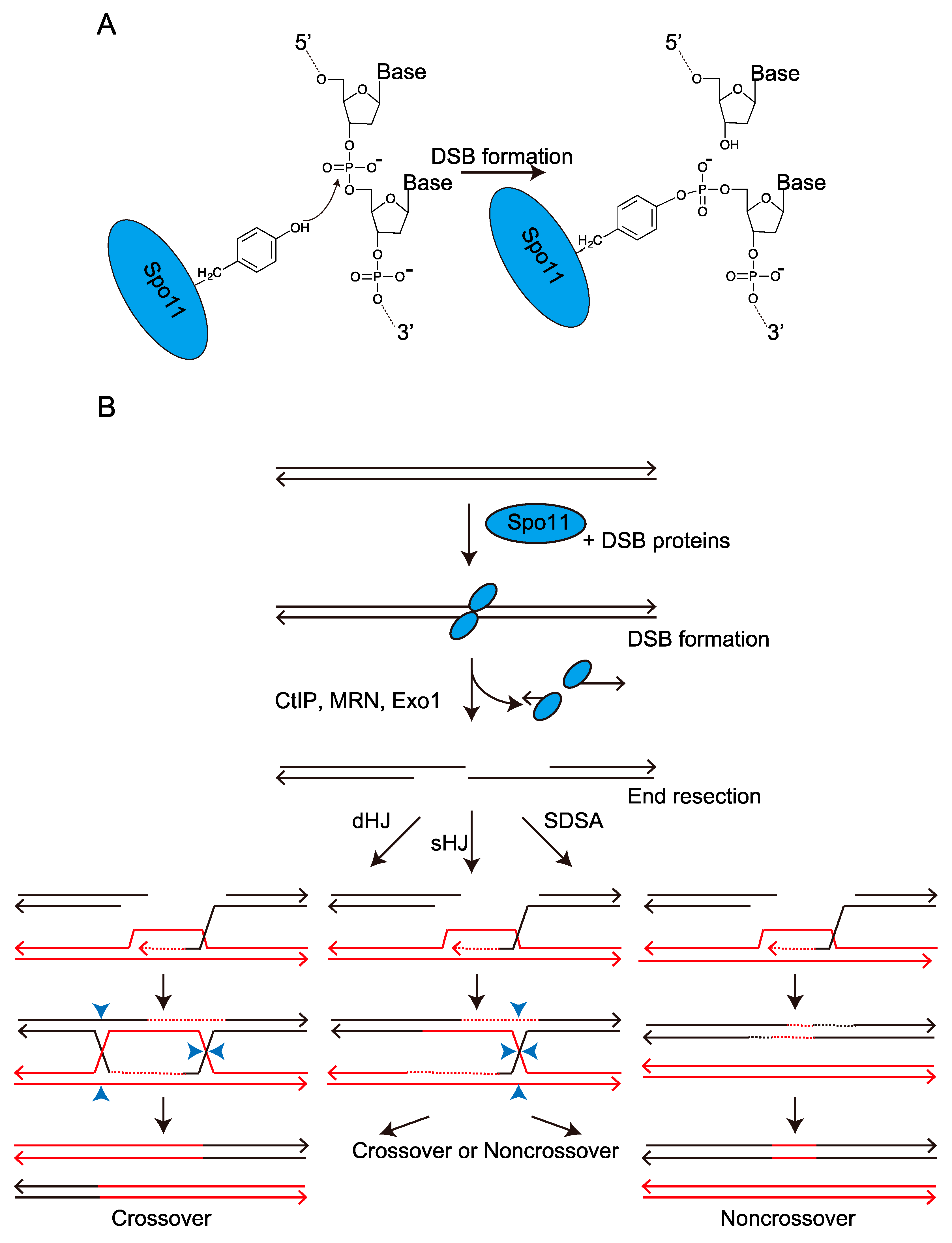
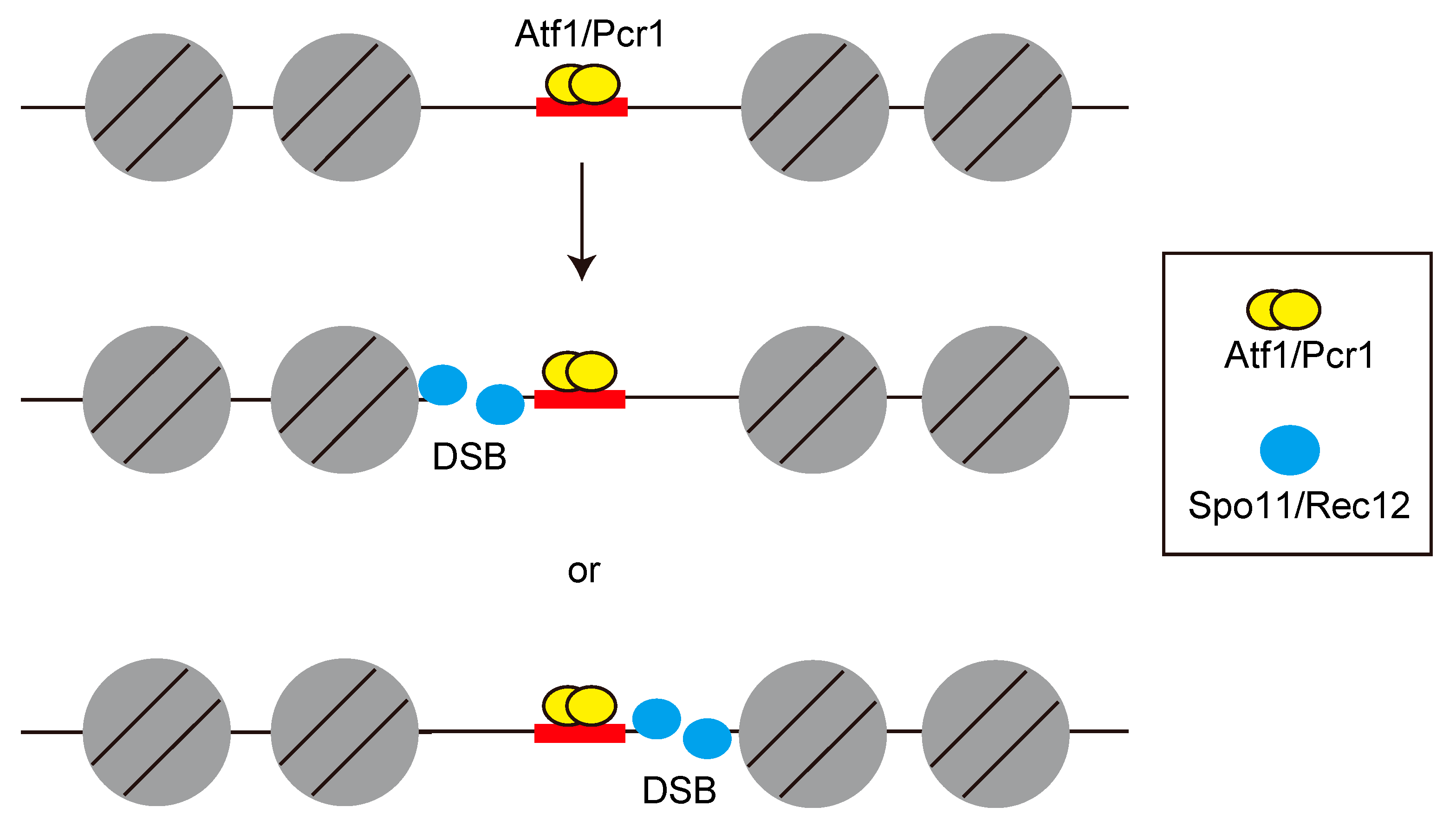
5. Meiotic Double-Strand Break Formation around M26 Mutation Site

Meiotic recombination is thought to be initiated by either single-strand breaks or double-strand breaks in the DNA [83,84]; however, which of the two is specifically responsible has been a long-standing question. This controversy was resolved by physical detection of double-strand breaks (DSBs) in budding yeast during meiosis [85]. The formation of DSBs in budding yeast requires multiple gene products that are also required for meiotic recombination and proper homologous chromosome segregation [86]. One such gene product, Spo11 is required for the formation of meiotic DSBs [87]. The fission yeast Spo11 homolog, Rec12 protein, is also essential for meiotic recombination [45], and thus this protein is hereafter referred to as Spo11/Rec12. The catalytic tyrosine residue of Spo11/Rec12 is pivotal for its action, suggesting that the DNA–Spo11/Rec12 covalent bond is mediated by this tyrosine residue [88,89] (Figure 1A). These results suggest a conserved mechanism for meiotic DSB formation and recombination. However, the initial attempt to assess meiotic DSBs in the fission yeast genome suffered due to a lack of a detectable level of DSBs at or near the ade6-M26 hotspot [89]. The unresolved question whether DSBs are associated with the ade6-M26 hotspot was resolved by using the rad50s mutant. This mutant, originally obtained from budding yeast, was found to be defective in end resection [90,91], and covalent Spo11/Rec12-DNA complexes were detected in this mutant [87,92] (DSB formation and end resection step in Figure 1B). In fission yeast, the rad50s mutant also shows accumulation of DSBs and no detectable repair [93]. Multiple DSBs were detected on both sides near the M26 mutation site (5′-ATGACGT-3′ heptanucleotide sequence) with closely spaced M26 sites [94]. Interestingly, the M26 site itself lies in a region of approximately 70 bp that is free of visible breaks (Figure 4), suggesting that Spo11/Rec12 is recruited to the genomic DNA where the chromatin configuration has been meiotically opened by the Atf1-Pcr1 and stochastic digest the surrounding sequences.

6. Induction of Meiotic DSB via the Expression of Long ncRNA

DSBs induced by Spo11/Rec12 are not uniformly distributed in the genome [87,92,93], and a significant overlap between the DSB sites and the regions of ncRNA expression was identified by bioinformatics analysis in fission yeast [36]. These facts suggest that the accessibility of Spo11/Rec12 to the genome could be controlled by ncRNAs. In this section, the role of ncRNA transcriptions in the induction of meiotic DSB is discussed.
The ncRNAs can be classified into two classes, small RNAs and long ncRNAs (lncRNAs), according to their length. The first class of ncRNAs, small RNAs are ncRNAs comprised of less than 200 nucleotides. siRNAs and miRNAs is well characterized examples of small RNAs. Small RNAs regulate gene expression via degradation of mRNA and inhibition of translation through their interaction with RNA-induced silencing complexes [95]. The second class of ncRNAs is long ncRNA (lncRNA) carrying over 200 nucleotides length. They are involved in various biological processes, including gene regulation, development, nuclear organization, and cancer development [96,97]. Deregulation of several lncRNAs in cancer cells has been identified and such lncRNAs can be used as a cancer marker in the diagnosis [98,99]. The expressed lncRNAs may serve as enhancers, scaffolds, or decoys by interacting with other RNAs or proteins, resulting in the aberrant activation of genes required for cancer proliferations [100]. In addition to the roles played by transcribed lncRNA molecules, several studies have suggested the importance of cis-acting RNA polymerase II-transcribing nascent lncRNAs in the regulation of neighboring genes through their effects on histone modifications [101,102]. The lnc RNAs expressed from gene promoter region are referred to as promoter-associated lncRNA, which are categorized into most abundant ncRNA, and play pivotal roles in the regulation of downstream genes [103].
The fission yeast fbp1 gene has been extensively analyzed for understanding gene regulation systems (reviewed in [104]), and several promoter-associated lncRNAs are identified in its promoter region [59,105]. These lncRNAs are referred to as metabolic stress-induced long-ncRNA (mlonRNA) and are expressed from specific cis-element, mlon-BOX, before induction of fbp1 mRNA upon glucose starvation stress [106,107]. Transcriptional initiation of mlonRNAs modulates chromatin structure around the fbp1-promoter and converts it into an open configuration, thereby promoting the binding of transcription factors to the promoter [59]. A significant overlap between the mlon-BOX and meiotic DSB sites in the fission yeast genome suggests interesting hypothesis that mlonRNA transcriptional initiations determine the DSB site via the modulation of chromatin architecture [65,108]. This hypothesis was assessed by inserting the mlon-BOX sequence into the ade6-M26 hotspot. To mimic fbp1 regulation, in which the mlon-BOX sequence is located 200 bp downstream from the Atf1 binding site in fbp1, the mlon-BOX sequence was placed 200 bp downstream from the M26 mutation site in ade6-M26 (Figure 5A,B). This insertion caused additional activation of transcription at the insertion site (Figure 5B) and stimulated meiotic recombination via local chromatin remodeling [65] (Figure 5C). These results suggest that mlonRNA transcription plays a universal role in chromatin remodeling and the regulation of transcription and recombination. Taken together with the regulatory mechanisms discussed in earlier sections, following model can be illustrated. The accessibility of Spo11/Rec12 to the genome could be one of the critical determinants of DSB sites and that Spo11/Rec12 might preferentially make DSBs at genomic regions showing open chromatin configurations induced by cis-acting RNA polymerase II transcribing nascent mlonRNAs.

7. Summary and Perspective

In this review, we summarized the multiple layers of regulation involved in meiotic recombination revealed from the analysis of the fission yeast ade6-M26 hotspot as a model system. Analyzed regulatory layers include transcription factors, chromatin modulators (histone modifiers and ADCRs), and lncRNA transcriptions. Although intensive analyses have been conducted using this model system, several important questions remained unsolved. The first question is what the proportion of meiotic recombination sites is dependent on transcription factors. The subset of naturally occurring M26 (binding sequence for the Atf1 transcription factor) sites that facilitate meiotic recombination supposedly mediate approximately 20% of all meiotic recombination events in the fission yeast genome [71]. The chromatin structure at prominent natural hotspots, such as major break site 1 (mbs1) in fission yeast, shows a constitutive open configuration independent of Atf1 [109,110]. These constitutive hotspots might be important for ensuring the basal recombination level required for CO, which is essential for the progression of meiosis. In contrast, recombination sites linked to transcription factor-binding sequences, such as M26 sites, might be important for the plasticity of meiotic recombination hotspots. Environmental conditions and stresses also affect the frequency and distribution of meiotic recombination [111]. The binding patterns of the transcription factors associated with meiotic recombination could possibly be affected by environmental conditions and thereby affecting the frequency and distribution of recombination. This was proved to be the case when hotspots created in the ade6 gene carrying a binding sequence for different transcription factors such as Atf1-Pcr1, Rst2, or Php2-Php3-Php5 were affected differently by distinct types of stresses [70]. The second unsolved question involves the factor(s) and/or conditions required for creating M26 sequence hotspots. The ade6 gene has a CRE sequence (5′-TGACGTCA-3′) at +1131 to +1136 bp starting from the first A in the ade6 ORF. However, Atf1-Pcr1 could not bind to this CRE sequence and could not facilitate recombination. Consistently, little meiotic DSB is introduced at CRE sequence in the regulatory region of fbp1 gene (our unpublished observation), indicating that not the all CRE sequences in fission yeast genome are meiotic recombination hotspot. Similarly, the HIS4 locus in budding yeast was initially shown to be linked to the activity of the transcription factors Bas1 and Bas2 [112], but subsequent genome-wide analyses failed to detect a clear correlation between Bas1/2 activity and DSB formation [113]. Thus, other factor(s) and/or condition(s) may be involved in making these transcription factors-binding sites meiotic recombination hotspot. Further research is required to clarify these questions.

Funding

This research was funded by Tokyo Metropolitan Government: Tokyo Metropolitan Government Advanced; Research Grant, Number (R3-2); Japan Society for the Promotion of Science: JP21K19235; Japan Society for the Promotion of Science: JP20H04337; Japan Society for the Promotion of Science: 19KK0210.

Institutional Review Board Statement

Not applicable.

Informed Consent Statement

Not applicable.

Acknowledgments

This work was supported by the Tokyo Metropolitan Government Advanced Research Grant Number (R3-2), JSPS KAKENHI (JP21K19235, JP20H04337, 19KK0210), Yamada Science Foundation, and Takeda Science Foundation (to K. H.).

Conflicts of Interest

The authors declare no conflict of interest.

References

  1. Láscarez-Lagunas, L.; Martinez-Garcia, M.; Colaiácovo, M. SnapShot: Meiosis—Prophase I. Cell 2020, 181, 1442–1442.e1. [Google Scholar] [CrossRef] [PubMed]
  2. Zickler, D.; Kleckner, N. Meiotic chromosomes: Integrating structure and function. Annu. Rev. Genet. 1999, 33, 603–754. [Google Scholar] [CrossRef] [PubMed]
  3. Keeney, S.; Neale, M.J. Initiation of meiotic recombination by formation of DNA double-strand breaks: Mechanism and regulation. Biochem. Soc. Trans. 2006, 34, 523–525. [Google Scholar] [CrossRef] [Green Version]
  4. Kim, S.; Peterson, S.E.; Jasin, M.; Keeney, S. Mechanisms of germ line genome instability. Semin. Cell Dev. Biol. 2016, 54, 177–187. [Google Scholar] [CrossRef] [PubMed]
  5. Lam, I.; Keeney, S. Mechanism and regulation of meiotic recombination initiation. Cold Spring Harb. Perspect. Biol. 2014, 7, a016634. [Google Scholar] [CrossRef] [Green Version]
  6. Arora, C.; Kee, K.; Maleki, S.; Keeney, S. Antiviral protein Ski8 is a direct partner of Spo11 in meiotic DNA break formation, independent of its cytoplasmic role in RNA metabolism. Mol. Cell 2004, 13, 549–559. [Google Scholar] [CrossRef] [PubMed]
  7. Li, J.; Hooker, G.W.; Roeder, G.S. Saccharomyces cerevisiae Mer2, Mei4 and Rec114 form a complex required for meiotic double-strand break formation. Genetics 2006, 173, 1969–1981. [Google Scholar] [CrossRef] [Green Version]
  8. Maleki, S.; Neale, M.J.; Arora, C.; Henderson, K.A.; Keeney, S. Interactions between Mei4, Rec114, and other proteins required for meiotic DNA double-strand break formation in Saccharomyces cerevisiae. Chromosoma 2007, 116, 471–486. [Google Scholar] [CrossRef] [Green Version]
  9. Miyoshi, T.; Ito, M.; Kugou, K.; Yamada, S.; Furuichi, M.; Oda, A.; Yamada, T.; Hirota, K.; Masai, H.; Ohta, K. A central coupler for recombination initiation linking chromosome architecture to S phase checkpoint. Mol. Cell 2012, 47, 722–733. [Google Scholar] [CrossRef] [Green Version]
  10. Panizza, S.; Mendoza, M.A.; Berlinger, M.; Huang, L.; Nicolas, A.; Shirahige, K.; Klein, F. Spo11-accessory proteins link double-strand break sites to the chromosome axis in early meiotic recombination. Cell 2011, 146, 372–383. [Google Scholar] [CrossRef]
  11. Sasanuma, H.; Hirota, K.; Fukuda, T.; Kakusho, N.; Kugou, K.; Kawasaki, Y.; Shibata, T.; Masai, H.; Ohta, K. Cdc7-dependent phosphorylation of Mer2 facilitates initiation of yeast meiotic recombination. Genes Dev. 2008, 22, 398–410. [Google Scholar] [CrossRef] [PubMed] [Green Version]
  12. Pommier, Y.; Sun, Y.; Huang, S.N.; Nitiss, J.L. Roles of eukaryotic topoisomerases in transcription, replication and genomic stability. Nat. Rev. Mol. Cell Biol. 2016, 17, 703–721. [Google Scholar] [CrossRef] [PubMed]
  13. Borde, V.; Lin, W.; Novikov, E.; Petrini, J.H.; Lichten, M.; Nicolas, A. Association of Mre11p with double-strand break sites during yeast meiosis. Mol. Cell 2004, 13, 389–401. [Google Scholar] [CrossRef] [PubMed]
  14. Farah, J.A.; Cromie, G.A.; Smith, G.R. Ctp1 and Exonuclease 1, alternative nucleases regulated by the MRN complex, are required for efficient meiotic recombination. Proc. Natl. Acad. Sci. USA 2009, 106, 9356–9361. [Google Scholar] [CrossRef] [Green Version]
  15. Mimitou, E.P.; Yamada, S.; Keeney, S. A global view of meiotic double-strand break end resection. Science 2017, 355, 40–45. [Google Scholar] [CrossRef] [PubMed] [Green Version]
  16. Ranjha, L.; Howard, S.M.; Cejka, P. Main steps in DNA double-strand break repair: An introduction to homologous recombination and related processes. Chromosoma 2018, 127, 187–214. [Google Scholar] [CrossRef] [Green Version]
  17. Shibata, T.; Iwasaki, W.; Hirota, K. The intrinsic ability of double-stranded DNA to carry out D-loop and R-loop formation. Comput. Struct. Biotechnol. J. 2020, 18, 3350–3360. [Google Scholar] [CrossRef]
  18. Shinohara, A.; Shinohara, M. Roles of RecA homologues Rad51 and Dmc1 during meiotic recombination. Cytogenet. Genome Res. 2004, 107, 201–207. [Google Scholar] [CrossRef]
  19. Haber, J.E. DNA Repair: The Search for Homology. Bioessays 2018, 40, e1700229. [Google Scholar] [CrossRef]
  20. Bannister, L.A.; Pezza, R.J.; Donaldson, J.R.; de Rooij, D.G.; Schimenti, K.J.; Camerini-Otero, R.D.; Schimenti, J.C. A dominant, recombination-defective allele of Dmc1 causing male-specific sterility. PLoS Biol. 2007, 5, e105. [Google Scholar] [CrossRef]
  21. Cao, D.; Shi, F.; Guo, C.; Liu, Y.; Lin, Z.; Zhang, J.; Li, R.H.W.; Yao, Y.; Liu, K.; Ng, E.H.Y.; et al. A pathogenic DMC1 frameshift mutation causes nonobstructive azoospermia but not primary ovarian insufficiency in humans. Mol. Hum. Reprod. 2021, 27, 58–70. [Google Scholar] [CrossRef] [PubMed]
  22. Chen, Z.; Yang, H.; Pavletich, N.P. Mechanism of homologous recombination from the RecA-ssDNA/dsDNA structures. Nature 2008, 453, 489–494. [Google Scholar] [CrossRef] [PubMed]
  23. Shibata, T.; Cunningham, R.P.; DasGupta, C.; Radding, C.M. Homologous pairing in genetic recombination: Complexes of recA protein and DNA. Proc. Natl. Acad. Sci. USA 1979, 76, 5100–5104. [Google Scholar] [CrossRef] [PubMed] [Green Version]
  24. Allers, T.; Lichten, M. Differential timing and control of noncrossover and crossover recombination during meiosis. Cell 2001, 106, 47–57. [Google Scholar] [CrossRef] [Green Version]
  25. Bishop, D.K.; Zickler, D. Early decision; meiotic crossover interference prior to stable strand exchange and synapsis. Cell 2004, 117, 9–15. [Google Scholar] [CrossRef] [Green Version]
  26. Cromie, G.A.; Hyppa, R.W.; Taylor, A.F.; Zakharyevich, K.; Hunter, N.; Smith, G.R. Single Holliday junctions are intermediates of meiotic recombination. Cell 2006, 127, 1167–1178. [Google Scholar] [CrossRef] [PubMed] [Green Version]
  27. Cooper, T.J.; Garcia, V.; Neale, M.J. Meiotic DSB patterning: A multifaceted process. Cell Cycle 2016, 15, 13–21. [Google Scholar] [CrossRef] [Green Version]
  28. de Massy, B. Initiation of meiotic recombination: How and where? Conservation and specificities among eukaryotes. Annu. Rev. Genet. 2013, 47, 563–599. [Google Scholar] [CrossRef]
  29. Ergoren, M.C. The Control of Meiotic Recombination in the Human Genome. Crit. Rev. Eukaryot. Gene Expr. 2018, 28, 187–204. [Google Scholar] [CrossRef]
  30. Tock, A.J.; Henderson, I.R. Hotspots for Initiation of Meiotic Recombination. Front. Genet. 2018, 9, 521. [Google Scholar] [CrossRef]
  31. Blitzblau, H.G.; Bell, G.W.; Rodriguez, J.; Bell, S.P.; Hochwagen, A. Mapping of meiotic single-stranded DNA reveals double-stranded-break hotspots near centromeres and telomeres. Curr. Biol. 2007, 17, 2003–2012. [Google Scholar] [CrossRef] [PubMed] [Green Version]
  32. Buhler, C.; Borde, V.; Lichten, M. Mapping meiotic single-strand DNA reveals a new landscape of DNA double-strand breaks in Saccharomyces cerevisiae. PLoS Biol. 2007, 5, e324. [Google Scholar] [CrossRef] [PubMed]
  33. Fowler, K.R.; Sasaki, M.; Milman, N.; Keeney, S.; Smith, G.R. Evolutionarily diverse determinants of meiotic DNA break and recombination landscapes across the genome. Genome. Res. 2014, 24, 1650–1664. [Google Scholar] [CrossRef] [PubMed] [Green Version]
  34. Gerton, J.L.; DeRisi, J.; Shroff, R.; Lichten, M.; Brown, P.O.; Petes, T.D. Global mapping of meiotic recombination hotspots and coldspots in the yeast Saccharomyces cerevisiae. Proc. Natl. Acad. Sci. USA 2000, 97, 11383–11390. [Google Scholar] [CrossRef] [PubMed] [Green Version]
  35. Pan, J.; Sasaki, M.; Kniewel, R.; Murakami, H.; Blitzblau, H.G.; Tischfield, S.E.; Zhu, X.; Neale, M.J.; Jasin, M.; Socci, N.D.; et al. A hierarchical combination of factors shapes the genome-wide topography of yeast meiotic recombination initiation. Cell 2011, 144, 719–731. [Google Scholar] [CrossRef] [Green Version]
  36. Wahls, W.P.; Siegel, E.R.; Davidson, M.K. Meiotic recombination hotspots of fission yeast are directed to loci that express non-coding RNA. PLoS ONE 2008, 3, e2887. [Google Scholar] [CrossRef] [Green Version]
  37. Yamada, S.; Okamura, M.; Oda, A.; Murakami, H.; Ohta, K.; Yamada, T. Correlation of Meiotic DSB Formation and Transcription Initiation Around Fission Yeast Recombination Hotspots. Genetics 2017, 206, 801–809. [Google Scholar] [CrossRef] [Green Version]
  38. Wolffe, A.P. Nucleosome positioning and modification: Chromatin structures that potentiate transcription. Trends Biochem. Sci. 1994, 19, 240–244. [Google Scholar] [CrossRef]
  39. Wolffe, A.P. Histones, nucleosomes and the roles of chromatin structure in transcriptional control. Biochem. Soc. Trans. 1997, 25, 354–358. [Google Scholar] [CrossRef] [Green Version]
  40. Modliszewski, J.L.; Copenhaver, G.P. Meiotic recombination gets stressed out: CO frequency is plastic under pressure. Curr. Opin. Plant. Biol. 2017, 36, 95–102. [Google Scholar] [CrossRef]
  41. Fisher, C.R. Enzymology of the pigmented adenine-requiring mutants of Saccharomyces and Schizosaccharomyces. Biochem. Biophys. Res. Commun 1969, 34, 306–310. [Google Scholar] [CrossRef] [PubMed]
  42. Gutz, H. Site Specific Induction of Gene Conversion in Schizosaccharomyces pombe. Genetics 1971, 69, 317–337. [Google Scholar] [CrossRef] [PubMed]
  43. Ponticelli, A.S.; Sena, E.P.; Smith, G.R. Genetic and physical analysis of the M26 recombination hotspot of Schizosaccharomyces pombe. Genetics 1988, 119, 491–497. [Google Scholar] [CrossRef] [PubMed]
  44. Szankasi, P.; Heyer, W.D.; Schuchert, P.; Kohli, J. DNA sequence analysis of the ade6 gene of Schizosaccharomyces pombe. Wild-type and mutant alleles including the recombination host spot allele ade6-M26. J. Mol. Biol. 1988, 204, 917–925. [Google Scholar] [CrossRef]
  45. De Veaux, L.C.; Hoagland, N.A.; Smith, G.R. Seventeen complementation groups of mutations decreasing meiotic recombination in Schizosaccharomyces pombe. Genetics 1992, 130, 251–262. [Google Scholar] [CrossRef]
  46. Ponticelli, A.S.; Smith, G.R. Meiotic recombination-deficient mutants of Schizosaccharomyces pombe. Genetics 1989, 123, 45–54. [Google Scholar] [CrossRef]
  47. Schuchert, P.; Kohli, J. The Ade6-M26 Mutation of Schizosaccharomyces pombe Increases the Frequency of Crossing over. Genetics 1988, 119, 507–515. [Google Scholar] [CrossRef]
  48. Kon, N.; Krawchuk, M.D.; Warren, B.G.; Smith, G.R.; Wahls, W.P. Transcription factor Mts1/Mts2 (Atf1/Pcr1, Gad7/Pcr1) activates the M26 meiotic recombination hotspot in Schizosaccharomyces pombe. Proc. Natl. Acad. Sci. USA 1997, 94, 13765–13770. [Google Scholar] [CrossRef] [Green Version]
  49. Kon, N.; Schroeder, S.C.; Krawchuk, M.D.; Wahls, W.P. Regulation of the Mts1-Mts2-dependent ade6-M26 meiotic recombination hot spot and developmental decisions by the Spc1 mitogen-activated protein kinase of fission yeast. Mol. Cell Biol. 1998, 18, 7575–7583. [Google Scholar] [CrossRef] [Green Version]
  50. Wahls, W.P.; Smith, G.R. A heteromeric protein that binds to a meiotic homologous recombination hot spot: Correlation of binding and hot spot activity. Genes Dev. 1994, 8, 1693–1702. [Google Scholar] [CrossRef]
  51. Kanoh, J.; Watanabe, Y.; Ohsugi, M.; Iino, Y.; Yamamoto, M. Schizosaccharomyces pombe gad7+ encodes a phosphoprotein with a bZIP domain, which is required for proper G1 arrest and gene expression under nitrogen starvation. Genes Cells 1996, 1, 391–408. [Google Scholar] [CrossRef] [PubMed]
  52. Shiozaki, K.; Russell, P. Conjugation, meiosis, and the osmotic stress response are regulated by Spc1 kinase through Atf1 transcription factor in fission yeast. Genes Dev. 1996, 10, 2276–2288. [Google Scholar] [CrossRef] [PubMed] [Green Version]
  53. Takeda, T.; Toda, T.; Kominami, K.; Kohnosu, A.; Yanagida, M.; Jones, N. Schizosaccharomyces pombe atf1+ encodes a transcription factor required for sexual development and entry into stationary phase. EMBO J. 1995, 14, 6193–6208. [Google Scholar] [CrossRef] [PubMed]
  54. Watanabe, Y.; Yamamoto, M. Schizosaccharomyces pombe pcr1+ encodes a CREB/ATF protein involved in regulation of gene expression for sexual development. Mol. Cell Biol. 1996, 16, 704–711. [Google Scholar] [CrossRef] [PubMed] [Green Version]
  55. Yamamoto, M. Regulation of meiosis in fission yeast. Cell Struct. Funct. 1996, 21, 431–436. [Google Scholar] [CrossRef] [Green Version]
  56. Adachi, A.; Senmatsu, S.; Asada, R.; Abe, T.; Hoffman, C.S.; Ohta, K.; Hirota, K. Interplay between chromatin modulators and histone acetylation regulates the formation of accessible chromatin in the upstream regulatory region of fission yeast fbp1. Genes Genet. Syst. 2018, 92, 267–276. [Google Scholar] [CrossRef] [Green Version]
  57. Hirota, K.; Hasemi, T.; Yamada, T.; Mizuno, K.I.; Hoffman, C.S.; Shibata, T.; Ohta, K. Fission yeast global repressors regulate the specificity of chromatin alteration in response to distinct environmental stresses. Nucleic Acids Res. 2004, 32, 855–862. [Google Scholar] [CrossRef]
  58. Hirota, K.; Hoffman, C.S.; Shibata, T.; Ohta, K. Fission yeast Tup1-like repressors repress chromatin remodeling at the fbp1+ promoter and the ade6-M26 recombination hotspot. Genetics 2003, 165, 505–515. [Google Scholar] [CrossRef]
  59. Hirota, K.; Miyoshi, T.; Kugou, K.; Hoffman, C.S.; Shibata, T.; Ohta, K. Stepwise chromatin remodelling by a cascade of transcription initiation of non-coding RNAs. Nature 2008, 456, 130–134. [Google Scholar] [CrossRef]
  60. Janoo, R.T.; Neely, L.A.; Braun, B.R.; Whitehall, S.K.; Hoffman, C.S. Transcriptional regulators of the Schizosaccharomyces pombe fbp1 gene include two redundant Tup1p-like corepressors and the CCAAT binding factor activation complex. Genetics 2001, 157, 1205–1215. [Google Scholar] [CrossRef]
  61. Koda, W.; Senmatsu, S.; Abe, T.; Hoffman, C.S.; Hirota, K. Reciprocal stabilization of transcription factor binding integrates two signaling pathways to regulate fission yeast fbp1 transcription. Nucleic Acids Res. 2021, 49, 9809–9820. [Google Scholar] [CrossRef]
  62. Neely, L.A.; Hoffman, C.S. Protein kinase A and mitogen-activated protein kinase pathways antagonistically regulate fission yeast fbp1 transcription by employing different modes of action at two upstream activation sites. Mol. Cell Biol. 2000, 20, 6426–6434. [Google Scholar] [CrossRef]
  63. Takemata, N.; Oda, A.; Yamada, T.; Galipon, J.; Miyoshi, T.; Suzuki, Y.; Sugano, S.; Hoffman, C.S.; Hirota, K.; Ohta, K. Local potentiation of stress-responsive genes by upstream noncoding transcription. Nucleic Acids Res. 2016, 44, 5174–5189. [Google Scholar] [CrossRef] [Green Version]
  64. Sánchez-Mir, L.; Fraile, R.; Ayté, J.; Hidalgo, E. Phosphorylation of the Transcription Factor Atf1 at Multiple Sites by the MAP Kinase Sty1 Controls Homologous Recombination and Transcription. J. Mol. Biol. 2020, 432, 5430–5446. [Google Scholar] [CrossRef] [PubMed]
  65. Senmatsu, S.; Asada, R.; Oda, A.; Hoffman, C.S.; Ohta, K.; Hirota, K. lncRNA transcription induces meiotic recombination through chromatin remodelling in fission yeast. Commun. Biol. 2021, 4, 295. [Google Scholar] [CrossRef]
  66. Ponticelli, A.S.; Smith, G.R. Chromosomal context dependence of a eukaryotic recombinational hot spot. Proc. Natl. Acad. Sci. USA 1992, 89, 227–231. [Google Scholar] [CrossRef] [Green Version]
  67. Zahn-Zabal, M.; Lehmann, E.; Kohli, J. Hot spots of recombination in fission yeast: Inactivation of the M26 hot spot by deletion of the ade6 promoter and the novel hotspot ura4-aim. Genetics 1995, 140, 469–478. [Google Scholar] [CrossRef] [PubMed]
  68. Steiner, W.W.; Davidow, P.A.; Bagshaw, A.T. Important characteristics of sequence-specific recombination hotspots in Schizosaccharomyces pombe. Genetics 2011, 187, 385–396. [Google Scholar] [CrossRef] [PubMed] [Green Version]
  69. Steiner, W.W.; Steiner, E.M.; Girvin, A.R.; Plewik, L.E. Novel nucleotide sequence motifs that produce hotspots of meiotic recombination in Schizosaccharomyces pombe. Genetics 2009, 182, 459–469. [Google Scholar] [CrossRef] [Green Version]
  70. Protacio, R.U.; Mukiza, T.O.; Davidson, M.K.; Wahls, W.P. Molecular mechanisms for environmentally induced and evolutionarily rapid redistribution (plasticity) of meiotic recombination. Genetics 2022, 220, iyab212. [Google Scholar] [CrossRef]
  71. Wahls, W.P.; Davidson, M.K. Discrete DNA sites regulate global distribution of meiotic recombination. Trends Genet. 2010, 26, 202–208. [Google Scholar] [CrossRef] [PubMed] [Green Version]
  72. Mukiza, T.O.; Protacio, R.U.; Davidson, M.K.; Steiner, W.W.; Wahls, W.P. Diverse DNA Sequence Motifs Activate Meiotic Recombination Hotspots Through a Common Chromatin Remodeling Pathway. Genetics 2019, 213, 789–803. [Google Scholar] [CrossRef] [PubMed] [Green Version]
  73. Grant, P.A.; Sterner, D.E.; Duggan, L.J.; Workman, J.L.; Berger, S.L. The SAGA unfolds: Convergence of transcription regulators in chromatin-modifying complexes. Trends Cell Biol. 1998, 8, 193–197. [Google Scholar] [CrossRef] [PubMed]
  74. Kuo, M.H.; Allis, C.D. Roles of histone acetyltransferases and deacetylases in gene regulation. Bioessays 1998, 20, 615–626. [Google Scholar] [CrossRef]
  75. Travers, A. An engine for nucleosome remodeling. Cell 1999, 96, 311–314. [Google Scholar] [CrossRef] [Green Version]
  76. Chandy, M.; Gutierrez, J.L.; Prochasson, P.; Workman, J.L. SWI/SNF displaces SAGA-acetylated nucleosomes. Eukaryot. Cell 2006, 5, 1738–1747. [Google Scholar] [CrossRef] [Green Version]
  77. Hassan, A.H.; Awad, S.; Prochasson, P. The Swi2/Snf2 bromodomain is required for the displacement of SAGA and the octamer transfer of SAGA-acetylated nucleosomes. J. Biol. Chem 2006, 281, 18126–18134. [Google Scholar] [CrossRef] [Green Version]
  78. Yamada, T.; Mizuno, K.; Hirota, K.; Kon, N.; Wahls, W.P.; Hartsuiker, E.; Murofushi, H.; Shibata, T.; Ohta, K. Roles of histone acetylation and chromatin remodeling factor in a meiotic recombination hotspot. EMBO J. 2004, 23, 1792–1803. [Google Scholar] [CrossRef] [Green Version]
  79. Mizuno, K.; Emura, Y.; Baur, M.; Kohli, J.; Ohta, K.; Shibata, T. The meiotic recombination hot spot created by the single-base substitution ade6-M26 results in remodeling of chromatin structure in fission yeast. Genes Dev. 1997, 11, 876–886. [Google Scholar] [CrossRef] [Green Version]
  80. Hirota, K.; Mizuno, K.; Shibata, T.; Ohta, K. Distinct chromatin modulators regulate the formation of accessible and repressive chromatin at the fission yeast recombination hotspot ade6-M26. Mol. Biol. Cell 2008, 19, 1162–1173. [Google Scholar] [CrossRef]
  81. Yamada, S.; Kugou, K.; Ding, D.Q.; Fujita, Y.; Hiraoka, Y.; Murakami, H.; Ohta, K.; Yamada, T. The histone variant H2A.Z promotes initiation of meiotic recombination in fission yeast. Nucleic Acids Res. 2018, 46, 609–620. [Google Scholar] [CrossRef] [PubMed] [Green Version]
  82. Storey, A.J.; Wang, H.P.; Protacio, R.U.; Davidson, M.K.; Tackett, A.J.; Wahls, W.P. Chromatin-mediated regulators of meiotic recombination revealed by proteomics of a recombination hotspot. Epigenetics Chromatin 2018, 11, 64. [Google Scholar] [CrossRef] [PubMed] [Green Version]
  83. Meselson, M.S.; Radding, C.M. A general model for genetic recombination. Proc. Natl. Acad. Sci. USA 1975, 72, 358–361. [Google Scholar] [CrossRef] [Green Version]
  84. Szostak, J.W.; Orr-Weaver, T.L.; Rothstein, R.J.; Stahl, F.W. The double-strand-break repair model for recombination. Cell 1983, 33, 25–35. [Google Scholar] [CrossRef] [PubMed]
  85. Sun, H.; Treco, D.; Schultes, N.P.; Szostak, J.W. Double-strand breaks at an initiation site for meiotic gene conversion. Nature 1989, 338, 87–90. [Google Scholar] [CrossRef]
  86. Roeder, G.S. Meiotic chromosomes: It takes two to tango. Genes Dev. 1997, 11, 2600–2621. [Google Scholar] [CrossRef] [Green Version]
  87. Keeney, S.; Giroux, C.N.; Kleckner, N. Meiosis-specific DNA double-strand breaks are catalyzed by Spo11, a member of a widely conserved protein family. Cell 1997, 88, 375–384. [Google Scholar] [CrossRef] [Green Version]
  88. Bergerat, A.; de Massy, B.; Gadelle, D.; Varoutas, P.C.; Nicolas, A.; Forterre, P. An atypical topoisomerase II from Archaea with implications for meiotic recombination. Nature 1997, 386, 414–417. [Google Scholar] [CrossRef]
  89. Cervantes, M.D.; Farah, J.A.; Smith, G.R. Meiotic DNA breaks associated with recombination in S. pombe. Mol. Cell 2000, 5, 883–888. [Google Scholar] [CrossRef]
  90. Alani, E.; Padmore, R.; Kleckner, N. Analysis of wild-type and rad50 mutants of yeast suggests an intimate relationship between meiotic chromosome synapsis and recombination. Cell 1990, 61, 419–436. [Google Scholar] [CrossRef]
  91. Cao, L.; Alani, E.; Kleckner, N. A pathway for generation and processing of double-strand breaks during meiotic recombination in S. cerevisiae. Cell 1990, 61, 1089–1101. [Google Scholar] [CrossRef] [PubMed]
  92. Keeney, S.; Kleckner, N. Covalent protein-DNA complexes at the 5′ strand termini of meiosis-specific double-strand breaks in yeast. Proc. Natl. Acad. Sci. USA 1995, 92, 11274–11278. [Google Scholar] [CrossRef] [PubMed] [Green Version]
  93. Young, J.A.; Schreckhise, R.W.; Steiner, W.W.; Smith, G.R. Meiotic recombination remote from prominent DNA break sites in S. pombe. Mol. Cell 2002, 9, 253–263. [Google Scholar] [CrossRef] [PubMed]
  94. Steiner, W.W.; Schreckhise, R.W.; Smith, G.R. Meiotic DNA breaks at the S. pombe recombination hot spot M26. Mol. Cell 2002, 9, 847–855. [Google Scholar] [CrossRef] [PubMed]
  95. Carthew, R.W.; Sontheimer, E.J. Origins and Mechanisms of miRNAs and siRNAs. Cell 2009, 136, 642–655. [Google Scholar] [CrossRef] [PubMed] [Green Version]
  96. Akkipeddi, S.M.K.; Velleca, A.J.; Carone, D.M. Probing the function of long noncoding RNAs in the nucleus. Chromosome Res. 2020, 28, 87–110. [Google Scholar] [CrossRef] [PubMed]
  97. Sanchez Calle, A.; Kawamura, Y.; Yamamoto, Y.; Takeshita, F.; Ochiya, T. Emerging roles of long non-coding RNA in cancer. Cancer Sci. 2018, 109, 2093–2100. [Google Scholar] [CrossRef] [Green Version]
  98. Akbari, A.; Abbasi, S.; Borumandnia, N.; Eshkiki, Z.S.; Sedaghat, M.; Tabaeian, S.P.; Kashani, A.F.; Talebi, A. Epigenetic regulation of gastrointestinal cancers mediated by long non-coding RNA. Cancer Biomark. 2022; 1–19, Prepress. [Google Scholar] [CrossRef]
  99. Zhou, X.; Wu, X.; Lai, K.; Zhou, R.; Chen, Z.; Yang, Z.; Gao, X. Discovery of the hidden coding information in cancers: Mechanisms and biological functions. Int. J. Cancer, 2022; Prepress. [Google Scholar] [CrossRef]
  100. Calin, G.A.; Liu, C.G.; Ferracin, M.; Hyslop, T.; Spizzo, R.; Sevignani, C.; Fabbri, M.; Cimmino, A.; Lee, E.J.; Wojcik, S.E.; et al. Ultraconserved regions encoding ncRNAs are altered in human leukemias and carcinomas. Cancer Cell 2007, 12, 215–229. [Google Scholar] [CrossRef]
  101. Adelman, K. Nascent transcription as a predictor and driver of histone modifications. Nat. Genet. 2022, 54, 223–224. [Google Scholar] [CrossRef]
  102. Wang, Z.; Chivu, A.G.; Choate, L.A.; Rice, E.J.; Miller, D.C.; Chu, T.; Chou, S.P.; Kingsley, N.B.; Petersen, J.L.; Finno, C.J.; et al. Prediction of histone post-translational modification patterns based on nascent transcription data. Nat. Genet. 2022, 54, 295–305. [Google Scholar] [CrossRef]
  103. An, B.; Kameda, T.; Imamura, T. The evolutionary acquisition and mode of functions of promoter-associated non-coding RNAs (pancRNAs) for mammalian development. Essays Biochem. 2021, 65, 697–708. [Google Scholar] [CrossRef]
  104. Asada, R.; Hirota, K. Multi-Layered Regulations on the Chromatin Architectures: Establishing the Tight and Specific Responses of Fission Yeast fbp1 Gene Transcription. Biomolecules 2022, 12, 1642. [Google Scholar] [CrossRef]
  105. Vassarotti, A.; Friesen, J.D. Isolation of the fructose-1,6-bisphosphatase gene of the yeast Schizosaccharomyces pombe. Evidence for transcriptional regulation. J. Biol. Chem. 1985, 260, 6348–6353. [Google Scholar] [CrossRef] [PubMed]
  106. Hirota, K.; Ohta, K. Transcription of mRNA-type long non-coding RNAs (mlonRNAs) disrupts chromatin array. Commun. Integr. Biol. 2009, 2, 25–26. [Google Scholar] [CrossRef] [PubMed]
  107. Oda, A.; Takemata, N.; Hirata, Y.; Miyoshi, T.; Suzuki, Y.; Sugano, S.; Ohta, K. Dynamic transition of transcription and chromatin landscape during fission yeast adaptation to glucose starvation. Genes Cells 2015, 20, 392–407. [Google Scholar] [CrossRef] [PubMed]
  108. Senmatsu, S.; Asada, R.; Abe, T.; Hoffman, C.S.; Ohta, K.; Hirota, K. lncRNA transcriptional initiation induces chromatin remodeling within a limited range in the fission yeast fbp1 promoter. Sci. Rep. 2019, 9, 299. [Google Scholar] [CrossRef] [PubMed] [Green Version]
  109. Cromie, G.A.; Rubio, C.A.; Hyppa, R.W.; Smith, G.R. A natural meiotic DNA break site in Schizosaccharomyces pombe is a hotspot of gene conversion, highly associated with crossing over. Genetics 2005, 169, 595–605. [Google Scholar] [CrossRef] [PubMed] [Green Version]
  110. Hirota, K.; Steiner, W.W.; Shibata, T.; Ohta, K. Multiple modes of chromatin configuration at natural meiotic recombination hot spots in fission yeast. Eukaryot. Cell 2007, 6, 2072–2080. [Google Scholar] [CrossRef] [PubMed]
  111. Plough, H.H. The Effect of Temperature on Linkage in the Second Chromosome of Drosophila. Proc. Natl. Acad. Sci. USA 1917, 3, 553–555. [Google Scholar] [CrossRef] [Green Version]
  112. White, M.A.; Dominska, M.; Petes, T.D. Transcription factors are required for the meiotic recombination hotspot at the HIS4 locus in Saccharomyces cerevisiae. Proc. Natl. Acad. Sci. USA 1993, 90, 6621–6625. [Google Scholar] [CrossRef] [PubMed] [Green Version]
  113. Mieczkowski, P.A.; Dominska, M.; Buck, M.J.; Gerton, J.L.; Lieb, J.D.; Petes, T.D. Global analysis of the relationship between the binding of the Bas1p transcription factor and meiosis-specific double-strand DNA breaks in Saccharomyces cerevisiae. Mol. Cell Biol. 2006, 26, 1014–1027. [Google Scholar] [CrossRef] [PubMed]
Figure 1. Meiotic double-strand break (DSB) formation and homologous recombination induced by the programed DSBs. (A) Mechanism of the DSB formation catalyzed by Spo11 protein. The tyrosine (Tyr) side chain on the Spo11 protein carries out nucleophilic attack on the DNA phosphodiester backbone. This transesterase reaction cuts the DNA backbone and covalently links the Spo11 protein to the DNA end via a tyrosyl phosphodiester linkage. Spo11 protein is represented by a blue circle. (B) Schematic representation of meiotic recombination. Spo11 protein is represented by a blue circle. Homologous donor DNA is indicated by the red line. Only one of the two sister chromatids from each homolog has been shown.
Biomolecules 12 01761 g001
Figure 2. ade6-M26 meiotic recombination hotspot created by single nucleotide substitution. (A) Image of pink colonies on a medium containing limited adenine concentration. (B) Schematic representation of ade6-M26 allele. The white box shows ade6 gene. The red vertical line shows the M26 mutation point. The sequences around M26 mutation are indicated. The red letter indicates the single nucleotide substitution in the ade6-M26 allele. The red square shows cyclic adenosine monophosphate (cAMP)-responsive element (CRE)-like heptanucleotide sequence. (C) Schematic representation of ade6-M26 and ade6-3049 alleles. White boxes show ade6 gene. Red vertical lines show each mutation. Yellow circles show Atf1 and Pcr1 (Mst1 and Mts2) transcription factors. Arrows indicate transcripts. In mitosis, ade6-M26 and ade6-3049 strains express ade6 gene from TATA box, while the transcriptional initiation site was shifted to Atf1/Pcr1 binding sites in meiosis.
Biomolecules 12 01761 g002
Figure 3. A model of meiotic recombination at ade6-M26 by histone acetylation-mediated chromatin remodeling. (A) During meiosis, the state of the chromatin at the M26 mutation site changes to open configuration. This chromatin alteration is dependent on Atf1/Pcr1 and CRE-like heptanucleotide sequence, 5′-ATGACGT-3′. The binding of Atf1 to M26 site is enhanced in meiosis. (B) Histones around M26 are acetylated by Gcn5 histone acetyl transferase and probably other histone acetyl transferases, which are recruited to the M26 site via Atf1/Pcr1. Acetylated histones are recognized by ATP-dependent chromatin remodelers including Snf22. DSB machinery is recruited to the open chromatin region created around M26, thereby activating meiotic recombination.
Biomolecules 12 01761 g003
Figure 4. Schematic representation showing the position of DSBs within ade6 gene. The red box indicates the position of the CRE-like heptanucleotide sequence generated due to M26 mutation. DSBs were detected on both sides near the M26 mutation site surrounded by closely spaced M26 sites, whereas the M26 site itself lies in a region free of visible breaks.
Biomolecules 12 01761 g004
Figure 5. The conserved role of promoter-lncRNAs, mlonRNAs identified in the upstream region of fbp1 during the regulation of chromatin. (A) Schematic representation of mlonRNA transcriptions from the fbp1 and ade6 loci. In the fbp1 locus, during glucose rich condition, the longest mlonRNA (mlonRNA-a) is weakly expressed. Initially, during glucose starvation (10–20 min of glucose starvation), mlonRNA-b and -c are progressively expressed. At 60–180 min of glucose starvation, fbp1-mRNA is massively induced. mlon-BOX (mlonRNA-c initiation element), is located approximately 100 bp upstream from mlonRNA-c transcription start site. Notably, mlon-BOX is around 200 bp downstream from the Atf1 binding site (CRE). Red, blue, and black boxes show CRE, mlon-BOX, and TATA-BOX, respectively. In the ade6 locus, during mitotic cell phase, mRNA is expressed from TATA-BOX. During meiosis, the transcription initiation site is shifted to the CRE site. (B) Schematic representation of ade6 gene locus. Position of TATA-box, M26, and the insertion point of mlon-BOX are indicated by black, red and blue boxes, respectively. The ade6 mRNA and transcripts induced from the M26 site or mlon-BOX are indicated by arrows. (C) Chromatin configuration around M26 site and mlon-BOX insertion point. Transcriptional activations occur form M26 site and mlon-BOX insertion point in meiosis. Then, chromatin configuration changes to open state and DSBs are induced at the M26 site and mlon-BOX insertion point. The DSB sites are shown.
Biomolecules 12 01761 g005
Publisher’s Note: MDPI stays neutral with regard to jurisdictional claims in published maps and institutional affiliations.

