Review

Comparative Research: Regulatory Mechanisms of Ribosomal Gene Transcription in Saccharomyces cerevisiae and Schizosaccharomyces pombe

1
Department of Life Sciences, Graduate School of Arts and Sciences, The University of Tokyo, Komaba 3-8-1, Tokyo 153-8902, Japan
2
Universal Biology Institute, The University of Tokyo, Hongo 7-3-1, Tokyo 113-0033, Japan
*
Author to whom correspondence should be addressed.
Biomolecules 2023, 13(2), 288; https://doi.org/10.3390/biom13020288
Submission received: 6 January 2023 / Revised: 31 January 2023 / Accepted: 1 February 2023 / Published: 3 February 2023
(This article belongs to the Special Issue Yeast Models for Gene Regulation)

Abstract

:
Restricting ribosome biosynthesis and assembly in response to nutrient starvation is a universal phenomenon that enables cells to survive with limited intracellular resources. When cells experience starvation, nutrient signaling pathways, such as the target of rapamycin (TOR) and protein kinase A (PKA), become quiescent, leading to several transcription factors and histone modification enzymes cooperatively and rapidly repressing ribosomal genes. Fission yeast has factors for heterochromatin formation similar to mammalian cells, such as H3K9 methyltransferase and HP1 protein, which are absent in budding yeast. However, limited studies on heterochromatinization in ribosomal genes have been conducted on fission yeast. Herein, we shed light on and compare the regulatory mechanisms of ribosomal gene transcription in two species with the latest insights.

1. Introduction

The ribosome is massive intracellular machinery translating mRNA, transcribed from DNA, into proteins in living organisms. This machinery is constructed by a small 40S subunit composed of 18S ribosomal RNA (rRNA) and 33 ribosomal proteins (RPs) and a large 60S subunit composed of three rRNAs (5S, 5.8S, and 25/28S) and 46 (in yeast) or 47 (in human) RPs [1]. It has been estimated that 2,000 ribosomes are synthesized per minute in the budding yeast Saccharomyces cerevisiae, and each cell contains 100,000–200,000 ribosomes in the fission yeast Schizosaccharomyces pombe [2,3]. To sustain large amounts of ribosomes, rRNA and RP genes must be vigorously transcribed. Indeed, rRNA accounts for about 80% of intracellular RNA in S. cerevisiae [2], and RP genes are highly transcribed by RNA polymerase II in both S. cerevisiae and S. pombe [3,4].
To achieve rapid and robust rRNA transcription, multiple copies of ribosomal DNA (rDNA) containing 18S, 5.8S, and 25S/28S regions exist tandemly in the genome (Figure 1). For example, S. cerevisiae carries approximately 150 rDNA tandem repeats on chromosome XII, S. pombe has 100–150 repeats on both ends of chromosome III, and humans carry a total of ~350 repeats on five different chromosomes [5,6,7]. The 5S rDNA gene is encoded on the same repeat of chromosome XII in S. cerevisiae [5], whereas multiple 5S rDNA genes are dispersed on three chromosomes in S. pombe [8]. About 100 repeats of 5S rDNA genes exist in humans on chromosome I [9]. Furthermore, some RP genes are found as paralogs due to genome duplication in S. cerevisiae [10].
Transcription of ribosome-associated genes requires three RNA polymerases. That is, consecutive sequences of 18S, 5.8S, and 25S/28S rRNA are transcribed by RNA polymerase I in the nucleolus as the precursor rRNA from 5′ETS (external transcribed spacer) to 3′ETS, including ITS (internal transcribed spacer) 1 and ITS 2 (Figure 1). 5S rRNA is transcribed by RNA polymerase III, and the RP genes are transcribed by RNA polymerase II [11]. Both small and large ribosomal subunit assemblies proceed in parallel with the removal of four transcribed spacers in the precursor rRNA by endonuclease and exonuclease processing [1,11,12]. These reactions require more than 200 assembly factors (Ribi) and 77 snoRNAs in S. cerevisiae and more than 500 assembly factors and 300 snoRNAs in humans [1].
In summary, numerous factors contribute to ribosome biogenesis, and these genes are actively transcribed. Accordingly, intracellular resources are substantially devoted to ribosome synthesis. Therefore, ribosome biosynthesis must be promptly halted to reduce intracellular energy consumption when the cells encounter nutrient depletion.
One way to cease ribosome biogenesis is epigenetic gene regulation through modifying histones and methylation of cytosine residues on DNA without altering the DNA sequences. DNA methylation does not occur in budding and fission yeasts [13,14]. Histone modifications, therefore, play a pivotal role in gene silencing. In fission yeast, the silent state of chromatin is accomplished by the methylation of histone H3 lysine 9 (H3K9me), followed by the recruitment of heterochromatin protein 1 (HP1) [15,16]. Recently, regions of heterochromatinization in response to environmental changes (namely, facultative heterochromatin) have been identified [17,18]. Some ribosomal genes are suppressed by heterochromatin formation in response to starvation [17]; however, the mechanisms of facultative heterochromatin formation have not been fully unveiled. Crucially, unlike fission yeast, there is no H3K9 methyltransferase Clr4 (SUV39H1 in humans) and HP1 in budding yeast [19]. Therefore, the mechanism of transcriptional repression is fundamentally distinct between budding and fission yeasts.
This review summarizes the findings on the transcriptional regulation mechanisms of ribosomal genes and compares the difference between budding and fission yeasts.

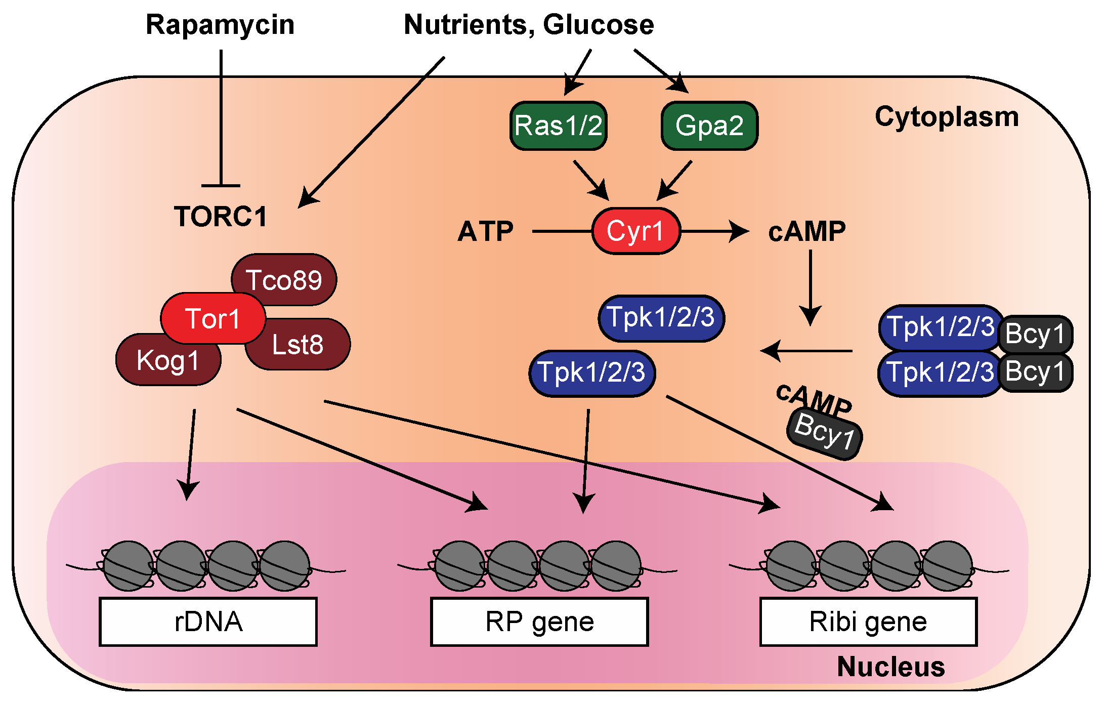
2. Transcription of the Ribosomal Genes in the Stress Response Is Regulated by the TOR and PKA Pathways

When the nutrient supply decreases, ribosome biosynthesis must be halted immediately to avoid the consumption of intracellular resources. Early studies using S. cerevisiae revealed that the concentration of active rDNA is reduced in the stationary phase (or when cells are cultured in a minimal medium) compared to that in the logarithmic growth phase (or when cells are cultured in a nutrient-rich medium) [20]. In addition, the expression of RP genes was repressed in the presence of 3-amino-1,2,4-triazole (3-AT), a metabolic antagonist that inhibits histidine synthesis [21], and tyrosine depletion reduced both rRNA and RP gene mRNA levels [22], indicating that amino acid starvation attenuates the transcription of ribosome-related genes. Subsequently, microarray analysis provided ample evidence that RP genes are repressed due to environmental stresses, including nitrogen and glucose starvation [23]. Jorgensen and colleagues also showed that the expression of RP and Ribi genes decreases 30 min after glucose depletion [24].
In S. pombe, the depletion of sulfur, an indispensable component for many coenzymes such as coenzyme A and biotin, diminishes the expression of ribosome-related genes [25]. A comprehensive analysis using RNA-seq indicated that nitrogen starvation reduces rRNA transcription levels by about 1/10 [3]. We also revealed that a group of gene expressions associated with ribosome biosynthesis was decreased within 30 min after glucose starvation [26,27].
Intriguingly, ribosomal genes can also be repressed by stresses other than nutritional starvation. For example, mild heat shock leads to quick but transient repression of RP genes in S. cerevisiae [22,28]. In S. pombe, oxidative, cadmium, heat, osmotic, and DNA damage stress commonly repress ribosomal genes [29]. Since nutrient signaling pathways, such as the target of rapamycin (TOR) and protein kinase A (PKA), are intensely involved in regulating ribosomal genes, it is, therefore, interesting to study how stresses, unlike starvation, can repress these genes.
TOR was initially identified as a target gene for the macrolide immunosuppressant drug rapamycin by genetic screening using S. cerevisiae. Two types of TOR proteins, Tor1 and Tor2, are found in budding yeast, forming TOR complex 1 (TORC1) and TOR complex 2 (TORC2), respectively [30,31]. Notably, starved and rapamycin-treated cells showed a similar phenotype, suggesting that the TOR signaling pathway recognizes nutrients and promotes cell proliferation [32,33]. With this background, the suppression of ribosome-associated genes by starvation was speculated to be due to TOR inactivation. Indeed, rapamycin’s inhibition of the TOR pathway rapidly downregulates the transcription of both rRNA and RP genes [34,35,36] (Figure 2). In fission yeast, TORC1 (composed of Tor2 protein) and TORC2 (of Tor1 protein) were identified, and TORC1 mainly regulates the expression of ribosome-related genes [37,38,39].
The PKA pathway regulates most glucose-dependent transcriptional responses [40]. PKA forms a heterotetramer consisting of two regulatory subunits of Bcy1 and two catalytic subunits of either of the three isoforms Tpk1/2/3. In the presence of glucose, the GTP-binding proteins Ras1/2 and Gpa2 activate the adenylate cyclase Cyr1, which converts ATP to cyclic AMP (cAMP), resulting in the inhibition of Bcy1, and free Tpk is activated [40,41,42,43] (Figure 2). In S. cerevisiae, the external addition of cAMP in a mutant capable of its uptake upregulates the expression of RP genes, and the depletion of cAMP downregulates their expression [44]. Furthermore, a comprehensive analysis using DNA microarrays showed that PKA and TOR cooperate in the transcriptional changes in ribosomal genes in response to fluctuating glucose levels [45].
Meanwhile, in S. pombe, the PKA pathway regulates the expression of some genes, including fbp1 and ste11 [46,47]. In contrast, there is no evidence that the pathway regulates the expression of ribosome-related genes. Therefore, a comprehensive expression analysis by RNA-seq using mutants that lack a functional PKA pathway could be highly informative.
Taken together, the TOR and PKA pathways, at least in S. cerevisiae, primarily regulate the expression of ribosome-associated genes by sensing external nutritional conditions. In the next section, we address specific mechanisms by which TOR and PKA regulate gene expression in budding yeast.

3. Regulatory Mechanisms of Ribosome-Related Gene Expression in S. cerevisiae

Ribosome-related genes can be classified into three categories: rRNA, RP genes, and Ribi genes. A universal pathway does not regulate these categories of genes; therefore, we present a summary of what is currently known about the regulatory mechanisms in each of these groups.

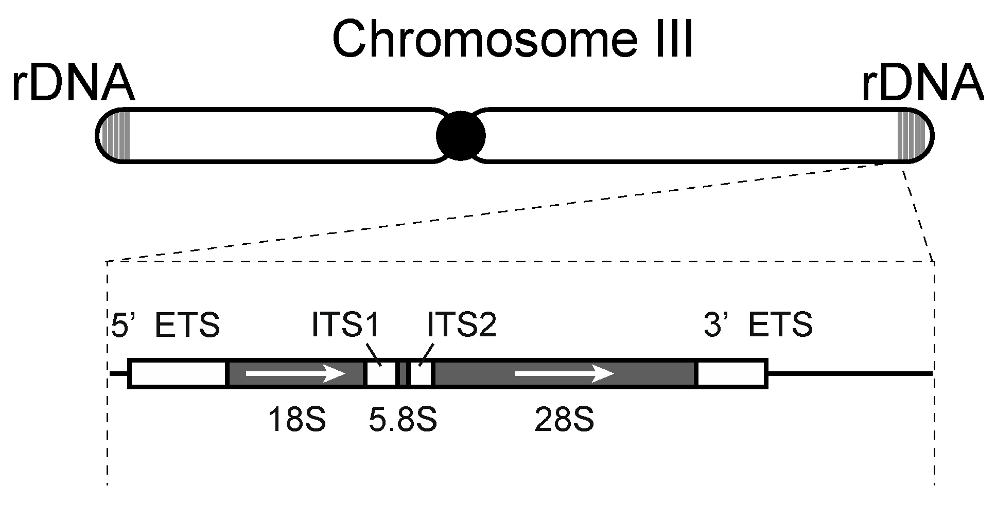
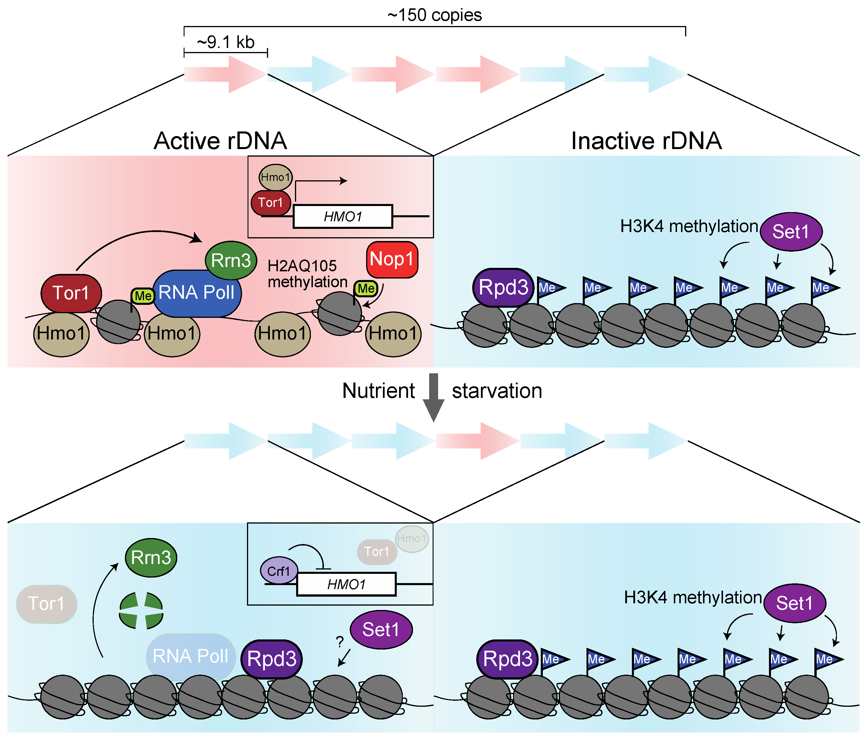
3.1. Regulatory Mechanisms of rRNA Expression

A region with the rDNA gene comprising approximately 9.1 kb of transcribed and non-transcribed spacers is repeated 150 times on chromosome XII in S. cerevisiae [5]. Analysis using a chromatin spread visualized by electron microscopy developed by Oscar Miller [48] suggests that active transcription only occurs in 50% of these rDNA repeats [20]. This analysis inferred that open and closed chromatin states couple with active and inactive transcription, respectively. The dynamics of chromatin structure are supposed to be defined by the distribution and modifications of its histone proteins. Staphylococcal nuclease and DNase I digestion assays implied that inactivated rDNA displays a homogenous chromatin pattern [49]. A study investigating the H3 and H2B profile using chromatin immunoprecipitation (ChIP) found that they are allocated throughout the 35S rDNA repeats. However, this strategy does not distinguish whether histones are homogenously distributed in both active and inactive rDNA. Notably, a mutant with fewer rDNA repeats showed activation of almost all rDNA accompanied by a reduction in histones on rDNA and a precarious chromatin structure [50,51]. These data suggest irregular and unstable chromatin structures are formed on actively transcribed rDNA units with low histone density (Figure 3).
Instead of histones, the HMG-box protein Hmo1 (which shares its function with linker histone H1) strongly accumulates on 35S rDNA through the DNA binding domain box A and box B, forming a fragile chromatin structure [52,53,54,55,56]. During DNA synthesis (S phase), nucleosomes fill the rDNA regions. However, as the cell cycle progresses, RNA polymerase I transcription displaces the nucleosome and facilitates the loading of Hmo1 into rDNA, thereby establishing open chromatin [57]. Since 18S and 25S rRNA transcript levels are reduced in hmo1∆ cells, nucleosome repackaging into rDNA may repress transcription [52,58]. An investigation of the Hmo1 behavior after rapamycin treatment mimicking nutrient starvation showed that it rapidly dissociates from 35S rDNA [53]. These findings suggest that TORC1 inactivation represses rRNA transcription by dissociating Hmo1 from rDNA. The level of Hmo1 expression may be able to explain this dissociation. Tor1 and Hmo1 localize on the HMO1 gene promoter in a nutrient medium. In contrast, they dissociate upon rapamycin treatment, leading to the recruitment of the repressor Crf1 to the promoter repression of the HMO1 gene [59,60] (Figure 3).
In summary, inhibiting canonical nucleosome formation by Hmo1 activates rRNA transcription. Upon starvation, TORC1 inactivation reduces the expression of the HMO1 gene, causing dissociation of Hmo1 and accumulation of nucleosomes on the rDNA, ultimately leading to the silencing of rRNA transcription (Figure 3). However, the dissociation of Hmo1 from rDNA upon starvation may not be sufficient to repress rRNA transcription rapidly.
The degradation of RNA polymerase I in response to starvation may be another mechanism for S. cerevisiae rRNA repression. RNA polymerase I, in concert with core factor CF [61], upstream activation factor UAF [62], TATA-binding protein TBP [63], and monomeric factor Rrn3 [64], are recruited to the rDNA promoter. Among them, rapamycin treatment diminishes RRN3 transcription and causes degradation of pre-existing Rrn3 [65]. Consequently, the complex of Rrn3 with RNA polymerase I is disrupted in the nucleolus, and rRNA transcription is attenuated [66] (Figure 3). Moreover, phosphorylation by TOR may promote the association of Rrn3 with RNA polymerase I since dephosphorylated RNA polymerase I dissociates with Rrn3 in vitro [67]. Artificial fusion of Rrn3 to RNA polymerase I subunit Rpa43 alleviates the reduction in rRNA transcription, albeit partially caused by nutrient starvation and rapamycin treatment, indicating that multilayered pathways contribute to rRNA repression [68].
In S. cerevisiae, the serine/threonine kinase TORC1 primarily localizes to the vacuolar membrane [69] and phosphorylates substrates, such as the homolog of mammalian S6 kinase, Sch9 [70]. If true, how can TORC1, located in the cytosol, swiftly regulate rRNA expression? Remarkably, Tor1 carries two nuclear localization signal (NLS) sequences and one nuclear export signal (NES) sequence and binds to the promoter of 35S rDNA and the entire 5S rDNA through its helix–turn–helix (HTH) domain in the presence of ambient nutrients [71,72]. Thus, TORC1 on rDNA might directly phosphorylate and activate Hmo1 and Rrn3.
In addition to these factors, the S. cerevisiae transcriptional repressor Maf1 is phosphorylated by Sch9, the downstream target of the TORC1 pathway, resulting in its dissociation from 5S rDNA, where transcription by RNA polymerase III is activated. Conversely, upon starvation or rapamycin treatment, dephosphorylated Maf1 quenches RNA polymerase III-dependent transcription as a negative cofactor [72,73,74,75].
Finally, we refer to a case where rDNA transcription is epigenetically regulated in S. cerevisiae. Crucially, the H3K9 methyltransferase SUV39H1 and heterochromatin protein HP1, essential for heterochromatin formation, are missing in budding yeast [19]. Hence, the histone deacetylase (HDAC) Sir2 and Rpd3 are supposed to function in chromatin condensation and silencing in S. cerevisiae rDNA. Although Rpd3, but not Sir2, accumulates in rDNA after the inactivation of TORC1 via rapamycin treatment and facilitates a closed chromatin formation in rDNA repeats (Figure 3), it does not contribute to the repression of rRNA production itself [76,77]. S. cerevisiae Sir2 prevents mitotic recombination by repressing transcription from the non-coding bidirectional promoter (E-pro) in the rDNA region rather than by repressing transcription of 35S rRNA [78]. Interestingly, in mammals, the deacetylation of the RNA polymerase I subunit PAF53 (A49 in S. cerevisiae) by SIRT7 (Sir2 in S. cerevisiae) enhances the association of histone and RNA polymerase I on rDNA, leading to an upregulation of rRNA transcription [79,80].
Instead, the S. cerevisiae COMPASS complex involving Set1 promotes histone H3K4 methylation and represses transcription from rDNA during nutrient-rich conditions, suggesting that the Set1 complex silences the dormant rDNA unit [81,82] (Figure 3). However, whether Set1 also contributes to S. cerevisiae rDNA silencing during starvation remains to be determined, and whether it is regulated by TORC1.
Remarkably, S. cerevisiae histone H2A can be methylated at a glutamine at position 105 (H2AQ105me) by methyltransferase Nop1 and is found to accumulate specifically in the nucleolus. In addition, the ChIP-seq profile indicates that H2AQ105me is enriched within the 35S rDNA regions. Since a mutant with an alanine substitution at Q105 mimicking methylation exhibits increased rRNA transcription, the enrichment in H2AQ105me is assumed to facilitate rRNA transcription [83]. This idea is supported by the finding that the inactivation of TORC1 eliminates H2A methylation from rDNA regions with a concomitant decrease in transcription [84] (Figure 3).
In summary, H3K4 methylation by Set1 and H2AQ105 methylation by Nop1 affect S. cerevisiae rRNA transcription negatively and positively, respectively.

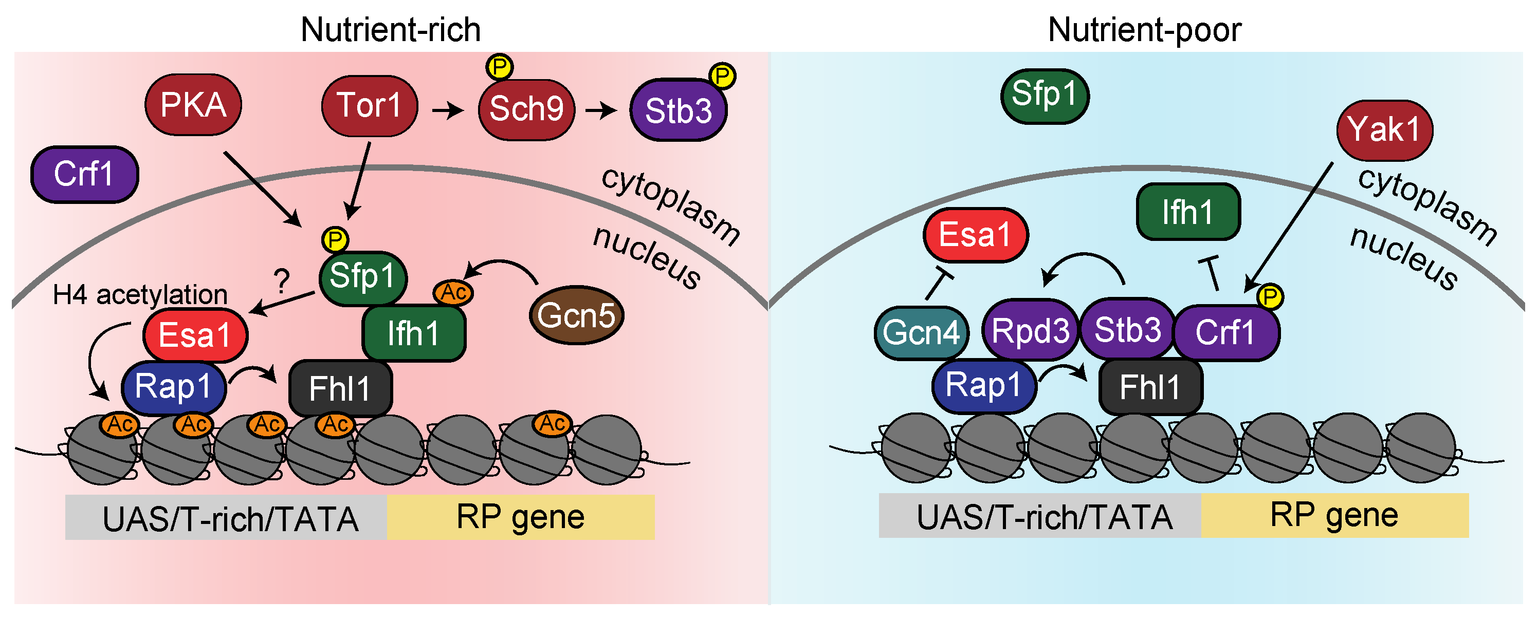
3.2. Regulatory Mechanisms of RP Gene Expression

The repressive mechanisms of S. cerevisiae RP genes are distinct from rDNA and add further complexity. The 137 genes encoding RP (except for 13 genes) carry a UAS (Upstream Activating Sequence), a T-rich element, and a TATA box as upstream regulatory elements. The transcriptional activator Rap1 (repressor/activator protein 1) recognizes the UAS and associates with ~90% of the RP genes [85,86,87,88]. Experiments measuring the expression levels of β-galactosidase from the lacZ gene, which is fused downstream of the RPL16 gene, revealed that transcription levels in cells lacking the Rap1 binding site at the RPL16 gene promoter were reduced to 10% of that in wild-type cells, confirming that Rap1 activates RP genes [21]. However, when S. cerevisiae cells are exposed to heat, osmotic stress, or rapamycin, the localization pattern of Rap1 is not altered despite the observed repression of RP gene transcription, suggesting that Rap1 is just a scaffold for other transcription factors governing their expression [89] (Figure 4).
A systematic study examined the genome-binding regions of 141 S. cerevisiae transcription factors identified as sharing DNA-binding and transcriptional activity in the Yeast Proteome Database yielded Fhl1, associated with RP genes, whose function was poorly investigated [90]. A more detailed study using ChIP-on-chip (chromatin immunoprecipitation combined with DNA microarray analysis) showed that Fhl1 accumulates to the promoter of 50–80% of the RP genes [89,91]. Fhl1 works as a transcriptional activator, as the expression of RP genes in cells lacking the FHL1 gene, was strongly reduced [91]. In addition, Fhl1 fails to bind at eight out of nine RP promoters to which Rap1 also does not bind, supporting the idea that Fhl1 localization depends on Rap1. Moreover, Fhl1 continuously associates with RP genes, as does Rap1, even if cells are exposed to rapamycin, indicating that both Rap1 and Fhl1 are scaffolds for other transcription factors [89] (Figure 4).
The binding sites of transcription factor Ifh1, which has a genetic interaction with those of Fhl1, were also examined by ChIP-on-chip analysis, revealing that both Ifh1 and Fhl1 bind to 46 of RP gene promoters [89,91]. Since overexpression of Ifh1 stimulates RP gene expression, Ifh1 recruited by Rap1 and Fhl1 probably acts as an activator. However, as opposed to Rap1 and Fhl1, Ifh1 is eliminated from almost all RP genes in the stationary phase or by rapamycin treatment [89,91]. Rapamycin treatment abrogates the physical interaction between Fhl1 and Ifh1 [92], which may be regulated by Ifh1 acetylation. Under nutritious conditions, Ifh1 is acetylated by acetyltransferase Gcn5 HAT, whereas rapamycin treatment removes Ifh1 acetylation [93] (Figure 4). This hypothesis could be verified by investigating whether the binding of Fhl1 and Ifh1 is weakened in the gcn5 deletion strain.
Almost concurrently, Sfp1 was found as a positive regulator of RP gene expression by a screen to isolate factors that change localization under rapamycin and a screen using a deletion library independently [94,95]. Recently, ChIP-exo analysis, capable of detecting protein binding sites at a single nucleotide resolution, has shown that Sfp1 coincides with the Fhl1 and Ifh1 at their binding sites [96]. Since depletion of Ifh1 reduces the association of Sfp1 with RP genes [97], Sfp1 is thought to associate with them via Ifh1 (Figure 4).
Sfp1 is phosphorylated not only by TORC1 directly but also by PKA. Therefore, Sfp1 is localized in the nucleus under nutrition-rich conditions, whereas starvation or rapamycin treatment promptly evacuates Sfp1 from the nucleus [94,98]. In this way, the accumulation of Ifh1 and Sfp1 activates the transcription of RP genes in nutrient-rich conditions, so how do these factors upregulate gene expression? Reid and colleagues provided a clue to answering this pivotal question. In S. cerevisiae, histone H4 acetylation, a marker of transcriptional activity, is catalyzed by the Esa1-containing NuA4 histone acetyltransferase [99]. ChIP combined with a microarray showed that Esa1 accumulates on the RP genes, and this binding is disrupted by rapamycin treatment [100,101]. Since depletion of Esa1 results in a reduction in acetylated H4 enrichment and transcription in RP genes, Esa1 likely upregulates RP gene transcriptions via H4 acetylation [100]. Given that Esa1 fails to associate with RP genes where Rap1 barely binds [100], it is possible that Ifh1 and Sfp1, both of which colocalize with Rap1, recruit Esa1 (Figure 4).
As described above, RP gene expression relies on complicated mechanisms, but the behavior of these factors is dynamically altered when cells shift into nutrient-poor conditions. Starvation and rapamycin treatment triggers the dissociation of Ifh1, Sfp1, and Esa1 from the RP gene. The dissociation of Esa1 was studied by Joo and coworkers [102]. A transcription factor Gcn4 competes with Esa1 for the binding to Rap1, thereby displacing Esa1 from the RP promoter, as demonstrated by in vitro and in vivo experiments [102]. It is expected that Gcn4 competes with Esa1 in a starvation-specific manner by a mechanism in which transcription of GCN4 is repressed by the general amino acids control (GAAC) pathway during nutritious conditions. In contrast, it is increased during amino acid starvation [103] (Figure 4).
In addition to the dissociation of Esa1, the downregulation of RP genes is enhanced by histone deacetylase Rpd3. While several studies reported that Rpd3 constitutively localizes to the RP genes [101,104], Humphery et al. suggested that Rpd3 only binds after rapamycin treatment [105]. In a seeming paradox, one may want to consider the binding of the transcriptional repressors Stb3 and Crf1 to these promoters [106]. Stb3, phosphorylated by Sch9, which acts downstream of TORC1, localizes to the cytoplasm, whereas it migrates to the nucleus after starvation or rapamycin treatment and accumulates at the RP gene promoters [106,107]. Rpd3 is then recruited onto RP genes through a scaffold of Stb3 [105] (Figure 4). Crf1 was identified by a two-hybrid screen in search of factors that bind to Fhl1. Since the transcriptional repression of RP genes by rapamycin treatment is alleviated in the crf1∆ mutant, Crf1 is considered a repressor of RP genes during starvation. Crf1 resides in the cytoplasm in nutritious conditions, whereas it is phosphorylated by the PKA-regulated kinase Yak1, which becomes activated upon starvation, migrates to the nucleus, and binds to Fhl1 [92]. Therefore, Crf1 might recruit Rpd3 to the RP genes in concert with Stb3 (Figure 4). Enigmatically, Crf1 functions as an RP gene repressor in the TB50 strain but not in the W303 strain background [108]. Therefore, it is plausible that the transcriptional repression of RP genes mainly relies on the recruitment of Rpd3 via Stb3.
In summary, the transcription of RP genes is strictly regulated by the dynamic localization of transcription factors and histone modification enzymes before and after starvation.

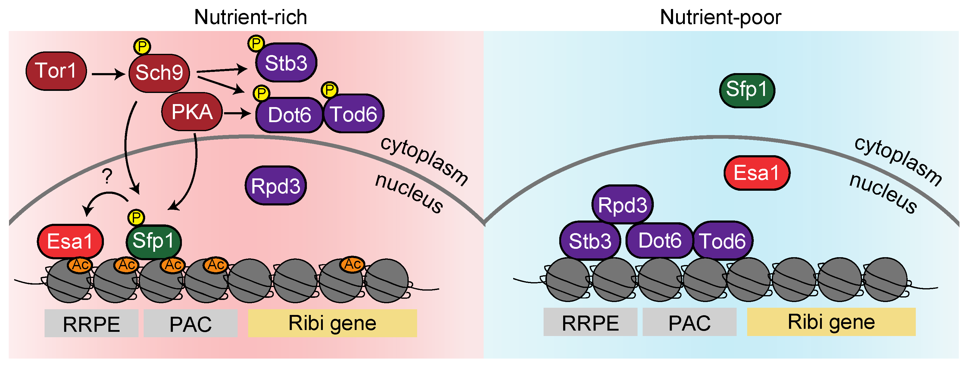
3.3. Regulatory Mechanisms of Ribi Gene Expression

In addition to the rRNAs and RPs that compose the ribosome, more than 200 other factors (Ribi) are involved in S. cerevisiae ribosome biogenesis [43]. The upstream sequence of the Ribi gene consists of RRPE and PAC sequences distinct from that of RP genes [109,110,111,112]. Therefore, Ribi gene transcription is regulated by a partially distinct mechanism from the RP genes. In a comprehensive analysis of proteins that bind to the RRPE sequence, Stb3 was identified as associating with RRPE during in vitro and in vivo experiments [113]. It is known that Stb3 contributes to the transcriptional repression of Ribi genes based on the finding that overexpression of Stb3 reduces their expression [107]. Additionally, Stb3 is sequestered to the cytoplasm by phosphorylation through the TORC1 pathway during nutrient-rich conditions. In contrast, upon starvation, it accumulates at the RRPE motif of Ribi gene promoters and represses their transcription [107] (Figure 5).
In a comprehensive analysis of DNA sequence motifs that bind transcriptional factors, Dot6 and Tod6 were found to bind to the PAC motif [114,115,116]. S. cerevisiae cells lacking both DOT6 and TOD6 genes exhibited mitigation of Ribi gene repression during starvation, suggesting that Dot6 and Tod6 downregulate their expression [117]. Moreover, a systematic analysis using mass spectrometry that detects proteins phosphorylated by TORC1 and its downstream factors, Sch9 and Tap42, revealed that Dot6 and Tod6 are phosphorylated by this pathway [75]. Huber and colleagues further indicated that R[R/K]x[S/T]* motifs of Dot6 and Tod6 are preferentially phosphorylated by Sch9 and PKA in vivo [106]. Dephosphorylation of Dot6 and Tod6 upon starvation and rapamycin treatment trigger the translocation of its localization from the cytoplasm to the nucleus, where they bind the Ribi gene promoters [118]. Taken together, Stb3 binds to the RRPE sequence and Dot6/Tod6 to the PAC sequence during starvation, respectively. These factors promote histone deacetylation by recruiting Rpd3 and repressing Ribi genes (Figure 5).
Then, how is the transcriptional activity of Ribi genes accomplished in nutritious environments? Although one report claimed that Sfp1, which is associated with RP genes, does not bind to Ribi gene promoters [96], ChEC-seq (endogenous chromatin cleavage-sequencing) analysis has recently revealed that Sfp1 accumulates on Ribi genes [97]. Importantly, Ihf1 hardly binds to Ribi gene promoters, although Sfp1 associates via a scaffold with Ifh1. This suggests that Sfp1 directly interacts with the Ribi gene sequence independent of Ifh1 [97]. It is, however, unclear whether Sfp1 recruits Esa1 since Rap1 is fundamentally absent on Ribi genes. Nevertheless, given an intriguing report that the depletion of Esa1 resulted in a decrease in both H4 acetylation and RNA polymerase II at most Ribi gene promoters, Esa1 is likely to accumulate on the Ribi genes [119] (Figure 5).
In summary, as shown in Figure 5, Ribi gene expression may be partially regulated by a common mechanism with RP gene regulation, while both can independently function.

4. Comparison of Regulatory Factors Repressing Ribosome-Related Genes between S. cerevisiae and S. pombe

In S. pombe, the molecular mechanisms underlying transcriptional activation during starvation have been extensively studied, mainly focusing on the gluconeogenic fbp1 gene [120,121]. However, research on S. pombe regarding the mechanism by which ribosome-associated genes are transcriptionally repressed is substantially lagging behind that of S. cerevisiae. Hereafter, we summarize the S. pombe homologs of transcription factors contributing to the ribosomal gene repression in S. cerevisiae.
Hmo1, which widely localizes on rDNA in S. cerevisiae, is also suggested to promote rDNA transcription in S. pombe [122]. However, the behavior of Hmo1 under starvation and whether it is regulated by TORC1 is unknown.
A homolog of the transcriptional activator Sfp1 is also present in S. pombe; however, it is currently unclear whether Sfp1 regulates ribosome-related gene transcription. Recently, a cap analysis of gene expression (CAGE) demonstrated that the Sfp1-binding motif is concentrated in the promoter of ribosome biosynthetic genes repressed during nitrogen starvation [123], indicating that Sfp1 may somehow contribute to the S. pombe ribosomal gene expression.
The forkhead protein Fhl1 is also regulated by TORC1 in S. pombe. However, Fhl1 predominantly contributes to the regulation of genes related to the transport process and mating/sporulation, with little or no influence on the ribosome-related genes [124].
Although S. cerevisiae Crf1 and Ifh1 are annotated with the S. pombe homolog Crf1, it has not been determined whether Crf1 regulates ribosome-related genes. A homolog of Stb3 is also present in S. pombe, but its function has yet to be discovered. Finally, there is no annotation and homolog information for Dot6 and Tod6 on PomBase (https://www.pombase.org/ accessed on 31 January 2023). Altogether, the analysis of transcription factors equivalent to those revealed in S. cerevisiae is poor in S. pombe.

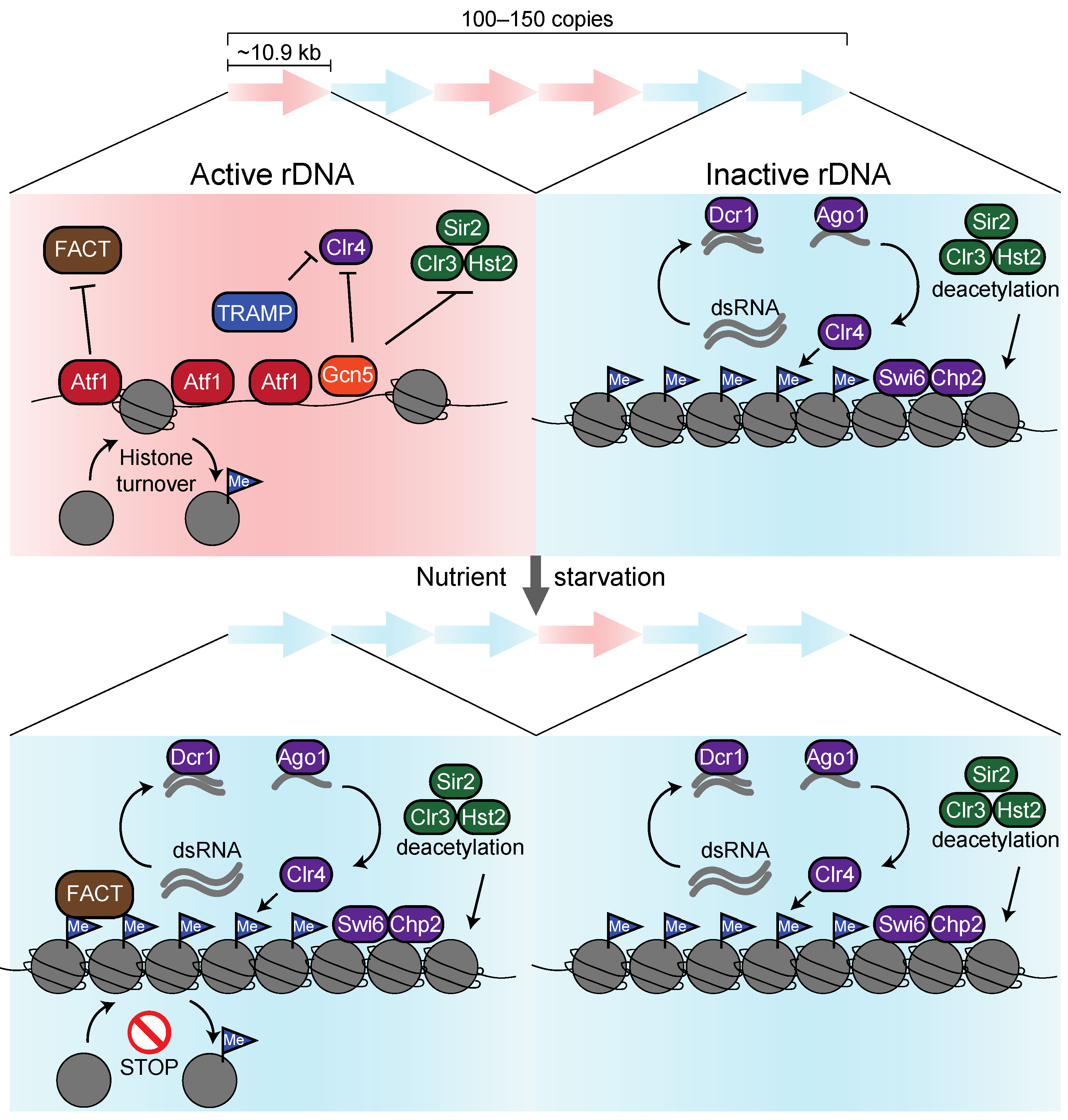
5. Epigenetic Regulatory Mechanisms of rDNA Regions in S. pombe

On the contrary, several studies have shown that some histone deacetylases contribute to S. pombe ribosomal gene repressions. Yet, histone deacetylase (class I) Clr6, homologs of Rpd3 repressing rRNA, RP, and Ribi genes in S. cerevisiae, do not contribute to the repression of ribosome-related genes. In contrast, class II deacetylase Clr3, a homolog of Hda1, represses at least rRNA gene expression [125,126]. Moreover, Sir2 and Hst2, among the three Sirtuin families in S. pombe, accumulate at rDNA regions to repress transcription [127]. These results were obtained under nutritious conditions, suggesting that part of the rDNA repeats is inactivated in S. pombe as in S. cerevisiae (Figure 6).
One significant discrepancy regarding the transcriptional repression mechanism between budding yeast and fission yeast is the heterochromatin formation machinery, which is only present in fission yeast [19]. Indeed, heterochromatinization by the H3K9 methyltransferase Clr4 and the heterochromatin proteins Swi6 and Chp2 (HP1 in humans) in concert with the RNAi-dependent pathway has been found in rDNA regions [128]. Such a heterochromatin formation is already observed in nutrient-rich environments and may prevent mitotic recombination between rDNA repeats, in addition to inactivating part of the rDNA repeats [129,130,131,132] (Figure 6).
In the actively transcribing rDNA repeats, ATF/CREB family transcription factor Atf1 (ATF-2 in humans) positively regulates rRNA gene expression [132]. Previous studies showed that stress-responsive transcription factor Atf1 accumulates at the promoter of the core environmental stress response (CESR) genes to activate the downstream gene in response to environmental cues, such as starvation [29,133,134,135]. On the other hand, it was reported that Atf1 directly binds to the histone methyltransferase Clr4 and silences the mating-type loci [136,137]. With this background, we conducted ChIP-seq for Atf1 binding sites before and after starvation, revealing that Atf1 broadly localizes on rDNA during nutrient-rich conditions but is dissociated upon starvation. Disruption of the atf1 gene caused an enhancement in H3K9 methylation and repression of rRNA genes, suggesting that Atf1 works as a transcriptional activator [132]. Additionally, the dissociation of Atf1 upon starvation triggers the accumulation of histone chaperone FACT [132], which maintains methylated histones by preventing histone turnover [138] (Figure 6). Since FACT potentially recognizes the Q105 site in H2A [83,139,140], it would be intriguing to study whether Atf1 regulates the modification of H2AQ105 in response to nutrient limitation.
In addition to, and independent of Atf1, acetyltransferase Gcn5 HAT accumulates in rDNA regions and dissociates upon starvation [132]. Inactivation of Gcn5 only in the nucleolus caused increased H3K9 methylation in rDNA chromatin, suggesting that Gcn5 competes with deacetylase and methyltransferase to prevent heterochromatin formation in the actively transcribing rDNA repeats [132]. Since Gcn5 is dissociated from rDNA upon starvation, histone H3 retained in the actively transcribing rDNA repeats is predominantly methylated by the RNAi pathway (Figure 6). Since Oya et al. showed that ubiquitination of H3K14 by CLRC complex is required for H3K9 methylation [141], H3K14 deacetylation by Clr3 is a prerequisite for H3K14 ubiquitination, leading to H3K9 methylation. Moreover, it is noteworthy that nitrogen starvation leads to the degradation of exosome/TRAMP, resulting in the enhancement in Ago1-associated sRNAs triggering heterochromatinization by the RNAi-dependent pathway on rDNA regions [17,142] (Figure 6).
As described above, transcription from rDNA in S. pombe is repressed by mechanisms quite different from those in S. cerevisiae. However, whether TORC1 and PKA participate in these mechanisms is unclear, and the same is true for whether mechanisms uncovered in rDNA also regulate ribosome-related genes. Intriguingly, H3K9 methylation in ribosome-related genes increased when S. pombe cells were exposed to nitrogen starvation since the sRNAs derived from transcriptions of ribosome-related genes were enhanced by Ago1, leading to the activation of the RNAi-dependent pathway [17]. Additionally, we found that the transcriptional activator Atf1 dissociated from ribosome-related genes upon glucose starvation, and transcription of rpl102, rlp7, and gar2 genes was decreased in atf1∆ cells, suggesting that the dissociation of Atf1 causes transcriptional repression of ribosome-related genes [132].

6. Perspectives

Mutations that cause complete loss of ribosomal function result in lethality in yeast and embryonic lethality in higher eukaryotes. Alternatively, partial loss of ribosomal function causes a variety of maladies called ribosomopathy in humans [143]. The etiology of Diamond–Blackfan anemia, a representative ribosomal disease, is a mutation of ribosomal genes [144,145]. Other diseases such as isolated congenital asplenia [146], childhood cirrhosis [147], chromosome 5q- syndrome [148], Treacher Collins syndrome [149], and Shwachman–Bodian–Diamond syndrome [150] are also caused by mutations in the ribosomal gene. Among these diseases, the etiology of Treacher Collins syndrome is a mutation in the TCOF1 gene, encoding the cofactor Treacle of RNA polymerase I, which transcribes rRNA [151]. This finding suggests that disrupting the rigorous regulation of rRNA expression levels may be the etiopathogenic factor. However, there are currently no findings on the transcription factors discussed in this paper regarding their role as etiologic agents of ribosomopathies.
In HEK293 cells and β-TC6 cells, a mouse pancreatic beta cell, mTOR binds to the rDNA promoter, and this binding is dissociated upon rapamycin treatments and amino acid starvation [152]. This suggests that the behavior of mTOR is equivalent, at least in S. cerevisiae. Additionally, methylated histone H3 and HP1 are accumulated in rDNA regions in NIH3T3 cells. NoRC, a nucleolar remodeling complex, mediates this, but it remains unknown whether another factor, such as ATF-2 (a homolog of Atf1 in S. pombe), also contributes [153]. In the future, we expect that defects in transcription factors that regulate the expression of ribosome-associated genes and cause ribosomopathies will be studied using D. melanogaster or mice as model organisms.

Author Contributions

Conceptualization, H.H. and K.O.; Writing—Original Draft Preparation, H.H.; Writing—Review and Editing, H.H. and K.O.; Figure Preparation, H.H.; Supervision, K.O.; Funding Acquisition, K.O. All authors have read and agreed to the published version of the manuscript.

Funding

This research was funded by JST CREST Grant [JPMJCR18S3] to K.O.

Institutional Review Board Statement

Not applicable.

Informed Consent Statement

Not applicable.

Data Availability Statement

Not applicable.

Conflicts of Interest

The authors declare no conflict of interest.

References

  1. de la Cruz, J.; Karbstein, K.; Woolford, J.L. Functions of ribosomal proteins in assembly of eukaryotic ribosomes in vivo. Annu. Rev. Biochem. 2015, 84, 93–129. [Google Scholar] [CrossRef] [PubMed]
  2. Warner, J.R. The economics of ribosome biosynthesis in yeast. Trends Biochem. Sci. 1999, 24, 437–440. [Google Scholar] [CrossRef] [PubMed]
  3. Marguerat, S.; Schmidt, A.; Codlin, S.; Chen, W.; Aebersold, R.; Bähler, J. Quantitative analysis of fission yeast transcriptomes and proteomes in proliferating and quiescent cells. Cell 2012, 151, 671–683. [Google Scholar] [CrossRef] [PubMed]
  4. Nagalakshmi, U.; Wang, Z.; Waern, K.; Shou, C.; Raha, D.; Gerstein, M.; Snyder, M. The transcriptional landscape of the yeast genome defined by RNA sequencing. Science 2008, 320, 1344–1349. [Google Scholar] [CrossRef]
  5. Kobayashi, T. Ribosomal RNA gene repeats, their stability and cellular senescence. Proc. Jpn. Acad. Ser. B Phys. Biol. Sci. 2014, 90, 119–129. [Google Scholar] [CrossRef]
  6. Toda, T.; Nakaseko, Y.; Niwa, O.; Yanagida, M. Mapping of rRNA genes by integration of hybrid plasmids in Schizosaccharomyces pombe. Curr. Genet. 1984, 8, 93–97. [Google Scholar] [CrossRef]
  7. Sanchez, J.A.; Kim, S.M.; Huberman, J.A. Ribosomal DNA replication in the fission yeast, Schizosaccharomyces pombe. Exp. Cell Res. 1998, 238, 220–230. [Google Scholar] [CrossRef]
  8. Wood, V.; Gwilliam, R.; Rajandream, M.-A.; Lyne, M.; Lyne, R.; Stewart, A.; Sgouros, J.; Peat, N.; Hayles, J.; Baker, S.; et al. The genome sequence of Schizosaccharomyces pombe. Nature 2002, 415, 871–880. [Google Scholar] [CrossRef]
  9. Stults, D.M.; Killen, M.W.; Pierce, H.H.; Pierce, A.J. Genomic architecture and inheritance of human ribosomal RNA gene clusters. Genome Res. 2008, 18, 13–18. [Google Scholar] [CrossRef]
  10. Wolfe, K.H.; Shields, D.C. Molecular evidence for an ancient duplication of the entire yeast genome. Nature 1997, 387, 708–713. [Google Scholar] [CrossRef]
  11. Klinge, S.; Woolford, J.L. Ribosome assembly coming into focus. Nat. Rev. Mol. Cell Biol. 2019, 20, 116–131. [Google Scholar] [CrossRef] [PubMed]
  12. Venema, J.; Tollervey, D. Processing of pre-ribosomal RNA in Saccharomyces cerevisiae. Yeast 1995, 11, 1629–1650. [Google Scholar] [CrossRef] [PubMed]
  13. Antequera, F.; Tamame, M.; Villanueva, J.R.; Santos, T. DNA methylation in the fungi. J. Biol. Chem. 1984, 259, 8033–8036. [Google Scholar] [CrossRef] [PubMed]
  14. Capuano, F.; Mülleder, M.; Kok, R.; Blom, H.J.; Ralser, M. Cytosine DNA methylation is found in Drosophila melanogaster but absent in Saccharomyces cerevisiae, Schizosaccharomyces pombe, and other yeast species. Anal. Chem. 2014, 86, 3697–3702. [Google Scholar] [CrossRef]
  15. Grewal, S.I.S.; Jia, S. Heterochromatin revisited. Nat. Rev. Genet. 2007, 8, 35–46. [Google Scholar] [CrossRef]
  16. Allshire, R.C.; Madhani, H.D. Ten principles of heterochromatin formation and function. Nat. Rev. Mol. Cell Biol. 2018, 19, 229–244. [Google Scholar] [CrossRef]
  17. Joh, R.I.; Khanduja, J.S.; Calvo, I.A.; Mistry, M.; Palmieri, C.M.; Savol, A.J.; Ho Sui, S.J.; Sadreyev, R.I.; Aryee, M.J.; Motamedi, M. Survival in Quiescence Requires the Euchromatic Deployment of Clr4/SUV39H by Argonaute-Associated Small RNAs. Mol. Cell 2016, 64, 1088–1101. [Google Scholar] [CrossRef]
  18. Torres-Garcia, S.; Yaseen, I.; Shukla, M.; Audergon, P.N.C.B.; White, S.A.; Pidoux, A.L.; Allshire, R.C. Epigenetic gene silencing by heterochromatin primes fungal resistance. Nature 2020, 585, 453–458. [Google Scholar] [CrossRef]
  19. Moazed, D. Common themes in mechanisms of gene silencing. Mol. Cell 2001, 8, 489–498. [Google Scholar] [CrossRef]
  20. Dammann, R.; Lucchini, R.; Koller, T.; Sogo, J.M. Chromatin structures and transcription of rDNA in yeast Saccharomyces cerevisiae. Nucleic Acids Res. 1993, 21, 2331–2338. [Google Scholar] [CrossRef] [Green Version]
  21. Moehle, C.M.; Hinnebusch, A.G. Association of RAP1 binding sites with stringent control of ribosomal protein gene transcription in Saccharomyces cerevisiae. Mol. Cell Biol. 1991, 11, 2723–2735. [Google Scholar] [PubMed]
  22. Warner, J.R.; Gorenstein, C. Yeast has a true stringent response. Nature 1978, 275, 338–339. [Google Scholar] [CrossRef] [PubMed]
  23. Gasch, A.P.; Spellman, P.T.; Kao, C.M.; Carmel-Harel, O.; Eisen, M.B.; Storz, G.; Botstein, D.; Brown, P.O. Genomic expression programs in the response of yeast cells to environmental changes. Mol. Biol. Cell 2000, 11, 4241–4257. [Google Scholar] [CrossRef]
  24. Jorgensen, P.; Rupes, I.; Sharom, J.R.; Schneper, L.; Broach, J.R.; Tyers, M. A dynamic transcriptional network communicates growth potential to ribosome synthesis and critical cell size. Genes Dev. 2004, 18, 2491–2505. [Google Scholar] [CrossRef] [PubMed]
  25. Ohtsuka, H.; Takinami, M.; Shimasaki, T.; Hibi, T.; Murakami, H.; Aiba, H. Sulfur restriction extends fission yeast chronological lifespan through Ecl1 family genes by downregulation of ribosome. Mol. Microbiol. 2017, 105, 84–97. [Google Scholar] [CrossRef]
  26. Oda, A.; Takemata, N.; Hirata, Y.; Miyoshi, T.; Suzuki, Y.; Sugano, S.; Ohta, K. Dynamic transition of transcription and chromatin landscape during fission yeast adaptation to glucose starvation. Genes Cells 2015, 20, 392–407. [Google Scholar] [CrossRef]
  27. Galipon, J.; Miki, A.; Oda, A.; Inada, T.; Ohta, K. Stress-induced lncRNAs evade nuclear degradation and enter the translational machinery. Genes Cells 2013, 18, 353–368. [Google Scholar] [CrossRef]
  28. Eisen, M.B.; Spellman, P.T.; Brown, P.O.; Botstein, D. Cluster analysis and display of genome-wide expression patterns. Proc. Natl. Acad. Sci. USA 1998, 95, 14863–14868. [Google Scholar] [CrossRef]
  29. Chen, D.; Toone, W.M.; Mata, J.; Lyne, R.; Burns, G.; Kivinen, K.; Brazma, A.; Jones, N.; Bähler, J. Global transcriptional responses of fission yeast to environmental stress. Mol. Biol. Cell 2003, 14, 214–229. [Google Scholar] [CrossRef]
  30. Heitman, J.; Movva, N.R.; Hall, M.N. Targets for cell cycle arrest by the immunosuppressant rapamycin in yeast. Science 1991, 253, 905–909. [Google Scholar] [CrossRef]
  31. Loewith, R.; Jacinto, E.; Wullschleger, S.; Lorberg, A.; Crespo, J.L.; Bonenfant, D.; Oppliger, W.; Jenoe, P.; Hall, M.N. Two TOR complexes, only one of which is rapamycin sensitive, have distinct roles in cell growth control. Mol. Cell 2002, 10, 457–468. [Google Scholar] [CrossRef] [PubMed]
  32. Barbet, N.C.; Schneider, U.; Helliwell, S.B.; Stansfield, I.; Tuite, M.F.; Hall, M.N. TOR controls translation initiation and early G1 progression in yeast. Mol. Biol. Cell 1996, 7, 25–42. [Google Scholar] [CrossRef] [PubMed]
  33. Thomas, G.; Hall, M.N. TOR signalling and control of cell growth. Curr. Opin. Cell Biol. 1997, 9, 782–787. [Google Scholar] [CrossRef]
  34. Zaragoza, D.; Ghavidel, A.; Heitman, J.; Schultz, M.C. Rapamycin induces the G0 program of transcriptional repression in yeast by interfering with the TOR signaling pathway. Mol. Cell Biol. 1998, 18, 4463–4470. [Google Scholar] [CrossRef] [PubMed]
  35. Powers, T.; Walter, P. Regulation of ribosome biogenesis by the rapamycin-sensitive TOR-signaling pathway in Saccharomyces cerevisiae. Mol. Biol. Cell 1999, 10, 987–1000. [Google Scholar] [CrossRef] [PubMed]
  36. Cardenas, M.E.; Cutler, N.S.; Lorenz, M.C.; Di Como, C.J.; Heitman, J. The TOR signaling cascade regulates gene expression in response to nutrients. Genes Dev. 1999, 13, 3271–3279. [Google Scholar] [CrossRef] [PubMed]
  37. Kawai, M.; Nakashima, A.; Ueno, M.; Ushimaru, T.; Aiba, K.; Doi, H.; Uritani, M. Fission yeast Tor1 functions in response to various stresses including nitrogen starvation, high osmolarity, and high temperature. Curr. Genet. 2001, 39, 166–174. [Google Scholar]
  38. Weisman, R.; Choder, M. The fission yeast TOR homolog, tor1+, is required for the response to starvation and other stresses via a conserved serine. J. Biol. Chem. 2001, 276, 7027–7032. [Google Scholar] [CrossRef]
  39. Alvarez, B.; Moreno, S. Fission yeast Tor2 promotes cell growth and represses cell differentiation. J. Cell Sci. 2006, 119, 4475–4485. [Google Scholar] [CrossRef]
  40. Zaman, S.; Lippman, S.I.; Schneper, L.; Slonim, N.; Broach, J.R. Glucose regulates transcription in yeast through a network of signaling pathways. Mol. Syst. Biol 2009, 5, 245. [Google Scholar] [CrossRef]
  41. Toda, T.; Cameron, S.; Sass, P.; Zoller, M.; Scott, J.D.; McMullen, B.; Hurwitz, M.; Krebs, E.G.; Wigler, M. Cloning and characterization of BCY1, a locus encoding a regulatory subunit of the cyclic AMP-dependent protein kinase in Saccharomyces cerevisiae. Mol. Cell Biol. 1987, 7, 1371–1377. [Google Scholar] [PubMed] [Green Version]
  42. Toda, T.; Cameron, S.; Sass, P.; Zoller, M.; Wigler, M. Three different genes in S. cerevisiae encode the catalytic subunits of the cAMP-dependent protein kinase. Cell 1987, 50, 277–287. [Google Scholar] [CrossRef]
  43. Broach, J.R. Nutritional control of growth and development in yeast. Genetics 2012, 192, 73–105. [Google Scholar] [CrossRef] [PubMed]
  44. Neuman-Silberberg, F.S.; Bhattacharya, S.; Broach, J.R. Nutrient availability and the RAS/cyclic AMP pathway both induce expression of ribosomal protein genes in Saccharomyces cerevisiae but by different mechanisms. Mol. Cell Biol. 1995, 15, 3187–3196. [Google Scholar] [CrossRef] [PubMed]
  45. Kunkel, J.; Luo, X.; Capaldi, A.P. Integrated TORC1 and PKA signaling control the temporal activation of glucose-induced gene expression in yeast. Nat. Commun. 2019, 10, 3558–3611. [Google Scholar] [CrossRef]
  46. Hoffman, C.S.; Winston, F. Glucose repression of transcription of the Schizosaccharomyces pombe fbp1 gene occurs by a cAMP signaling pathway. Genes Dev. 1991, 5, 561–571. [Google Scholar] [CrossRef]
  47. Higuchi, T.; Watanabe, Y.; Yamamoto, M. Protein kinase A regulates sexual development and gluconeogenesis through phosphorylation of the Zn finger transcriptional activator Rst2p in fission yeast. Mol. Cell Biol. 2002, 22, 1–11. [Google Scholar] [CrossRef]
  48. Miller, O.L.; Beatty, B.R. Visualization of nucleolar genes. Science 1969, 164, 955–957. [Google Scholar] [CrossRef]
  49. Lohr, D. Chromatin structure differs between coding and upstream flanking sequences of the yeast 35S ribosomal genes. Biochemistry 1983, 22, 927–934. [Google Scholar] [CrossRef]
  50. French, S.L.; Osheim, Y.N.; Cioci, F.; Nomura, M.; Beyer, A.L. In exponentially growing Saccharomyces cerevisiae cells, rRNA synthesis is determined by the summed RNA polymerase I loading rate rather than by the number of active genes. Mol. Cell Biol. 2003, 23, 1558–1568. [Google Scholar] [CrossRef]
  51. Jones, H.S.; Kawauchi, J.; Braglia, P.; Alen, C.M.; Kent, N.A.; Proudfoot, N.J. RNA polymerase I in yeast transcribes dynamic nucleosomal rDNA. Nat. Struct. Mol. Biol. 2007, 14, 123–130. [Google Scholar] [CrossRef] [PubMed]
  52. Hall, D.B.; Wade, J.T.; Struhl, K. An HMG protein, Hmo1, associates with promoters of many ribosomal protein genes and throughout the rRNA gene locus in Saccharomyces cerevisiae. Mol. Cell Biol. 2006, 26, 3672–3679. [Google Scholar] [CrossRef] [PubMed]
  53. Berger, A.B.; Decourty, L.; Badis, G.; Nehrbass, U.; Jacquier, A.; Gadal, O. Hmo1 is required for TOR-dependent regulation of ribosomal protein gene transcription. Mol. Cell Biol. 2007, 27, 8015–8026. [Google Scholar] [CrossRef] [PubMed]
  54. Kasahara, K.; Ohtsuki, K.; Ki, S.; Aoyama, K.; Takahashi, H.; Kobayashi, T.; Shirahige, K.; Kokubo, T. Assembly of regulatory factors on rRNA and ribosomal protein genes in Saccharomyces cerevisiae. Mol. Cell Biol. 2007, 27, 6686–6705. [Google Scholar] [CrossRef] [PubMed]
  55. Panday, A.; Grove, A. Yeast HMO1: Linker Histone Reinvented. Microbiol. Mol. Biol. Rev. 2017, 81, e00037-16. [Google Scholar] [CrossRef]
  56. Higashino, A.; Shiwa, Y.; Yoshikawa, H.; Kokubo, T.; Kasahara, K. Both HMG boxes in Hmo1 are essential for DNA binding in vitro and in vivo. Biosci. Biotechnol. Biochem. 2015, 79, 384–393. [Google Scholar] [CrossRef]
  57. Wittner, M.; Hamperl, S.; Stöckl, U.; Seufert, W.; Tschochner, H.; Milkereit, P.; Griesenbeck, J. Establishment and maintenance of alternative chromatin states at a multicopy gene locus. Cell 2011, 145, 543–554. [Google Scholar] [CrossRef]
  58. Gadal, O.; Labarre, S.; Boschiero, C.; Thuriaux, P. Hmo1, an HMG-box protein, belongs to the yeast ribosomal DNA transcription system. EMBO J. 2002, 21, 5498–5507. [Google Scholar] [CrossRef]
  59. Xiao, L.; Kamau, E.; Donze, D.; Grove, A. Expression of yeast high mobility group protein HMO1 is regulated by TOR signaling. Gene 2011, 489, 55–62. [Google Scholar] [CrossRef]
  60. Panday, A.; Gupta, A.; Srinivasa, K.; Xiao, L.; Smith, M.D.; Grove, A. DNA damage regulates direct association of TOR kinase with the RNA polymerase II-transcribed HMO1 gene. Mol. Biol. Cell 2017, 28, 2449–2459. [Google Scholar] [CrossRef]
  61. Keys, D.A.; Vu, L.; Steffan, J.S.; Dodd, J.A.; Yamamoto, R.T.; Nogi, Y.; Nomura, M. RRN6 and RRN7 encode subunits of a multiprotein complex essential for the initiation of rDNA transcription by RNA polymerase I in Saccharomyces cerevisiae. Genes Dev. 1994, 8, 2349–2362. [Google Scholar] [CrossRef] [Green Version]
  62. Keys, D.A.; Lee, B.S.; Dodd, J.A.; Nguyen, T.T.; Vu, L.; Fantino, E.; Burson, L.M.; Nogi, Y.; Nomura, M. Multiprotein transcription factor UAF interacts with the upstream element of the yeast RNA polymerase I promoter and forms a stable preinitiation complex. Genes Dev. 1996, 10, 887–903. [Google Scholar] [CrossRef] [PubMed]
  63. Steffan, J.S.; Keys, D.A.; Dodd, J.A.; Nomura, M. The role of TBP in rDNA transcription by RNA polymerase I in Saccharomyces cerevisiae: TBP is required for upstream activation factor-dependent recruitment of core factor. Genes Dev. 1996, 10, 2551–2563. [Google Scholar] [CrossRef] [PubMed]
  64. Peyroche, G.; Milkereit, P.; Bischler, N.; Tschochner, H.; Schultz, P.; Sentenac, A.; Carles, C.; Riva, M. The recruitment of RNA polymerase I on rDNA is mediated by the interaction of the A43 subunit with Rrn3. EMBO J. 2000, 19, 5473–5482. [Google Scholar] [CrossRef]
  65. Philippi, A.; Steinbauer, R.; Reiter, A.; Fath, S.; Leger-Silvestre, I.; Milkereit, P.; Griesenbeck, J.; Tschochner, H. TOR-dependent reduction in the expression level of Rrn3p lowers the activity of the yeast RNA Pol I machinery, but does not account for the strong inhibition of rRNA production. Nucleic Acids Res. 2010, 38, 5315–5326. [Google Scholar] [CrossRef]
  66. Claypool, J.A.; French, S.L.; Johzuka, K.; Eliason, K.; Vu, L.; Dodd, J.A.; Beyer, A.L.; Nomura, M. Tor pathway regulates Rrn3p-dependent recruitment of yeast RNA polymerase I to the promoter but does not participate in alteration of the number of active genes. Mol. Biol. Cell 2004, 15, 946–956. [Google Scholar] [CrossRef]
  67. Fath, S.; Milkereit, P.; Peyroche, G.; Riva, M.; Carles, C.; Tschochner, H. Differential roles of phosphorylation in the formation of transcriptional active RNA polymerase I. Proc. Natl. Acad. Sci. USA 2001, 98, 14334–14339. [Google Scholar] [CrossRef]
  68. Laferté, A.; Favry, E.; Sentenac, A.; Riva, M.; Carles, C.; Chédin, S. The transcriptional activity of RNA polymerase I is a key determinant for the level of all ribosome components. Genes Dev. 2006, 20, 2030–2040. [Google Scholar] [CrossRef]
  69. Binda, M.; PEli-Gulli, M.-P.; Bonfils, G.; Panchaud, N.; Urban, J.; Sturgill, T.W.; Loewith, R.; De Virgilio, C. The Vam6 GEF controls TORC1 by activating the EGO complex. Mol. Cell 2009, 35, 563–573. [Google Scholar] [CrossRef]
  70. Takeda, E.; Jin, N.; Itakura, E.; Kira, S.; Kamada, Y.; Weisman, L.S.; Noda, T.; Matsuura, A. Vacuole-mediated selective regulation of TORC1-Sch9 signaling following oxidative stress. Mol. Biol. Cell 2018, 29, 510–522. [Google Scholar] [CrossRef]
  71. Li, H.; Tsang, C.K.; Watkins, M.; Bertram, P.G.; Zheng, X.F.S. Nutrient regulates Tor1 nuclear localization and association with rDNA promoter. Nature 2006, 442, 1058–1061. [Google Scholar] [CrossRef]
  72. Wei, Y.; Tsang, C.K.; Zheng, X.F.S. Mechanisms of regulation of RNA polymerase III-dependent transcription by TORC1. EMBO J. 2009, 28, 2220–2230. [Google Scholar] [CrossRef]
  73. Oficjalska-Pham, D.; Harismendy, O.; Smagowicz, W.J.; Gonzalez de Peredo, A.; Boguta, M.; Sentenac, A.; Lefebvre, O. General repression of RNA polymerase III transcription is triggered by protein phosphatase type 2A-mediated dephosphorylation of Maf1. Mol. Cell 2006, 22, 623–632. [Google Scholar] [CrossRef]
  74. Roberts, D.N.; Wilson, B.; Huff, J.T.; Stewart, A.J.; Cairns, B.R. Dephosphorylation and genome-wide association of Maf1 with Pol III-transcribed genes during repression. Mol. Cell 2006, 22, 633–644. [Google Scholar] [CrossRef]
  75. Huber, A.; Bodenmiller, B.; Uotila, A.; Stahl, M.; Wanka, S.; Gerrits, B.; Aebersold, R.; Loewith, R. Characterization of the rapamycin-sensitive phosphoproteome reveals that Sch9 is a central coordinator of protein synthesis. Genes Dev. 2009, 23, 1929–1943. [Google Scholar] [CrossRef]
  76. Sandmeier, J.J.; French, S.; Osheim, Y.; Cheung, W.L.; Gallo, C.M.; Beyer, A.L.; Smith, J.S. RPD3 is required for the inactivation of yeast ribosomal DNA genes in stationary phase. EMBO J. 2002, 21, 4959–4968. [Google Scholar] [CrossRef]
  77. Oakes, M.L.; Siddiqi, I.; French, S.L.; Vu, L.; Sato, M.; Aris, J.P.; Beyer, A.L.; Nomura, M. Role of histone deacetylase Rpd3 in regulating rRNA gene transcription and nucleolar structure in yeast. Mol. Cell Biol. 2006, 26, 3889–3901. [Google Scholar] [CrossRef]
  78. Kobayashi, T.; Ganley, A.R.D. Recombination regulation by transcription-induced cohesin dissociation in rDNA repeats. Science 2005, 309, 1581–1584. [Google Scholar] [CrossRef]
  79. Ford, E.; Voit, R.; Liszt, G.; Magin, C.; Grummt, I.; Guarente, L. Mammalian Sir2 homolog SIRT7 is an activator of RNA polymerase I transcription. Genes Dev. 2006, 20, 1075–1080. [Google Scholar] [CrossRef]
  80. Chen, S.; Seiler, J.; Santiago-Reichelt, M.; Felbel, K.; Grummt, I.; Voit, R. Repression of RNA polymerase I upon stress is caused by inhibition of RNA-dependent deacetylation of PAF53 by SIRT7. Mol. Cell 2013, 52, 303–313. [Google Scholar] [CrossRef]
  81. Briggs, S.D.; Bryk, M.; Strahl, B.D.; Cheung, W.L.; Davie, J.K.; Dent, S.Y.; Winston, F.; Allis, C.D. Histone H3 lysine 4 methylation is mediated by Set1 and required for cell growth and rDNA silencing in Saccharomyces cerevisiae. Genes Dev. 2001, 15, 3286–3295. [Google Scholar] [CrossRef] [PubMed] [Green Version]
  82. Bryk, M.; Briggs, S.D.; Strahl, B.D.; Curcio, M.J.; Allis, C.D.; Winston, F. Evidence that Set1, a factor required for methylation of histone H3, regulates rDNA silencing in S. cerevisiae by a Sir2-independent mechanism. Curr. Biol. 2002, 12, 165–170. [Google Scholar] [CrossRef] [PubMed]
  83. Tessarz, P.; Santos-Rosa, H.; Robson, S.C.; Sylvestersen, K.B.; Nelson, C.J.; Nielsen, M.L.; Kouzarides, T. Glutamine methylation in histone H2A is an RNA-polymerase-I-dedicated modification. Nature 2014, 505, 564–568. [Google Scholar] [CrossRef]
  84. Mawer, J.S.P.; Massen, J.; Reichert, C.; Grabenhorst, N.; Mylonas, C.; Tessarz, P. Nhp2 is a reader of H2AQ105me and part of a network integrating metabolism with rRNA synthesis. EMBO Rep. 2021, 22, e52435. [Google Scholar] [CrossRef]
  85. Shore, D. RAP1: A protean regulator in yeast. Trends Genet. 1994, 10, 408–412. [Google Scholar] [CrossRef]
  86. Mager, W.H.; Planta, R.J. Multifunctional DNA-binding proteins mediate concerted transcription activation of yeast ribosomal protein genes. Biochim. Biophys. Acta 1990, 1050, 351–355. [Google Scholar] [CrossRef] [PubMed]
  87. Lascaris, R.F.; Mager, W.H.; Planta, R.J. DNA-binding requirements of the yeast protein Rap1p as selected in silico from ribosomal protein gene promoter sequences. Bioinformatics 1999, 15, 267–277. [Google Scholar] [CrossRef]
  88. Lieb, J.D.; Liu, X.; Botstein, D.; Brown, P.O. Promoter-specific binding of Rap1 revealed by genome-wide maps of protein-DNA association. Nat. Genet. 2001, 28, 327–334. [Google Scholar] [CrossRef]
  89. Wade, J.T.; Hall, D.B.; Struhl, K. The transcription factor Ifh1 is a key regulator of yeast ribosomal protein genes. Nature 2004, 432, 1054–1058. [Google Scholar] [CrossRef]
  90. Lee, T.I.; Rinaldi, N.J.; Robert, F.; Odom, D.T.; Bar-Joseph, Z.; Gerber, G.K.; Hannett, N.M.; Harbison, C.T.; Thompson, C.M.; Simon, I.; et al. Transcriptional regulatory networks in Saccharomyces cerevisiae. Science 2002, 298, 799–804. [Google Scholar] [CrossRef]
  91. Schawalder, S.B.; Kabani, M.; Howald, I.; Choudhury, U.; Werner, M.; Shore, D. Growth-regulated recruitment of the essential yeast ribosomal protein gene activator Ifh1. Nature 2004, 432, 1058–1061. [Google Scholar] [CrossRef] [PubMed]
  92. Martin, D.E.; Soulard, A.; Hall, M.N. TOR regulates ribosomal protein gene expression via PKA and the Forkhead transcription factor FHL1. Cell 2004, 119, 969–979. [Google Scholar] [CrossRef] [PubMed]
  93. Downey, M.; Knight, B.; Vashisht, A.A.; Seller, C.A.; Wohlschlegel, J.A.; Shore, D.; Toczyski, D.P. Gcn5 and sirtuins regulate acetylation of the ribosomal protein transcription factor Ifh1. Curr. Biol. 2013, 23, 1638–1648. [Google Scholar] [CrossRef]
  94. Marion, R.M.; Regev, A.; Segal, E.; Barash, Y.; Koller, D.; Friedman, N.; O’Shea, E.K. Sfp1 is a stress- and nutrient-sensitive regulator of ribosomal protein gene expression. Proc. Natl. Acad. Sci. USA 2004, 101, 14315–14322. [Google Scholar] [CrossRef] [PubMed]
  95. Jorgensen, P.; Nishikawa, J.L.; Breitkreutz, B.-J.; Tyers, M. Systematic identification of pathways that couple cell growth and division in yeast. Science 2002, 297, 395–400. [Google Scholar] [CrossRef]
  96. Reja, R.; Vinayachandran, V.; Ghosh, S.; Pugh, B.F. Molecular mechanisms of ribosomal protein gene coregulation. Genes Dev. 2015, 29, 1942–1954. [Google Scholar] [CrossRef]
  97. Albert, B.; Tomassetti, S.; Gloor, Y.; Dilg, D.; Mattarocci, S.; Kubik, S.; Hafner, L.; Shore, D. Sfp1 regulates transcriptional networks driving cell growth and division through multiple promoter-binding modes. Genes Dev. 2019, 33, 288–293. [Google Scholar] [CrossRef]
  98. LempiAinen, H.; Uotila, A.; Urban, J.; Dohnal, I.; Ammerer, G.; Loewith, R.; Shore, D. Sfp1 interaction with TORC1 and Mrs6 reveals feedback regulation on TOR signaling. Mol. Cell 2009, 33, 704–716. [Google Scholar] [CrossRef]
  99. Smith, E.R.; Eisen, A.; Gu, W.; Sattah, M.; Pannuti, A.; Zhou, J.; Cook, R.G.; Lucchesi, J.C.; Allis, C.D. ESA1 is a histone acetyltransferase that is essential for growth in yeast. Proc. Natl. Acad. Sci. USA 1998, 95, 3561–3565. [Google Scholar] [CrossRef]
  100. Reid, J.L.; Iyer, V.R.; Brown, P.O.; Struhl, K. Coordinate regulation of yeast ribosomal protein genes is associated with targeted recruitment of Esa1 histone acetylase. Mol. Cell 2000, 6, 1297–1307. [Google Scholar] [CrossRef]
  101. Rohde, J.R.; Cardenas, M.E. The Tor pathway regulates gene expression by linking nutrient sensing to histone acetylation. Mol. Cell Biol. 2003, 23, 629–635. [Google Scholar] [CrossRef] [PubMed] [Green Version]
  102. Joo, Y.J.; Kim, J.-H.; Kang, U.-B.; Yu, M.-H.; Kim, J. Gcn4p-mediated transcriptional repression of ribosomal protein genes under amino-acid starvation. EMBO J. 2011, 30, 859–872. [Google Scholar] [CrossRef] [PubMed]
  103. Conrad, M.; Schothorst, J.; Kankipati, H.N.; Van Zeebroeck, G.; Rubio-Texeira, M.; Thevelein, J.M. Nutrient sensing and signaling in the yeast Saccharomyces cerevisiae. FEMS Microbiol. Rev. 2014, 38, 254–299. [Google Scholar] [CrossRef] [PubMed]
  104. Kurdistani, S.K.; Robyr, D.; Tavazoie, S.; Grunstein, M. Genome-wide binding map of the histone deacetylase Rpd3 in yeast. Nat. Genet. 2002, 31, 248–254. [Google Scholar] [CrossRef]
  105. Humphrey, E.L.; Shamji, A.F.; Bernstein, B.E.; Schreiber, S.L. Rpd3p relocation mediates a transcriptional response to rapamycin in yeast. Chem. Biol. 2004, 11, 295–299. [Google Scholar] [CrossRef]
  106. Huber, A.; French, S.L.; Tekotte, H.; Yerlikaya, S.; Stahl, M.; Perepelkina, M.P.; Tyers, M.; Rougemont, J.; Beyer, A.L.; Loewith, R. Sch9 regulates ribosome biogenesis via Stb3, Dot6 and Tod6 and the histone deacetylase complex RPD3L. EMBO J. 2011, 30, 3052–3064. [Google Scholar] [CrossRef]
  107. Liko, D.; Conway, M.K.; Grunwald, D.S.; Heideman, W. Stb3 plays a role in the glucose-induced transition from quiescence to growth in Saccharomyces cerevisiae. Genetics 2010, 185, 797–810. [Google Scholar] [CrossRef]
  108. Zhao, Y.; McIntosh, K.B.; Rudra, D.; Schawalder, S.; Shore, D.; Warner, J.R. Fine-structure analysis of ribosomal protein gene transcription. Mol. Cell Biol. 2006, 26, 4853–4862. [Google Scholar] [CrossRef]
  109. Dequard-Chablat, M.; Riva, M.; Carles, C.; Sentenac, A. RPC19, the gene for a subunit common to yeast RNA polymerases A (I) and C (III). J. Biol. Chem. 1991, 266, 15300–15307. [Google Scholar] [CrossRef]
  110. Hughes, J.D.; Estep, P.W.; Tavazoie, S.; Church, G.M. Computational identification of cis-regulatory elements associated with groups of functionally related genes in Saccharomyces cerevisiae. J. Mol. Biol. 2000, 296, 1205–1214. [Google Scholar] [CrossRef]
  111. Wade, C.; Shea, K.A.; Jensen, R.V.; McAlear, M.A. EBP2 is a member of the yeast RRB regulon, a transcriptionally coregulated set of genes that are required for ribosome and rRNA biosynthesis. Mol. Cell Biol. 2001, 21, 8638–8650. [Google Scholar] [CrossRef] [PubMed] [Green Version]
  112. Beer, M.A.; Tavazoie, S. Predicting gene expression from sequence. Cell 2004, 117, 185–198. [Google Scholar] [CrossRef] [PubMed]
  113. Liko, D.; Slattery, M.G.; Heideman, W. Stb3 binds to ribosomal RNA processing element motifs that control transcriptional responses to growth in Saccharomyces cerevisiae. J. Biol. Chem. 2007, 282, 26623–26628. [Google Scholar] [CrossRef] [PubMed]
  114. Badis, G.; Chan, E.T.; van Bakel, H.; Pena-Castillo, L.; Tillo, D.; Tsui, K.; Carlson, C.D.; Gossett, A.J.; Hasinoff, M.J.; Warren, C.L.; et al. A library of yeast transcription factor motifs reveals a widespread function for Rsc3 in targeting nucleosome exclusion at promoters. Mol. Cell 2008, 32, 878–887. [Google Scholar] [CrossRef]
  115. Zhu, C.; Byers, K.J.R.P.; McCord, R.P.; Shi, Z.; Berger, M.F.; Newburger, D.E.; Saulrieta, K.; Smith, Z.; Shah, M.V.; Radhakrishnan, M.; et al. High-resolution DNA-binding specificity analysis of yeast transcription factors. Genome Res. 2009, 19, 556–566. [Google Scholar] [CrossRef]
  116. Freckleton, G.; Lippman, S.I.; Broach, J.R.; Tavazoie, S. Microarray profiling of phage-display selections for rapid mapping of transcription factor-DNA interactions. PLoS Genet. 2009, 5, e1000449. [Google Scholar] [CrossRef]
  117. Lippman, S.I.; Broach, J.R. Protein kinase A and TORC1 activate genes for ribosomal biogenesis by inactivating repressors encoded by Dot6 and its homolog Tod6. Proc. Natl. Acad. Sci. USA 2009, 106, 19928–19933. [Google Scholar] [CrossRef]
  118. Hughes Hallett, J.E.; Luo, X.; Capaldi, A.P. State transitions in the TORC1 signaling pathway and information processing in Saccharomyces cerevisiae. Genetics 2014, 198, 773–786. [Google Scholar] [CrossRef]
  119. Bruzzone, M.J.; Grünberg, S.; Kubik, S.; Zentner, G.E.; Shore, D. Distinct patterns of histone acetyltransferase and Mediator deployment at yeast protein-coding genes. Genes Dev. 2018, 32, 1252–1265. [Google Scholar] [CrossRef]
  120. Hirota, K.; Miyoshi, T.; Kugou, K.; Hoffman, C.S.; Shibata, T.; Ohta, K. Stepwise chromatin remodelling by a cascade of transcription initiation of non-coding RNAs. Nature 2008, 456, 130–134. [Google Scholar] [CrossRef]
  121. Asada, R.; Hirota, K. Multi-Layered Regulations on the Chromatin Architectures: Establishing the Tight and Specific Responses of Fission Yeast fbp1 Gene Transcription. Biomolecules 2022, 12, 1642. [Google Scholar] [CrossRef]
  122. Albert, B.; Colleran, C.; Leger-Silvestre, I.; Berger, A.B.; Dez, C.; Normand, C.; Perez-Fernandez, J.; McStay, B.; Gadal, O. Structure-function analysis of Hmo1 unveils an ancestral organization of HMG-Box factors involved in ribosomal DNA transcription from yeast to human. Nucleic Acids Res. 2013, 41, 10135–10149. [Google Scholar] [CrossRef] [PubMed]
  123. Thodberg, M.; Thieffry, A.; Bornholdt, J.; Boyd, M.; Holmberg, C.; Azad, A.; Workman, C.T.; Chen, Y.; Ekwall, K.; Nielsen, O.; et al. Comprehensive profiling of the fission yeast transcription start site activity during stress and media response. Nucleic Acids Res. 2019, 47, 1671–1691. [Google Scholar] [CrossRef] [PubMed]
  124. Pataki, E.; Weisman, R.; Sipiczki, M.; Miklos, I. fhl1 gene of the fission yeast regulates transcription of meiotic genes and nitrogen starvation response, downstream of the TORC1 pathway. Curr. Genet. 2017, 63, 91–101. [Google Scholar] [CrossRef] [PubMed]
  125. Bjerling, P.; Silverstein, R.A.; Thon, G.; Caudy, A.; Grewal, S.; Ekwall, K. Functional Divergence between Histone Deacetylases in Fission Yeast by Distinct Cellular Localization and In Vivo Specificity. Mol. Cell Biol. 2002, 22, 2170–2181. [Google Scholar] [CrossRef]
  126. Sugiyama, T.; Cam, H.P.; Sugiyama, R.; Noma, K.-I.; Zofall, M.; Kobayashi, R.; Grewal, S.I.S. SHREC, an effector complex for heterochromatic transcriptional silencing. Cell 2007, 128, 491–504. [Google Scholar] [CrossRef]
  127. Durand-Dubief, M.; Sinha, I.; Fagerström-Billai, F.; Bonilla, C.; Wright, A.; Grunstein, M.; Ekwall, K. Specific functions for the fission yeast Sirtuins Hst2 and Hst4 in gene regulation and retrotransposon silencing. EMBO J. 2007, 26, 2477–2488. [Google Scholar] [CrossRef] [PubMed]
  128. Cam, H.P.; Sugiyama, T.; Chen, E.S.; Chen, X.; FitzGerald, P.C.; Grewal, S.I.S. Comprehensive analysis of heterochromatin- and RNAi-mediated epigenetic control of the fission yeast genome. Nat. Genet. 2005, 37, 809–819. [Google Scholar] [CrossRef] [PubMed]
  129. Thon, G.; Verhein-Hansen, J. Four chromo-domain proteins of Schizosaccharomyces pombe differentially repress transcription at various chromosomal locations. Genetics 2000, 155, 551–568. [Google Scholar] [CrossRef]
  130. Sugiyama, T.; Thillainadesan, G.; Chalamcharla, V.R.; Meng, Z.; Balachandran, V.; Dhakshnamoorthy, J.; Zhou, M.; Grewal, S.I.S. Enhancer of Rudimentary Cooperates with Conserved RNA-Processing Factors to Promote Meiotic mRNA Decay and Facultative Heterochromatin Assembly. Mol. Cell 2016, 61, 747–759. [Google Scholar] [CrossRef]
  131. Roche, B.; Arcangioli, B.; Martienssen, R.A. RNA interference is essential for cellular quiescence. Science 2016, 354, 6313. [Google Scholar] [CrossRef] [Green Version]
  132. Hirai, H.; Takemata, N.; Tamura, M.; Ohta, K. Facultative heterochromatin formation in rDNA is essential for cell survival during nutritional starvation. Nucleic Acids Res. 2022, 50, 3727–3744. [Google Scholar] [CrossRef] [PubMed]
  133. Takeda, T.; Toda, T.; Kominami, K.; Kohnosu, A.; Yanagida, M.; Jones, N. Schizosaccharomyces pombe atf1+ encodes a transcription factor required for sexual development and entry into stationary phase. EMBO J. 1995, 14, 6193–6208. [Google Scholar] [CrossRef] [PubMed]
  134. Salat-Canela, C.; Paulo, E.; Sánchez-Mir, L.; Carmona, M.; Ayté, J.; Oliva, B.; Hidalgo, E. Deciphering the role of the signal- and Sty1 kinase-dependent phosphorylation of the stress-responsive transcription factor Atf1 on gene activation. J. Biol. Chem. 2017, 292, 13635–13644. [Google Scholar] [CrossRef]
  135. Takemata, N.; Oda, A.; Yamada, T.; Galipon, J.; Miyoshi, T.; Suzuki, Y.; Sugano, S.; Hoffman, C.S.; Hirota, K.; Ohta, K. Local potentiation of stress-responsive genes by upstream noncoding transcription. Nucleic Acids Res. 2016, 44, 5174–5189. [Google Scholar] [CrossRef] [PubMed]
  136. Jia, S.; Noma, K.-I.; Grewal, S.I.S. RNAi-independent heterochromatin nucleation by the stress-activated ATF/CREB family proteins. Science 2004, 304, 1971–1976. [Google Scholar] [CrossRef]
  137. Yamada, T.; Fischle, W.; Sugiyama, T.; Allis, C.D.; Grewal, S.I.S. The Nucleation and Maintenance of Heterochromatin by a Histone Deacetylase in Fission Yeast. Mol. Cell 2005, 20, 173–185. [Google Scholar] [CrossRef]
  138. Holla, S.; Dhakshnamoorthy, J.; Folco, H.D.; Balachandran, V.; Xiao, H.; Sun, L.-L.; Wheeler, D.; Zofall, M.; Grewal, S.I.S. Positioning Heterochromatin at the Nuclear Periphery Suppresses Histone Turnover to Promote Epigenetic Inheritance. Cell 2020, 180, 150–164.e15. [Google Scholar] [CrossRef]
  139. VanDemark, A.P.; Xin, H.; McCullough, L.; Rawlins, R.; Bentley, S.; Heroux, A.; Stillman, D.J.; Hill, C.P.; Formosa, T. Structural and functional analysis of the Spt16p N-terminal domain reveals overlapping roles of yFACT subunits. J. Biol. Chem. 2008, 283, 5058–5068. [Google Scholar] [CrossRef]
  140. McCullough, L.; Rawlins, R.; Olsen, A.; Xin, H.; Stillman, D.J.; Formosa, T. Insight into the mechanism of nucleosome reorganization from histone mutants that suppress defects in the FACT histone chaperone. Genetics 2011, 188, 835–846. [Google Scholar] [CrossRef]
  141. Oya, E.; Nakagawa, R.; Yoshimura, Y.; Tanaka, M.; Nishibuchi, G.; Machida, S.; Shirai, A.; Ekwall, K.; Kurumizaka, H.; Tagami, H.; et al. H3K14 ubiquitylation promotes H3K9 methylation for heterochromatin assembly. EMBO Rep. 2019, 20, e48111. [Google Scholar] [CrossRef]
  142. Bühler, M.; Spies, N.; Bartel, D.P.; Moazed, D. TRAMP-mediated RNA surveillance prevents spurious entry of RNAs into the Schizosaccharomyces pombe siRNA pathway. Nat. Struct. Mol. Biol. 2008, 15, 1015–1023. [Google Scholar] [CrossRef]
  143. Farley-Barnes, K.I.; Ogawa, L.M.; Baserga, S.J. Ribosomopathies: Old Concepts, New Controversies. Trends Genet. 2019, 35, 754–767. [Google Scholar] [CrossRef]
  144. Draptchinskaia, N.; Gustavsson, P.; Andersson, B.; Pettersson, M.; Willig, T.N.; Dianzani, I.; Ball, S.; Tchernia, G.; Klar, J.; Matsson, H.; et al. The gene encoding ribosomal protein S19 is mutated in Diamond-Blackfan anaemia. Nat. Genet. 1999, 21, 169–175. [Google Scholar] [CrossRef]
  145. Boria, I.; Garelli, E.; Gazda, H.T.; Aspesi, A.; Quarello, P.; Pavesi, E.; Ferrante, D.; Meerpohl, J.J.; Kartal, M.; Da Costa, L.; et al. The ribosomal basis of Diamond-Blackfan Anemia: Mutation and database update. Hum. Mutat. 2010, 31, 1269–1279. [Google Scholar] [CrossRef]
  146. Bolze, A.; Mahlaoui, N.; Byun, M.; Turner, B.; Trede, N.; Ellis, S.R.; Abhyankar, A.; Itan, Y.; Patin, E.; Brebner, S.; et al. Ribosomal protein SA haploinsufficiency in humans with isolated congenital asplenia. Science 2013, 340, 976–978. [Google Scholar] [CrossRef]
  147. Chagnon, P.; Michaud, J.; Mitchell, G.; Mercier, J.; Marion, J.-F.; Drouin, E.; Rasquin-Weber, A.; Hudson, T.J.; Richter, A. A missense mutation (R565W) in cirhin (FLJ14728) in North American Indian childhood cirrhosis. Am. J. Hum. Genet. 2002, 71, 1443–1449. [Google Scholar] [CrossRef]
  148. Ebert, B.L.; Pretz, J.; Bosco, J.; Chang, C.Y.; Tamayo, P.; Galili, N.; Raza, A.; Root, D.E.; Attar, E.; Ellis, S.R.; et al. Identification of RPS14 as a 5q- syndrome gene by RNA interference screen. Nature 2008, 451, 335–339. [Google Scholar] [CrossRef]
  149. Trainor, P.A.; Dixon, J.; Dixon, M.J. Treacher Collins syndrome: Etiology, pathogenesis and prevention. Eur. J. Hum. Genet. 2009, 17, 275–283. [Google Scholar] [CrossRef]
  150. Warren, A.J. Molecular basis of the human ribosomopathy Shwachman-Diamond syndrome. Adv. Biol. Regul. 2018, 67, 109–127. [Google Scholar] [CrossRef]
  151. Valdez, B.C.; Henning, D.; So, R.B.; Dixon, J.; Dixon, M.J. The Treacher Collins syndrome (TCOF1) gene product is involved in ribosomal DNA gene transcription by interacting with upstream binding factor. Proc. Natl. Acad. Sci. USA 2004, 101, 10709–10714. [Google Scholar] [CrossRef] [Green Version]
  152. Tsang, C.K.; Liu, H.; Zheng, X.F.S. mTOR binds to the promoters of RNA polymerase I- and III-transcribed genes. Cell Cycle 2010, 9, 953–957. [Google Scholar] [CrossRef]
  153. Santoro, R.; Li, J.; Grummt, I. The nucleolar remodeling complex NoRC mediates heterochromatin formation and silencing of ribosomal gene transcription. Nat. Genet. 2002, 32, 393–396. [Google Scholar] [CrossRef]
Figure 1. The structure of rDNA in S. pombe. rDNA regions exist on both ends of chromosome III in S. pombe. A single repeat of rDNA is shown in the dotted black box. 18S, 5.8S, and 28S rRNAs are transcribed by RNA polymerase I as the precursor rRNA from 5′ETS (external transcribed spacer) to 3′ETS. Four transcribed spacers (white boxes), including ITS (internal transcribed spacer) 1 and ITS 2, are removed by endonuclease and exonuclease processing.
Biomolecules 13 00288 g001
Figure 2. Regulatory mechanism of ribosomal genes by TOR and PKA pathways. Of the two TOR complexes, TORC1, composed of Tor1, Kog1, Lst8, and Tco89, primarily regulates the transcription of ribosomal genes. Nutrient limitation or rapamycin treatments causes the inactivation of TORC1, leading to the downregulation of ribosomal gene expressions. The PKA pathway also contributes to the expression of ribosomal genes. In the presence of glucose, Cyr1, activated by Ras1/2 and Gpa2, converts ATP to cAMP. As a result of cAMP inhibiting Bcy1, free Tpk activates the transcription of ribosomal genes.
Biomolecules 13 00288 g002
Figure 3. Regulatory mechanisms of rRNA expression in S. cerevisiae. A graphical view of how rDNA genes are transcriptionally regulated before and after starvation. In S. cerevisiae, half of the 150 copies of rDNA genes are active (the pink area). In contrast, nutrient starvation inactivates many repeats by the degradation of Rrn3, dissociation of Hmo1, and loss of H2AQ105 methylation (the blue area). Upon starvation, TORC1 deprivation leads to the accumulation of repressor Crf1 at the HMO1 promoter, resulting in the downregulation of HMO1 expression (in black square boxes). The deacetylase Rpd3 maintains a closed-chromatin formation, and H3K4 methyltransferase Set1 represses rRNA transcription in inactive rDNA repeats.
Biomolecules 13 00288 g003
Figure 4. Regulatory mechanisms of RP gene expression in S. cerevisiae. In nutrient-rich environments (the pink area on the left), histone acetyltransferase Esa1 is recruited to the promoter of RP genes via interaction with Rap1. Ifh1 and Sfp1, which are recruited via the Fhl1 scaffold, also contribute to the expression of RP genes. In addition, the repressor Stb3 is phosphorylated by Sch9, which acts downstream of TORC1 (Tor1), preventing its accumulation at RP gene promoters. In nutrient-poor environments (the blue area on the right), Gcn4, interacting with Rap1, prevents the recruitment of Esa1 to RP genes. Moreover, Ifh1 and Sfp1 are dissociated from Fhl1, and the repressors Crf1 and Stb3 are recruited instead, leading to the accumulation of Rpd3.
Biomolecules 13 00288 g004
Figure 5. Regulatory mechanisms of Ribi gene expression in S. cerevisiae. In nutrient-rich conditions (the pink area on the left), Sfp1 resides at Ribi gene promoters without Ifh1. Sfp1 might recruit Esa1 and upregulate the transcription of Ribi genes. Stb3, Dot6, and Tod6 phosphorylated by Sch9 and PKA are in the cytoplasm. During nutrient starvation (the blue area on the right), Stb3 binds to the RRPE motif (AAAAATTT) of Ribi genes, and Dot6/Tod6 accumulates at the PAC motif (CTCATCG). As a result of the recruitment of Rpd3 by these factors, Ribi gene expression is repressed.
Biomolecules 13 00288 g005
Figure 6. Regulatory mechanisms of rDNA gene expression in S. pombe. In nutrient-rich conditions (the pink area on the left), transcription factor Atf1 prevents the accumulation of histone chaperone FACT. Gcn5 HAT competes with histone deacetylase and methyltransferase to inhibit hypermethylation of H3K9. Without FACT, histone turnover is facilitated so that H3K9 methylation is quickly removed. TRAMP/exosome prevents the deposition of RNAi-related proteins in rDNA, preventing H3K9 methylation and heterochromatinization. Disruption of RNAi-related genes or deacetylase genes causes an enhancement in rRNA expression, suggesting that an RNAi-dependent pathway accelerates heterochromatin formation in dormant rDNA repeats. In nutrient starvation (the blue area on the right), Atf1 and Gcn5 dissociate from rDNA, leading to the recruitment of FACT. Methylated histones are maintained by FACT, which prevents histone turnover. Removal of TRAMP/exosome from rDNA accelerates Ago1-dependent sRNA generation, resulting in the heterochromatin formation by RNAi-dependent pathway.
Biomolecules 13 00288 g006
Disclaimer/Publisher’s Note: The statements, opinions and data contained in all publications are solely those of the individual author(s) and contributor(s) and not of MDPI and/or the editor(s). MDPI and/or the editor(s) disclaim responsibility for any injury to people or property resulting from any ideas, methods, instructions or products referred to in the content.

