
X-ray Spectroscopy Based on Micro-Calorimeters at Internal Targets of Storage Rings
 , , ,
, , ,
Abstract
:1. Introduction
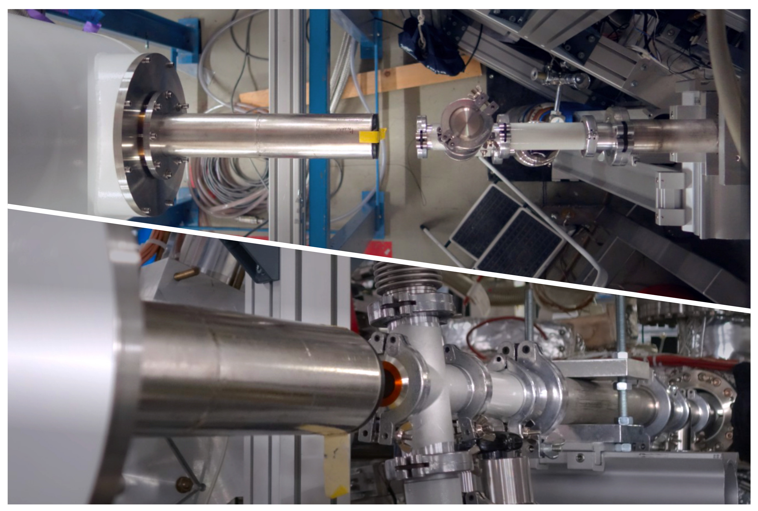
2. Experiment
3. Detector Performance
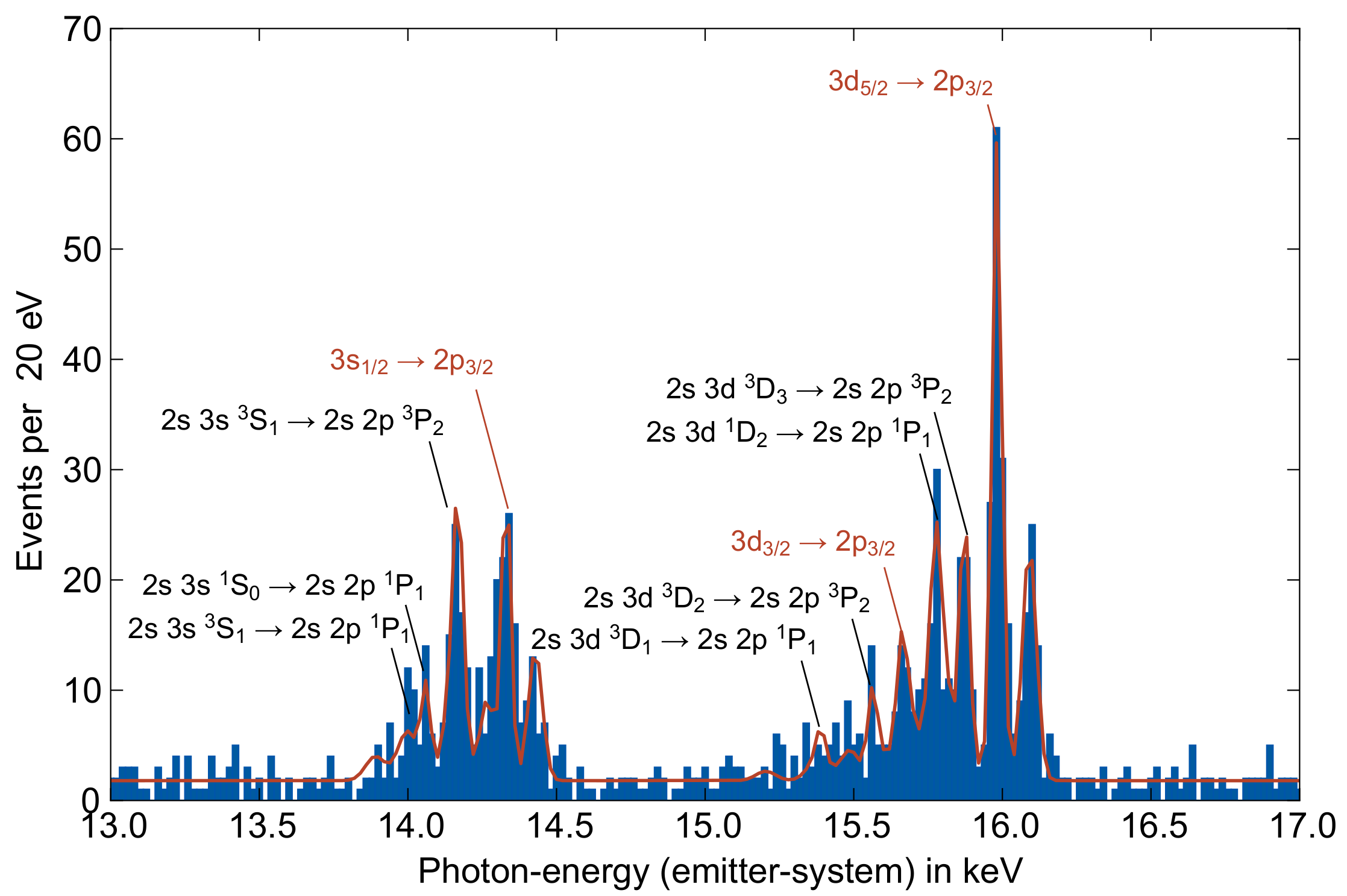
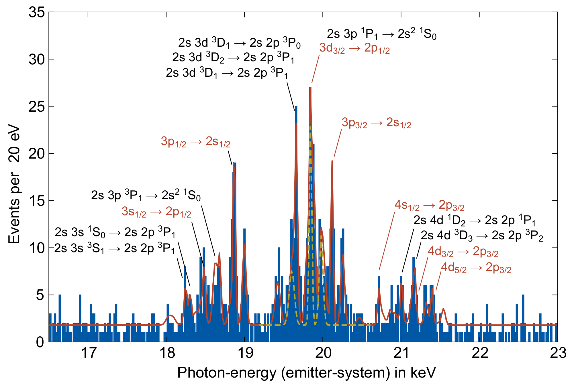
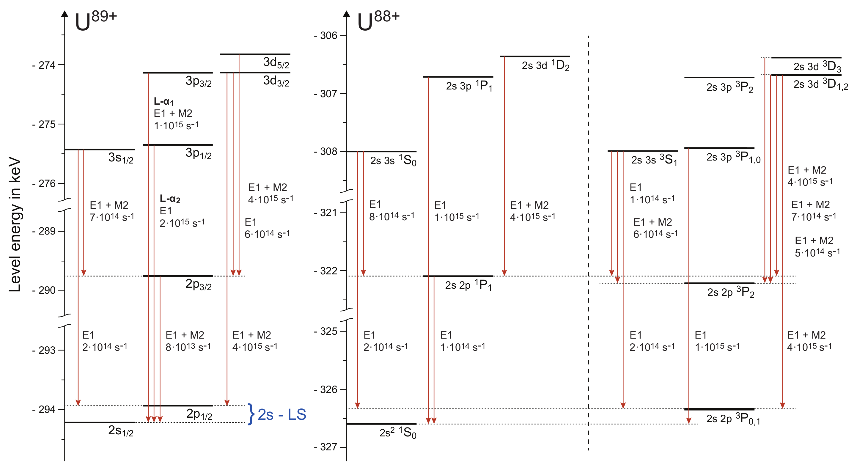
4. Experimental Data and Results
4.1. Transitions into the L-Shell
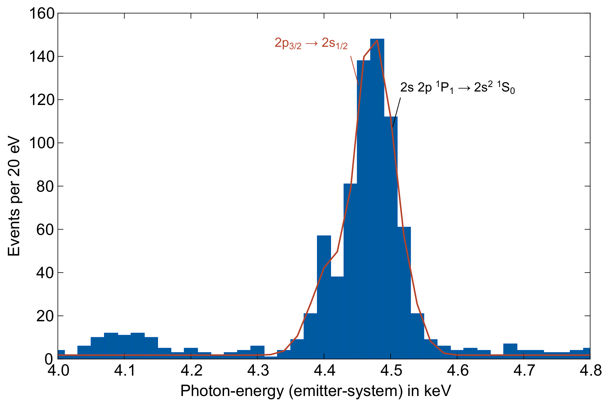
4.2. Transitions within the L-Shell
5. Conclusions and Outlook
Author Contributions
Funding
Data Availability Statement
Acknowledgments
Conflicts of Interest
References
- Fleischmann, A. Magnetische Mikrokalorimeter: Hochauflösende Röntgenspektroskopie Mit Energiedispersiven Detektoren. Ph.D. Thesis, Ruprecht-Karls-Universität Heidelberg, Heidelberg, Germany, 2003. [Google Scholar]
- Pies, C. maXs-200: Entwicklung Und Charakterisierung Eines Röntgendetektors Basierend Auf Magnetischen Kalorimetern Für Die Hochauflösende Spektroskopie Hochgeladener Ionen. Ph.D. Thesis, Ruprecht-Karls-Universität Heidelberg, Heidelberg, Germany, 2012. [Google Scholar]
- Hengstler, D. Development and Characterization of Two-Dimensional Metallic Magnetic Calorimeter Arrays for the High-Resolution X-ray Spectroscopy. Ph.D. Thesis, Ruprecht-Karls-Universität Heidelberg, Heidelberg, Germany, 2017. [Google Scholar]
- Geist, J. Bestimmung Der Isomerenergie von 229Th Mit Dem Hochauflösenden Mikrokalorimeter-Array maXs30. Ph.D. Thesis, Ruprecht-Karls-Universität Heidelberg, Heidelberg, Germany, 2020. [Google Scholar]
- Pies, C.; Schäfer, S.; Heuser, S.; Kempf, S.; Pabinger, A.; Porst, J.P.; Ranitsch, P.; Foerster, N.; Hengstler, D.; Kampkötter, A.; et al. maXs: Microcalorimeter Arrays for High-Resolution X-Ray Spectroscopy at GSI/FAIR. J. Low Temp. Phys. 2012, 167, 269–279. [Google Scholar] [CrossRef]
- Fleischmann, A.; Enss, C.; Seidel, G.M. Metallic Magnetic Calorimeters. In Cryogenic Particle Detection; Enss, C., Ed.; Topics in Applied Physics; Springer: Berlin/Heidelberg, Germany, 2005; pp. 151–216. [Google Scholar] [CrossRef]
- Thorn, D.B.; Gu, M.F.; Brown, G.V.; Beiersdorfer, P.; Porter, F.S.; Kilbourne, C.A.; Kelley, R.L. Precision Measurement of the K -Shell Spectrum from Highly Charged Xenon with an Array of X-Ray Calorimeters. Phys. Rev. Lett. 2009, 103, 0163001. [Google Scholar] [CrossRef] [PubMed]
- Porter, F.S.; Adams, J.S.; Beiersdorfer, P.; Brown, G.V.; Clementson, J.; Frankel, M.; Kahn, S.M.; Kelley, R.L.; Kilbourne, C.A. High-resolution x-ray spectroscopy with the EBIT Calorimeter Spectrometer. AIP Conf. Proc. 2009, 1185, 454–457. [Google Scholar] [CrossRef]
- Shen, Y.; Silver, E.; Hutton, R.; Zou, Y. The status of the micro-calorimeter at the Shanghai EBIT. Phys. Scr. 2011, 2011, 014060. [Google Scholar] [CrossRef]
- Szypryt, P.; O’Neil, G.C.; Takacs, E.; Tan, J.N.; Buechele, S.W.; Naing, A.S.; Bennett, D.A.; Doriese, W.B.; Durkin, M.; Fowler, J.W.; et al. A transition-edge sensor-based X-ray spectrometer for the study of highly charged ions at the National Institute of Standards and Technology electron beam ion trap. Rev. Sci. Instrum. 2019, 90, 123107. [Google Scholar] [CrossRef] [PubMed]
- Hillenbrand, P.M.; Hagmann, S.; Groshev, M.E.; Banaś, D.; Benis, E.P.; Brandau, C.; De Filippo, E.; Forstner, O.; Glorius, J.; Grisenti, R.E.; et al. Radiative Electron Capture to the Continuum in U89+ + N2 Collisions: Experiment and Theory. Phys. Rev. A 2020, 101, 022708. [Google Scholar] [CrossRef] [Green Version]
- Hillenbrand, P.M.; Lyashchenko, K.N.; Hagmann, S.; Andreev, O.Y.; Banaś, D.; Benis, E.P.; Bondarev, A.I.; Brandau, C.; De Filippo, E.; Forstner, O.; et al. Electron-Loss-to-Continuum Cusp in Collisions of U 89 + with N 2 and Xe. Phys. Rev. A 2021, 104, 012809. [Google Scholar] [CrossRef]
- Chu, S.Y.F.; Ekström, L.P.; Firestone, R.B. Table of Radioactive Isotopes. 1999. Available online: http://nucleardata.nuclear.lu.se/toi/ (accessed on 10 March 2021).
- Gu, M.F. The Flexible Atomic Code. Can. J. Phys. 2008, 86, 675–689. [Google Scholar] [CrossRef]
- Fritzsche, S. A fresh computational approach to atomic structures, processes and cascades. Comput. Phys. Commun. 2019, 240, 1–14. [Google Scholar] [CrossRef]
- Eichler, J.; Stöhlker, T. Radiative Electron Capture in Relativistic Ion–Atom Collisions and the Photoelectric Effect in Hydrogen-like High-Z Systems. Phys. Rep. 2007, 439, 1–99. [Google Scholar] [CrossRef]
- Yerokhin, V.A.; Indelicato, P.; Shabaev, V.M. Nonperturbative Calculation of the Two-Loop Lamb Shift in Li-Like Ions. Phys. Rev. Lett. 2006, 97, 0253004. [Google Scholar] [CrossRef] [PubMed] [Green Version]
- Schweppe, J.; Belkacem, A.; Blumenfeld, L.; Claytor, N.; Feinberg, B.; Gould, H.; Kostroun, V.E.; Levy, L.; Misawa, S.; Mowat, J.R.; et al. Measurement of the Lamb Shift in Lithiumlike Uranium (U89+). Phys. Rev. Lett. 1991, 66, 1434–1437. [Google Scholar] [CrossRef] [PubMed]
- Brandau, C.; Kozhuharov, C.; Müller, A.; Shi, W.; Schippers, S.; Bartsch, T.; Böhm, S.; Böhme, C.; Hoffknecht, A.; Knopp, H.; et al. Precise Determination of the 2s1/2 - 2p1/2 Splitting in Very Heavy Lithiumlike Ions Utilizing Dielectronic Recombination. Phys. Rev. Lett. 2003, 91, 073202. [Google Scholar] [CrossRef] [PubMed]
- Beiersdorfer, P.; Chen, H.; Thorn, D.B.; Träbert, E. Measurement of the Two-Loop Lamb Shift in Lithiumlike U 89 +. Phys. Rev. Lett. 2005, 95, 0233003. [Google Scholar] [CrossRef] [PubMed] [Green Version]
- Beiersdorfer, P.; Knapp, D.; Marrs, R.E.; Elliott, S.R.; Chen, M.H. Structure and Lamb Shift of 2s1/2-2p3/2 Levels in Lithiumlike U89+ through Neonlike U82+. Phys. Rev. Lett. 1993, 71, 3939–3942. [Google Scholar] [CrossRef] [PubMed]
- Beiersdorfer, P. Spectral Measurements of Few-Electron Uranium Ions Produced and Trapped in a High-Energy Electron Beam Ion Trap. Nucl. Instruments Methods Phys. Res. Sect. Beam Interact. Mater. Atoms 1995, 99, 114–116. [Google Scholar] [CrossRef] [Green Version]
- Pfäfflein, P.; Allgeier, S.; Bernitt, S.; Fleischmann, A.; Friedrich, M.; Hahn, C.; Hengstler, D.; Herdrich, M.O.; Kalinin, A.; Kröger, F.M.; et al. Integration of maXs-type Microcalorimeter Detectors for High-Resolution x-Ray Spectroscopy into the Experimental Environment at the CRYRING@ESR Electron Cooler. Physica Scripta 2022, 97, 0114005. [Google Scholar] [CrossRef]


| Transition | ||
|---|---|---|
| Emitter-System | Calculation FAC | |
Disclaimer/Publisher’s Note: The statements, opinions and data contained in all publications are solely those of the individual author(s) and contributor(s) and not of MDPI and/or the editor(s). MDPI and/or the editor(s) disclaim responsibility for any injury to people or property resulting from any ideas, methods, instructions or products referred to in the content. |
© 2023 by the authors. Licensee MDPI, Basel, Switzerland. This article is an open access article distributed under the terms and conditions of the Creative Commons Attribution (CC BY) license (https://creativecommons.org/licenses/by/4.0/).
Share and Cite
Herdrich, M.O.; Hengstler, D.; Fleischmann, A.; Enss, C.; Gumberidze, A.; Hillenbrand, P.-M.; Indelicato, P.; Fritzsche, S.; Stöhlker, T. X-ray Spectroscopy Based on Micro-Calorimeters at Internal Targets of Storage Rings. Atoms 2023, 11, 13. https://doi.org/10.3390/atoms11010013
Herdrich MO, Hengstler D, Fleischmann A, Enss C, Gumberidze A, Hillenbrand P-M, Indelicato P, Fritzsche S, Stöhlker T. X-ray Spectroscopy Based on Micro-Calorimeters at Internal Targets of Storage Rings. Atoms. 2023; 11(1):13. https://doi.org/10.3390/atoms11010013
Chicago/Turabian StyleHerdrich, Marc Oliver, Daniel Hengstler, Andreas Fleischmann, Christian Enss, Alexandre Gumberidze, Pierre-Michel Hillenbrand, Paul Indelicato, Stephan Fritzsche, and Thomas Stöhlker. 2023. "X-ray Spectroscopy Based on Micro-Calorimeters at Internal Targets of Storage Rings" Atoms 11, no. 1: 13. https://doi.org/10.3390/atoms11010013