
Electron Capture by Proton Beam in Collisions with Water Vapor

Abstract
:1. Introduction
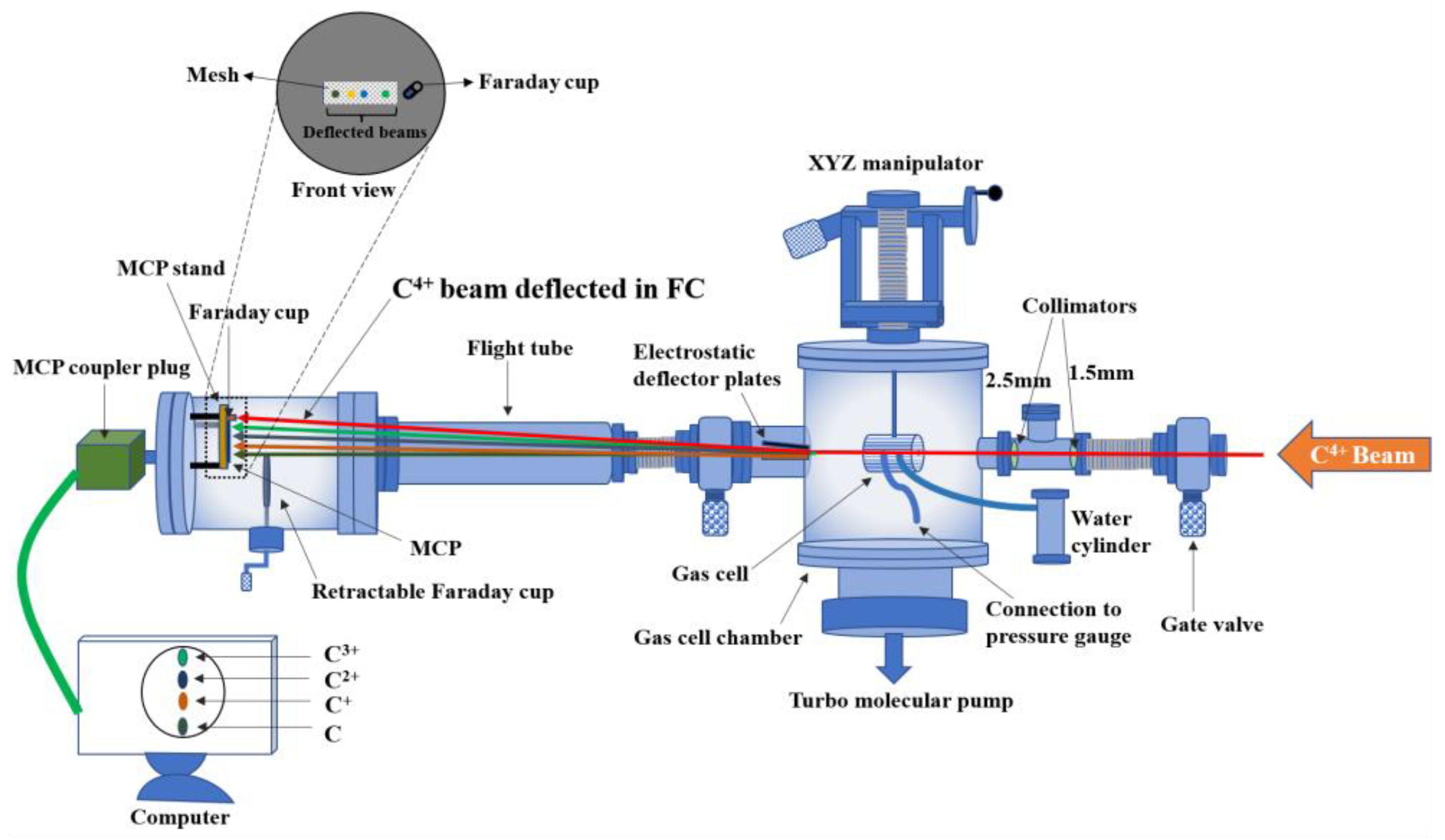
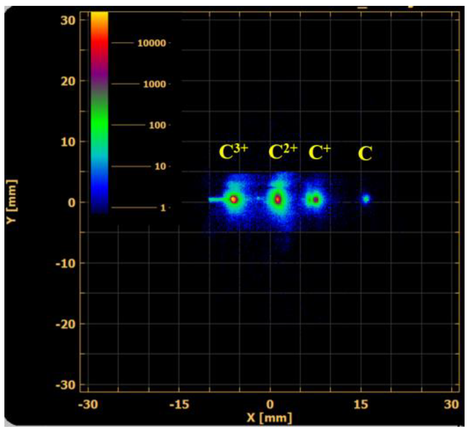
2. Experimental Details
3. Results and Discussion
4. Conclusions
Author Contributions
Funding
Data Availability Statement
Acknowledgments
Conflicts of Interest
References
- McDaniel, E.W.; Mitchell, J.B.A.; Rudd, M.E. Atomic Collisions; Wiley: New York, NY, USA, 1993. [Google Scholar]
- Enos, C.S.; Lee, A.R.; Brenton, A.G. Electronic excitation of atmospheric molecules by proton impact. Int. J. Mass Spectrom. Ion Process 1991, 104, 137. [Google Scholar] [CrossRef]
- Townsend, L.W.; Wilson, J.W.; Cucinotta, F.A.; Shinn, J.L. Galactic Cosmic Ray Transport Methods and Radiation Quality Issues. Int. J. Radiat. Appl. Instrum Part D. Nucl. Tracks Radiat. Meas. 1992, 20, 65–72. [Google Scholar] [CrossRef] [PubMed]
- Amaldi, U.; Kraft, G. Radiotherapy with Beams of Carbon Ions. Rep. Prog. Phys. 2005, 68, 1861–1882. [Google Scholar] [CrossRef]
- Inokuti, M. Inelastic Collisions of Fast Charged Particles with Atoms and Molecules- The Bethe Theory Revisited. Rev. Mod. Phys. 1971, 43, 297–347. [Google Scholar] [CrossRef]
- Champion, C.; Weck, P.F.; Lekadir, H.; Galassi, M.E.; Fojón, O.A.; Abufager, P.; Rivarola, R.D.; Hanssen, J. Proton-induced single electron capture on DNA/RNA bases. Phys. Med. Biol. 2012, 57, 3039. [Google Scholar] [CrossRef] [PubMed]
- Tachino, C.A.; Monti, J.M.; Fojón, O.A.; Champion, C.; Rivarola, R.D. Ionization of water molecules by ion beams. On the relevance of dynamic screening and the influence of the description of the initial state. J. Phys. B At. Mol. Opt. Phys. 2014, 47, 035203. [Google Scholar] [CrossRef]
- Mendez, A.M.P.; Montanari, C.C.; Miraglia, J.E. Ionization of biological molecules by multicharged ions using the stoichiometric model. J. Phys. B At. Mol. Opt. Phys. 2020, 53, 055201, Correction: J. Phys. B At. Mol. Opt. Phys. 2020, 53, 249501. [Google Scholar] [CrossRef] [Green Version]
- Levin, W.P.; Kooy, H.; Loeffler, J.S.; DeLaney, T.F. Proton beam therapy. Brit. J. Cancer 2005, 93, 849. [Google Scholar] [CrossRef] [PubMed] [Green Version]
- Smith, A.R. Proton therapy. Phys. Med. Biol. 2006, 51, R491. [Google Scholar] [CrossRef] [PubMed]
- Schardt, D.; Elsasser, T.; Schulz-Ertner, D. Heavy-ion tumor therapy: Physical and radiobiological benefits. Rev. Mod. Phys. 2010, 82, 383–425. [Google Scholar] [CrossRef]
- Ohsawa, D.; Tawara, H.; Soga, F.; Galassi, M.E.; Rivarola, R.D. 6.0 MeV u−1 carbon ion (C6+ and C4+)-induced secondary electron emission from water vapor. Phys. Scr. 2013, T156, 014039. [Google Scholar] [CrossRef]
- Bhattacharjee, S.; Biswas, S.; Monti, J.M.; Rivarola, R.D.; Tribedi, L.C. Double-differential cross section for ionization of H2O molecules by 4-MeV/u C6+ and Si13+ ions. Phys. Rev. A 2017, 96, 052707. [Google Scholar] [CrossRef]
- Luna, H.; Wolff, W.; Montenegro, E.C.; Tavares, A.C.; Ludde, H.J.; Schenk, G.; Horbatsch, M.; Kirchner, T. Ionization and electron-capture cross sections for single- and multiple-electron removal from H2O by Li3+ impact. Phys. Rev. A 2016, 93, 052705. [Google Scholar] [CrossRef]
- Nikjoo, H.; Emfietzoglou, D.; Liamsuwan, T.; Taleei, R.; Liljequist, D.; Uehara, S. Radiation track, DNA damage and response- A review. Rep. Prog. Phys. 2016, 79, 116601. [Google Scholar] [CrossRef]
- Toburen, L.H.; Nakai, M.Y.; Langley, R.A. Measurement of High-Energy Charge-Transfer Cross Sections for Incident Protons and Atomic Hydrogen in Various Gases*. Phys. Rev. 1968, 171, 114. [Google Scholar] [CrossRef]
- Rudd, M.E.; Goffe, T.V.; DuBois, R.D.; Toburen, L.H. Cross sections for ionization of water vapor by 7-4000-keV protons. Phys. Rev. A 1985, 31, 492. [Google Scholar] [CrossRef] [PubMed] [Green Version]
- Maurya, S.K.; Bhattacharjee, S. Variable gaseous ion beams from plasmas driven by electromagnetic waves for nano-micro structuring: A tutorial and an overview of recent works and future prospects. Plasma Res. Express 2020, 2, 033001. [Google Scholar] [CrossRef]
- Maurya, S.K.; Paul, S.; Shah, J.K.; Chatterjee, S.; Bhattacharjee, S. Momentum transfer using variable gaseous plasma ion beams and creation of high aspect ratio microstructures. J. Appl. Phys. 2017, 121, 123302. [Google Scholar] [CrossRef]
- Maurya, S.K.; Barman, S.; Pan, N.; Bhattacharjee, S. Effect of plasma and beam parameters on focal dimensions in micrometer charged particle optics: Enhanced nonlinear demagnification below the Debye length. Phys. Plasmas 2019, 26, 063103. [Google Scholar] [CrossRef]
- Maurya, S.K.; Barman, S.; Bhattacharjee, S. Charge dissipation and self focusing limit in high current density ion beam transport through a micro glass capillary. J. Phys. D Appl. Phys. 2019, 52, 055205. [Google Scholar] [CrossRef]
- Barman, S.; Maurya, S.K.; Bhattacharjee, S. Experimental realization of nonlinear demagnification in plasma-based charged particle optics. Plasma Res. Express 2022, 4, 025003. [Google Scholar] [CrossRef]
- Chatterjee, S.; Bhattacharjee, S.; Maurya, S.K.; Srinivasan, V.; Khare, K.; Khandekar, S. Surface wettability of an atomically heterogeneous system and the resulting intermolecular forces. Euro Phy. Lett. 2017, 118, 68006. [Google Scholar] [CrossRef]
- Agnihotri, A.N.; Kelkar, A.H.; Kasthurirangan, S.; Thulasiram, K.V.; Desai, C.A.; Fernandez, W.A.; Tribedi, L.C. An ECR ion source-based low-energy ion accelerator: Development and performance. Phys. Scr. 2011, T144, 014038. [Google Scholar] [CrossRef]
- Available online: https://www.roentdek.com (accessed on 10 January 2023).
- Krems, M.; Zirbel, J.; Thomason, M.; DuBoisa, R.D. Channel electron multiplier and channelplate efficiencies for detecting positive ions. Rev. Sci. Instrum. 2005, 76, 093305. [Google Scholar] [CrossRef]
- Dunseath, K.M.; Crothers, D.S.F. Transfer and ionization processes during the collision of fast H+, He2+ nuclei with helium. J. Phys. B At. Mol. Opt. Phys. 1991, 24, 5003–5022. [Google Scholar] [CrossRef]


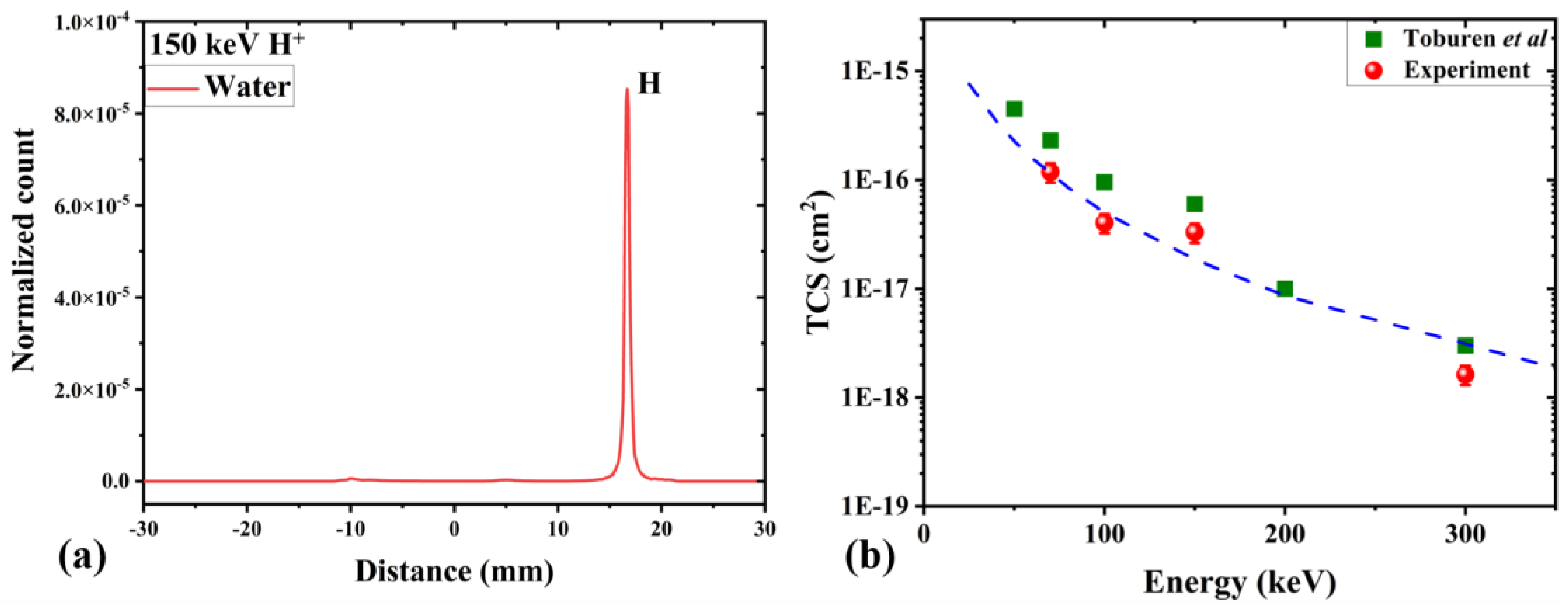
| Energy (keV) | TCS (cm2) | |
|---|---|---|
| Present | Toburen et al. | |
| 70 | 2.4 (−16) | 2.3 (−16) |
| 100 | 8.1 (−17) | 9.5 (−17) |
| 150 | 6.6 (−17) | 6.0 (−17) |
| 300 | 3.2 (−18) | 3.0 (−18) |
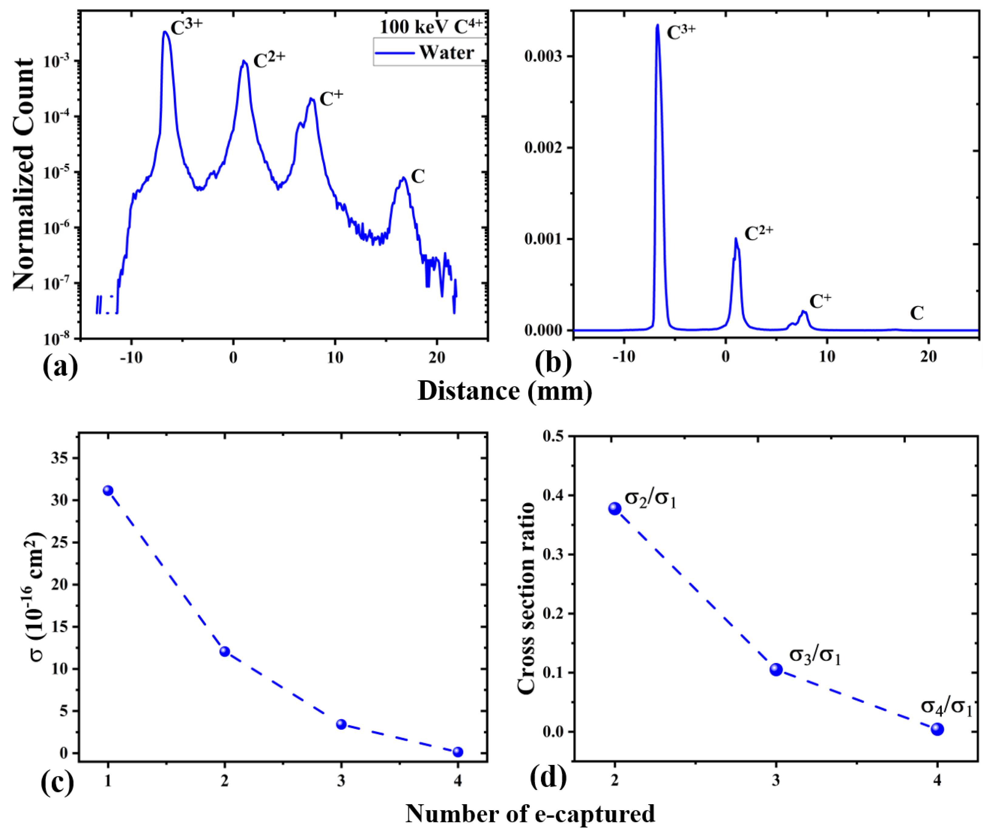
| Number of Captured Electrons | Cross-Section (10−16 cm2) | Ratio of Multi E-Capture to Single Capture |
|---|---|---|
| 1 | 13.1 | 1.000 |
| 2 | 12.0 | 0.377 |
| 3 | 3.41 | 0.105 |
| 4 | 0.132 | 0.004 |
Disclaimer/Publisher’s Note: The statements, opinions and data contained in all publications are solely those of the individual author(s) and contributor(s) and not of MDPI and/or the editor(s). MDPI and/or the editor(s) disclaim responsibility for any injury to people or property resulting from any ideas, methods, instructions or products referred to in the content. |
© 2023 by the authors. Licensee MDPI, Basel, Switzerland. This article is an open access article distributed under the terms and conditions of the Creative Commons Attribution (CC BY) license (https://creativecommons.org/licenses/by/4.0/).
Share and Cite
Maurya, S.K.; Bhogale, A.; Tribedi, L.C. Electron Capture by Proton Beam in Collisions with Water Vapor. Atoms 2023, 11, 21. https://doi.org/10.3390/atoms11020021
Maurya SK, Bhogale A, Tribedi LC. Electron Capture by Proton Beam in Collisions with Water Vapor. Atoms. 2023; 11(2):21. https://doi.org/10.3390/atoms11020021
Chicago/Turabian StyleMaurya, Sanjeev Kumar, Abhijeet Bhogale, and Lokesh C. Tribedi. 2023. "Electron Capture by Proton Beam in Collisions with Water Vapor" Atoms 11, no. 2: 21. https://doi.org/10.3390/atoms11020021