
Environment, Endocrine Disruptors, and Fatty Liver Disease Associated with Metabolic Dysfunction (MASLD)
 , ,
, ,  and
and
Abstract
:1. Introduction

| Countries | Sample Type | Compounds | Concentration (ng/L or ng/g) | Reference |
|---|---|---|---|---|
| Taiwan (China) | River water | BPA | 302 | Tao et al., 2021 [6] |
| Kuwait | River water | DMP | 16.9 | Saeed et al., 2017 [7] |
| DEP | 524.8 | |||
| DBP | 899.0 | |||
| China | River water | NP | 634.8 | Wang et al., 2018 [8] |
| BPA | 1573.1 | |||
| 17 β-estradiol | 23.9 | |||
| Vietnam | River water | PAEs | 2.78–412.27 | Quynh et al., 2019 [9] |
| PBDEs | 1.92–7.08 | |||
| Malaysia | River water | 3,4,4-Trichlorocarbanilide | 261.67 | Aziz et al., 2014 [10] |
| Methylparaben | 4.93 | |||
| South Africa | River water | NP | 2550 | Farounbi et al., 2020 [11] |
| Dichlorophenol | 737 | |||
| BPA | 477 | |||
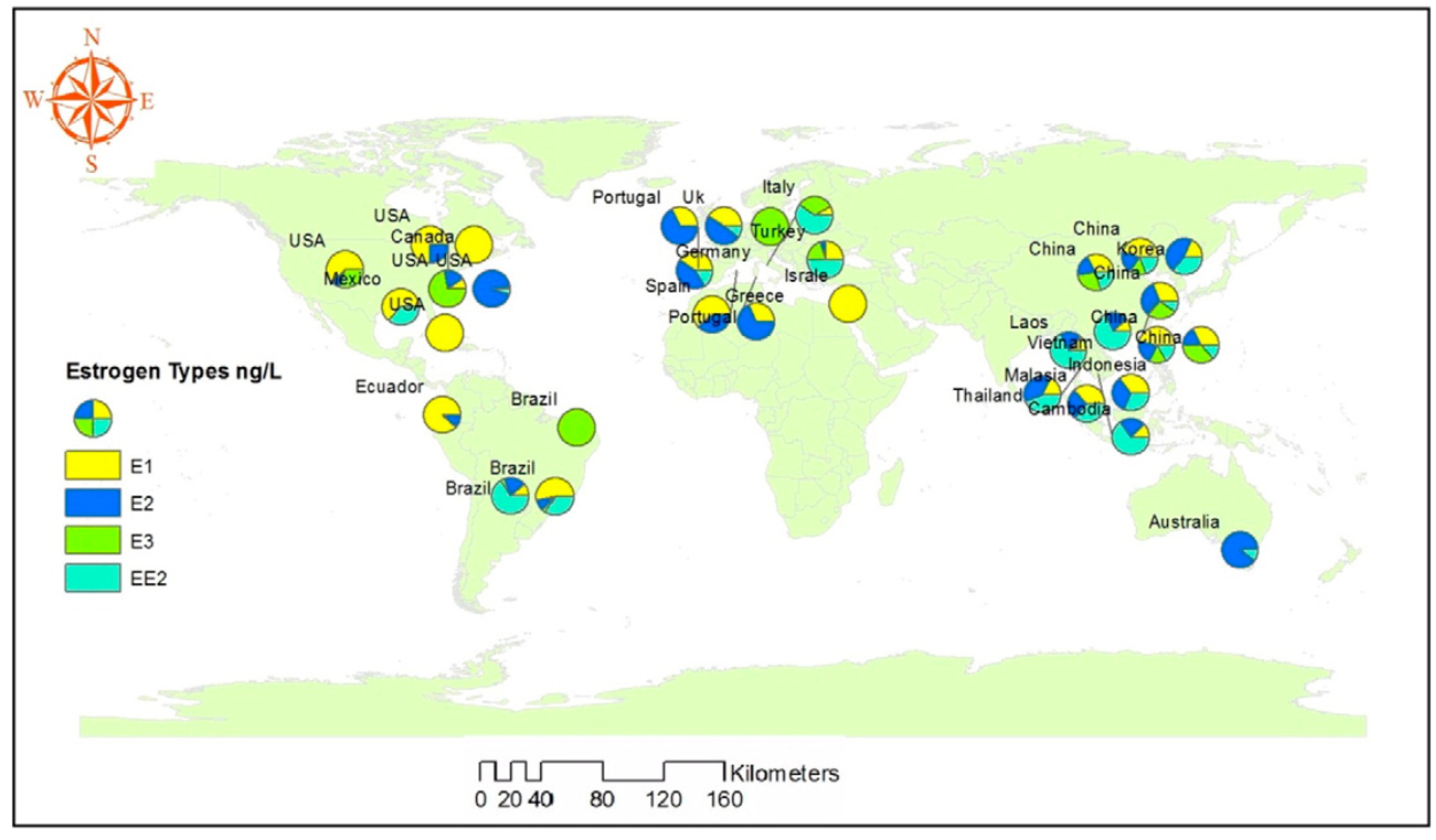
| Italy | River water | E1 | 28 | Pignotti et al., 2017 [12] |
| E2 | 39.7 | |||
| America | River water | E1 | 21.1–37.7 | Sweeney et al., 2021 [13] |
| E3 | 38.2–79.6 | |||
| EE2 | 5.1–6.7 | |||
| Thailand | River water | E2 | 62.98 ± 5.03 | Ocharoen et al., 2018 [14] |
| EE2 | 35.53 ± 2.99 | |||
| BPA | 50.67 ± 4.19 | |||
| Brazil | River sediment | E1 | 187.39 | Froehner et al., 2012 [15] |
| DES | 453.69 | |||
| α-E2 | 21.36 | |||
| β-E2 | 52.82 | |||
| EE2 | 70.28 | |||
| E3 | 34.68 |

2. Methods
3. Environmental EDCs and Sources of Exposure
4. The Effects of Obesogens across the Lifespan
5. EDC and MASLD
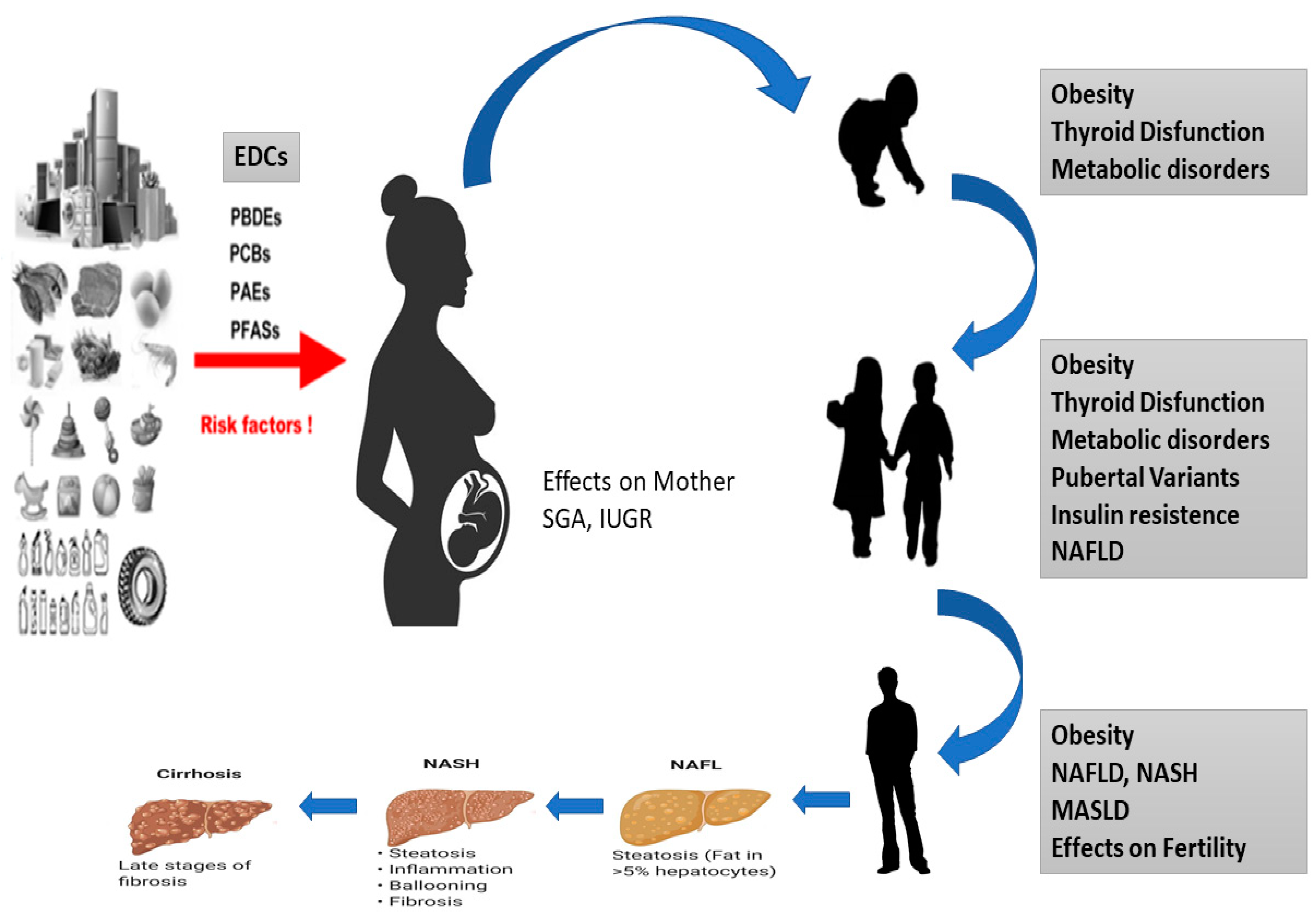
5.1. Prenatal Life
5.2. Young Adult Life
6. Endocrine Disruptors and the Development of MASLD
Studies in Human Population with Obesity and MASLD
7. Future Prospective
8. Conclusions
Author Contributions
Funding
Informed Consent Statement
Data Availability Statement
Conflicts of Interest
Abbreviations
| Alanine aminotransferase | (ALT) |
| Androgen receptors | (ARs) |
| Aspartate aminotransferase | (AST) |
| Atrazine | (ATZ) |
| Body mass index | (BMI) |
| Plasticizers bisphenol A | (BPA) |
| Bisphenol A | (BPA) |
| Confidence interval | (CI) |
| Credible interval | (CrI) |
| Cypermethrin | (CYP) |
| Cytochrome P450 | (CYP) |
| Organochlorines dichlorodiphenyltrichloroethane | (DDT) |
| Phthalates di-2-ethylhexyl phthalate | (DEHP) |
| Developmental Origins of Health and Disease | (DOHaD) |
| Estrone | (E1) |
| 17-β-estradiol | (E2) |
| Estriol | (E3) |
| European Chemicals Agency | (ECHA) |
| Endocrine-disrupting chemicals | (EDCs) |
| Ethinylestradiol | (EE2) |
| European Food Safety Authority | (EFSA) |
| Protection Agency | (EPA) |
| Estrogen receptors | (ERα and ERβ) |
| Free Fatty Acids | (FFAs) |
| Farnesoid X α receptor | (FXRα) |
| Gamma-glutamyl transferase | (GGT) |
| Hepatitis C Virus virus | (HCV) |
| High-density lipoprotein | (HDL) |
| Janus kinase 2/Signal Transducer and Transcription Activator 5 | (JAK2/STAT5) |
| Lipid accumulation product | (LAP) |
| Low birth weight | (LBW) |
| Liver X receptors α and β | (LXR) |
| Steatohepatitis associated with metabolic dysfunction | (MASH), |
| Metabolic dysfunction-associated steatotic liver disease | (MASLD) |
| Mono-(2-ethylhexyl) phthalate | (MEHP) |
| Monoethyl phthalate | (MEP) |
| Mono-(2-ethylhexyl) phthalate | (MEPH) |
| Metabolite mono-(2-ethylhexyl) phthalate | (MEPH) |
| Nonalcoholic fatty liver disease | (NAFLD) |
| Non-alcoholic Steatosteato-hepatitis | (NASH) |
| Dioxin-like (DL) and non-dioxin-like | (NDL) |
| Dioxin-like (DL) and non-dioxin-like | (NDL) |
| Nonylphenols | (NPs) |
| Occhlorinated dibenzodioxin | (OCDD) |
| Odds ratio | (OR) |
| Polycyclic aromatic hydrocarbons | (PAHs) |
| Polybrominated diphenyl ethers | (PBDEs) |
| 2-polybrominated diphenyl ethers | (PBDEs) |
| Polychlorinated biphenyls | (PCBs) |
| Perfluoroalkyl substances | (PFAS) |
| Fluorinated chemicals | (PFCs) |
| Perfluorohexane sulfonic acid | (PFHxS) |
| Perfluorooctanoic acid | (PFOA) |
| Organotins tributyltin polyfluoroalkyls and/or perfluorooctane sulfonate | (PFOS) |
| Perfluorooctane sulfonate | (PFOS) |
| Persistent Organic Pollutants | (POPs) |
| Peroxisome proliferators | (PPARs) |
| Peroxisome proliferator-activated γ receptor | (PPARγ) |
| Progesterone receptors | (PRs) |
| Retinoic receptor isoforms | (RARα and RARγ) |
| Retinoid X receptors | (RXRα, RXRβ, and RXRγ) |
| Retinoid α X receptor | (RXRα) |
| Steroid receptor coactivator complexes | (SRCs) |
| Type 2 diabetes mellitus | (T2DM) |
| Triacylglyceride | (TAG) |
| Thyroid receptors | (TR) |
| Thyroid α receptor isoforms | (TRα) |
| UDP-glucuronosyltransferase | (UGT) |
| Visceral adiposity index | (VAI) |
| Very Low-Density Lipoprotein | VLDL |
References
- Swinburn, B.A.; Kraak, V.I.; Allender, S.; Atkins, V.J.; Baker, P.I.; Bogard, J.R.; Brinsden, H.; Calvillo, A.; De Schutter, O.; Devarajan, R.; et al. The global syndemia of obesity, undernutrition and climate change: The report of the Lancet Commission. Lancet 2019, 393, 791–846. [Google Scholar] [CrossRef]
- Lamb, J.C.; Boffetta, P.; Foster, W.G.; Goodman, J.E.; Hentz, K.L.; Rhomberg, L.R.; Staveley, J.; Swaen, G.; Van Der Kraak, G.; Williams, A.L. Critical comments on the state of the science of endocrine disrupting chemicals by WHO and UNEP—2012. Regul. Toxicol. Pharmacol. 2012, 69, 22–40. [Google Scholar] [CrossRef]
- Ahn, C.; Jeung, E.-B. Endocrine-disrupting chemicals and disease endpoints. Int. J. Mol. Sci. 2023, 24, 5342. [Google Scholar] [CrossRef]
- Gupta, R.; Kumar, P.; Fahmi, N.; Garg, B.; Dutta, S.; Sachar, S.; Matharu, A.S.; Vimaleswaran, K.S. Endocrine Disruption and Obesity: A Current Review on the Obesogenic Environment. Curr. Res. Green Sustain. Chem. 2020, 3, 100009. [Google Scholar] [CrossRef]
- European Chemical Agency (ECHA) and European Food Safety Authority (EFSA) with the technical support of the Joint Research Centre (JRC); Andersson, N.; Arena, M.; Auteri, D.; Barmaz, S.; Grignard, E.; Kienzler, A.; Lepper, P.; Lostia, A.M.; Munn, S.; et al. Guidance for the identification of endocrine disruptors in the context of Regulations (EU) No 528/2012 and (EC) No 1107/2009. EFSA J. 2018, 16, e05311. [Google Scholar] [CrossRef] [PubMed]
- Tao, H.-Y.; Zhang, J.; Shi, J.; Guo, W.; Liu, X.; Zhang, M.; Ge, H.; Li, X.-Y. Occurrence and emission of phthalates, bisphenol A, and oestrogenic compounds in concentrated animal feeding operations in Southern China. Ecotoxicol. Environ. Saf. 2021, 207, 111521. [Google Scholar] [CrossRef]
- Saeed, T.; Al-Jandal, N.; Abusam, A.; Taqi, H.; Al-Khabbaz, A.; Zafar, J. Sources and levels of endocrine disrupting compounds (EDCs) in Kuwait’s coastal areas. Mar. Pollut. Bull. 2017, 118, 407–412. [Google Scholar] [CrossRef]
- Wang, J.; Cheng, L.-M.; Su, T. Multivariate Cryptography Based on Clipped Hopfield Neural Network. IEEE Trans. Neural Netw. Learn. Syst. 2018, 29, 353–363. [Google Scholar] [CrossRef]
- Quynh, T.X.; Toan, V.D. Endocrine Disrupting Compounds (EDCs) in Surface Waters of the KimNguu River, Vietnam. Bull. Environ. Contam. Toxicol. 2019, 103, 734–738. [Google Scholar] [CrossRef]
- Aziz, A.N.; Taha, M.; Ismail, N.H.; Anouar, E.H.; Yousuf, S.; Jamil, W.; Awang, K.; Ahmat, N.; Khan, K.M.; Kashif, S.M. Synthesis, Crystal Structure, DFT Studies and Evaluation of the Antioxidant Activity of 3,4-Dimethoxybenzenamine Schiff Bases. Molecules 2014, 19, 8414–8433. [Google Scholar] [CrossRef]
- Farounbi, A.I.; Ngqwala, N.P. Occurrence of selected endocrine disrupting compounds in the eastern cape province of South Africa. Environ. Sci. Pollut. Res. Int. 2020, 27, 17268–17279. [Google Scholar] [CrossRef] [PubMed]
- Pignotti, E.; Farré, M.; Barceló, D.; Dinelli, E. Occurrence and distribution of six selected endocrine disrupting compounds in surface- and groundwaters of the Romagna area (North Italy). Environ. Sci. Pollut. Res. Int. 2017, 24, 21153–21167. [Google Scholar] [CrossRef] [PubMed]
- Sweeney, C.L.; Bennett, J.L.; Brown, C.A.M.; Ross, N.W.; Gagnon, G.A. Validation of a QuEChERS method for extraction of estrogens from a complex water matrix and quantitation via high-performance liquid chromatography-mass spectrometry. Chemosphere 2021, 263, 128315. [Google Scholar] [CrossRef] [PubMed]
- Ocharoen, Y.; Boonphakdee, C.; Boonphakdee, T.; Shinn, A.P.; Moonmangmee, S. High levels of the endocrine disruptors bisphenol-A and 17β-estradiol detected in populations of green mussel, Perna viridis, cultured in the Gulf of Thailand. Aquaculture 2018, 497, 348–356. [Google Scholar] [CrossRef]
- Froehner, S.; Machado, K.S.; Stefan, E.; Bleninger, T.; da Rosa, E.C.; de Castro Martins, C. Occurrence of selected estrogens in mangrove sediments. Mar. Pollut. Bull. 2012, 64, 75–79. [Google Scholar] [CrossRef] [PubMed]
- Adeel, M.; Song, X.; Wang, Y.; Francis, D.; Yang, Y. Environmental impact of estrogens on human, animal and plant life: A critical review. Environ. Int. 2017, 99, 107–119. [Google Scholar] [CrossRef] [PubMed]
- Moher, D.; Liberati, A.; Tetzlaff, J.; Altman, D.G.; PRISMA Group. Preferred reporting items for systematic reviews and meta-analyses: The PRISMA statement. Int. J. Surg. 2010, 8, 336–341. [Google Scholar] [CrossRef]
- McCabe, C.F.; Padmanabhan, V.; Dolinoy, D.C.; Domino, S.E.; Jones, T.R.; Bakulski, K.M.; Goodrich, J.M. Maternal Environmental Exposure to Bisphenols and Epigenome-Wide DNA Methylation in Infant Cord Blood. Environ. Epigenetics 2020, 6, dvaa021. [Google Scholar] [CrossRef]
- Ke, Z.-H.; Pan, J.-X.; Jin, L.-Y.; Xu, H.-Y.; Yu, T.-T.; Ullah, K.; Rahman, T.U.; Ren, J.; Cheng, Y.; Dong, X.-Y.; et al. Bisphenol A Exposure May Induce Hepatic Lipid Accumulation via Reprogramming the DNA Methylation Patterns of Genes Involved in Lipid Metabolism. Sci. Rep. 2016, 6, 31331. [Google Scholar] [CrossRef]
- Manikkam, M.; Tracey, R.; Guerrero-Bosagna, C.; Skinner, M.K. Plastics Derived Endocrine Disruptors (BPA, DEHP and DBP) Induce Epigenetic Transgenerational Inheritance of Obesity, Reproductive Disease and Sperm Epimutations. PLoS ONE 2013, 8, e55387. [Google Scholar] [CrossRef]
- Ditzel, E.J.; Nguyen, T.; Parker, P.; Camenisch, T.D. Effects of Arsenite Exposure during Fetal Development on Energy Metabolism and Susceptibility to Diet-Induced Fatty Liver Disease in Male Mice. Environ. Health Perspect. 2016, 124, 201–209. [Google Scholar] [CrossRef]
- Maranghi, F.; Lorenzetti, S.; Tassinari, R.; Moracci, G.; Tassinari, V.; Marcoccia, D.; Di Virgilio, A.; Eusepi, A.; Romeo, A.; Magrelli, A.; et al. In Utero Exposure to Di-(2-Ethylhexyl) Phthalate Affects Liver Morphology and Metabolism in Post-Natal CD-1 Mice. Reprod. Toxicol. 2010, 29, 427–432. [Google Scholar] [CrossRef] [PubMed]
- Boverhof, D.R.; Burgoon, L.D.; Tashiro, C.; Sharratt, B.; Chittim, B.; Harkema, J.R.; Mendrick, D.L.; Zacharewski, T.R. Comparative Toxicogenomic Analysis of the Hepatotoxic Effects of TCDD in Sprague Dawley Rats and C57BL/6 Mice. Toxicol. Sci. 2006, 94, 398–416. [Google Scholar] [CrossRef]
- Lei, R.; Xue, B.; Tian, X.; Liu, C.; Li, Y.; Zheng, J.; Luo, B. The association between endocrine disrupting chemicals and MAFLD: Evidence from NHANES survey. Ecotoxicol. Environ. Saf. 2023, 256, 114836. [Google Scholar] [CrossRef] [PubMed]
- Midya, V.; Colicino, E.; Conti, D.V.; Berhane, K.; Garcia, E.; Stratakis, N.; Andrusaityte, S.; Basagaña, X.; Casas, M.; Fossati, S.; et al. Association of Prenatal Exposure to Endocrine-Disrupting Chemicals With Liver Injury in Children. JAMA Netw. Open 2022, 5, e2220176. [Google Scholar] [CrossRef] [PubMed]
- Heindel, J.J.; Blumberg, B.; Cave, M.; Machtinger, R.; Mantovani, A.; Mendez, M.A.; Nadal, A.; Palanza, P.; Panzica, G.; Sargis, R.; et al. Lacerating metabolic chemicals and metabolic disorders. Reprod. Toxicol. 2016, 68, 3–33. [Google Scholar] [CrossRef] [PubMed]
- Guo, W.; Pan, B.; Sakkiah, S.; Yavas, G.; Ge, W.; Zou, W.; Tong, W.; Hong, H. Persistent Organic Pollutants in Food: Contamination Sources, Health Effects and Detection Methods. Int. J. Environ. Res. Public Health 2019, 16, 4361. [Google Scholar] [CrossRef] [PubMed]
- Palade, L.M.; Negoiță, M.; Adascălului, A.C.; Mihai, A.L. Polycyclic Aromatic Hydrocarbon Occurrence and Formation in Processed Meat, Edible Oils, and Cereal-Derived Products: A Review. Appl. Sci. 2023, 13, 7877. [Google Scholar] [CrossRef]
- Thoene, M.; Rytel, L.; Dzika, E.; Włodarczyk, A.; Kruminis-Kaszkiel, E.; Konrad, P.; Wojtkiewicz, J. Bisphenol A Causes Liver Damage and Selectively Alters the Neurochemical Coding of Intrahepatic Parasympathetic Nerves in Juvenile Porcine Models under Physiological Conditions. Int. J. Mol. Sci. 2017, 18, 2726. [Google Scholar] [CrossRef]
- Ma, N.; Yip, R.; Lewis, S.; Dinani, A.; Wyatt, C.; Crane, M.; Jirapatnakul, A.; Li, L.; Aloman, C.; Bansal, M.B.; et al. Environmental exposures are important risk factors for advanced liver fibrosis in African American adults. JHEP Rep. 2023, 5, 100696. [Google Scholar] [CrossRef]
- Program, N.T. NTP Toxicology and Carcinogenesis Studies of Polybrominated Biphenyls (CAS No. 67774-32-7)(Firemaster FF-1(R)) in F344/N Rats and B6C3F1 Mice (Feed Studies). Natl. Toxicol. Program Tech. Rep. Ser. 1993, 398, 1–235. [Google Scholar]
- Costello, E.; Rock, S.; Stratakis, N.; Eckel, S.P.; Walker, D.I.; Valvi, D.; Cserbik, D.; Jenkins, T.; Xanthakos, S.A.; Kohli, R.; et al. Exposure to per- and Polyfluoroalkyl Substances and Markers of Liver Injury: A Systematic Review and Meta-Analysis. Environ. Health Perspect. 2022, 130, 46001. [Google Scholar] [CrossRef] [PubMed]
- Karri, K.; Waxman, D.J. TCDD dysregulation of lncRNA expression, liver zonation and intercellular communication across the liver lobule. bioRxiv 2023. [Google Scholar] [CrossRef] [PubMed]
- Zhang, Y.; Han, S.; Li, T.; Zhu, L.; Wei, F. Bisphenol A induces non-alcoholic fatty liver disease by promoting the O-GlcNAcylation of NLRP3. Arch. Physiol. Biochem. 2023, 1–9. [Google Scholar] [CrossRef] [PubMed]
- Chen, X.; Tian, F.; Wu, J.; Liu, L.; Li, Y.; Yu, G.; Duan, H.; Jiang, Y.; Liu, S.; He, Y.; et al. Associations of phthalates with NAFLD and liver fibrosis: A nationally representative cross-sectional study from NHANES 2017 to 2018. Front. Nutr. 2022, 9, 1059675. [Google Scholar] [CrossRef] [PubMed]
- Shi, H.; Zhao, X.-H.; Peng, Q.; Zhou, X.-L.; Liu, S.-S.; Sun, C.-C.; Cao, Q.-Y.; Zhu, S.-P.; Sun, S.-Y. Green tea polyphenols alleviate di-(2-ethylhexyl) phthalate-induced liver injury in mice. World J. Gastroenterol. 2023, 29, 5054–5074. [Google Scholar] [CrossRef] [PubMed]
- Jin, Y.; Lin, X.; Miao, W.; Wang, L.; Wu, Y.; Fu, Z. Oral exposure of pubertal male mice to endocrine-disrupting chemicals alters fat metabolism in adult livers. Environ. Toxicol. 2015, 30, 1434–1444. [Google Scholar] [CrossRef]
- La Merrill, M.A.; Johnson, C.L.; Smith, M.T.; Kandula, N.R.; Macherone, A.; Pennell, K.D.; Kanaya, A.M. Exposure to Persistent Organic Pollutants (POPs) and Their Relationship to Hepatic Fat and Insulin Insensitivity among Asian Indian Immigrants in the United States. Environ. Sci. Technol. 2019, 53, 13906–13918. [Google Scholar] [CrossRef]
- Yang, J.S.; Qi, W.; Farias-Pereira, R.; Choi, S.; Clark, J.M.; Kim, D.; Park, Y. Permethrin and ivermectin modulate lipid metabolism in steatosis-induced HepG2 hepatocyte. Food Chem. Toxicol. 2019, 125, 595–604. [Google Scholar] [CrossRef]
- Peyre, L.; Rouimi, P.; de Sousa, G.; Héliès-Toussaint, C.; Carré, B.; Barcellini, S.; Chagnon, M.-C.; Rahmani, R. Comparative study of bisphenol A and its analogue bisphenol S on human hepatic cells: A focus on their potential involvement in nonalcoholic fatty liver disease. Food Chem. Toxicol. 2014, 70, 9–18. [Google Scholar] [CrossRef]
- Li, W.; Jiang, X.; Qian, H.; Li, X.; Su, J.; Zhang, G.; Li, X. Associations of arsenic exposure with liver injury in US adults: NHANES 2003–2018. Environ. Sci. Pollut. Res. 2023, 30, 48260–48269. [Google Scholar] [CrossRef] [PubMed]
- Zhong, G.; Wan, F.; Wu, S.; Jiang, X.; Tang, Z.; Zhang, X.; Huang, R.; Hu, L. Arsenic or/and antimony induced mitophagy and apoptosis associated with metabolic abnormalities and oxidative stress in the liver of mice. Sci. Total Environ. 2021, 777, 146082. [Google Scholar] [CrossRef] [PubMed]
- Nguyen, H.D.; Kim, M.-S. Cadmium, lead, and mercury mixtures interact with non-alcoholic fatty liver diseases. Environ. Pollut. 2022, 309, 119780. [Google Scholar] [CrossRef] [PubMed]
- Tinkov, A.A.; Aschner, M.; Santamaria, A.; Bogdanov, A.R.; Tizabi, Y.; Virgolini, M.B.; Zhou, J.-C.; Skalny, A.V. Dissecting the role of cadmium, lead, arsenic, and mercury in non-alcoholic fatty liver disease and non-alcoholic steatohepatitis. Environ. Res. 2023, 238, 117134. [Google Scholar] [CrossRef]
- Han, S.; Sung, G.-H.; Lee, S.; Han, K.J.; Han, H.-J. Serum cadmium is associated with hepatic steatosis and fibrosis: Korean national health and nutrition examination survey data IV–VII. Medicine 2022, 101, e28559. [Google Scholar] [CrossRef]
- Naomi, R.; Yazid, M.D.; Bahari, H.; Keong, Y.Y.; Rajandram, R.; Embong, H.; Teoh, S.H.; Halim, S.; Othman, F. Bisphenol A (BPA) Leading to Obesity and Cardiovascular Complications: A Compilation of Current In Vivo Study. Int. J. Mol. Sci. 2022, 23, 2969. [Google Scholar] [CrossRef]
- Hafezi, S.A.; Abdel-Rahman, W.M. The Endocrine Disruptor Bisphenol A (BPA) Exerts a Wide Range of Effects in Carcinogenesis and Response to Therapy. Curr. Mol. Pharmacol. 2019, 12, 230–238. [Google Scholar] [CrossRef]
- Dallio, M.; Diano, N.; Masarone, M.; Gravina, A.G.; Patanè, V.; Romeo, M.; Di Sarno, R.; Errico, S.; Nicolucci, C.; Abenavoli, L.; et al. Chemical Effect of Bisphenol A on Non-Alcoholic Fatty Liver Disease. Int. J. Environ. Res. Public Health 2019, 16, 3134. [Google Scholar] [CrossRef]
- Grün, F.; Blumberg, B. Obesogenic endocrine disruptors. Mol. Cell. Endocrinol. 2009, 304, 19–29. [Google Scholar] [CrossRef]
- Braun, J.M. Early-life exposure to EDCs: Role in childhood obesity and neurodevelopment. Nat. Rev. Endocrinol. 2017, 13, 161–173. [Google Scholar] [CrossRef]
- Heindel, J.J.; Blumberg, B. Environmental Obesogens: Mechanisms and Controversies. Annu. Rev. Pharmacol. Toxicol. 2019, 59, 89–106. [Google Scholar] [CrossRef] [PubMed]
- Cano, R.; Pérez, J.L.; Dávila, L.A.; Ortega, Á.; Gómez, Y.; Valero-Cedeño, N.J.; Parra, H.; Manzano, A.; Castro, T.I.V.; Albornoz, M.P.D.; et al. Role of Endocrine-Disrupting Chemicals in the Pathogenesis of Non-Alcoholic Fatty Liver Disease: A Comprehensive Review. Int. J. Mol. Sci. 2021, 22, 4807. [Google Scholar] [CrossRef] [PubMed]
- Rinella, M.E.; Lazarus, J.V.; Ratziu, V.; Francque, S.M.; Sanyal, A.J.; Kanwal, F.; Romero, D.; Abdelmalek, M.F.; Anstee, Q.M.; Arab, J.P.; et al. A multi-company Delphi consensus statement on the new nomenclature of fatty liver disease. J. Hepatol. 2023, 79, 1542–1556. [Google Scholar] [CrossRef] [PubMed]
- Xing, Y.; Fan, J.; Wang, H.-J.; Wang, H. Comparison of characteristics of MAFLD and NAFLD in children. Children 2023, 10, 560. [Google Scholar] [CrossRef] [PubMed]
- Mandy, M.; Nyirenda, M. Developmental Origins of Health and Disease: Relevance to Developing Nations. Int. Health 2018, 10, 66–70. [Google Scholar] [CrossRef] [PubMed]
- Loomba, R.; Friedman, S.L.; Shulman, G.I. Mechanisms and consequences of non-alcoholic fatty liver disease. Cell 2021, 184, 2537–2564. [Google Scholar] [CrossRef]
- Li, X.; Zhang, M.; Pan, X.; Xu, Z.; Sun, M. “Three-shot” hypothesis for the origins of health and disease development in view of cardiovascular abnormalities. Birth Defects Res. 2017, 109, 744–757. [Google Scholar] [CrossRef] [PubMed]
- Heindel, J.J.; Howard, S.; Agay-Shay, K.; Arrebola, J.P.; Audouze, K.; Babin, P.J.; Barouki, R.; Bansal, A.; Blanc, E.; Cave, M.C.; et al. Obesity II: Establishing a causal golf course between chemical exposures and obesity. Biochem. Pharmacol. 2022, 199, 115015. [Google Scholar] [CrossRef]
- Lucas, A.; Herrmann, S.; Lucas, M. The role of endocrine-disrupting phthalates and bisphenols in cardiometabolic disease: The evidence is mounting. Curr. Opin. Endocrinol. Diabetes Obes. 2022, 29, 87–94. [Google Scholar] [CrossRef]
- Mohajer, N.; From, C.Y.; Checkcinco, C.; Blumberg, B. Obesogens: How they are identified and the molecular mechanisms underlying their action. Anterior Endocrinol. 2021, 12, 780888. [Google Scholar] [CrossRef]
- Foulds, C.E.; Treviño, L.S.; York, B.; Walker, C.L. Endocrine-disrupting chemicals and fatty liver disease. Nat. Rev. Endocrinol. 2017, 13, 445–457. [Google Scholar] [CrossRef] [PubMed]
- Hammes, S.R.; Levin, E.R. Minireview: Recent advances in the actions of the extranuclear steroid receptor. Endocrinology 2011, 152, 4489–4495. [Google Scholar] [CrossRef] [PubMed]
- Wilkenfeld, S.R.; Lin, C.; Frigo, D.E. Communication between genomic and non-genomic signaling events coordinates the actions of steroid hormones. Steroids 2018, 133, 2–7. [Google Scholar] [CrossRef] [PubMed]
- Ballestri, S.; Nascimbeni, F.; Romagnoli, D.; Baldelli, E.; Lonardo, U. The role of nuclear receptors in the pathophysiology, natural course and pharmacological treatment of NAFLD in humans. Avv. ter. 2016, 33, 291–319. [Google Scholar]
- Schultz, J.R.; Tu, H.; Luk, A.; Repa, J.J.; Medina, J.C.; Li, L.; Schwendner, S.; Wang, S.; Thoolen, M.; Mangelsdorf, D.J.; et al. Role of LXRs in the control of lipogenesis. Genes Dev. 2000, 14, 2831–2838. [Google Scholar] [CrossRef]
- Al-Eryani, L.; Wahlang, B.; Falkner, K.C.; Guardiola, J.J.; Clair, H.B.; Prough, R.A.; Cave, M. Identification of environmental chemicals associated with the development of toxicant-associated fatty liver disease in rodents. Toxicol. Pathol. 2015, 43, 482–497. [Google Scholar] [CrossRef]
- 67 Montjean, D.; Neyroud, A.-S.; Yefimova, M.G.; Benkhalifa, M.; Cabry, R.; Ravel, C. Impact of endocrine disruptors on non-genetic inheritance. Int. J. Mol. Sci. 2022, 23, 3350. [Google Scholar] [CrossRef]
- Du, J.; Xiang, X.; Xu, D.; Zhang, J.; Fang, W.; Xu, W.; Mai, K.; Ai, Q. FXR, a Key Regulator of Lipid Metabolism, Is Inhibited by ER Stress-Mediated Activation of JNK and p38 MAPK in Large Yellow Croakers (Larimichthys crocea) Fed High Fat Diets. Nutrients 2021, 13, 4343. [Google Scholar] [CrossRef]
- Jin, B.; Li, Y.; Robertson, K.D. DNA Methylation: Superior or Subordinate in the Epigenetic Hierarchy? Cancer Genes 2011, 2, 607–617. [Google Scholar] [CrossRef]
- Mooli, R.G.R.; Ramakrishnan, S.K. Emergent role of hepatic ketogenesis in hepatic steatosis. Front. Physiol. 2022, 13, 946474. [Google Scholar] [CrossRef]
- Kortenkamp, A.; Scholze, M.; Ermler, S.; Priskorn, L.; Jørgensen, N.; Andersson, A.-M.; Frederiksen, H. Combined exposures to bisphenols, polychlorinated dioxins, paracetamol, and phthalates as drivers of deteriorating semen quality. Environ. Int. 2022, 165, 107322. [Google Scholar] [CrossRef]
- Liu, R.; Liu, B.; Tian, L.; Jiang, X.; Li, X.; Cai, D.; Sun, J.; Bai, W.; Jin, Y. Exposure to bisphenol a caused hepatoxicity and intestinal flora disorder in rats. Int. J. Mol. Sci. 2022, 23, 8042. [Google Scholar] [CrossRef]
- Puttabyatappa, M.; Saadat, N.; Elangovan, V.R.; Dou, J.; Bakulski, K.; Padmanabhan, V. Developmental programming: Impact of prenatal bisphenol-A exposure on liver and muscle transcriptome of female sheep. Toxicol. Appl. Pharmacol. 2022, 451, 116161. [Google Scholar] [CrossRef]
- Lang, I.A.; Galloway, T.S.; Scarlett, A.; Henley, W.E.; Depledge, M.; Wallace, R.B.; Melzer, D. Association of urinary bisphenol A concentration with medical disorders and laboratory abnormalities in adults. JAMA. 2008, 300, 1303–1310. [Google Scholar] [CrossRef]
- Verstraete, S.G.; Wojcicki, J.M.; Perito, E.R.; Rosenthal, P. Bisphenol a increases risk for presumed non-alcoholic fatty liver disease in Hispanic adolescents in NHANES 2003–2010. Environ. Health 2018, 17, 12. [Google Scholar] [CrossRef]
- Shipley, H.J.; Sokoly, D.; Johnson, D.W. Historical data review and source analysis of PCBs/Arochlors in the Lower Leon Creek Watershed. Environ. Monit. Assess. 2017, 189, 75. [Google Scholar] [CrossRef]
- Shin, M.Y.; Shin, C.; Choi, J.W.; Lee, J.; Lee, S.; Kim, S. Pharmacokinetic profile of propyl paraben in humans after oral administration. Environ. Int. 2019, 130, 104917. [Google Scholar] [CrossRef]
- Shi, H.; Jan, J.; Hardesty, J.E.; Falkner, K.C.; Prough, R.A.; Balamurugan, A.N.; Mokshagundam, S.P.; Chari, S.T.; Cave, M.C. Polychlorinated biphenyl exposures differentially regulate hepatic metabolism and pancreatic function: Implications for nonalcoholic steatohepatitis and diabetes. Toxicol. Appl. Pharmacol. 2019, 363, 22–33. [Google Scholar] [CrossRef]
- Chen, Y.; Wang, Y.; Cui, Z.; Liu, W.; Liu, B.; Zeng, Q.; Zhao, X.; Dou, J.; Cao, J. Endocrine disrupting chemicals: A promoter of non-alcoholic fatty liver disease. Front. Public Health 2023, 11, 1154837. [Google Scholar] [CrossRef]
- Cave, M.; Appana, S.; Patel, M.; Falkner, K.C.; McClain, C.J.; Brock, G. Polychlorinated biphenyls, lead, and mercury are associated with liver disease in American adults: NHANES 2003–2004. Environ. Health Perspect. 2010, 118, 1735–1742. [Google Scholar] [CrossRef] [PubMed]
- Milošević, N.; Milić, N.; Bosić, D.Ž.; Bajkin, I.; Perčić, I.; Abenavoli, L.; Stojanoska, M.M. Potential influence of the phthalates on normal liver function and cardiometabolic risk in males. Environ. Monit. Assess. 2017, 190, 17. [Google Scholar] [CrossRef]
- Zhang, Y.; Wang, S.; Zhao, T.; Yang, L.; Guo, S.; Shi, Y.; Zhang, X.; Zhou, L.; Ye, L. Mono-2-ethylhexyl phthalate (MEHP) promoted lipid accumulation via JAK2/STAT5 and aggravated oxidative stress in BRL-3A cells. Ecotoxicol. Environ. Saf. 2019, 184, 109611. [Google Scholar] [CrossRef]
- Yang, Y.J.; Kim, T.; Hong, Y.P. Urinary phthalate levels associated with the risk of nonalcoholic fatty liver disease in adults: The Korean National Environmental Health Survey (KoNEHS) 2012–2014. Int. J. Environ. Res. Public Health 2021, 18, 6035. [Google Scholar] [CrossRef]
- Terrault, N.A.; Francoz, C.; Berenguer, M.; Charlton, M.; Heimbach, J. Liver Transplantation 2023: Status Report, Current and Future Challenges. Clin. Gastroenterol. Hepatol. 2023, 21, 2150–2166. [Google Scholar] [CrossRef]
- Burra, P.; Becchetti, C.; Germani, G. NAFLD and liver transplantation: Disease burden, current management and future challenges. JHEP Rep. 2020, 2, 100192. [Google Scholar] [CrossRef]
- Hadžić, N.; Baumann, U.; McKiernan, P.; McLin, V.; Nobili, V. Challenges and long-term perspectives of preadolescent liver disease. Lancet Gastroenterol. Hepatol. 2017, 2, 435–445. [Google Scholar] [CrossRef]
- Sun, L.; Zhang, H.; Gao, P. Metabolic reprogramming and epigenetic modifications on the road to cancer. Protein Cell 2022, 13, 877–919. [Google Scholar] [CrossRef]
- Madrigal, D.S.; Minkler, M.; Parra, K.L.; Mundo, C.; Gonzalez, J.E.C.; Jimenez, R.; Vera, C.; Harley, K.G. Improving Latino Youth Environmental Health Literacy and Leadership Skills Through Participatory Research on Chemical Exposures in Cosmetics: The HERMOSA Study. Int. Q. Community Health Educ. 2016, 36, 231–240. [Google Scholar] [CrossRef]
- Boronow, K.E.; Cohn, B.; Havas, L.; Plumb, M.; Brody, J.G. The effect of an individual or study-level report on knowledge, concern, and exposure-reducing behaviors related to endocrine-disrupting chemicals. Environ. Perspect. Health 2023, 131, 97005. [Google Scholar] [CrossRef]

| Study | Authors | EDCs | Results |
|---|---|---|---|
| Cross-sectional cohort study | McCabe et al. [18] | PCBs | Twenty PCBs positively associated with elevated ALT levels (p < 0.05) in 436 adults |
| Cross-sectional study | Ke et al. [19] | Dioxins | Risk of fatty liver significantly increased in adults with higher BMI and higher serum PCDD/Fs (OR = 27.00, 95% CI = 4.47–229.58) |
| Cross-sectional study | Manikkam et al. [20] | PFAS | NASH significantly increased in 74 children, with increase in plasma concentrations of PFOS (OR: 3.32, 95% CI: 1.40–7.87), PFHxS (OR: 4.18, 95% CI: 1.64–10.7), and PFAS composite variable (OR: 4.89, 95% CI: 1.86–12.8). |
| Cross-sectional study | Ditzel et al. [21] | PFOA | Obesity and pathologies following DDT-induced epigenetic transgenerational inheritance of disease in 2216 adults |
| Cross-sectional study | Maranghi et al. [22] | BPA | Higher serum levels of BPA associated with higher grades of hepatic steatosis and AST, ALT, and GGT (p < 0.05) in women |
| Cross-sectional study | Boverhof et al. [23] | Phthalates | Correlations found between MEP concentration in urine and TAG serum levels (r2 = 0.33; p < 0.01), VAI (r2 = 0.41; p < 0.01), LAP (r2 = 0.32; p < 0.01), and TAG-to-HDL ratio (r2 = 0.40, p < 0.01) among 102 obese males |
| Cross-sectional study | Lei et al., 2021 [24] | 3 EDCs metabolites (As, DiNP and PFOA) | In 5073 American adults the 3 EDCs metabolites significantly associated with MAFLD. ORs: 1.819 (95% CI: 1.224, 2.702), 1.959 (95% CI: 1.224, 3.136) and 2.148 (95% CI: 1.036, 4.456), respectively |
| Longitudinal population-based cohort studies | Midya et al., 2022 [25] | 5 polychlorinated biphenyls, 2 polybrominated diphenyl ethers (PBDEs), 3 phenols, 4 parabens, 10 phthalates, 4 organophosphate pesticides, 5 perfluoroalkyl substances, and 9 metals. | A total of 1108 children, 253 of which (22.8%) classified as at high risk for liver injury. Increased ORs of liver injury per exposure-mixture quartile: 1.44 (95% CrI, 1.21–1.71) for organochlorine pesticides; 1.57 (95% CrI, 1.34–1.84) for PBDEs; 1.73 (95% CrI, 1.45–2.09) for PFAS; 2.21 (95% CrI, 1.65–3.02) for metals. Decreased ORs of liver injury associated with high-molecular-weight phthalates and phenols: 0.74 (95% CrI, 0.60–0.91) and 0.66 (95% CrI, 0.54–0.78), respectively. |
| EDCs | Name | Molecular Targets | Study Model |
|---|---|---|---|
| Solvents/lubricants | Bisphenyl polychlorinates (PCBs) | Corticosterone levels, liver fibrosis | Mice, male [30] |
| Bisphenyl polybrominated (PBBs) | Fecundit cells, liver steatosis | Mice [31] | |
| Per- and polyfluoroalkyl substances (PFOS) | Liver injury, PFASs | mice [32] | |
| Dioxins | Cell-mediated immunity, liver injury | Human, mice [33] | |
| Plasticizers | Bisphenol A (BPA) | Fertility, liver steatosis | Mice, male female [34] |
| Phthalates | Insulin resistance and type II diabetes, overweight and obesity, liver steatosis | Male, female and children [35] | |
| Di(2-ethylhexyl) phthalate (DEHP) | Liver injury | Mice [36] | |
| Pesticides | Cypermethrin (CYP), atrazine (ATZ) | liver injury, growth parameters | Mice [37] |
| Dichlorophenyltrichloroethane (DDT) | neonatal body weight, liver damage | Mice, male, children [38] | |
| Permethrin | Dopamine transport, fatty liver | Mice, human [39] | |
| Drugs | Diethylstilbestrol (DES) | Expression of PDGF receptor, neonatal body weight, fatty liver | male and female/mice [40] |
| Heavy Metals | Arsenic | apoptotic index, liver injury | Mice [41,42] |
| Cadmium | Expression of metallothionein, pS2/TFF1, liver steatosis | Mice [43,44,45] | |
| Mercury | Growth, hepatic steatosis | Mice, human [44] |
Disclaimer/Publisher’s Note: The statements, opinions and data contained in all publications are solely those of the individual author(s) and contributor(s) and not of MDPI and/or the editor(s). MDPI and/or the editor(s) disclaim responsibility for any injury to people or property resulting from any ideas, methods, instructions or products referred to in the content. |
© 2024 by the authors. Licensee MDPI, Basel, Switzerland. This article is an open access article distributed under the terms and conditions of the Creative Commons Attribution (CC BY) license (https://creativecommons.org/licenses/by/4.0/).
Share and Cite
Mosca, A.; Manco, M.; Braghini, M.R.; Cianfarani, S.; Maggiore, G.; Alisi, A.; Vania, A. Environment, Endocrine Disruptors, and Fatty Liver Disease Associated with Metabolic Dysfunction (MASLD). Metabolites 2024, 14, 71. https://doi.org/10.3390/metabo14010071
Mosca A, Manco M, Braghini MR, Cianfarani S, Maggiore G, Alisi A, Vania A. Environment, Endocrine Disruptors, and Fatty Liver Disease Associated with Metabolic Dysfunction (MASLD). Metabolites. 2024; 14(1):71. https://doi.org/10.3390/metabo14010071
Chicago/Turabian StyleMosca, Antonella, Melania Manco, Maria Rita Braghini, Stefano Cianfarani, Giuseppe Maggiore, Anna Alisi, and Andrea Vania. 2024. "Environment, Endocrine Disruptors, and Fatty Liver Disease Associated with Metabolic Dysfunction (MASLD)" Metabolites 14, no. 1: 71. https://doi.org/10.3390/metabo14010071