Review

Sulfur-Containing Amino Acids, Hydrogen Sulfide, and Sulfur Compounds on Kidney Health and Disease

1
Department of Pediatrics, Kaohsiung Chang Gung Memorial Hospital, Kaohsiung 833, Taiwan
2
Department of Pharmacy, Kaohsiung Chang Gung Memorial Hospital, Kaohsiung 833, Taiwan
3
School of Pharmacy, Kaohsiung Medical University, Kaohsiung 807, Taiwan
4
Institute for Translational Research in Biomedicine, Kaohsiung Chang Gung Memorial Hospital, Kaohsiung 833, Taiwan
5
College of Medicine, Chang Gung University, Taoyuan 333, Taiwan
*
Author to whom correspondence should be addressed.
These authors contributed equally to this work.
Metabolites 2023, 13(6), 688; https://doi.org/10.3390/metabo13060688
Submission received: 21 April 2023 / Revised: 23 May 2023 / Accepted: 24 May 2023 / Published: 25 May 2023

Abstract

:
Hydrogen sulfide (H2S) plays a decisive role in kidney health and disease. H2S can ben synthesized via enzymatic and non-enzymatic pathways, as well as gut microbial origins. Kidney disease can originate in early life induced by various maternal insults throughout the process, namely renal programming. Sulfur-containing amino acids and sulfate are essential in normal pregnancy and fetal development. Dysregulated H2S signaling behind renal programming is linked to deficient nitric oxide, oxidative stress, the aberrant renin–angiotensin–aldosterone system, and gut microbiota dysbiosis. In animal models of renal programming, treatment with sulfur-containing amino acids, N-acetylcysteine, H2S donors, and organosulfur compounds during gestation and lactation could improve offspring’s renal outcomes. In this review, we summarize current knowledge regarding sulfide/sulfate implicated in pregnancy and kidney development, current evidence supporting the interactions between H2S signaling and underlying mechanisms of renal programming, and recent advances in the beneficial actions of sulfide-related interventions on the prevention of kidney disease. Modifying H2S signaling is the novel therapeutic and preventive approach to reduce the global burden of kidney disease; however, more work is required to translate this into clinical practice.

1. Introduction

Sulfur-containing amino acids cover methionine, cysteine, homocysteine, and taurine. Methionine and cysteine are precursors of glutathione, which play a prominent role in oxidative stress [1]. It is known that oxidative stress is involved in the development of kidney disease [2]. Homocysteine is a non-protein-bound sulfur amino acid implicated in one-carbon metabolism and kidney disease [3]. Taurine, a major end-product of methionine metabolism, is also linked to kidney disease [4]. Additionally, hydrogen sulfide (H2S) is endogenously generated from the metabolic pathway of sulfur-containing amino acids and plays a key role in kidney health and disease [5,6].
The main sources of sulfur in the diet are sulfur-containing amino acids and inorganic sulfate. During pregnancy, sulfate is an important nutrient for fetal development [7]. As fetal tissues have a limited capacity to produce sulfate, the source of sulfate for the fetus is mainly dependent on maternal circulation. Apart from the metabolism of sulfur-containing amino acids in pregnant mothers, sulfate can be obtained from sulfur compounds in the maternal diet. Maternal nutrition is the major determinant of fetal morphology and function via a process known as developmental programming [8]. An imbalanced process may provoke renal programming, resulting in kidney disease later in life [9]. This concept is recognized as the developmental origins of health and disease (DOHaD) [10].
According to the DOHaD theory, renal programming processes are able to be reversed or postponed in early life by reprogramming to prevent adulthood kidney disease [9]. Emerging evidence suggests sulfur-containing amino acids, their derivatives, and sulfur compounds may serve as reprogramming strategies to avert kidney disease and promote kidney health [11].
Nowadays, chronic kidney disease (CKD) is still on the rise all over the world [12], despite medical advances made in recent decades. This situation raises questions about whether more attention is required on global kidney health policy, mostly emphasizing early prevention of kidney disease from occurring in early life [12].
Therefore, the purpose of this review is to give an overview of the roles of sulfur-containing amino acids, organosulfur compounds, and sulfate in maternal diets involved in kidney health and disease (Figure 1). Additionally, the uses of sulfide-related interventions as reprogramming interventions to prevent adulthood kidney disease are reviewed.
A literature review was carried out by searching the databases Embase, MEDLINE, and Cochrane Library using keywords relevant to hydrogen sulfide, sulfur-containing amino acid, sulfur, sulfide, organosulfur compound, cysteine, pregnancy, lactation, kidney disease, hypertension, developmental programming, and DOHaD. We found that there are more than 2000 publications related to kidney disease and hydrogen sulfide/sulfur. However, less than 4% belong to DOHaD research. Both positive and negative studies were included. Original articles account for nearly 90% of searchable publications. In total, we screened 71 full-text reports for eligibility. The reference lists of articles were also examined to identify any additional references that would be related to this review.

2. Sulfur, Pregnancy, and Fetal Development

Sulfur, a fundamental element, is the third most abundant mineral in our body. The human diet covers a diverse spectrum of inorganic and organic dietary-derived sulfur compounds [13]. Inorganic sulfate (SO42−) and sulfites (SO32−) are common in foods and water, sulfur-containing amino acids present in meat products, and other organosulfur compounds, such as garlic and onions. The maternal diet is recognized as a critical factor for determining the life-long health of the offspring [8]. Here, we summarize the physiological roles and regulation of sulfur-containing amino acids and sulfate during pregnancy, with a particular focus on their impacts on fetal development.

2.1. Sulfur-Containing Amino Acids

During pregnancy, amino acids represent one of the major nutrients for fetal development [8]. A net gain in protein by increasing the demand for amino acids during gestation is required by both the mother and the fetus. These amino acids are derived from the diet, as well as from the turnover of maternal proteins. Sulfur-containing amino acids methionine and cysteine account for approximately 4% of maternal proteins [14].
Methionine is essential for protein synthesis and methylation reactions. In both human and animal studies, low dietary consumption of methionine is related to fetal growth retardation [15,16,17]. In pregnant women, the transsulfuration rate of methionine in early gestation and its transmethylation rate in late gestation were higher than those in nonpregnant women [17]. The high rate of transsulfuration in the first trimester is necessary for supplying cysteine and glutathione to the fetus, suggesting a higher demand for methionine.
One-carbon metabolism maintains the critical function of synthesis of purines, thymidylate, and methylation via multiple methyl transferases driven by the methyl donor s-adenosylmethionine (SAM) [18]. Methionine is a key element of the one-carbon metabolism essential for the transfer of methyl groups from folate to SAM. One-carbon metabolism has profound effects on fetal growth and development, implicating long-term morbidity in the offspring [18]. A high rate of transmethylation during late gestation proposes a greater demand for methyl donors [17]. Although deficit methionine is linked to adverse pregnancy and offspring outcomes, excess dietary methionine may lead to a deficiency of glycine and serine [17]. As any imbalance may worsen the supply of particular amino acids to the fetus, one would need to be extremely cautious in considering maternal methionine supplementation to improve fetal growth and development.
High levels of homocysteine, an intermediate of methionine metabolism, in humans, was associated with adverse pregnancy and fetal outcomes, including spontaneous abortion and premature delivery [19]. Compared to nonpregnant women, plasma concentration of homocysteine was lower in normal pregnancies [17]. Nevertheless, the exact mechanism of homocysteine-lowering during pregnancy remains unclear.
Plasma cysteine levels are lower in the third trimester [20], suggesting cysteine is essential for the fetus. As the fetus is incapable of synthesizing adequate cysteine, transsulfuration in the maternal compartment becomes a great source, other than protein breakdown and diet, of cysteine for the fetus. Cysteine is utilized not only in protein synthesis, but also for the biosynthesis of various sulfur-containing molecules. One important product of cysteine is hydrogen sulfide (H2S). H2S is a gasotransmitter, which regulates placentation, vascular adaptation, and fetal development during normal pregnancy [21]. In addition, cysteine is the precursor for glutathione synthesis. As glutathione is considered the most abundant endogenous antioxidant, this antioxidant response maintains cellular homeostasis during pregnancy [22].
Taurine, a non-protein amino acid, has long been considered an end-product of the metabolism of sulfur-containing amino acids. Prenatal taurine deficiency induces low birth weights and, in later life, risk of adult disease [23]. Emerging evidence supports the notion that taurine coming from the maternal compartment is crucial for fetal development, resulting in different adult phenotypes [24].

2.2. Sulfate

In addition to sulfur-containing amino acids, the major dietary sources of sulfur are inorganic sulfur (sulfate and sulfite) and other forms of organic sulfur present in foods such as onion, garlic, broccoli, etc. Sulfate is present in foods, beverages, and drinking water. In the gut, sulfate-reducing bacteria (SRB) can reduce sulfate to sulfide [25]. Sulfate reduction uses sulfate as the electron acceptor, producing H2S as a metabolic end-product [26]. Sulfate is an important nutrient for fetal growth and development [27]. In pregnant women, plasma sulfate concentrations are higher than nonpregnant women and increased by twofold with levels peaking in late gestation [28].
Increased plasma sulfate concentrations originate in increased tubular sulfate reabsorption, which was mediated by increased SLC13A1 expression (encoded for sodium-dependent sulfate transmembrane transporter) in the mother’s kidneys [29]. Sulfate can be actively transported from mother to fetus via the placenta. As sulfate is essential for sulfonation reactions to maintain normal structure and the development of tissues [30], maternal sulfate deficiency is detrimental to fetal development [28]. The findings above provide significant insights into the importance of sulfur-containing amino acids and sulfate in normal pregnancy and fetal development.

2.3. Organosulfur Compounds

Organosulfur compounds have shown health-promotion benefits due to their ability to participate in metabolism, cellular functions, and protection of cells from oxidative damage [31]. Vegetables in the Allium and Brassica genus, i.e., garlic, onion, broccoli, cauliflower, cabbage, etc., are good sources of organosulfur compounds.
Organosulfur compounds contain sulfur atoms that are bound with a cyanate group or a carbon atom in a chain or cyclic configuration. Allium species contain diverse bioactive compounds, such as alk(en)yl cysteine sulfoxides; S-allyl cysteine; diallyl; mono-, di-, and tri-sulfides; thiosulfinates; and vinyldithiins. Cruciferous vegetables consist of a diverse group of vegetables containing glucosinolates (GLCs), the precursors of ITCs [32].
So far, only one cohort study has demonstrated that intake of garlic in pregnancy was associated with a decreased risk of spontaneous preterm delivery [33]. Garlic contains diverse organosulfur compounds, such as alliin, dialyllsulfides, and allicin [34]. However, safe doses of organosulfur compounds that could be used by pregnant and lactating women await further clarification.

3. Hydrogen Sulfide in Kidney Health and Disease

3.1. H2S Biosynthesis and Metabolism

H2S is a colorless gas with a distinctive smell of rotten eggs. In the 1700s, H2S was identified as an environmental toxin [35]. Investigations on the biological effects of H2S began around the turn of the 20th century. The production of H2S can occur via three origin pathways: enzymatic, non-enzymatic, and bacterial. Figure 2 summarizes enzymatic and non-enzymatic H2S synthesis pathways and gut microbial H2S production that have been described.
H2S is synthesized from l-cysteine via three enzymes, namely cystathionine β-synthase (CBS), cystathionine γ-lyase (CSE), and 3-mercaptopyruvate sulfurtransferase (3-MST) [26]. 3-MST exists in both the mitochondria and cytoplasm, while CBS and CSE are primarily located in the cytosol.
CBS and CSE can decompose l-cysteine and generate H2S. They both can also produce H2S using other substrates. Homocysteine can be catalyzed by CBS to generate cystathionine, followed by CSE to produce cysteine. 3-MST can also produce H2S through a reaction involving the generation of pyruvate from 3-mercaptopyruvate (3-MP), which is provided by cysteine aminotransferase (CAT) and d-amino acid oxidase (DAO). H2S can also be derived from d-cysteine by DAO in peroxisomes [36]. Figure 2 illustrates how these enzymes all together regulate physiological H2S concentrations in a complex and overlapping manner.
In addition to enzymatic pathways, H2S can also be generated via non-enzymatic reactions. Non-enzymatic H2S production occurs through thiosulfate, glutathione, glucose, inorganic sulfur, and organic polysulfides (e.g., garlic). Thiosulfate is not only an intermediate of sulfur metabolism, but also a metabolite of H2S that can contribute to H2S production [37]. Thiosulfate generates H2S through a reductive reaction involving pyruvate, which acts as a hydrogen donor. H2S can also be formed from glucose, either from phosphogluconate via NADPH oxidase or through glycolysis. Glucose interacts with cysteine, methionine, or homocysteine to yield gaseous sulfur compounds—H2S and methanethiol. Additionally, H2S is produced through a direct reduction in glutathione and inorganic sulfur. Organic polysulfides can undergo nucleophilic substitution at a sulfur atom, yielding H2S and hydropolysulfide [38].
H2S can also be produced in the gut by SRB, which obtains energy from the oxidation of organic compounds, reducing sulfate to H2S [25]. Approximately 66% of all SRB account for Desulfovibrio in the human colon [39]. Other gut bacteria can also generate H2S by sulfite reduction, covering species E. coli, Salmonella, Enterobacter, Bacillus, Corynebacterium, Klebsiella, Rhodococcus, Staphylococcus, etc. [39]. On the other hand, fecal H2S can be removed by sulfur-oxidizing bacteria (SOB) via sulfur oxidation. In addition, gut-bacteria-derived H2S can also be generated through the fermentation of sulfur-containing amino acids. Large amounts of H2S are oxidized by colonocytes into thiosulfate [39].
As shown in Figure 2, H2S can be metabolized by a series of enzymatic reactions. Sulfide is oxidized to sulfite in a two-step reaction [40,41]. First, sulfide quinone oxidoreductase (SQR) oxidizes sulfide to generate persulfide [42]. Then, persulfide is oxidized by persulfide dioxygenase (ETHE1) to yield sulfite. As a result, sulfite can be converted to sulfate or thiosulfate by sulfite oxidase (SUOX) and thiosulfate sulfurtransferase (TST), respectively [41]. Sulfide is excreted primarily as sulfate and thiosulfate in the urine.

3.2. Biological Function of H2S in Kidney

H2S has multi-faceted biological functions, including but not limited to antioxidant, anti-inflammation, vasodilatation, mitochondria bioenergetics, metabolic modulation, angiogenesis, and anti-apoptosis [43,44,45]. In the kidney, H2S increases the glomerular filtration rate (GFR), inhibits tubular sodium reabsorption, regulates renin release, controls blood pressure (BP), and increases ATP production as a sensor for oxygen [45,46].
All H2S-generating enzymes are localized in the kidney. Dual inhibition of CSE and CBS decreased GFR, urinary sodium, and potassium excretion [47]. Conversely, exogenous NaHS administered for 4 weeks increased GFR, urinary sodium excretion, and fractional sodium excretion in spontaneously hypertensive rats (SHRs) [48]. In a two-kidney-one-clip (2K1C) model of renovascular hypertension, NaHS prevented hypertension from accompanying by inhibiting the upregulation of renin mRNA and protein levels in the clipped kidneys [49]. Additionally, H2S is able to enhance ATP production and prevent ischemia-reperfusion (IR)-induced kidney damage [50]. Total, cortical, and medullary renal blood flow were reduced in rats with inhibition of CSE and CBS [51]. Meanwhile, renal blood flow can be increased by intrarenal arterial infusion of NaHS [52]. Additionally, a CSE inhibitor decreased blood flow in the renal artery in rats, suggesting CSE-derived H2S has a prominent role in regulating renal blood flow and vascular resistance in renal circulation [53].
H2S-induced vasodilation has been attributed to several mechanisms, covering the reduction in oxidative stress and inflammation [54], improvement in endothelial function [55], opening of vascular potassium channels [56], augmented NO signaling [57], and activation of vascular endothelial growth factor receptor-2 (VEGFR-2) [58]. The results above reveal that H2S is involved in renal physiology and that deficient H2S may participate in the pathogenesis of kidney disease.

3.3. Impact of H2S on Renal Programming

As kidney disease can take its origins in early life via renal programming [59,60], a deeper understanding of how H2S impacts renal programming will aid in targeted therapy and the prevention of adult kidney disease. Developing kidneys are vulnerable to adverse environmental stimuli that disrupt fetal development during gestation, resulting in structural changes and functional adaption [59,60]. These risk factors cover imbalanced nutrition, maternal illness, environmental toxins, medication use, etc.
Maternal protein restriction results in harm to kidney development and causes a permanently low nephron endowment [9]. Because the nephron is the basic functional unit of the kidney, a low nephron number can result in glomerular hyperfiltration and compensatory glomerular hypertrophy, and lead to further loss of nephrons [61]. Although methionine and cysteine are essential for protein synthesis [14], whether their deficiencies in the maternal diet are related to low nephron number in renal programming remains unknown. One previous study demonstrated that a maternal methyl-deficient diet caused 938 renal transcripts to be modified and programmed hypertension in adult progeny [62]. In consideration of the view that methionine is part of methyl-donor nutrients [63], its link to H2S signaling in renal programming deserves further clarification.
As reviewed elsewhere [59], several maternal illnesses have been related to renal programming, just like hypertensive disorders of pregnancy, preeclampsia, CKD, and diabetes. Interestingly, these maternal diseases are more or less related to abnormal H2S signaling [64]. Furthermore, emerging evidence from human evidence and animal models supports the link between environmental toxin exposure during gestation and the developmental programming of kidney disease later in life [65]. It is needless to mention that H2S has traditionally been viewed as a toxic gas at high concentrations devoid of any physiological function [66]. Another risk factor for renal programming is medication use. Existing research demonstrates that several drugs administrated during pregnancy may induce renal programming [67]. One example is glucocorticoids. Antenatal glucocorticoid exposure has been relevant to low nephron numbers and renal programming [68]. As glucocorticoids can inhibit CSE expression and H2S production [69], glucocorticoid-induced renal programming might be related to abnormal H2S signaling. Together, the findings presented above point toward the roles played by abnormal H2S signaling in renal programming.

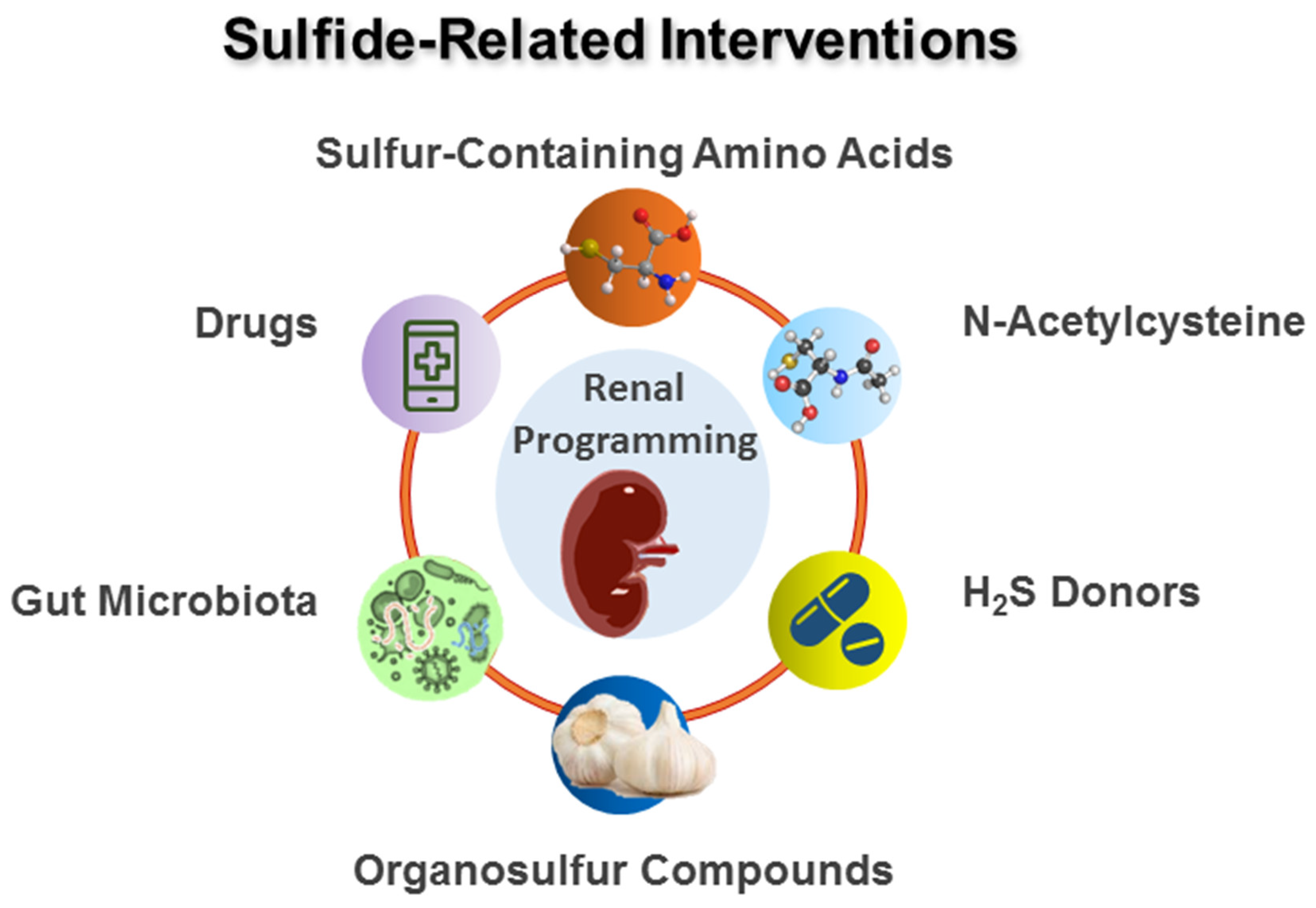
4. Sulfide-Related Reprogramming Intervention

The utilization of sulfide-related therapy has been proven to yield benefits in several kidney diseases, such as acute kidney injury [70], CKD [71], diabetic nephropathy [72], drug-induced nephropathy [73], obstructive nephropathy [74], glomerulosclerosis [75], urolithiasis [76], and kidney transplant [77,78]. Still, little attention has been paid to understanding H2S signaling pathway during pregnancy and lactation for the prevention of offspring kidney disease. Early intervention, even prior to the disease appearing, is key to preventing the development of adult kidney disease [9]. Studies documenting sulfide-related interventions in animal models for renal reprogramming are summarized in Table 1, restricting interventions to start before the onset of disease [79,80,81,82,83,84,85,86,87,88,89,90,91,92,93].
Table 1 illustrates that rats are the most frequently used animal species. Several developmental programming models have been used to study renal programming, covering the genetic spontaneously hypertensive rat (SHR) model [79,82,83,89,90], maternal CKD model [80,93], maternal high-sugar-diet model [81], prenatal dexamethasone and postnatal high-fat diet [84], NG-nitro-l-arginine-methyl-ester (L-NAME) exposure model [85], maternal suramin administration model [86], maternal hypertension [87], maternal nicotine exposure [88], maternal renovascular hypertension model [91], and maternal high-fat-diet model [92]. Hypertension is the major renal-programming-induced adverse outcome being evaluated. Reported sulfide-related interventions include sulfur-containing amino acids, N-acetylcysteine (NAC), H2S donors, and organosulfur compounds. It has been reported that sulfide-related interventions have reprogramming effects in rat offspring aged 8 weeks to 8 months, which is in line for adolescents to middle adulthood in humans [94].

4.1. Sulfur-Containing Amino Acids

l-cysteine is a substrate for the production of H2S. Another substrate for H2S generation is d-cysteine [95]. Prior work reported that the d-cysteine pathway is 80-fold greater at H2S-producing activity than the l-cysteine pathway in the kidneys [36]. Prior work revealed that high-salt-treated young SHRs supplemented with d- or l-cysteine over a period of 2 weeks were protected against hypertension and kidney damage at 12 weeks old [79]. Another study evaluated whether l- or d-cysteine supplementation in pregnancy can prevent maternal CKD-primed offspring hypertension [80]. Administration of l-cysteine has been shown to enhance renal H2S-generating enzyme CBS and CSE expression, increase renal H2S-releasing activity, and increase plasma concentration of H2S and thiosulfate [80]. Furthermore, d-cysteine supplementation restored CKD-primed reduction in plasma thiosulfate levels, while it had a negligible effect on renal H2S-generating enzymes [80].
Another sulfur-containing amino acid used for reprogramming is taurine. Perinatal taurine supplementation was able to protect adult rat offspring against hypertension and kidney dysfunction induced by a maternal high-sugar diet [81]. In SHRs and stroke-prone spontaneously hypertensive rats (SHRSPs), taurine supplementation during pregnancy and lactation had antihypertensive effects on adult offspring [82,83]. Taurine treatment has shown benefits for several kidney diseases, such as diabetic nephropathy [96], renal ischemia/reperfusion injury [97], glomerulonephritis [98], and nephrotic syndrome [99]. Nevertheless, further clarification is needed regarding the reprogramming effects of perinatal taurine supplementation on offspring’s kidney disease.

4.2. N-Acetylcysteine

NAC, an N-acetyl derivative of l-cysteine, can also be used to produce H2S in experimental studies. Similar to cysteine, early NAC therapy at age 4–12 weeks displayed protection against hypertension in adult SHRs [89]. In addition, administration of NAC during gestation and lactation has been shown to prevent offspring hypertension in several models of renal programming, covering antenatal dexamethasone administration plus post-weaning high-fat diet [85], maternal L-NAME exposure [86], maternal suramin administration [87], maternal hypertension [88], and maternal nicotine exposure [89]. Although several animal models in response to different early-life insults presented protection against hypertension, data are still lacking regarding other reno-protective benefits. It should be noted, however, that NAC is widely used as a pharmacological antioxidant [100].

4.3. H2S Donors

Inorganic sulfide salts such as sodium hydrosulfide (NaHS) and sodium sulfide (Na2S) are the most commonly utilized exogenous H2S donors [101,102]. NaHS therapy between 4–8 weeks of age prevented the development of hypertension in 12-week-old SHRs [90]. Another study demonstrated that maternal NaHS therapy protects adult progeny against hypertension in a 2K1C hypertensive model [91].
Inorganic sulfide salts provide direct and prompt release of free H2S. As a result, these H2S donors might be unsuitable for clinical use due to the rapid increase in H2S concentration to supraphysiological concentration. Later on, some organic slow-releasing H2S donors are synthesized to better mimic the physiological H2S production and overcome this limitation [101,102].
GYY4137 was produced as one of the first slow-releasing H2S donors [102]. Even though GYY4137 exerted protective action against hypertension in a CSE inhibition model and an L-NAME-treated SHR model [103,104], organic slow-releasing H2S donors have not yet been assessed in terms of their reno-protective effects on renal-programming-induced models. Moreover, thiosulfate can be considered a H2S mimetic, which presents the therapeutic potential of sodium thiosulfate for kidney disease [105,106]. We recently found that sodium thiosulfate therapy can produce H2S and prevent hypertension concurrently in an adenine-induced CKD model [107]. However, there is little knowledge on whether sodium thiosulfate treatment during gestation and lactation can prevent renal-programming-related adverse offspring’s outcomes.

4.4. Organosulfur Compounds

In addition to synthetic H2S donors, researchers have focused their attention on natural H2S donors. These organosulfur compounds include polysulfides derived from Alliaceae—diallyl di- and tri-sulfide—and GLS-derived ITCs [108].
Garlic-derived organic polysulfides have shown potential benefits as a treatment option in kidney disease and related complications [109,110,111]. Supplementation of garlic oil during gestation and lactation protected against maternal CKD-primed offspring hypertension at 12 weeks of age [92]. In another study examining the reprograming effect of garlic oil in a maternal high-fat model, the rise of BP in 16-week-old offspring was prevented by perinatal garlic oil supplementation [93].
Though interest in exploring the potential therapeutic effects of ITCs has grown with the finding of their ability to release H2S [108], their beneficial effect against renal programming has not yet been explored.

4.5. Others

The impact of gut-derived H2S on renal programming has not been studied, while gut microbiota denotes the greatest source of H2S in the body. Abundant SRB and SOB control the generation and degradation of H2S in the gut [112]. High concentrations of H2S are toxic for the gut epithelium and may contribute to bowel disease. Therapeutic targeting of SRB has been tested to regulate gut-inflammation-related H2S production [113]. More research on gut-bacteria-derived H2S is required as they may turn into a potential therapeutic target for renal-programming-related diseases.
H2S is also regulated by several presently used drugs, such as aspirin, amlodipine, atorvastatin, carvedilol, testosterone, digoxin, metformin, paracetamol, ramipril, vitamin D, and 17β-estradiol [114]. Although metformin was reported to protect maternal high-fructose plus post-weaning high-fat-diet-induced offspring [115], whether it is beneficial for kidney health and related to H2S signaling is unclear. It would be interesting to see whether targeting H2S-signal-related mechanisms by these drugs would become a practical approach to prevent renal programming for further clinical translations. A summary of potential sulfide-related interventions as reprogramming strategies for renal programming is illustrated in Figure 3.

5. Mechanisms behind Protective Actions of H2S on Renal Programming

Investigation of the potential mechanisms underlying renal programming has gained increasing attention [9,60]. Currently, the mechanisms accounting for renal programming include deficient NO [116], oxidative stress [2,117], the aberrant renin–angiotensin–aldosterone system (RAAS) [118], and gut microbiota dysbiosis [119,120]. The results of animal experiments indicate that the H2S signaling pathway interacts with the abovementioned mechanisms. A summary of the link between H2S and other mechanisms involved in renal programming and reprogramming by sulfide-related interventions for the prevention of kidney disease is depicted in Figure 3. Each of these mechanisms are discussed in turn.

5.1. Deficient NO

NO, a vasodilator, plays a key role in embryogenesis, regulation of fetoplacental vascular reactivity, and fetal development during gestation [121]. NO deficiency participates in the development of kidney disease, as well as hypertension [122,123]. The critical role of deficient NO implicated in renal programming is supported by several animal models, as we reviewed elsewhere [116]. Renal NO deficiency can be attributed to l-arginine deficiency (the substrate for NOS), diminished NOS activity and abundance, NO inactivation by oxidative stress, and inhibition by asymmetric or symmetric dimethylarginine (ADMA or SDMA) [116].
As revealed in Table 1, renal programming induced by maternal L-NAME administration [85], maternal suramin administration [86], and maternal CKD [92] is associated with impaired NO pathways. Prior work revealed that maternal NO deficiency induced by L-NAME caused renal programming and hypertension in adult offspring [85]. Protective actions of maternal NAC therapy against L-NAME-induced offspring hypertension were associated with increases in renal H2S synthesis and H2S-producing enzyme expression [85]. In another maternal suramin-induced hypertension model [86], the beneficial effects of NAC were accompanied by increased renal 3MST protein abundance, an increase in plasma glutathione level, and restoration of NO. Another line of evidence for the interplay between H2S and NO implications in renal programming was obtained in a maternal CKD model, which showed that the protective effects of perinatal garlic oil supplementation against offspring hypertension coincided with enhanced H2S signaling and increased NO bioavailability [92].
Increasing evidence supports the assumption that H2S and NO affect not only the production of each other but also the further downstream signaling pathway [124]. H2S causes the increase in NO bioavailability in several ways, such as reduction in ADMA [125], activation of eNOS via calcium influx or Akt activation [126,127], diminished cGMP degradation [128], reduction in nitrite [129], and augmenting eNOS activity by S-sulfhydration [130]. Though there is a lot of evidence pointing towards their close connection, additional research is needed to explore the crosstalk between H2S and NO in renal programming and reprogramming.

5.2. Oxidative Stress

The developing kidney is vulnerable to oxidative damage stress due to the low antioxidant capacity of the fetus [131]. Oxidative stress is a phenomenon caused by an imbalance between oxidants and antioxidants in favor of the oxidants. Oxidative stress and renal programming are intertwined in several animal models, covering maternal CKD [80], prenatal dexamethasone plus post-weaning high-fat diet [84], maternal suramin administration [84], and maternal nicotine exposure [88], as listed in Table 1.
The role of H2S as an antioxidant in the renal oxidative stress response has been widely noted [45]. This occurs through scavenging ROS; increasing antioxidants, such as glutathione, superoxide dismutase (SOD), and nuclear factor E2-related factor 2 (Nrf2); and downregulating ROS-generating enzymes, such as NADPH oxidase [45].
Accumulative evidence has supported the reprogramming effects of perinatal antioxidant therapy on renal programming and how this may prevent adult-onset kidney disease [132]. In a maternal CKD model [80], the protective effect of both l- and d-cysteine against hypertension in adult rat offspring was accompanied by the reduction in renal oxidative damage. Additionally, the utilization of NAC during gestation and lactation was reported to reprogram hypertension and reduce renal oxidative stress concurrently in animal models of prenatal dexamethasone plus post-weaning high-fat diet [84], maternal suramin administration [86], and maternal nicotine exposure [88].
Although some sulfide-related interventions have previously been shown to counterbalance oxidative stress to protect offspring against renal programming, whether the antioxidant property of H2S has the greatest impact in preserving kidney health compared to other mechanisms still awaits further elucidation.

5.3. Aberrant RAAS

The RAAS is a key hormone cascade regulating BP and the renal system [133]. There are two pathways of the RAAS: classic and non-classic systems. The classic RAAS is composed of angiotensin-converting enzyme (ACE), angiotensin (Ang) II, and angiotensin type 1 receptor (AT1R). On the other hand, the ACE2–angiotensin (1–7)–Mas receptor pathway is a counter-regulatory RAAS system that offsets the harmful effects of Ang II signaling.
H2S is known to influence several elements of the RAAS system, including decreasing the release of renin [134], inhibiting ACE activity [135], and reducing AT1R expression [136]. Conversely, pharmacological inhibition of CSE leads to increases in ACE and AT1R expression [137]. Taken together, existing evidence indicates that H2S suppresses the biological effects of the classic RAAS.
During kidney development, RAAS genes are highly expressed and have a transient biphasic response with the downregulation of the classic RAAS in neonates that becomes normalized over time [60,138]. Various early-life environmental insults interrupt this normalization and improperly initiate the classic RAAS, resulting in kidney disease and hypertension later in life [118]. Meanwhile, early blockade of the classic RAAS has revealed benefits against offspring hypertension in several models of renal programming [118]. These observations can provide support for the role of aberrant RAAS in renal programming.
In SHR, downregulated H2S-generating enzymes and low concentrations of H2S were reported in hypertensive rats, accompanied by activation of the classic RAAS [90]. NaHS treatment protected against hypertension coincided with the downregulation of classic-RAAS-related gene expression [87]. In a maternal renovascular hypertensive model, NaHS treatment also prevented the rise in BP in adult offspring, together with reducing the AT1R protein level [136]. Although the beneficial action of H2S has been linked to the activation of non-classic RAAS systems [139], no information currently exists regarding whether the reprogramming effect of H2S on renal programming is due to non-classic RAAS.

5.4. Gut Microbiota Dysbiosis

Gut microbiota have been implicated in the regulation of the absorption and metabolism of dietary nutrients that influence human health and disease [140]. The bidirectional link between the gut microbiota and kidney disease is termed the gut–kidney axis [141]. Gut–kidney axis dysfunction due to gut microbiota dysbiosis is implicated in kidney disease [119,120]. So far, some mechanisms underlying gut microbiota dysbiosis have been connected to kidney disease, including increases in trimethylamine-N-oxide (TMAO), alterations of short-chain fatty acids (SCFAs), and increases in tryptophan-derived uremic toxins [142,143]. Kidney disease can be treated or modified through agents that modulate the gut microbes and their metabolites, including prebiotics, probiotics, postbiotics, etc. [142,143,144].
Maternal nutritional insults alter gut microbiota composition and function, resulting in an increased risk of developing adult diseases [145]. Nevertheless, whether early gut-microbiota-targeted therapy may serve as a reprogramming strategy to prevent the developmental programming of kidney disease remains largely unknown [120]. In a maternal CKD model, l-cysteine supplementation protection against offspring hypertension is related to reshaping the gut microbiome [80]. Tryptophan metabolites, such as indole derivatives, are well-known uremic toxins [146]. The beneficial actions of l-cysteine supplementation are associated with the depletion of indole-producing genera Akkermansia and Alistipes, reduction in several indole metabolites, and enhancement of beneficial genera Butyricicoccus and Oscillibacter.
Another study reported that maternal NAC therapy protects male SHR progeny against hypertension and is connected to increased fecal thiosulfate levels and alterations of gut microbiota compositions [87]. NAC therapy increased the abundance of genus Bifidobacterium and its related phylum Actinobacteria, a common SOB [147]. Given that NAC enhanced Actinobacteria abundance and fecal thiosulfate levels concurrently, and that SOB can oxidize H2S to thiosulfate, it is possible that the beneficial actions of NAC are relevant to increased SOB and their derived thiosulfate production.
Maternal garlic oil supplementation prevented maternal CKD, and high-fat-diet-primed offspring hypertension was also relevant to modifications in gut microbiota [92,93]. Apart from the increased abundance of the genus Lactobacillus, a known probiotic, garlic oil supplementation increases plasma concentrations of SCFAs [93].
Together, these results establish a tight connection between H2S and other important mechanisms behind renal programming. The advantageous effects of sulfide-related therapy on renal programming are associated widely with deficient NO, oxidative stress, aberrant RAAS, and gut microbiota dysbiosis. Nevertheless, additional research is required to gain an understanding of how H2S may play a major role in mediating other mechanisms to develop a specific reprogramming strategy for the prevention of kidney disease.

6. Conclusions and Perspectives

The kidney is a major contributor to overall endogenous H2S generation, and H2S appears to play a significant role in kidney health and disease. Similar to adult kidney disease, deficient H2S is present in early life, resulting in renal programming. The dysregulated H2S signaling underlying renal programming is connected to deficient NO, oxidative stress, aberrant RAAS, and gut microbiota dysbiosis. The importance of sulfide-related interventions during gestation and lactation in reprogramming kidney disease is highlighted by the observations that sulfur-containing amino acids, NAC, H2S donors, and organosulfur compounds prevent offspring’s renal adverse outcomes in a variety of animal models.
One crucial aspect to consider is that research carried out so far has mainly focused on H2S-releasing drugs. However, how gut-bacteria-derived H2S participate in renal programming is largely unclear. Whether gut-derived H2S is beneficial for kidney health and whether gut-microbiota-targeted therapies may alter SRB/SOB to affect gut-derived H2S seems worthy of investigation. Another important aspect of H2S biology that remains unexplored is the identity of the molecular targets of H2S in the kidney, especially during kidney development. As H2S can impact multiple proteins and signaling pathways via sulfhydration in the kidney [130], it may act through the crosstalk with other molecular mechanisms to induce renal programming. It should be kept in mind that H2S at supraphysiologic concentrations is toxic. Clinical trials should be performed to test whether promising data from animal studies can be translated into human therapies. Attention needs to be paid to accurately monitor the concentration of H2S in vivo, to increase the efficiency of sulfide-related interventions, and improve kidney-targeting properties.

Author Contributions

Funding acquisition, Y.-L.T. and C.-N.H.; conceptualization, C.-N.H., C.-J.C., M.-C.C. and Y.-L.T.; data curation, C.-N.H., C.-J.C., M.-C.C. and Y.-L.T.; writing—original draft, C.-N.H., C.-J.C., M.-C.C. and Y.-L.T.; writing—review and editing, C.-N.H., C.-J.C., M.-C.C. and Y.-L.T. All authors have read and agreed to the published version of the manuscript.

Funding

This research was funded by grants MOST 110-2314-B-182-020-MY3 (Y.-L.T.) and MOST 111-2314-B-182A-021 (C.-N.H.) from the Ministry of Science and Technology, Taiwan.

Conflicts of Interest

The authors declare no conflict of interest.

References

  1. Martínez, Y.; Li, X.; Liu, G.; Bin, P.; Yan, W.; Más, D.; Valdivié, M.; Hu, C.A.; Ren, W.; Yin, Y. The role of methionine on metabolism, oxidative stress, and diseases. Amino Acids 2017, 49, 2091–2098. [Google Scholar] [CrossRef] [PubMed]
  2. Tain, Y.L.; Hsu, C.N. Perinatal Oxidative Stress and Kidney Health: Bridging the Gap between Animal Models and Clinical Reality. Antioxidants 2022, 12, 13. [Google Scholar] [CrossRef] [PubMed]
  3. Angelini, A.; Cappuccilli, M.L.; Magnoni, G.; Croci Chiocchini, A.L.; Aiello, V.; Napoletano, A.; Iacovella, F.; Troiano, A.; Mancini, R.; Capelli, I.; et al. The link between homocysteine, folic acid and vitamin B12 in chronic kidney disease. G. Ital. Nefrol. 2021, 38, 2021-vol4. [Google Scholar] [PubMed]
  4. Chesney, R.W.; Han, X.; Patters, A.B. Taurine and the renal system. J. Biomed. Sci. 2010, 17, S4. [Google Scholar] [CrossRef] [PubMed]
  5. Kasinath, B.S.; Feliers, D.; Lee, H.J. Hydrogen sulfide as a regulatory factor in kidney health and disease. Biochem. Pharmacol. 2018, 149, 29–41. [Google Scholar] [CrossRef]
  6. Peleli, M.; Zampas, P.; Papapetropoulos, A. Hydrogen Sulfide and the Kidney: Physiological Roles, Contribution to Pathophysiology, and Therapeutic Potential. Antioxid. Redox Signal. 2022, 36, 220–243. [Google Scholar] [CrossRef]
  7. Dawson, P.A.; Elliott, A.; Bowling, F.G. Sulphate in pregnancy. Nutrients 2015, 7, 1594–1606. [Google Scholar] [CrossRef]
  8. Hsu, C.N.; Tain, Y.L. The Good, the Bad, and the Ugly of Pregnancy Nutrients and Developmental Programming of Adult Disease. Nutrients 2019, 11, 894. [Google Scholar] [CrossRef]
  9. Tain, Y.L.; Hsu, C.N. Developmental Origins of Chronic Kidney Disease: Should We Focus on Early Life? Int. J. Mol. Sci. 2017, 18, 381. [Google Scholar] [CrossRef]
  10. Hanson, M. The birth and future health of DOHaD. J. Dev. Orig. Health Dis. 2015, 6, 434–437. [Google Scholar] [CrossRef]
  11. Hsu, C.N.; Tain, Y.L. Hydrogen Sulfide in Hypertension and Kidney Disease of Developmental Origins. Int. J. Mol. Sci. 2018, 19, 1438. [Google Scholar] [CrossRef] [PubMed]
  12. Luyckx, V.A.; Tonelli, M.; Stanifer, J.W. The global burden of kidney disease and the sustainable development goals. Bull. World Health Organ. 2018, 96, 414–422. [Google Scholar] [CrossRef] [PubMed]
  13. Rose, P.; Moore, P.K.; Whiteman, M.; Kirk, C.; Zhu, Y.Z. Diet and Hydrogen Sulfide Production in Mammals. Antioxid. Redox Signal. 2021, 34, 1378–1393. [Google Scholar] [CrossRef] [PubMed]
  14. Brand, E. Amino acid composition of simple proteins. Ann. N. Y. Acad. Sci. 1946, 47, 187–228. [Google Scholar] [CrossRef] [PubMed]
  15. Shaw, G.M.; Velie, E.M.; Schaffer, D.M. Is dietary intake of methionine associated with a reduction in risk for neural tube defect-affected pregnancies? Teratology 1997, 56, 295–299. [Google Scholar] [CrossRef]
  16. Rees, W.D.; Hay, S.M.; Cruickshank, M. An imbalance in the methionine content of the maternal diet reduces postnatal growth in the rat. Metabolism 2006, 55, 763–770. [Google Scholar] [CrossRef]
  17. Dasarathy, J.; Gruca, L.L.; Bennett, C.; Parimi, P.S.; Duenas, C.; Marczewski, S.; Fierro, J.L.; Kalhan, S.C. Methionine metabolism in human pregnancy. Am. J. Clin. Nutr. 2010, 91, 357–365. [Google Scholar] [CrossRef]
  18. Kalhan, S.C. One carbon metabolism in pregnancy: Impact on maternal, fetal and neonatal health. Mol. Cell. Endocrinol. 2016, 435, 48–60. [Google Scholar] [CrossRef]
  19. Gaiday, A.N.; Tussupkaliyev, A.B.; Bermagambetova, S.K.; Zhumagulova, S.S.; Sarsembayeva, L.K.; Dossimbetova, M.B.; Daribay, Z.Z. Effect of homocysteine on pregnancy: A systematic review. Chem. Biol. Interact. 2018, 293, 70–76. [Google Scholar] [CrossRef]
  20. Viskova, H.; Vesela, K.; Janosikova, B.; Krijt, J.; Visek, J.A.; Calda, P. Plasma cysteine concentrations in uncomplicated pregnancies. Fetal Diagn. Ther. 2007, 22, 254–258. [Google Scholar] [CrossRef]
  21. Guerra, D.D.; Hurt, K.J. Gasotransmitters in pregnancy: From conception to uterine involution. Biol. Reprod. 2019, 101, 4–25. [Google Scholar] [CrossRef] [PubMed]
  22. Knapen, M.F.; Zusterzeel, P.L.; Peters, W.H.; Steegers, E.A. Glutathione and glutathione-related enzymes in reproduction. A review. Eur. J. Obstet. Gynecol. Reprod. Biol. 1999, 82, 171–184. [Google Scholar] [CrossRef] [PubMed]
  23. Lerdweeraphon, W.; Wyss, J.M.; Boonmars, T.; Roysommuti, S. Perinatal taurine exposure affects adult oxidative stress. Am. J. Physiol. Regul. Integr. Comp. Physiol. 2013, 305, R95–R97. [Google Scholar] [CrossRef] [PubMed]
  24. Tochitani, S. Taurine: A Maternally Derived Nutrient Linking Mother and Offspring. Metabolites 2022, 12, 228. [Google Scholar] [CrossRef] [PubMed]
  25. Linden, D.R. Hydrogen Sulfide Signaling in the Gastrointestinal Tract. Antioxid. Redox Signal. 2014, 20, 818–830. [Google Scholar] [CrossRef] [PubMed]
  26. Kimura, H. Signaling molecules: Hydrogen sulfide and polysulfide. Antioxid. Redox Signal. 2015, 22, 362–376. [Google Scholar] [CrossRef]
  27. Dawson, P.A. Sulfate in fetal development. Semin. Cell Dev. Biol. 2011, 22, 653–659. [Google Scholar] [CrossRef]
  28. Dawson, P.A.; McIntyre, H.D.; Petersen, S.; Gibbons, K.; Bowling, F.G.; Hurrion, E. Sulfate in human pregnancy and preterm babies: What we ought to know. J. Ped. Child Health 2014, 50, 46. [Google Scholar]
  29. Dawson, P.A.; Rakoczy, J.; Simmons, D.G. Placental, renal, and ileal sulfate transporter gene expression in mouse gestation. Biol. Reprod. 2012, 87, 1–9. [Google Scholar] [CrossRef]
  30. Strott, C.A. Sulfonation and molecular action. Endocr. Rev. 2002, 23, 703–732. [Google Scholar] [CrossRef]
  31. Lu, Y.; Zhang, M.; Huang, D. Dietary Organosulfur-Containing Compounds and Their Health-Promotion Mechanisms. Annu. Rev. Food Sci. Technol. 2022, 13, 287–313. [Google Scholar] [CrossRef] [PubMed]
  32. Barba, F.J.; Orlien, V. Processing, bioaccessibility and bioavailability of bioactive sulfur compounds: Facts and gaps. J. Food Compos. Anal. 2017, 61, 1–3. [Google Scholar] [CrossRef]
  33. Myhre, R.; Brantsæter, A.L.; Myking, S.; Eggesbø, M.; Meltzer, H.M.; Haugen, M.; Jacobsson, B. Intakes of garlic and dried fruits are associated with lower risk of spontaneous preterm delivery. J. Nutr. 2013, 143, 1100–1108. [Google Scholar] [CrossRef] [PubMed]
  34. Shang, A.; Cao, S.Y.; Xu, X.Y.; Gan, R.Y.; Tang, G.Y.; Corke, H.; Mavumengwana, V.; Li, H.B. Bioactive Compounds and Biological Functions of Garlic (Allium sativum L.). Foods 2019, 8, 246. [Google Scholar] [CrossRef]
  35. Szabo, C. A timeline of hydrogen sulfide (H2S) research: From environmental toxin to biological mediator. Biochem. Pharmacol. 2018, 149, 5–19. [Google Scholar] [CrossRef]
  36. Shibuya, N.; Kimura, H. Production of hydrogen sulfide from d-cysteine and its therapeutic potential. Front. Endocrinol. 2013, 4, 87. [Google Scholar] [CrossRef]
  37. Yang, G.; Wu, L. Trend in H2S Biology and Medicine Research—A Bibliometric Analysis. Molecules 2017, 22, 2087. [Google Scholar] [CrossRef]
  38. Benavides, G.A.; Squadrito, G.L.; Mills, R.W.; Patel, H.D.; Isbell, T.S.; Patel, R.P.; Darley-Usmar, V.M.; Doeller, J.E.; Kraus, D.W. Hydrogen sulfide mediates the vasoactivity of garlic. Proc. Natl. Acad. Sci. USA 2007, 104, 17977–17982. [Google Scholar] [CrossRef]
  39. Blachier, F.; Davila, A.-M.; Mimoun, S.; Benetti, P.-H.; Atanasiu, C.; Andriamihaja, M.; Benamouzig, R.; Bouillaud, F.; Tomé, D. Luminal sulfide and large intestine mucosa: Friend or foe? Amino Acids 2009, 39, 335–347. [Google Scholar] [CrossRef]
  40. Filipovic, M.R.; Zivanovic, J.; Alvarez, B.; Banerjee, R. Chemical biology of H2S signaling through persulfidation. Chem. Rev. 2018, 118, 1253–1337. [Google Scholar] [CrossRef]
  41. Kabil, O.; Banerjee, R. Enzymology of H2S biogenesis, decay and signaling. Antioxid. Redox Signal. 2014, 20, 770–782. [Google Scholar] [CrossRef] [PubMed]
  42. Murphy, B.; Bhattacharya, R.; Mukherjee, P. Hydrogen sulfide signaling in mitochondria and disease. FASEB J. 2019, 33, 13098–13125. [Google Scholar] [CrossRef] [PubMed]
  43. Kimura, H. The physiological role of hydrogen sulfide and beyond. Nitric Oxide 2014, 41, 4–10. [Google Scholar] [CrossRef] [PubMed]
  44. Olas, B. Medical functions of hydrogen sulfide. Adv. Clin. Chem. 2016, 74, 195–210. [Google Scholar] [PubMed]
  45. Scammahorn, J.J.; Nguyen, I.T.N.; Bos, E.M.; Van Goor, H.; Joles, J.A. Fighting oxidative stress with sulfur: Hydrogen sulfide in the renal and cardiovascular systems. Antioxidants 2021, 10, 373. [Google Scholar] [CrossRef]
  46. Feliers, D.; Lee, H.J.; Kasinath, B.S. Hydrogen sulfide in renal physiology and disease. Antioxid. Redox Signal. 2016, 25, 720–731. [Google Scholar] [CrossRef]
  47. Xia, M.; Chen, L.; Muh, R.W.; Li, P.L.; Li, N. Production and actions of hydrogen sulfide, a novel gaseous bioactive substance, in the kidneys. J. Pharmacol. Exp. Ther. 2009, 329, 1056–1062. [Google Scholar] [CrossRef]
  48. Ahmad, F.U.; Sattar, M.A.; Rathore, H.A.; Tan, Y.C.; Akhtar, S.; Jin, O.H.; Pei, Y.P.; Abdullah, N.A.; Johns, E.J. Hydrogen sulphide and tempol treatments improve the blood pressure and renal excretory responses in spontaneously hypertensive rats. Ren. Fail. 2014, 36, 598–605. [Google Scholar] [CrossRef]
  49. Lu, M.; Liu, Y.H.; Goh, H.S.; Wang, J.J.; Yong, Q.C.; Wang, R.; Bian, J.S. Hydrogen sulfide inhibits plasma renin activity. J. Am. Soc. Nephrol. 2010, 21, 993–1002. [Google Scholar] [CrossRef]
  50. Roorda, M.; Miljkovic, J.L.; van Goor, H.; Henning, R.H.; Bouma, H.R. Spatiotemporal regulation of hydrogen sulfide signaling in the kidney. Redox Biol. 2021, 43, 101961. [Google Scholar] [CrossRef]
  51. Roy, A.; Khan, A.H.; Islam, M.T.; Prieto, M.C.; Majid, D.S. Interdependency of cystathione gamma-lyase and cystathione beta-synthase in hydrogen sulfide-induced blood pressure regulation in rats. Am. J. Hypertens. 2012, 25, 74–81. [Google Scholar] [CrossRef] [PubMed]
  52. Ahmad, A.; Druzhyna, N.; Szabo, C. Delayed Treatment with Sodium Hydrosulfide Improves Regional Blood Flow and Alleviates Cecal Ligation and Puncture (CLP)-Induced Septic Shock. Shock 2016, 46, 183–193. [Google Scholar] [CrossRef] [PubMed]
  53. Morales-Loredo, H.; Barrera, A.; Garcia, J.M.; Pace, C.E.; Naik, J.S.; Gonzalez Bosc, L.V.; Kanagy, N.L. Hydrogen sulfide regulation of renal and mesenteric blood flow. Am. J. Physiol. Heart Circ. Physiol. 2019, 317, H1157–H1165. [Google Scholar] [CrossRef] [PubMed]
  54. Li, J.; Teng, X.; Jin, s.; Dong, J.; Guo, Q.; Tian, D.; Wu, Y. Hydrogen sulfide improves endothelial dysfunction by inhibiting the vicious cycle of NLRP3 inflammasome and oxidative stress in spontaneously hypertensive rats. J. Hypertens. 2019, 37, 1633–1643. [Google Scholar] [CrossRef]
  55. Xiao, L.; Dong, J.; Jin, S.; Xue, H.M.; Guo, Q.; Teng, X.; Wu, Y.M. Hydrogen sulfide improves endothelial dysfunction via downregulating BMP4/COX-2 pathway in rats with hypertension. Oxid. Med. Cell. Longev. 2016, 2016, 8128957. [Google Scholar] [CrossRef]
  56. Kang, M.; Hashimoto, A.; Gade, A.; Akbarali, H.I. Interaction between hydrogen sulfide-induced sulfhydration and tyrosine nitration in the KATP channel complex. Am. J. Physiol. Gastrointest. Liver Physiol. 2015, 308, G532. [Google Scholar] [CrossRef]
  57. Bucci, M.; Papapetropoulos, A.; Vellecco, V.; Zhou, Z.; Pyriochou, A.; Roussos, C.; Roviezzo, F.; Brancaleone, V.; Cirino, G. Hydrogen sulfide is an endogenous inhibitor of phosphodiesterase activity. Arterioscler. Thromb. Vasc. Biol. 2010, 30, 1998–2004. [Google Scholar] [CrossRef]
  58. Zhu, M.L.; Zhao, F.R.; Zhu, T.T.; Wang, Q.Q.; Wu, Z.Q.; Song, P.; Xu, J.; Wan, G.R.; Yin, Y.L.; Li, P. The antihypertension effect of hydrogen sulfide (H2S) is induced by activating VEGFR2 signaling pathway. Life Sci. 2021, 267, 118831. [Google Scholar] [CrossRef]
  59. Hsu, C.N.; Tain, Y.L. The First Thousand Days: Kidney Health and Beyond. Healthcare 2021, 9, 1332. [Google Scholar] [CrossRef]
  60. Kett, M.M.; Denton, K. Renal programming: Cause for concern? Am. J. Physiol. Integr. Comp. Physiol. 2011, 300, R791–R803. [Google Scholar] [CrossRef]
  61. Bertram, J.F.; Douglas-Denton, R.N.; Diouf, B.; Hughson, M.; Hoy, W. Human nephron number: Implications for health and disease. Pediatr. Nephrol. 2011, 26, 1529–1533. [Google Scholar] [CrossRef] [PubMed]
  62. Tain, Y.L.; Chan, J.Y.H.; Lee, C.T.; Hsu, C.N. Maternal Melatonin Therapy Attenuates Methyl-Donor Diet-Induced Programmed Hypertension in Male Adult Rat Offspring. Nutrients 2018, 10, 1407. [Google Scholar] [CrossRef] [PubMed]
  63. O’Neill, R.J.; Vrana, P.B. Rosenfeld CS. Maternal methyl supplemented diets and effects on offspring health. Front. Genet. 2014, 5, 289. [Google Scholar] [PubMed]
  64. Rengarajan, A.; Mauro, A.K.; Boeldt, D.S. Maternal disease and gasotransmitters. Nitric Oxide 2020, 96, 1–12. [Google Scholar] [CrossRef]
  65. Hsu, C.N.; Tain, Y.L. Adverse Impact of Environmental Chemicals on Developmental Origins of Kidney Disease and Hypertension. Front. Endocrinol. 2021, 12, 745716. [Google Scholar] [CrossRef]
  66. Chan, Y.H.; Lock, S.S.M.; Wong, M.K.; Yiin, C.L.; Loy, A.C.M.; Cheah, K.W.; Chai, S.Y.W.; Li, C.; How, B.S.; Chin, B.L.F.; et al. A state-of-the-art review on capture and separation of hazardous hydrogen sulfide (H2S): Recent advances, challenges and outlook. Environ. Pollut. 2022, 314, 120219. [Google Scholar] [CrossRef]
  67. Schreuder, M.F.; Bueters, R.R.; Huigen, M.C.; Russel, F.G.; Masereeuw, R.; van den Heuvel, L.P. Effect of drugs on renal development. Clin. J. Am. Soc. Nephrol. 2011, 6, 212–217. [Google Scholar] [CrossRef]
  68. Tain, Y.-L.; Sheen, J.M.; Chen, C.C.; Yu, H.-R.; Tiao, M.-M.; Kuo, H.C.; Huang, L.T. Maternal citrulline supplementation prevents prenatal dexamethasone-induced programmed hypertension. Free Radic. Res. 2014, 48, 580–586. [Google Scholar] [CrossRef]
  69. Zhu, X.Y.; Liu, S.J.; Liu, Y.J.; Wang, S.; Ni, X. Glucocorticoids suppress cystathionine gamma-lyase expression and H2S production in lipopolysaccharide-treated macrophages. Cell. Mol. Life Sci. 2010, 67, 1119–1132. [Google Scholar] [CrossRef]
  70. Chen, Y.; Jin, S.; Teng, X.; Hu, Z.; Zhang, Z.; Qiu, X.; Tian, D.; Wu, Y. Hydrogen sulfide attenuates LPS-induced acute kidney injury by inhibiting inflammation and oxidative stress. Oxid. Med. Cell. Longev. 2018, 2018, 6717212. [Google Scholar] [CrossRef]
  71. Dugbartey, G.J. The smell of renal protection against chronic kidney disease: Hydrogen sulfide offers a potential stinky remedy. Pharm. Rep. 2018, 70, 196–205. [Google Scholar] [CrossRef] [PubMed]
  72. Xue, R.; Hao, D.D.; Sun, J.P.; Li, W.W.; Zhao, M.M.; Li, X.H.; Chen, Y.; Zhu, J.H.; Ding, Y.J.; Liu, J.; et al. Hydrogen sulfide treatment promotes glucose uptake by increasing insulin receptor sensitivity and ameliorates kidney lesions in type 2 diabetes. Antioxid. Redox Signal. 2013, 19, 5–23. [Google Scholar] [CrossRef] [PubMed]
  73. Cao, X.; Zhang, W.; Moore, P.K.; Bian, J. Protective smell of hydrogen sulfide and polysulfide in cisplatin-induced nephrotoxicity. Int. J. Mol. Sci. 2019, 20, 313. [Google Scholar] [CrossRef] [PubMed]
  74. Lin, S.; Visram, F.; Liu, W.; Haig, A.; Jiang, J.; Mok, A.; Lian, D.; Wood, M.E.; Torregrossa, R.; Whiteman, M.; et al. GYY4137, a slow-releasing hydrogen sulfide donor, ameliorates renal damage associated with chronic obstructive uropathy. J. Urol. 2016, 196, 1778–1787. [Google Scholar] [CrossRef] [PubMed]
  75. Lee, H.J.; Feliers, D.; Barnes, J.L.; Oh, S.; Choudhury, G.G.; Diaz, V.; Galvan, V.; Strong, R.; Nelson, J.; Salmon, A.; et al. Hydrogen sulfide ameliorates aging-associated changes in the kidney. GeroScience 2018, 40, 163–176. [Google Scholar] [CrossRef] [PubMed]
  76. Vaitheeswari, S.; Sriram, R.; Brindha, P.; Kurian, G.A. Studying inhibition of calcium oxalate stone formation: An in vitro approach for screening hydrogen sulfide and its metabolites. Int. Braz J. Urol. 2015, 41, 503–510. [Google Scholar] [CrossRef] [PubMed]
  77. van den Berg, E.; Pasch, A.; Westendorp, W.H.; Navis, G.; Brink, E.J.; Gans, R.O.; van Goor, H.; Bakker, S.J. Urinary sulfur metabolites associate with a favorable cardiovascular risk profile and survival benefit in renal transplant recipients. J. Am. Soc. Nephrol. 2014, 25, 1303–1312. [Google Scholar] [CrossRef]
  78. McFarlane, L.; Nelson, P.; Dugbartey, G.J.; Sener, A. Pre-Treatment of Transplant Donors with Hydrogen Sulfide to Protect against Warm and Cold Ischemia-Reperfusion Injury in Kidney and Other Transplantable Solid Organs. Int. J. Mol. Sci. 2023, 24, 3518. [Google Scholar] [CrossRef]
  79. Hsu, C.N.; Lin, Y.J.; Lu, P.C.; Tain, Y.L. Early supplementation of d-cysteine or l-cysteine prevents hypertension and kidney damage in spontaneously hypertensive rats exposed to high-salt intake. Mol. Nutr. Food Res. 2018, 62, 2. [Google Scholar] [CrossRef] [PubMed]
  80. Hsu, C.N.; Hou, C.Y.; Chang-Chien, G.P.; Lin, S.; Tain, Y.L. Dietary Supplementation with Cysteine during Pregnancy Rescues Maternal Chronic Kidney Disease-Induced Hypertension in Male Rat Offspring: The Impact of Hydrogen Sulfide and Microbiota-Derived Tryptophan Metabolites. Antioxidants 2022, 11, 483. [Google Scholar] [CrossRef]
  81. Roysommuti, S.; Lerdweeraphon, W.; Malila, P.; Jirakulsomchok, D.; Wyss, J.M. Perinatal taurine alters arterial pressure control and renal function in adult offspring. Adv. Exp. Med. Biol. 2009, 643, 145–156. [Google Scholar] [PubMed]
  82. Thaeomor, A.; Teangphuck, P.; Chaisakul, J.; Seanthaweesuk, S.; Somparn, N.; Roysommuti, S. Perinatal Taurine Supplementation Prevents Metabolic and Cardiovascular Effects of Maternal Diabetes in Adult Rat Offspring. Adv. Exp. Med. Biol. 2017, 975, 295–305. [Google Scholar] [PubMed]
  83. Horie, R.; Yamori, Y.; Nara, Y.; Sawamura, M.; Mano, M. Effects of sulphur amino acids on the development of hypertension and atherosclerosis in stroke-prone spontaneously hypertensive rats. J. Hypertens. Suppl. 1987, 5, S223–S225. [Google Scholar] [PubMed]
  84. Tai, I.-H.; Sheen, J.-M.; Lin, Y.-J.; Yu, H.-R.; Tiao, M.-M.; Chen, C.-C.; Huang, L.-T.; Tain, Y.-L. Maternal N-acetylcysteine therapy regulates hydrogen sulfide-generating pathway and prevents programmed hypertension in male offspring exposed to prenatal dexamethasone and postnatal high-fat diet. Nitric Oxide 2016, 53, 6–12. [Google Scholar] [CrossRef]
  85. Tain, Y.L.; Lee, C.T.; Chan, J.Y.; Hsu, C.N. Maternal melatonin or N-acetylcysteine therapy regulates hydrogen sulfide-generating pathway and renal transcriptome to prevent prenatal N(G)-Nitro-l-argininemethyl ester (L-NAME)-induced fetal programming of hypertension in adult male offspring. Am. J. Obstet. Gynecol. 2016, 215, 636. [Google Scholar] [CrossRef]
  86. Tain, Y.L.; Hsu, C.N.; Lee, C.T.; Lin, Y.J.; Tsai, C.C. N-Acetylcysteine Prevents Programmed Hypertension in Male Rat Offspring Born to Suramin-Treated Mothers. Biol. Reprod. 2016, 95, 8. [Google Scholar] [CrossRef]
  87. Hsu, C.-N.; Hou, C.-Y.; Chang-Chien, G.-P.; Lin, S.; Tain, Y.-L. Maternal N-Acetylcysteine Therapy Prevents Hypertension in Spontaneously Hypertensive Rat Offspring: Implications of Hydrogen Sulfide-Generating Pathway and Gut Microbiota. Antioxidants 2020, 9, 856. [Google Scholar] [CrossRef]
  88. Xiao, D.; Huang, X.; Li, Y.; Dasgupta, C.; Wang, L.; Zhang, L. Antenatal Antioxidant Prevents Nicotine-Mediated Hypertensive Response in Rat Adult Offspring. Biol. Reprod. 2015, 93, 66. [Google Scholar] [CrossRef]
  89. Fan, N.C.; Tsai, C.M.; Hsu, C.N.; Huang, L.T.; Tain, Y.L. N-acetylcysteine prevents hypertension via regulation of the ADMA-DDAH pathway in young spontaneously hypertensive rats. Biomed. Res. Int. 2013, 2013, 696317. [Google Scholar] [CrossRef]
  90. Tain, Y.-L.; Hsu, C.-N.; Lu, P.-C. Early short-term treatment with exogenous hydrogen sulfide postpones the transition from prehypertension to hypertension in spontaneously hypertensive rat. Clin. Exp. Hypertens. 2017, 40, 58–64. [Google Scholar] [CrossRef]
  91. Feng, X.; Guo, Q.; Xue, H.; Duan, X.; Jin, S.; Wu, Y. Hydrogen Sulfide Attenuated Angiotensin II-Induced Sympathetic Excitation in Offspring of Renovascular Hypertensive Rats. Front. Pharmacol. 2020, 11, 565726. [Google Scholar] [CrossRef] [PubMed]
  92. Tain, Y.L.; Hou, C.Y.; Chang-Chien, G.P.; Lin, S.; Hsu, C.N. Perinatal Garlic Oil Supplementation Averts Rat Offspring Hypertension Programmed by Maternal Chronic Kidney Disease. Nutrients 2022, 14, 4624. [Google Scholar] [CrossRef] [PubMed]
  93. Hsu, C.N.; Hou, C.Y.; Chang-Chien, G.P.; Lin, S.; Tain, Y.L. Maternal Garlic Oil Supplementation Prevents High-Fat Diet-Induced Hypertension in Adult Rat Offspring: Implications of H2S-Generating Pathway in the Gut and Kidneys. Mol. Nutr. Food Res. 2021, 65, e2001116. [Google Scholar] [CrossRef] [PubMed]
  94. Sengupta, P. The Laboratory Rat: Relating Its Age with Human’s. Int. J. Prev. Med. 2013, 4, 624–630. [Google Scholar]
  95. Shibuya, N.; Koike, S.; Tanaka, M.; Ishigami-Yuasa, M.; Kimura, Y.; Ogasawara, Y.; Fukui, K.; Nagahara, N.; Kimura, H. A novel pathway for the production of hydrogen sulfide from d-cysteine in mammalian cells. Nat. Commun. 2013, 4, 1366. [Google Scholar] [CrossRef]
  96. Trachtman, H.; Futterweit, S.; Maesaka, J.; Ma, C.; Valderrama, E.; Fuchs, A.; Tarectecan, A.A.; Rao, P.S.; Sturman, J.A.; Boles, T.H. Taurine ameliorates chronic streptozocin-induced diabetic nephropathy in rats. Am. J. Physiol. 1995, 269, F429–F438. [Google Scholar] [CrossRef]
  97. Michalk, D.V.; Hoffmann, B.; Minor, T. Taurine reduces renal ischemia/ reperfusion injury in the rat. Adv. Exp. Med. Biol. 2003, 526, 49–56. [Google Scholar]
  98. Lian, X.; Yang, L.; Chen, Q.; Sheng, A.; Zhao, J. Effects of taurine on platelet activating factor in rats with Masugi glomerulonephritis. Chin. J. Microcirc. 2003, 7, 151–153. [Google Scholar]
  99. Trachtman, H.; Del Pizzo, R.; Futterweit, S.; Levine, D.; Rao, P.S.; Valderrama, E.; Sturman, J.A. Taurine attenuates renal disease in chronic puromycin aminonucleoside nephropathy. Am. J. Physiol. 1992, 262, F117–F123. [Google Scholar] [CrossRef]
  100. Ezeriņa, D.; Takano, Y.; Hanaoka, K.; Urano, Y.; Dick, T.P. N-Acetyl Cysteine Functions as a Fast-Acting Antioxidant by Triggering Intracellular H2S and Sulfane Sulfur Production. Cell Chem. Biol. 2018, 25, 447–459. [Google Scholar] [CrossRef]
  101. Li, Z.; Polhemus, D.J.; Lefer, D.J. Evolution of Hydrogen Sulfide Therapeutics to Treat Cardiovascular Disease. Circ. Res. 2018, 123, 590–600. [Google Scholar] [CrossRef] [PubMed]
  102. Wen, Y.-D.; Wang, H.; Zhu, Y.Z. The Drug Developments of Hydrogen Sulfide on Cardiovascular Disease. Oxidative Med. Cell. Longev. 2018, 2018, 4010395. [Google Scholar] [CrossRef] [PubMed]
  103. Wang, K.; Ahmad, S.; Cai, M.; Rennie, J.; Fujisawa, T.; Crispi, F.; Baily, J.; Miller, M.R.; Cudmore, M.; Hadoke, P.W.F.; et al. Dysregulation of Hydrogen Sulfide Producing Enzyme Cystathionine γ-lyase Contributes to Maternal Hypertension and Placental Abnormalities in Preeclampsia. Circulation 2013, 127, 2514–2522. [Google Scholar] [CrossRef]
  104. Sharma, D.K.; Manral, A.; Saini, V.; Singh, A.; Srinivasan, B.; Tiwari, M. Novel diallyldisulfide analogs ameliorate cardiovascular remodeling in rats with L-NAME-induced hypertension. Eur. J. Pharmacol. 2012, 691, 198–208. [Google Scholar] [CrossRef] [PubMed]
  105. Nguyen, I.T.; Klooster, A.; Minnion, M.; Feelisch, M.; Verhaar, M.C.; Van Goor, H.; Joles, J.A. Sodium thiosulfate improves renal function and oxygenation in L-NNA–induced hypertension in rats. Kidney Int. 2020, 98, 366–377. [Google Scholar] [CrossRef]
  106. Snijder, P.M.; Frenay, A.-R.S.; Koning, A.M.; Bachtler, M.; Pasch, A.; Kwakernaak, A.J.; Berg, E.V.D.; Bos, E.M.; Hillebrands, J.-L.; Navis, G.; et al. Sodium thiosulfate attenuates angiotensin II-induced hypertension, proteinuria and renal damage. Nitric Oxide 2014, 42, 87–98. [Google Scholar] [CrossRef]
  107. Hsu, C.N.; Hou, C.Y.; Chang-Chien, G.P.; Lin, S.; Yang, H.W.; Tain, Y.L. Sodium Thiosulfate Improves Hypertension in Rats with Adenine-Induced Chronic Kidney Disease. Antioxidants 2022, 11, 147. [Google Scholar] [CrossRef]
  108. Piragine, E.; Citi, V.; Lawson, K.; Calderone, V.; Martelli, A. Potential Effects of Natural H2S-Donors in Hypertension Management. Biomolecules 2022, 12, 581. [Google Scholar] [CrossRef]
  109. Shouk, R.; Abdou, A.; Shetty, K.; Sarkar, D.; Eid, A.H. Mechanisms underlying the antihypertensive effects of garlic bioactives. Nutr. Res. 2014, 34, 106–115. [Google Scholar] [CrossRef]
  110. Ried, K.; Fakler, P. Potential of garlic (Allium sativum) in lowering high blood pressure: Mechanisms of action and clinical relevance. Integr. Blood Press. Control 2014, 7, 71–82. [Google Scholar] [CrossRef]
  111. Ribeiro, M.; Alvarenga, L.; Cardozo, L.F.M.F.; Chermut, T.R.; Sequeira, J.; de Souza Gouveia Moreira, L.; Teixeira, K.T.R.; Shiels, P.G.; Stenvinkel, P.; Mafra, D. From the distinctive smell to therapeutic effects: Garlic for cardiovascular, hepatic, gut, diabetes and chronic kidney disease. Clin. Nutr. 2021, 40, 4807–4819. [Google Scholar] [CrossRef] [PubMed]
  112. Tomasova, L.; Konopelski, P.; Ufnal, M. Gut Bacteria and Hydrogen Sulfide: The New Old Players in Circulatory System Homeostasis. Molecules 2016, 21, 1558. [Google Scholar] [CrossRef] [PubMed]
  113. Dostal Webster, A.; Staley, C.; Hamilton, M.J.; Huang, M.; Fryxell, K.; Erickson, R.; Kabage, A.J.; Sadowsky, M.J.; Khoruts, A. Influence of short-term changes in dietary sulfur on the relative abundances of intestinal sulfate-reducing bacteria. Gut Microbes 2019, 10, 447–457. [Google Scholar] [CrossRef] [PubMed]
  114. Bełtowski, J. Hydrogen sulfide in pharmacology and medicine—An update. Pharmacol. Rep. 2015, 67, 647–658. [Google Scholar] [CrossRef]
  115. Tain, Y.L.; Wu, K.L.H.; Lee, W.C.; Leu, S.; Chan, J.Y.H. Prenatal Metformin Therapy Attenuates Hypertension of Developmental Origin in Male Adult Offspring Exposed to Maternal High-Fructose and Post-Weaning High-Fat Diets. Int. J. Mol. Sci. 2018, 19, 1066. [Google Scholar] [CrossRef]
  116. Hsu, C.N.; Tain, Y.L. Regulation of nitric oxide production in the developmental programming of hypertension and kidney disease. Int. J. Mol. Sci. 2019, 20, 681. [Google Scholar] [CrossRef]
  117. Thompson, L.P.; Al-Hasan, Y. Impact of oxidative stress in fetal programming. J. Pregnancy 2012, 2012, 582748. [Google Scholar] [CrossRef]
  118. Hsu, C.N.; Tain, Y.L. Targeting the renin–angiotensin–aldosterone system to prevent hypertension and kidney disease of developmental origins. Int. J. Mol. Sci. 2021, 22, 2298. [Google Scholar] [CrossRef]
  119. Yang, T.; Richards, E.M.; Pepine, C.J.; Raizada, M.K. The gut microbiota and the brain–gut–kidney axis in hypertension and chronic kidney disease. Nat. Rev. Nephrol. 2018, 14, 442–456. [Google Scholar] [CrossRef]
  120. Hsu, C.N.; Tain, Y.L. Chronic Kidney Disease and Gut Microbiota: What Is Their Connection in Early Life? Int. J. Mol. Sci. 2022, 23, 3954. [Google Scholar] [CrossRef]
  121. Sladek, S.M.; Magness, R.R.; Conrad, K.P. Nitric oxide and pregnancy. Am. J. Physiol. Regul. Integr. Comp. Physiol. 1997, 272, R441–R463. [Google Scholar] [CrossRef] [PubMed]
  122. Wilcox, C.S. Oxidative stress and nitric oxide deficiency in the kidney: A critical link to hypertension? Am. J. Physiol.-Regul. Integr. Comp. Physiol. 2005, 289, R913–R935. [Google Scholar] [CrossRef] [PubMed]
  123. Baylis, C. Nitric oxide synthase derangements and hypertension in kidney disease. Curr. Opin. Nephrol. Hypertens. 2012, 21, 1–6. [Google Scholar] [CrossRef] [PubMed]
  124. Wu, D.; Hu, Q.; Zhu, D. An Update on Hydrogen Sulfide and Nitric Oxide Interactions in the Cardiovascular System. Oxidative Med. Cell. Longev. 2018, 2018, 4579140. [Google Scholar] [CrossRef] [PubMed]
  125. Mostafa, D.K.; El Azhary, N.M.; Nasra, R.A. The hydrogen sulfide releasing compounds ATB-346 and diallyl trisulfide attenuate streptozotocin-induced cognitive impairment, neuroinflammation, and oxidative stress in rats: Involvement of asymmetric dimethylarginine. Can. J. Physiol. Pharmacol. 2016, 94, 699–708. [Google Scholar] [CrossRef] [PubMed]
  126. Ping, N.N.; Li, S.; Mi, Y.N.; Cao, L.; Cao, Y.X. Hydrogen sulphide induces vasoconstriction of rat coronary artery via activation of Ca(2+) influx. Acta Physiol. 2015, 214, 88–96. [Google Scholar] [CrossRef]
  127. Pardue, S.; Kolluru, G.K.; Shen, X.; Lewis, S.E.; Saffle, C.B.; Kelley, E.E.; Kevil, C.G. Hydrogen sulfide stimulates xanthine oxidoreductase conversion to nitrite reductase and formation of NO. Redox Biol. 2020, 34, 101447. [Google Scholar] [CrossRef]
  128. Forrester, S.J.; Booz, G.W.; Sigmund, C.D.; Coffman, T.M.; Kawai, T.; Rizzo, V.; Scalia, R.; Eguchi, S. Angiotensin II Signal Transduction: An Update on Mechanisms of Physiology and Pathophysiology. Physiol. Rev. 2018, 98, 1627–1738. [Google Scholar] [CrossRef]
  129. Bogdarina, I.; Welham, S.; King, P.J.; Burns, S.P.; Clark, A.J.L. Epigenetic Modification of the Renin-Angiotensin System in the Fetal Programming of Hypertension. Circ. Res. 2007, 100, 520–526. [Google Scholar] [CrossRef]
  130. Ju, Y.; Fu, M.; Stokes, E.; Wu, L.; Yang, G. H2S-Mediated Protein S-Sulfhydration: A Prediction for Its Formation and Regulation. Molecules 2017, 22, 1334. [Google Scholar] [CrossRef]
  131. Vento, M. Oxidative stress in the perinatal period. Free Radic. Biol. Med. 2019, 142, 1–2. [Google Scholar] [CrossRef] [PubMed]
  132. Hsu, C.N.; Tain, Y.L. Developmental Origins of Kidney Disease: Why Oxidative Stress Matters? Antioxidants 2020, 10, 33. [Google Scholar] [CrossRef] [PubMed]
  133. Yang, T.; Xu, C. Physiology and Pathophysiology of the Intrarenal Renin-Angiotensin System: An Update. J. Am. Soc. Nephrol. 2017, 28, 1040–1049. [Google Scholar] [CrossRef] [PubMed]
  134. Liu, Y.H.; Lu, M.; Xie, Z.Z.; Hua, F.; Xie, L.; Gao, J.H.; Koh, Y.H.; Bian, J.S. Hydrogen sulfide prevents heart failure development via inhibition of renin release from mast cells in isoproterenol-treated rats. Antioxid. Redox Signal. 2014, 20, 759–769. [Google Scholar] [CrossRef]
  135. Laggner, H.; Hermann, M.; Esterbauer, H.; Muellner, M.K.; Exner, M.; Gmeiner, B.M.; Kapiotis, S. The novel gaseous vasorelaxant hydrogen sulfide inhibits angiotensin-converting enzyme activity of endothelial cells. J. Hypertens. 2007, 25, 2100–2104. [Google Scholar] [CrossRef]
  136. Guo, Q.; Feng, X.; Xue, H.; Teng, X.; Jin, S.; Duan, X.; Xiao, L.; Wu, Y. Maternal renovascular hypertensive rats treatment with hydrogen sulfide increased the methylation of AT1b gene in offspring. Am. J. Hypertens. 2017, 30, 1220–1227. [Google Scholar] [CrossRef]
  137. Xue, H.; Yuan, P.; Ni, J.; Li, C.; Shao, D.; Liu, J.; Shen, Y.; Wang, Z.; Zhou, L.; Zhang, W.; et al. H2S inhibits hyperglycemia-induced intrarenal reninangiotensin system activation via attenuation of reactive oxygen species generation. PLoS ONE 2013, 8, e74366. [Google Scholar] [CrossRef]
  138. Yosypiv, I.V. Renin-angiotensin system in ureteric bud branching morphogenesis: Insights into the mechanisms. Pediatr. Nephrol. 2011, 26, 1499–1512. [Google Scholar] [CrossRef]
  139. Silva-Velasco, D.L.; Beltran-Ornelas, J.H.; Tapia-Martínez, J.; Sánchez-López, A.; de la Cruz, S.H.; Cervantes-Pérez, L.G.; Del Valle-Mondragón, L.; Sánchez-Mendoza, A.; Centurión, D. NaHS restores the vascular alterations in the renin-angiotensin system induced by hyperglycemia in rats. Peptides 2023, 164, 171001. [Google Scholar] [CrossRef]
  140. Lynch, S.V.; Pedersen, O. The Human Intestinal Microbiome in Health and Disease. N. Engl. J. Med. 2016, 375, 2369–2379. [Google Scholar] [CrossRef]
  141. Evenepoel, P.; Poesen, R.; Meijers, B. The gut-kidney axis. Pediatr. Nephrol. 2017, 32, 2005–2014. [Google Scholar] [CrossRef] [PubMed]
  142. Chen, Y.Y.; Chen, D.Q.; Chen, L.; Liu, J.R.; Vaziri, N.D.; Guo, Y.; Zhao, Y.Y. Microbiome-metabolome reveals the contribution of gut-kidney axis on kidney disease. J. Transl. Med. 2019, 17, 5. [Google Scholar] [CrossRef] [PubMed]
  143. Kim, M.G.; Yang, J.; Jo, S.K. Intestinal microbiota and kidney diseases. Kidney Res. Clin. Pract. 2021, 40, 335–343. [Google Scholar] [CrossRef] [PubMed]
  144. Harkins, C.P.; Kong, H.H.; Segre, J.A. Manipulating the Human Microbiome to Manage Disease. JAMA 2020, 323, 303–304. [Google Scholar] [CrossRef]
  145. Chu, D.M.; Meyer, K.M.; Prince, A.L.; Aagaard, K.M. Impact of maternal nutrition in pregnancy and lactation on offspring gut microbial composition and function. Gut Microbes 2016, 7, 459–470. [Google Scholar] [CrossRef]
  146. Hsu, C.N.; Tain, Y.L. Developmental Programming and Reprogramming of Hypertension and Kidney Disease: Impact of Tryptophan Metabolism. Int. J. Mol. Sci. 2020, 21, 8705. [Google Scholar] [CrossRef]
  147. Di Masi, A.; Ascenzi, P. H2S: A “double face” molecule in health and disease. Biofactors 2013, 39, 186–196. [Google Scholar] [CrossRef]
Figure 1. Schematic representation of impact of sulfur-containing amino acids, hydrogen sulfide (H2S), and sulfur compounds on kidney health and disease. GSH = glutathione.
Metabolites 13 00688 g001
Figure 2. Three major H2S synthesis pathways are of enzymatic, non-enzymatic, and gut microbial origin. Cystathionine β-synthase (CBS) or cystathionine γ-lyase (CSE) catalyzes homocysteine to produce l-cysteine. Both CBS and CSE can catalyze l-cysteine to generate H2S. 3-mercaptopyruvate sulfurtransferase (3MST) produces H2S from 3-mercaptopyruvate (3-MP), which is formed by d-amino acid oxidase (DAO) cysteine aminotransferase (CAT) from d-cysteine and l-cysteine. Another source of endogenous H2S is derived from the non-enzymatic synthesis pathway. The other source of H2S comes from intestinal bacteria, mainly from sulfate-reducing bacteria. H2S is metabolized by sulfide quinone oxidoreductase (SQR) to form persulfide, which can be oxidized by persulfide dioxy-genase (ETHE1) to yield sulfite. Sulfite is converted to sulfate or thiosulfate, which can be excreted into the urine.
Metabolites 13 00688 g002
Figure 3. Schema outlining potential sulfide-related interventions used for renal programming.
Metabolites 13 00688 g003
Table 1. Summary of sulfide-related interventions utilized as reprogramming strategies in animal models of renal programming.
Sulfide-Related InterventionAnimal ModelsSpecies/
Gender
Age at EvaluationReprogramming
Effects
Ref.
Sulfur-containing amino acids
l-cysteine (8 mmol/kg/day) from 4 to 6 weeks of ageHigh-salt SHRSHR/M12 weeksPrevented hypertension and kidney damage[79]
d-cysteine (8 mmol/kg/day) from 4 to 6 weeks of ageHigh-salt SHRSHR/M12 weeksPrevented hypertension and kidney damage[79]
l-cysteine (8 mmol/kg/day) during gestationMaternal CKDSD rat/M12 weeksPrevented hypertension and reduced renal oxidative stress[80]
d-cysteine (8 mmol/kg/day) during gestationMaternal CKDSD rat/M12 weeksPrevented hypertension and reduced renal oxidative stress[80]
3% taurine in drinking water during gestation and lactationMaternal high-sugar dietSD rat/F8 weeksPrevented hypertension and improved renal function[81]
3% taurine in drinking water during gestation and lactationGenetic hypertension modelSHR/M22 weeksPrevented hypertension[82]
5% taurine in drinking water during gestation and lactationGenetic hypertension modelSHRSP/M3 monthsPrevented hypertension[83]
N-acetylcysteine
1% NAC in drinking
water during gestation and lactation
Prenatal
dexamethasone
plus post-weaning high-fat diet
SD rat/M12 weeksPrevented hypertension and reduced renal oxidative stress[84]
1% NAC in drinking
water during gestation and lactation
Maternal L-NAME
exposure
SD rat/M12 weeksPrevented hypertension and altered renal transcriptome[85]
1% NAC in drinking
water during gestation and lactation
Maternal suramin administrationSD rat/M12 weeksPrevented hypertension[86]
1% NAC in drinking
water during gestation and lactation
Maternal hypertension SHR rat/M12 weeksPrevented hypertension[87]
NAC (500 mg/kg/day) in drinking water from gestational day 4 to postnatal day 10Maternal nicotine exposureSD rat/M8 monthsPrevented hypertension and reduced oxidative stress[88]
2% NAC in drinking
water from 4 to 12 weeks of age
Genetic hypertension modelSHR/M12 weeksPrevented hypertension[89]
H2S donors
NaHS (14 μmol/kg/day) daily intraperitoneal injection from 4 to 8 weeks of ageGenetic hypertension modelSHR/M12 weeksPrevented hypertension[90]
NaHS (56 μmol/kg/day) daily intraperitoneal injection during gestation and lactation2-kidney, 1-clip renovascular hypertension modelSD rat/M and F16 weeksPrevented hypertension [91]
Organosulfur compounds
Garlic oil (100 mg/kg/day) during gestation and lactationMaternal CKDSD rat/M12 weeksPrevented hypertension[92]
Garlic oil (100 mg/kg/day) during gestation and lactationMaternal high-fat dietSD rat/M16 weeksPrevented hypertension[93]
NAC = N-acetylcysteine. NaHS = sodium hydrosulfide. CKD = chronic kidney disease. L-NAME = NG-nitro-l-arginine-methyl ester. M = male. F = female. SHR = spontaneously hypertensive rat. SD = Sprague–Dawley.
Disclaimer/Publisher’s Note: The statements, opinions and data contained in all publications are solely those of the individual author(s) and contributor(s) and not of MDPI and/or the editor(s). MDPI and/or the editor(s) disclaim responsibility for any injury to people or property resulting from any ideas, methods, instructions or products referred to in the content.

