

What Do NAFLD, Liver Fibrosis, and Inflammatory Bowel Disease Have in Common? Review of the Current Literature

Abstract
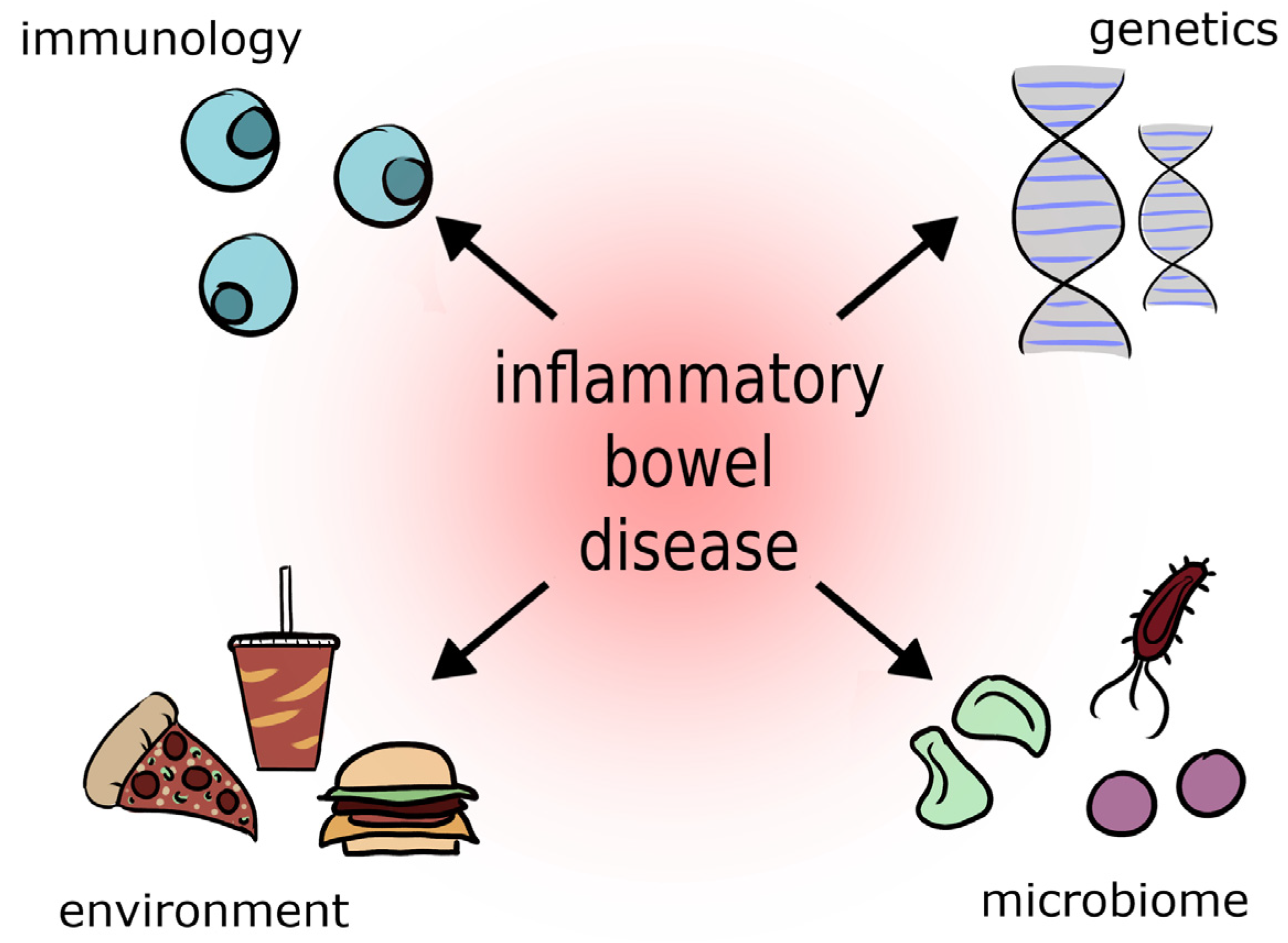
:1. Introduction
2. Nonalcoholic Fatty Liver Disease (NAFLD) and Liver Fibrosis (LF)
3. Inflammatory Bowel Diseases (IBD)
Symptoms and Diagnostics of Inflammatory Bowel Disease
4. Primary Sclerosing Cholangitis (PSC) and Inflammatory Bowel Disease (IBD)
5. Liver Fibrosis and Inflammatory Bowel Disease
5.1. Studies in Humans
5.2. Animal Studies
6. Treatment
7. Conclusions
Author Contributions
Funding
Conflicts of Interest
References
- Marcellin, P.; Kutala, B.K. Liver diseases: A major, neglected global public health problem requiring urgent actions and large-scale screening. Liver Int. 2018, 38, 2–6. [Google Scholar] [CrossRef] [Green Version]
- Mokdad, A.A.; Lopez, A.D.; Shahraz, S.; Lozano, R.; Mokdad, A.H.; Stanaway, J.; Murray, C.J.; Naghavi, M. Liver cirrhosis mortality in 187 countries between 1980 and 2010: A systematic analysis. BMC Med. 2014, 12, 145. [Google Scholar] [CrossRef] [Green Version]
- Stepanova, M.; De Avila, L.; Afendy, M.; Younossi, I.; Pham, H.; Cable, R.; Younossi, Z.M. Direct and Indirect Economic Burden of Chronic Liver Disease in the United States. Clin. Gastroenterol. Hepatol. 2017, 15, 759–766.e5. [Google Scholar] [CrossRef]
- Kim, D.; Li, A.A.; Gadiparthi, C.; Khan, M.A.; Cholankeril, G.; Glenn, J.S.; Ahmed, A. Changing Trends in Etiology-Based Annual Mortality From Chronic Liver Disease, From 2007 Through 2016. Gastroenterology 2018, 155, 1154–1163.e3. [Google Scholar] [CrossRef] [Green Version]
- Younossi, Z.; Tacke, F.; Arrese, M.; Chander Sharma, B.; Mostafa, I.; Bugianesi, E.; Wai-Sun Wong, V.; Yilmaz, Y.; George, J.; Fan, J.; et al. Global Perspectives on Nonalcoholic Fatty Liver Disease and Nonalcoholic Steatohepatitis. Hepatology 2019, 69, 2672–2682. [Google Scholar] [CrossRef] [PubMed] [Green Version]
- Haas, J.; Vonghia, L.; Mogilenko, D.; Verrijken, A.; Molendi-Coste, O.; Fleury, S.; Deprince, A.; Nikitin, A.; Woitrain, E.; Ducrocq-Geoffroy, L.; et al. Transcriptional Network Analysis Implicates Altered Hepatic Immune Function in NASH development and resolution. Nat. Metab. 2019, 1, 604–614. [Google Scholar] [CrossRef] [PubMed]
- Leslie, M. The liver’s weighty problem. Science 2015, 349, 18–20. [Google Scholar] [CrossRef]
- Zeitz, J.; Mullhaupt, B.; Fruehauf, H.; Rogler, G.; Vavricka, S.R. Hepatic failure due to hepati-tis B reactivation in a patient with ulcerative colitis treated with prednisone. Hepatology 2009, 50, 653–654. [Google Scholar] [CrossRef] [PubMed] [Green Version]
- Dean, G.; Hanauer, S.; Levitsky, J. The Role of the Intestine in the Pathogenesis of Primary Sclerosing Cholangitis: Evidence and Therapeutic Implications. Hepatology 2020, 72, 1127–1138. [Google Scholar] [CrossRef] [PubMed]
- Younossi, Z.; Anstee, Q.M.; Marietti, M.; Hardy, T.; Henry, L.; Eslam, M.; George, J.; Bugianesi, E. Global burden of NAFLD and NASH: Trends, predictions, risk factors and prevention. Nat. Rev. Gastroenterol. Hepatol. 2018, 15, 11–20. [Google Scholar] [CrossRef]
- National Health Survey: First Results, 2014–2015. Available online: http://www.abs.gov.au/ausstats/abs@.nsf/mf/4364.0.55.001 (accessed on 21 December 2022).
- Coppell, K.J.; Miller, J.C.; Gray, A.R.; Schultz, M.; Mann, J.I.; Parnell, W.R. Obesity and the extent of liver damage among adult New Zealanders: Findings from a national survey. Obes. Sci. Pract. 2015, 1, 67–77. [Google Scholar] [CrossRef] [PubMed]
- Kalia, H.S.; Gaglio, P.J. The prevalence and pathobiology of nonalcoholic fatty liver disease in patients of different races or ethnicities. Clin. Liver Dis. 2016, 20, 215–224. [Google Scholar] [CrossRef] [PubMed]
- Singh, S.; Allen, A.M.; Wang, Z.; Prokop, L.J.; Murad, M.H.; Loomba, R. Fibrosis progression in nonalcoholic fatty liver vs nonalcoholic steatohepatitis: A systematic review and meta-analysis of paired-biopsy studies. Clin. Gastroenterol. Hepatol. 2015, 13, 643–654. [Google Scholar] [CrossRef] [PubMed] [Green Version]
- Musso, G.; Gambino, R.; Tabibian, J.H.; Ekstedt, M.; Kechagias, S.; Hamaguchi, M.; Hultcrantz, R.; Hagström, H.; Yoon, S.K.; Charatcharoenwitthaya, P.; et al. Association of non-alcoholic fatty liver disease with chronic kidney disease: A systematic review and meta-analysis. PLoS Med. 2014, 11, e001680. [Google Scholar] [CrossRef] [Green Version]
- Fazel, Y.; Koenig, A.B.; Sayiner, M.; Goodman, Z.D.; Younossi, Z.M. Epidemiology and natural history of non-alcoholic fatty liver disease. Metabolism 2016, 65, 1017–1025. [Google Scholar] [CrossRef] [PubMed] [Green Version]
- Ralston, S.H.; Strachan, M.W.J.; Penman, I.; Hobson, R. Choroby Wewnętrzne Davidson t. 2; Edra Urban & Partner: Wrocław, Poland, 2020. [Google Scholar]
- Szczeklik, A.; Gajewski, P. Interna Szczeklika; Medycyna Praktyczna: Kraków, Poland, 2020. [Google Scholar]
- Berkan-Kawińska, A.; Piekarska, A. Hepatocellular carcinoma in non-alcohol fatty liver disease—changing trends and specific challenges. Curr. Med. Res. Opin. 2020, 36, 235–243. [Google Scholar] [CrossRef]
- Pierantonelli, I.; Svegliati-Baroni, G. Nonalcoholic Fatty Liver Disease: Basic Pathogenetic Mechanisms in the Progression From NAFLD to NASH. Transplantation 2019, 103, e1–e13. [Google Scholar] [CrossRef] [PubMed]
- Sheka, A.C.; Adeyi, O.; Thompson, J.; Hameed, B.; Crawford, P.A.; Ikramuddin, S. Nonalcoholic Steatohepatitis: A Review. JAMA 2020, 323, 1175–1183. [Google Scholar] [CrossRef]
- Powell, E.E.; Wong, V.W.; Rinella, M. Non-alcoholic fatty liver disease. Lancet 2021, 397, 2212–2224. [Google Scholar] [CrossRef]
- Tsuchida, T.; Friedman, S.L. Mechanisms of activation of liver stellate cells. Nat. Rev. Gastroenterol. Hepatol. 2017, 14, 397–411. [Google Scholar] [CrossRef]
- Kumar, S.; Duan, Q.; Wu, R.; Harris, E.N.; Su, Q. Pathophysiological communication between hepatocytes and nonparenchymal cells in liver injury from NAFLD to liver fibrosis. Adv. Drug Deliv. Rev. 2021, 176, 113869. [Google Scholar] [CrossRef]
- Schuppan, D.; Surabattula, R.; Wang, X.Y. Determinants of the progression and regression of fibrosis in NASH. J. Hepatol. 2018, 68, 238–250. [Google Scholar] [CrossRef]
- Radwan, P.; Radwan, K. Nieswoiste choroby zapalne jelit u osób w podeszłym wieku. Gastroenterol. Klin. 2016, 8, 27–35. [Google Scholar]
- de Zoeten, E.F.; Pasternak, B.A.; Mattei, P.; Kramer, R.E.; Kader, H.A. Diagnosis and treatment of perianal Crohn disease: NASPGHAN clinical report and consensus statement. J. Pediatr. Gastroenterol. Nutr. 2013, 57, 401–412. [Google Scholar] [CrossRef]
- Haac, B.; Palmateer, N.; Seaton, M.; VanYPeren, R.; Fraser, C.; Bafford, A. A Distinct Gut Microbiota Exists Within Crohn’s Disease–Related Perianal Fistulae. Gastrointestinal 2019, 242, 118–128. [Google Scholar] [CrossRef] [PubMed]
- Fiocchi, C. Inflammatory bowel disease: Etiology and pathogenesis. Gastroenterology 1998, 115, 182–205. [Google Scholar] [CrossRef] [PubMed]
- Chreptowicz, A. Rośnie liczba osób z rozpoznaniem mikroskopowego zapalenia jelit—Co nowego w leczeniu? Gastroenterol. Klin. 2016, 8, 107–112. [Google Scholar]
- Soubieres, A.A.; Poullis, A. Emerging role of novel biomarkers in the diagnosis of inflammatory bowel disease. World J. Gastrointest. Pharmacol. Ther. 2016, 7, 41–50. [Google Scholar] [CrossRef] [PubMed]
- Yan, J.B.; Luo, M.M.; Chen, Z.Y.; He, B.H. The Function and Role of the Th17 (T helper 17)/Treg (T regulatory) Cell Balance in Inflammatory Bowel Disease. J. Immunol. Res. 2020, 2020, 8813558. [Google Scholar] [CrossRef] [PubMed]
- Van Herck, M.A.; Weyler, J.; Kwanten, W.J.; Dirinck, E.L.; De Winter, B.Y.; Francque, S.M.; Vonghia, L. The Differential Roles of T Cells in Non-alcoholic Fatty Liver Disease and Obesity. Front. Immunol. 2019, 10, 82. [Google Scholar] [CrossRef] [Green Version]
- De la Fuente, M.; MacDonald, T.T.; Hermoso, M.A. Intestinal Homeostasis and Disease: A Complex Partnership Between Immune Cells, Non-Immune Cells, and the Microbiome. Front. Immunol. 2019, 10, 2775. [Google Scholar] [CrossRef] [PubMed] [Green Version]
- Garcia-Hernandez, V.; Quiros, M.; Nusrat, A. Intestinal epithelial claudins: Expression and regulation in homeostasis and inflammation. Ann. N. Y. Acad. Sci. 2017, 1397, 66–79. [Google Scholar] [CrossRef]
- Hu, C.A.; Hou, Y.; Yi, D.; Qiu, Y.; Wu, G.; Kong, X.; Yin, Y. Autophagy and tight junction proteins in the intestine and intestinal diseases. Anim. Nutr. 2015, 1, 123–127. [Google Scholar] [CrossRef]
- Yamamoto-Furusho, J.K.; Mendivil, E.J.; Fonseca-Camarillo, G. Differential expression of occludin in patients with ulcerative colitis and healthy controls. Inflamm. Bowel Dis. 2012, 18, E1999. [Google Scholar] [CrossRef] [PubMed]
- Van Itallie, C.M.; Tietgens, A.J.; Anderson, J.M. Visualizing the dynamic coupling of claudin strands to the actin cytoskeleton through ZO-1. Mol. Biol. Cell 2017, 28, 524–534. [Google Scholar] [CrossRef] [PubMed]
- Sugita, K.; Kabashima, K. Tight junctions in the development of asthma, chronic rhinosinusitis, atopic dermatitis, eosinophilic esophagitis, and inflammatory bowel diseases. J. Leukoc. Biol. 2020, 107, 749–762. [Google Scholar] [CrossRef] [PubMed]
- Østvik, A.E.; Svendsen, T.D.; Granlund, A.V.B.; Doseth, B.; Skovdahl, H.K.; Bakke, I.; Thorsvik, S.; Afroz, W.; Walaas, G.A.; Mollnes, T.E.; et al. Intestinal epithelial cells express immunomodulatory ISG15 during active ulcerative colitis and Crohn’s disease. J. Crohns Colitis 2020, 14, 920–934. [Google Scholar] [CrossRef] [PubMed]
- Danilova, N.A.; Abdulkhakov, S.R.; Grigoryeva, T.V.; Markelova, M.I.; Vasilyev, I.Y.; Boulygina, E.A.; Ardatskaya, M.D.; Pavlenko, A.V.; Tyakht, A.V.; Odintsova, A.K.; et al. Markers of dysbiosis in patients with ulcerative colitis and Crohn’s disease. Ter. Arkh. 2019, 91, 17–24. [Google Scholar] [CrossRef]
- Glassner, K.L.; Abraham, B.P.; Quigley, E.M.M. The microbiome and inflammatory bowel disease. J. Allergy Clin. Immunol. 2020, 145, 16–27. [Google Scholar] [CrossRef] [Green Version]
- Zuo, T.; Ng, S.C. The Gut Microbiota in the Pathogenesis and Therapeutics of Inflammatory Bowel Disease. Front. Microbiol. 2018, 9, 2247. [Google Scholar] [CrossRef] [Green Version]
- Chu, Y.; Jiang, M.Z.; Xu, B.; Wang, W.J.; Chen, D.; Li, X.W.; Zhang, Y.J.; Liang, J. Specific changes of enteric mycobiota and virome in inflammatory bowel disease. J. Dig. Dis. 2018, 19, 2–7. [Google Scholar] [CrossRef]
- Gonciarz, M.; Szkudłapski, D.; Mularczyk, A.; Radwan, P.; Kłopocka, M.; Bartnik, W.; Rydzewska, G. Wytyczne postępowania z chorymi na nieswoiste choroby zapalne jelit w praktyce lekarza rodzinnego. Lekarz POZ 2017, 3, 1–11. [Google Scholar]
- Nowakowski, J.; Chrobak, A.; Dudek, D. Psychiatric illnesses in inflammatory bowel diseases—psychiatric comorbidity and biological underpinnings. Psychiatr. Pol. 2016, 50, 1157–1166. [Google Scholar] [CrossRef] [PubMed]
- Man, S.; Kaakoush, N.; Mitchell, H. The role of bacteria and pattern-recognition receptors in Crohn’s disease. Nat. Rev. Gastroenterol. Hepatol. 2011, 8, 152–168. [Google Scholar] [CrossRef] [PubMed]
- Bartnik, W. Wytyczne postępowania w nieswoistych chorobach zapalnych jelit. Przegląd Gastroenterol. 2007, 2, 215–229. [Google Scholar]
- Eder, P. Przydatność biomarkerów w ocenie aktywności nieswoistych chorób zapalnych jelit -wskazówki praktyczne. Varia Med. 2018, 2, 371–380. [Google Scholar]
- Arai, T.; Takeuchi, K.; Miyamura, M.; Ishikawa, R.; Yamada, A.; Katsumata, M.; Igarashi, Y.; Suzuki, Y. Level of Fecal Calprotectin Correlates With Severity of Small Bowel Crohn’s Disease, Measured by Balloon-assisted Enteroscopy and Computed Tomography Enterography. Clin. Gastroenterol. Hepatol. 2017, 15, 56–62. [Google Scholar] [CrossRef]
- Molander, P.; Färkkilä, M.; Ristimäki, A.; Salminen, K.; Kemppainen, H.; Blomster, T.; Koskela, R.; Jussila, A.; Rautiainen, H.; Nissinen, M.; et al. Does fecal calprotectin predict short-term relapse after stopping TNFα-blocking agents in inflammatory bowel disease patients in deep remission? J. Crohn’s. Colitis 2015, 9, 33–40. [Google Scholar] [CrossRef] [Green Version]
- Hao, L.; Shan, Q.; Wei, J.; Ma, F.; Sun, P. Lactoferrin: Major Physiological Functions and Applications. Curr. Protein Pept. Sci. 2019, 20, 139–144. [Google Scholar] [CrossRef]
- Niaz, B.; Saeed, F.; Ahmed, A.; Imran, M.; Maan, A.A.; Khan, M.K.I.; Tufail, T.; Anjum, F.M.; Hussain, S.; Suleria, H.A.R. Lactoferrin (LF): A natural antimicrobial protein. Int. J. Food Prop. 2019, 22, 1626–1641. [Google Scholar] [CrossRef] [Green Version]
- Pan, S.; Weng, H.; Hu, G.; Wang, S.; Zhao, T.; Yao, X.; Liao, L.; Zhu, X.; Ge, Y. Lactoferrin may inhibit the development of cancer via its immunostimulatory and immunomodulatory activities (Review). Int. J. Oncol. 2021, 59, 85. [Google Scholar] [CrossRef] [PubMed]
- Boyapati, R.K.; Torres, J.; Palmela, C.; Parker, C.E.; Silverberg, O.M.; Upadhyaya, S.D.; Nguyen, T.M.; Colombel, J.F. Withdrawal of immunosuppressant or biologic therapy for patients with quiescent Crohn’s disease. Cochrane Database Syst. Rev. 2018, 5, CD012540. [Google Scholar] [CrossRef]
- Rabiee, A.; Silveira, M. Primary sclerosing cholangitis. Transl. Gastroenterol. Hepatol. 2021, 6, 29. [Google Scholar] [CrossRef]
- Williamson, K.; Chapman, R. Primary sclerosing cholangitis. Dig. Dis. 2014, 32, 438–445. [Google Scholar] [CrossRef]
- Chung, B.; Hirschfield, G. Immunogenetics in primary sclerosing cholangitis. Curr. Opin. Gastroenterol. 2017, 33, 93–98. [Google Scholar] [CrossRef]
- O’Toole, A.; Alakkari, A.; Keegan, D.; Doherty, G.; Mulcahy, H.; O’Donoghue, D. Primary Sclerosing Cholangitis and Disease Distribution in Inflammatory Bowel Disease. Clin. Gastroenterol. Hepatol. 2012, 10, 439–441. [Google Scholar] [CrossRef] [PubMed]
- Karlsen, T.H.; Folseraas, T.; Thorburn, D.; Vesterhus, M. Primary sclerosing cholangitis—A comprehensive review. J. Hepatol. 2017, 67, 1298–1323. [Google Scholar] [CrossRef] [PubMed] [Green Version]
- Eksteen, B. The Gut-Liver Axis in Primary Sclerosing Cholangitis. Clin. Liver Dis. 2016, 20, 1–14. [Google Scholar] [CrossRef]
- Ali, A.H.; Damman, J.; Shah, S.B.; Davies, Y.; Hurwitz, M.; Stephen, M.; Lemos, L.M.; Carey, E.J.; Lindor, K.D.; Buness, C.W.; et al. Open-label prospective therapeutic clinical trials: Oral vancomycin in children and adults with primary sclerosing cholangitis. Scand. J. Gastroenterol. 2020, 55, 941–950. [Google Scholar] [CrossRef]
- Bajer, L.; Kverka, M.; Kostovcik, M.; Macinga, P.; Dvorak, J.; Stehlikova, Z.; Brezina, J.; Wohl, P.; Spicak, J.; Drastich, P. Distinct gut microbiota profiles in patients with primary sclerosing cholangitis and ulcerative colitis. World J. Gastroenterol. 2017, 23, 4548–4558. [Google Scholar] [CrossRef]
- Ostadmohammadi, S.; Azimirad, M.; Houri, H.; Naseri, K.; Javanmard, E.; Mirjalali, H.; Yadegar, A.; Sadeghi, A.; Asadzadeh Aghdaei, H.; Zali, M. Characterization of the gut microbiota in patients with primary sclerosing cholangitis compared to inflammatory bowel disease and healthy controls. Mol. Biol. Rep. 2021, 48, 5519–5529. [Google Scholar] [CrossRef] [PubMed]
- Mehta, T.; Weissman, S.; Fung, B.; Sotiriadis, J.; Lindor, K.; Tabibian, J. Global incidence, prevalence and features of primary sclerosing cholangitis: A systematic review and meta-analysis. Liver Int. 2021, 41, 2418–2426. [Google Scholar] [CrossRef] [PubMed]
- Ji, S.G.; Juran, B.D.; Mucha, S.; Folseraas, T.; Jostins, L.; Melum, E.; Kumasaka, N.; Atkinson, E.J.; Schlicht, E.M.; Liu, J.Z.; et al. Genome-wide association study of primary sclerosing cholangitis identifies new risk loci and quantifies the genetic relationship with inflammatory bowel disease. Nat. Genet 2017, 49, 269–273. [Google Scholar] [CrossRef] [PubMed] [Green Version]
- de Vries, A.; Janse, M.; Blokzijl, H.; Weersma, R. Distinctive inflammatory bowel disease phenotype in primary sclerosing cholangitis. World J. Gastroenterol. 2015, 21, 1956–1971. [Google Scholar] [CrossRef] [Green Version]
- Sørensen, J.; Nielsen, O.; Andersson, M.; Ainsworth, M.; Ytting, H.; Bélard, E.; Jess, T. Inflammatory bowel disease with primary sclerosing cholangitis: A Danish population-based cohort study 1977–2011. Liver Int. 2018, 38, 532–541. [Google Scholar] [CrossRef]
- Ricciuto, A.; Kamath, B.; Griffiths, A. The IBD and PSC Phenotypes of PSC-IBD. Curr. Gastroenterol. Rep. 2018, 20, 16. [Google Scholar] [CrossRef]
- Palmela, C.; Peerani, F.; Castaneda, D.; Torres, J.; Itzkowitz, S. Inflammatory Bowel Disease and Primary Sclerosing Cholangitis: A Review of the Phenotype and Associated Specific Features. Gut Liver 2018, 12, 17–29. [Google Scholar] [CrossRef] [Green Version]
- Loftus, E.J.; Harewood, G.; Loftus, C.; Tremaine, W.; Harmsen, W.; Zinsmeister, A.; Jewell, D.; Sandborn, W. PSC-IBD: A unique form of inflammatory bowel disease associated with primary sclerosing cholangitis. Gut 2005, 54, 91–96. [Google Scholar] [CrossRef]
- Nordenvall, C.; Olén, O.; Nilsson, P.; von Seth, E.; Ekbom, A.; Bottai, M.; Myrelid, P.; Bergquist, A. Colectomy prior to diagnosis of primary sclerosing cholangitis is associated with improved prognosis in a nationwide cohort study of 2594 PSC-IBD patients. Aliment. Pharmacol. Ther. 2018, 47, 238–245. [Google Scholar] [CrossRef]
- de Krijger, M.; Wildenberg, M.E.; de Jonge, W.J.; Ponsioen, C.Y. Return to sender: Lymphocyte trafficking mechanisms as contributors to primary sclerosing cholangitis. J. Hepatol. 2019, 71, 603–615. [Google Scholar] [CrossRef] [Green Version]
- Hirschfield, G.M.; Karlsen, T.H.; Lindor, K.D.; Adams, D.H. Primary sclerosing cholangitis. Lancet 2013, 382, 1587–1599. [Google Scholar] [CrossRef]
- Rinella, M.E.; Tacke, F.; Sanyal, A.J.; Anstee, Q.M. participants of the AASLD/EASL Workshop. Report on the AASLD/EASL Joint Workshop on Clinical Trial Endpoints in NAFLD. Hepatology 2019, 70, 1424–1436. [Google Scholar] [CrossRef]
- Greuter, T.; Vavricka, S.R. Extraintestinal manifestations in inflammatory bowel disease—Epidemiology, genetics, and pathogenesis. Expert. Rev. Gastroenterol. Hepatol. 2019, 13, 307–317. [Google Scholar] [CrossRef] [Green Version]
- Rogler, G.; Singh, A.; Kavanaugh, A.; Rubin, D. Extraintestinal Manifestations of Inflammatory Bowel Disease: Current Concepts, Treatment, and Implications for Disease Management. Gastroenterology 2021, 161, 1118–1132. [Google Scholar] [CrossRef] [PubMed]
- Ritaccio, G.; Stoleru, G.; Abutaleb, A.; Cross, R.; Shetty, K.; Sakiani, S.; Wong, U. Nonalcoholic Fatty Liver Disease Is Common in IBD Patients However Progression to Hepatic Fibrosis by Noninvasive Markers Is Rare. Dig. Dis. Sci. 2021, 66, 3186–3191. [Google Scholar] [CrossRef] [PubMed]
- Barbero-Villares, A.; Mendoza Jiménez-Ridruejo, J.; Taxonera, C.; López-Sanromán, A.; Pajares, R.; Bermejo, F.; Pérez-Calle, J.; Mendoza, J.; Algaba, A.; Moreno-Otero, R.; et al. Evaluation of liver fibrosis by transient elastography (Fibroscan®) in patients with inflammatory bowel disease treated with methotrexate: A multicentric trial. Scand. J. Gastroenterol. 2012, 47, 575–579. [Google Scholar] [CrossRef]
- Laharie, D.; Zerbib, F.; Adhoute, X.; Boué-Lahorgue, X.; Foucher, J.; Castéra, L.; Rullier, A.; Bertet, J.; Couzigou, P.; Amouretti, M.; et al. Diagnosis of liver fibrosis by transient elastography (FibroScan) and non-invasive methods in Crohn’s disease patients treated with methotrexate. Aliment. Pharmacol. Ther. 2006, 23, 1621–1628. [Google Scholar] [CrossRef]
- Wang, Y.; Li, Y.; Liu, Y.; Zhang, Y.; Ke, Z.; Zhang, Y.; Liu, Y. Patients With IBD Receiving Methotrexate Are at Higher Risk of Liver Injury Compared With Patients With Non-IBD Diseases: A Meta-Analysis and Systematic Review. Front. Med. 2021, 8, 774824. [Google Scholar] [CrossRef] [PubMed]
- Magrì, S.; Paduano, D.; Chicco, F.; Cingolani, A.; Farris, C.; Delogu, G.; Tumbarello, F.; Lai, M.; Melis, A.; Casula, L.; et al. Nonalcoholic fatty liver disease in patients with inflammatory bowel disease: Beyond the natural history. World J. Gastroenterol. 2019, 25, 5676–5686. [Google Scholar] [CrossRef]
- Carr, R.; Patel, A.; Bownik, H.; Oranu, A.; Kerner, C.; Praestgaard, A.; Forde, K.; Reddy, K.; Lichtenstein, G. Intestinal Inflammation Does Not Predict Nonalcoholic Fatty Liver Disease Severity in Inflammatory Bowel Disease Patients. Dig. Dis. Sci. 2017, 62, 1354–1361. [Google Scholar] [CrossRef]
- Rodriguez-Duque, J.C.; Calleja, J.L.; Iruzubieta, P.; Hernández-Conde, M.; Rivas-Rivas, C.; Vera, M.I.; Garcia, M.J.; Pascual, M.; Castro, B.; García-Blanco, A.; et al. Increased risk of MAFLD and Liver Fibrosis in Inflammatory Bowel Disease Independent of Classic Metabolic Risk Factors. Clin. Gastroenterol. Hepatol. 2022, 21, 406–414. [Google Scholar] [CrossRef]
- Veltkamp, C.; Lan, S.; Korompoki, E.; Weiss, K.; Schmidt, H.; Seitz, H. Hepatic Steatosis and Fibrosis in Chronic Inflammatory Bowel Disease. J. Clin. Med. 2022, 11, 2623. [Google Scholar] [CrossRef]
- Rieder, F.; Bettenworth, D.; Imai, J.; Inagaki, Y. Intestinal Fibrosis and Liver Fibrosis: Consequences of Chronic Inflammation or Independent Pathophysiology? Inflamm. Intest. Dis. 2016, 1, 41–49. [Google Scholar] [CrossRef] [PubMed]
- Inagaki, Y.; Okazaki, I. Emerging insights into Transforming growth factor beta Smad signal in hepatic fibrogenesis. Gut 2007, 56, 284–292. [Google Scholar] [CrossRef] [Green Version]
- Fiocchi, C.; Lund, P. Themes in fibrosis and gastrointestinal inflammation. Am. J. Physiol. Gastrointest. Liver Physiol. 2011, 300, G677–G683. [Google Scholar] [CrossRef]
- Steen, E.; Wang, X.; Balaji, S.; Butte, M.; Bollyky, P.; Keswani, S. The Role of the Anti-Inflammatory Cytokine Interleukin-10 in Tissue Fibrosis. Adv. Wound Care 2020, 9, 184–198. [Google Scholar] [CrossRef] [PubMed] [Green Version]
- Baumann, A.; Hernández-Arriaga, A.; Brandt, A.; Sánchez, V.; Nier, A.; Jung, F.; Kehm, R.; Höhn, A.; Grune, T.; Frahm, C.; et al. Microbiota profiling in aging-associated inflammation and liver degeneration. Int. J. Med. Microbiol. 2021, 311, 151500. [Google Scholar] [CrossRef]
- Xie, G.; Wang, X.; Liu, P.; Wei, R.; Chen, W.; Rajani, C.; Hernandez, B.; Alegado, R.; Dong, B.; Li, D.; et al. Distinctly altered gut microbiota in the progression of liver disease. Oncotarget 2016, 7, 19355–19366. [Google Scholar] [CrossRef] [Green Version]
- Sultan, S.; El-Mowafy, M.; Elgaml, A.; Ahmed, T.; Hassan, H.; Mottawea, W. Metabolic Influences of Gut Microbiota Dysbiosis on Inflammatory Bowel Disease. Front. Physiol. 2021, 12, 715506. [Google Scholar] [CrossRef] [PubMed]
- Tilg, H.; Cani, P.; Mayer, E. Gut microbiome and liver diseases. Gut 2016, 65, 2035–2044. [Google Scholar] [CrossRef]
- Liu, Y.; Chen, K.; Li, F.; Gu, Z.; Liu, Q.; He, L.; Shao, T.; Song, Q.; Zhu, F.; Zhang, L.; et al. Probiotic Lactobacillus rhamnosus GG Prevents Liver Fibrosis Through Inhibiting Hepatic Bile Acid Synthesis and Enhancing Bile Acid Excretion in Mice. Hepatology 2020, 71, 2050–2066. [Google Scholar] [CrossRef] [Green Version]
- Li, Y.; Yang, S.; Lun, J.; Gao, J.; Gao, X.; Gong, Z.; Wan, Y.; He, X.; Cao, H. Inhibitory Effects of the Lactobacillus rhamnosus GG Effector Protein HM0539 on Inflammatory Response through the TLR4/MyD88/NF-кB Axis. Front. Immunol. 2020, 11, 551449. [Google Scholar] [CrossRef] [PubMed]
- Ruemmele, F.M.; Veres, G.; Kolho, K.L.; Griffiths, A.; Levine, A.; Escher, J.C.; Amil Dias, J.; Barabino, A.; Braegger, C.P.; Bronsky, J.; et al. European Crohn’s and Colitis Organisation; European Society of Pediatric Gastroenterology, Hepatology and Nutrition. Consensus guidelines of ECCO/ESPGHAN on the medical management of pediatric Crohn’s disease. J. Crohns Colitis 2014, 8, 1179–1207. [Google Scholar] [CrossRef] [Green Version]
- Rawa, T. Doustne preparaty kwasu 5-aminosalicylowego w leczeniu wrzodziejącego zapalenia jelita grubego. Gastroenterol. Klin. 2012, 4, 98–104. [Google Scholar]
- Radwan, P. Steroidooporność i steroidozależność w nieswoistych chorobach zapal-nych jelit. Gastroenterol. Klin. 2015, 7, 46–52. [Google Scholar]
- Amin, J.; Huang, B.; Yoon, J.; Shih, D.Q. Update 2014: Advances to optimize 6-mercaptopurine and azathioprine to reduce toxicity and improve efficacy in the management of IBD. Inflamm. Bowel Dis. 2015, 21, 445–452. [Google Scholar] [CrossRef] [PubMed]
- Danese, S.; Gomollon, F. Governing Board and Operational Board of ECCO. ECCO position statement: The use of biosimilar medicines in the treatment of inflammatory bowel disease (IBD). J. Crohns Colitis 2013, 7, 586–589. [Google Scholar] [CrossRef] [PubMed] [Green Version]
- Huldani, H.; Margiana, R.; Ahmad, F.; Opulencia, M.J.C.; Ansari, M.J.; Bokov, D.O.; Abdullaeva, N.N.; Siahmansouri, H. Immunotherapy of inflammatory bowel disease (IBD) through mesenchymal stem cells. Int. Immunopharmacol. 2022, 107, 108698. [Google Scholar] [CrossRef] [PubMed]
- Hu, C.; Zhao, L.; Zhang, L.; Bao, Q.; Li, L. Mesenchymal stem cell-based cell-free strategies: Safe and effective treatments for liver injury. Stem Cell Res. Ther. 2020, 11, 377. [Google Scholar] [CrossRef] [PubMed]
- Zhang, L.; Ma, X.J.; Fei, Y.Y.; Han, H.T.; Xu, J.; Cheng, L.; Li, X. Stem cell therapy in liver regeneration: Focus on mesenchymal stem cells and induced pluripotent stem cells. Pharmacol. Ther. 2022, 232, 108004. [Google Scholar] [CrossRef] [PubMed]
- European Association for the Study of the Liver. EASL Clinical Practice Guidelines on nutrition in chronic liver disease. J. Hepatol. 2019, 70, 172–193. [Google Scholar] [CrossRef] [PubMed] [Green Version]
- Sasson, A.N.; Ananthakrishnan, A.N.; Raman, M. Diet in Treatment of Inflammatory Bowel Diseases. Clin. Gastroenterol. Hepatol. 2021, 19, 425–435.e3. [Google Scholar] [CrossRef] [PubMed]


| Intestinal Epithelial Barrier Disturbances | Intestinal Microbiota Disturbances |
|---|---|
| -increased expression of claudin-1–reduction of mucus secretion, which increases the risk of enteritis (CD and UC) -increased expression of claudin-2–Increased pores in TJ (CD and UC) -decreased expression of claudin–3,5,8 (CD)–reduction of intestinal barrier properties -decreased expression of claudin–3,4,7 (UC)–reduction of intestinal barrier properties -increased expression of occludin (active UC) -ZO1 dysfunction–decrease in stabilised claudin strands -TNF-α–decrease in normal TJ function and increase in intestinal epithelial apoptosis -IL-1𝛽 i IFN-𝛾–reduction of TJ integrity by affecting occludins and ZO1 -IFN-𝜆–occurrence of Paneth cell defect -dysfunction of ILC3 -secrection of chemokines by EIC–causes and maintains inflammation in IBD (e.g. CXCR1 (+) CXCR2 (+) IL-23) | Increase in the amount of the following bacteria: -Proteobacteria (degradation of intestinal mucus) np. Haemophilus, Pasteurellaceae -Streptococcus -Fusobacterium (degradation of intestinal mucus) -Enterobacteriaceae np. E. Coli (AIEC) Decrease in the amount of the following bacteria: -Firmicutes np. Faecalibacterium prausnitzii, Roseburia (SCFA producing bacteria) -Euryarchaeota -Bacteroidetes np. Prevotella spp. -Bifidobacterium Increase in the amount of following fungus: -C. albicans -C. parapsilosis -Aspergillus clavatus (CD) -Cryptococcus neoformans (CD) -Ascomycota |
Disclaimer/Publisher’s Note: The statements, opinions and data contained in all publications are solely those of the individual author(s) and contributor(s) and not of MDPI and/or the editor(s). MDPI and/or the editor(s) disclaim responsibility for any injury to people or property resulting from any ideas, methods, instructions or products referred to in the content. |
© 2023 by the authors. Licensee MDPI, Basel, Switzerland. This article is an open access article distributed under the terms and conditions of the Creative Commons Attribution (CC BY) license (https://creativecommons.org/licenses/by/4.0/).
Share and Cite
Jarmakiewicz-Czaja, S.; Gruszecka, J.; Filip, R. What Do NAFLD, Liver Fibrosis, and Inflammatory Bowel Disease Have in Common? Review of the Current Literature. Metabolites 2023, 13, 378. https://doi.org/10.3390/metabo13030378
Jarmakiewicz-Czaja S, Gruszecka J, Filip R. What Do NAFLD, Liver Fibrosis, and Inflammatory Bowel Disease Have in Common? Review of the Current Literature. Metabolites. 2023; 13(3):378. https://doi.org/10.3390/metabo13030378
Chicago/Turabian StyleJarmakiewicz-Czaja, Sara, Jolanta Gruszecka, and Rafał Filip. 2023. "What Do NAFLD, Liver Fibrosis, and Inflammatory Bowel Disease Have in Common? Review of the Current Literature" Metabolites 13, no. 3: 378. https://doi.org/10.3390/metabo13030378