Article

Data Independent Acquisition Reveals In-Depth Serum Proteome Changes in Canine Leishmaniosis

1
Faculty of Veterinary Medicine, University of Zagreb, Heinzelova 55, HR-10000 Zagreb, Croatia
2
Department of Safety and Protection, Karlovac University of Applied Sciences, Trg Josipa Juraja Strossmayera 9, HR-47000 Karlovac, Croatia
3
School of Biodiversity, One Health and Veterinary Medicine, University of Glasgow, Bearsden Rd, Glasgow G61 1QH, UK
4
Interdisciplinary Laboratory of Clinical Analysis of the University of Murcia (Interlab-UMU), Department of Animal Medicine and Surgery, Veterinary School, University of Murcia, 30100 Murcia, Spain
5
Faculty of Food Technology and Biotechnology, University of Zagreb, Pierottijeva 6, HR-10000 Zagreb, Croatia
*
Author to whom correspondence should be addressed.
Metabolites 2023, 13(3), 365; https://doi.org/10.3390/metabo13030365
Submission received: 1 February 2023 / Revised: 19 February 2023 / Accepted: 26 February 2023 / Published: 28 February 2023
(This article belongs to the Special Issue Advances in Functional Proteomics and Metabolomics)

Abstract

:
Comprehensive profiling of serum proteome provides valuable clues of health status and pathophysiological processes, making it the main strategy in biomarker discovery. However, the high dynamic range significantly decreases the number of detectable proteins, obstructing the insights into the underlying biological processes. To circumvent various serum enrichment methods, obtain high-quality proteome wide information using the next-generation proteomic, and study host response in canine leishmaniosis, we applied data-independent acquisition mass spectrometry (DIA-MS) for deep proteomic profiling of clinical samples. The non-depleted serum samples of healthy and naturally Leishmania-infected dogs were analyzed using the label-free 60-min gradient sequential window acquisition of all theoretical mass spectra (SWATH-MS) method. As a result, we identified 554 proteins, 140 of which differed significantly in abundance. Those were included in lipid metabolism, hematological abnormalities, immune response, and oxidative stress, providing valuable information about the complex molecular basis of the clinical and pathological landscape in canine leishmaniosis. Our results show that DIA-MS is a method of choice for understanding complex pathophysiological processes in serum and serum biomarker development.

Graphical Abstract

1. Introduction

Blood serum reflects an individual’s phenotype providing a valuable information of physiological and pathological processes in organism making it a valuable source of biomarkers [1]. However, the high dynamic range aggravates the routine identification and quantification of low abundant proteins which are of a great interest to decipher proteome-wide protein functions and their interactions [2]. For that reason, various pre-analytical strategies such as fractionation, as well as enrichment methods including affinity depletion using immobilized antibodies or other molecules that specifically remove up to 22 high abundant proteins, and combinatorial peptide ligand library for proteome normalization are introduced [3,4]. Current depletion methods are costly, time-consuming, lack reproducibility, and often require additional processing (e.g., desalting or sample concentration) as a part of the MS-compatible proteomic analysis workflow. Additional separation steps are time consuming and introduce experimental variability making the results less reliable, especially regarding protein quantification [3]. Furthermore, most of the specific antibody-based depletion columns are constructed to remove the most abundant proteins from human samples unlike animal samples.
Mass spectrometry (MS)-based proteomics has become a powerful tool in biomedical research allowing the characterization of thousands of proteins as well as their posttranslational modifications [5]. Over the past couple of decades, MS-based proteomics has been mostly performed in a data-dependent acquisition (DDA) mode using shotgun strategy where proteins are digested by a sequence specific protease and resulting peptides are analyzed by liquid chromatography tandem mass spectrometry (LC- MS/MS) [4]. Herein, the top N most intense peptide’s fragments in MS2 produce a pattern of unique fragment ions spectra used to be assigned to their corresponding peptide sequences for unambiguous protein identification. However, low intensity ions often remain unidentified. Advances in MS-based technology and bioinformatics enabled to overcome the detectable dynamic range restrictions of peptides that ionize the best. Data-independent acquisition (DIA) using sequential window acquisition of all theoretical mass spectra (SWATH-MS) approach has been developed enabling the deep proteome coverage through confident peptide identification over a dynamic range of four orders of magnitude with quantitative consistency and accuracy [6]. The application of SWATH-MS enables the systematic and unbiased fragmentation of all ions within the overlapping precursor isolation windows leading to highly complex fragment ion spectra. Peptide identification is based on sample-specific spectral libraries constructed using DDA data, containing chromatographic and mass spectrometric coordinates determined by using normalized retention time [7]. Unlike DDA-based sample-specific spectral libraries, in silico spectral libraries are built directly from protein sequence databases (FASTA files) without previous DDA data requirements providing better proteome coverage in unbiased way [8]. For that purpose, deep learning algorithms are applied, selecting the list of target peptides from protein sequence databases by predicting the MS detectability of candidate proteotypic peptides [8]. It is worth mentioning that sample-specific spectral libraries (e.g., pan-human DIA libraries) are still the main choice in cancer research, reflecting tumor heterogeneity [9].
Zoonotic visceral leishmaniosis (VL) is a potentially fatal vector-borne disease endemic in South America, South-East Asia, Eastern Africa, and the Mediterranean Region [10]. In Croatia, human and canine leishmaniosis caused by Leishmania infantum has been present in central and southern Dalmatia since the beginning of the 20th century [11], and the dog has remained the main reservoir. Limited drug development and emerging drug resistance, as well as long duration of treatment [12] are making the understanding of the host-related processes in leishmaniosis even more important. In our previous studies, we applied tandem mass tag (TMT)-based DDA-MS methods for treatment monitoring in canine leishmaniosis by analyzing serum, and to understand the disease-related processes in canine saliva [11,12].
In that light, in order to obtain high-quality proteome wide information using the next-generation proteomics and study host response in canine leishmaniosis which could be observed in serum, the aim of this experiment was to construct canine serum proteome spectral library using DDA-MS approach and establish fast and label-free short gradient SWATH-MS method. Our results indicate the DIA-MS approach is extremely sensitive and informative, enabling the identification of canine serum proteins affected by canine leishmaniosis with biomarker potential, their functions and molecular pathways mostly included in lipid metabolism, hematological abnormalities, immune response, and oxidative stress.

2. Materials and Methods

2.1. Sample Description

The study was carried out using 10 archived canine sera previously used in routine leishmaniosis diagnostics at Department for Parasitology and Parasitic Diseases with Clinics, Faculty of Veterinary Medicine, University of Zagreb collected during six month- time interval. It involved sera classified into two groups: sera from healthy control group (N = 5) were mix breeds, age 2.5–9 years, while the Leishmania-infected group (N = 5) were mixed breeds, age 2–8 years. Control dogs were considered healthy based on previous serological tests. The infection of Leishmania infantum was diagnosed using three serological tests [13]: indirect fluorescence antibody test (IFAT), enzyme-linked immunosorbent assay (ELISA), and kinesin-related conserved recombinant antigen (rK39 rapid immunochromatographic test evaluating the presence of anti-Leishmania antibody) following the manufacturers’ instructions (Supplementary file S1). All naturally infected dogs (originating from the enzootic region of Croatia) showed clinical signs of Leishmania infection. The sera of infected dogs were taken before the treatment. All canine sera used in this study were tested negative for the presence of Dirofilaria immitis antigen using the antigen rapid test (Fast test HWantigen, Megacor Diagnostik, Vorarlberg, Austria).

2.2. Sample Collection

Archived, 10 canine sera samples were received from different veterinary stations sent to Department for Parasitology and Parasitic Diseases with Clinics, Faculty of Veterinary Medicine, University of Zagreb, for routine serological leishmaniosis diagnostics. After the diagnostics, sera samples were aliquoted and stored at −80 °C until analyzed to avoid multiple freeze-thaw cycles. Total protein concentration in serum was determined using a Pierce BCA Protein Assay Kit (Thermo Scientific, Rockford, IL, USA).

2.3. Sample Preparation for Spectral Library Generation

2.3.1. In-Solution Digestion

The pooled sample was prepared by mixing equal protein amounts of all ten samples involved in this study and used for in-solution digestion [14]. A total of 50 μg of total protein was mixed with 50 mM NH4HCO3 to a final concentration of 1 mg/mL. After the addition of 5 μL DTT (50 mM) sample was incubated for 30 min at 55 °C and 5 μL IAA (200 mM) was added subsequently. After 30 min, overnight digestion at 37 °C using trypsin gold (1:50 w/w) was performed. Tryptic digest was stored at −20 °C for further analysis [14]. Before the analysis, samples were mixed with 10× iRT peptide solution (Biognosys AG, Schlieren, Switzerland) for subsequent LC-MS/MS analysis.

2.3.2. Strong Cation Exchange (SCX) Chromatography

SCX was performed using pipette tips [15] with some modifications. Before the SCX chromatography, digested sample was desalted using Cleanup C18 pipette tips (10 μL, Agilent Technologies, Santa Clara, CA, USA) according to manufacturer’s instructions. Finally, peptides were eluted with 80% ACN/0.1% (v/v) formic acid, dried using vacuum concentrator and dissolved in 10 μL 0.1% formic acid. Strong cation exchange chromatography was performed using OMIX SCX pipette tips (10 μL, Agilent Technologies, Santa Clara, CA, USA). The tips were washed by mixture of 25% ACN in 0.05% formic acid and peptides were subsequently eluted with 100, 200 and 400 mM NH4HCO3 in 25%ACN/0.05% formic acid (v/v). Finally, samples were dried in vacuum concentrator, desalted using Cleanup C18 pipette tips, dried again, and finally mixed with 18 μL loading buffer and 2 μL of 10× iRT peptide solution (Biognosys AG, Schlieren, Switzerland) for DDA analysis.

2.3.3. Combinatorial Peptide Ligand Library (ProteoMiner)

The ProteoMiner Small-Capacity Kit (Bio-Rad, Hercules, CA, USA) containing a hexapeptide library [16] was used to equalize the protein concentration dynamic range of canine serum samples according to manufacturer’s procedure. Briefly, ProteoMiner column containing 20 μL beads was washed three times with 200 μL phosphate-buffered saline (PBS) buffer by 5 min incubations followed by 1000× g centrifugation. After adding 200 μL of pooled sample, bead-sample slurry was incubated with rotation (2 h at room temperature) and washed repeatedly three times before the elution. The protein elution was conducted by adding 20 µL of elution reagent to the column and vortexing for 15 min, followed by centrifugation at 1000× g.
As a part of MS-compatible workflow, eluted proteins were desalted using Microcon filter (10 kD MWCO, Millipore, Darmstadt, Germany). The eluate after ProteoMiner enrichment was mixed with 200 μL of milliQ water followed by centrifugation at 8000× g for 45 min. Proteins were washed twice with milliQ water by centrifugation at 8000× g for 30 min and once with 200 μL 50 mM NH4HCO3. Finally, proteins were collected by inverting the filter assembly followed by centrifugation at 2800× g for 5 min. Due to the reduced sample volume, the total protein concentration was determined by NanoDrop. Finally, in-solution digestion was performed.

2.4. MS-Based Proteomic Analysis

High resolution LC-MS/MS analysis was carried out using an Ultimate 3000 RSLCnano system (Dionex, Germering, Germany) coupled to a Q Exactive Plus mass spectrometer (Thermo Fisher Scientific, Bremen, Germany). Prior to analysis, tryptic peptides were mixed with iRT peptides (Biognosys) (1:10 v/v), desalted on the trap column and separated on the analytical column (PepMap™ RSLC C18, 50 cm × 75 μm). Ionization was achieved using nanospray Flex ion source (Thermo Fisher Scientific, Bremen, Germany) equipped with a 10 μm-inner diameter SilicaTip emitter (New Objective, Littleton, MA, USA).

DDA Proteomic Analysis for Spectral Library Generation

DDA analysis was performed as reported [14] with some changes. In short, for peptide separation, linear gradient 5–45% mobile phase B (0.1% formic acid in 80% ACN, v/v) over 120 min, at the flow rate of 300 nL/min was used. Mobile phase A contained 0.1% formic acid in water. The MS operated in positive ion mode using DDA Top10 method. Full scan MS spectra were acquired in range from m/z 350.0 to m/z 1800.0 with a resolution of 70,000, 120 ms injection time, AGC target 1 × 10⁶, a ±2.0 Da isolation window and the dynamic exclusion 30 s. For HCD fragmentation, step collision energy (29% and 35% NCE) with a resolution of 17,500 and AGC target of 2 × 10⁵ was used. Precursor ions with charge states of +1 and more than +7, as well as unassigned charge states, were excluded from HCD fragmentation.
For DDA protein identification, the SEQUEST algorithm implemented into Proteome Discoverer (version 2.3., Thermo Fisher Scientific) was applied. Database search against database containing Canis lupus familiaris FASTA files (downloaded from UniprotKB database 14/10/2021, 49,889 entries) combined with Biognosys iRT fusion peptide FASTA file (available at https://biognosys.com/shop/iRT-Kit (accessed on 1 September 2022)) was performed using parameters as follows: two trypsin missed cleavage sites, precursor and fragment mass tolerances of 10 ppm and 0.02 Da, respectively; carbamidomethyl (C) fixed peptide modification, oxidation (M), dynamic modifications. The false discovery rate (FDR) for peptide identification was calculated by the Percolator algorithm within the Proteome Discoverer workflow and was set at 1%.
The DDA mass spectrometry proteomics data have been deposited to the Consortium via the PRIDE partner repository [17] with the dataset PXD039763.

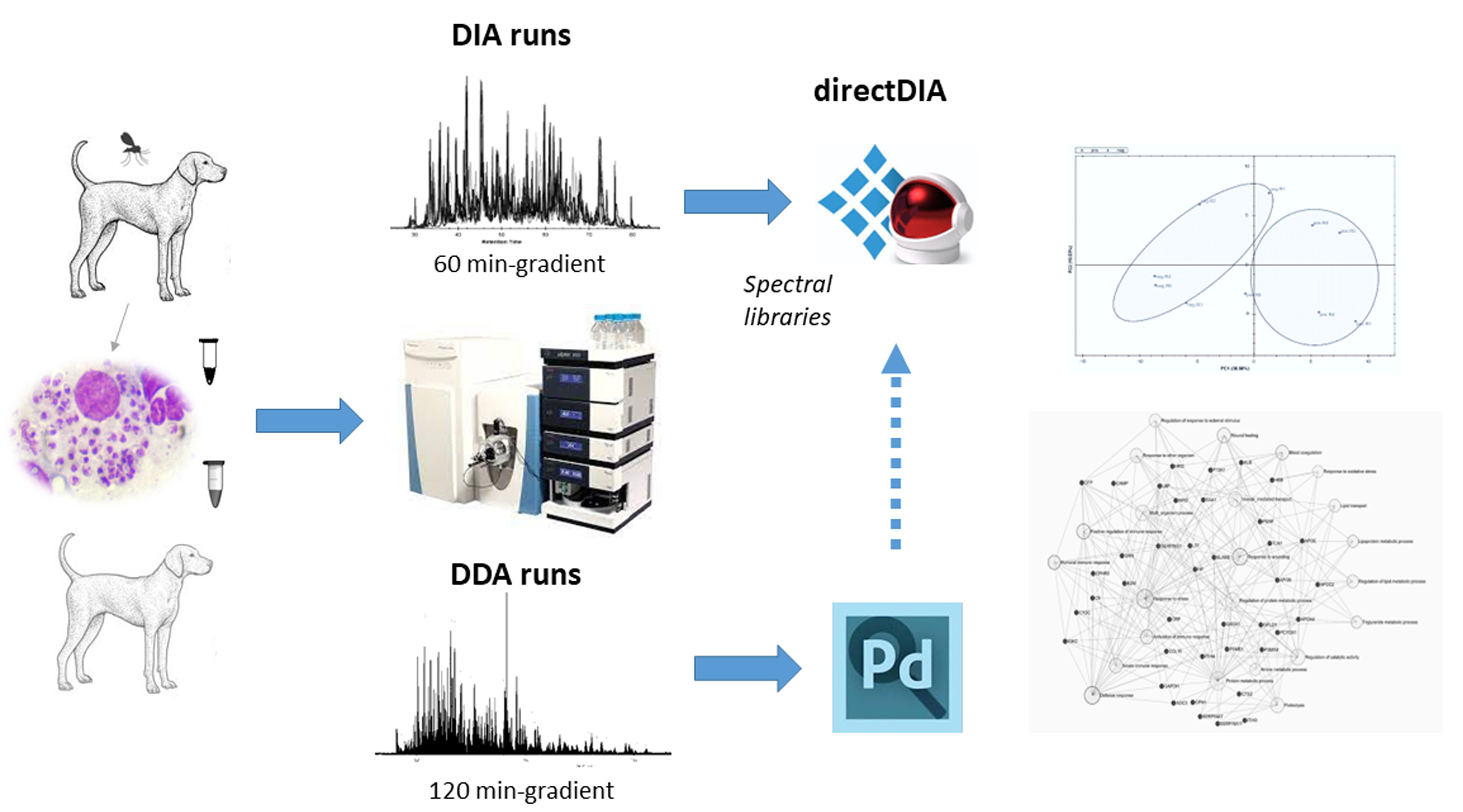
2.5. DIA Proteomic Analysis

For DIA analysis, a linear gradient 5–45% mobile phase B (0.1% formic acid in 80% ACN, v/v) over 60 min, at the flow rate of 300 nL/min was used. Mobile phase A contained 0.1% formic acid in water. The MS operated in positive ion mode using DIA Full MS method. MS spectra representing 44 overlapping sequential DIA windows with an isolation width of m/z 15 between m/z 450 and m/z 1100 were acquired using resolution of 35,000 and AGC target 1 × 10⁶, MSX 1, and charge state +2. For HCD, fragmentation collision energy 29% (NCE) with a resolution of 17,500 and AGC target of 2 × 10⁵ was used. The raw and analyzed DIA data files have been deposited to the ProteomeXchange Consortium via the PRIDE partner repository [17] with the dataset PXD039765.
Obtained data were analyzed using Spectronaut 16 (v. 16.2.220203.530000, Biognosys AG, Schlieren, Switzerland) [18]. Protein identification and quantification was performed using directDIA workflow. Identification was improved by combining of in-house generated spectral library and FASTA files (as described in Section 2.5). Factory settings were used to perform differential expression analysis between two different conditions, e.g., healthy and Leishmania-infected dogs and calculate adjusted p-values (q-values) based on multiple hypotheses testing corrections using Benjamini–Hochberg method. Proteins having adjusted p-value (q-value) < 0.05 were considered as significantly abundant. Principal component analysis (PCA) plot and volcano plot were generated in Spectronaut software. Gene ontology (GO) terms were enriched based on Welch’s t-test with p-value < 0.05 using bioinformatics web platform ExpressAnalyst (https://www.expressanalyst.ca/ (accessed on 30 November 2022)). For the network-based and enrichment analyses, Homo sapiens was selected as model organism.

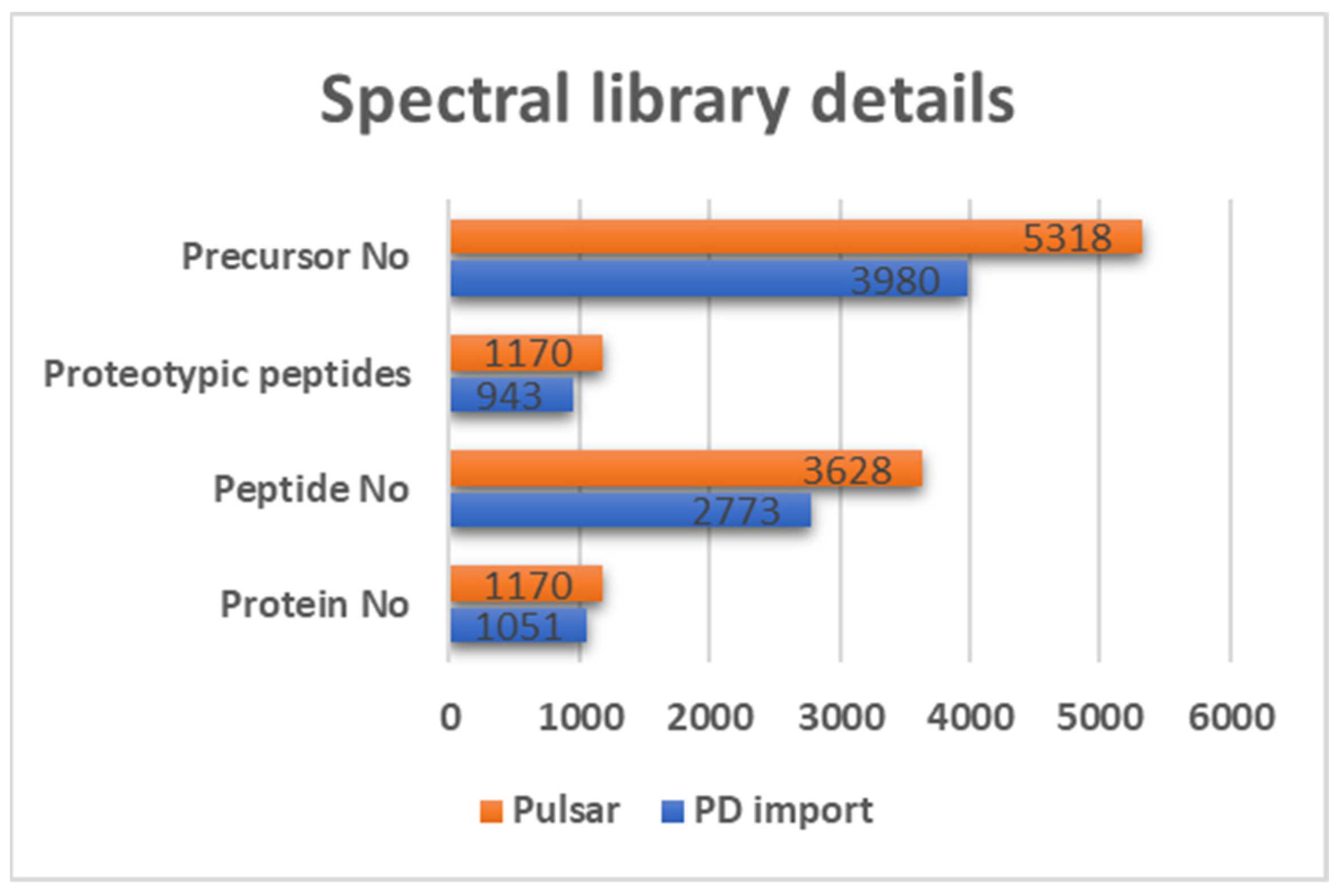
2.6. Spectral Library Generation

For spectral libraries generation, DDA raw files and belonging Proteome Discoverer search results of analyzed samples (namely pooled, mixed control, mixed case, ProteoMiner enriched, and 3 SCX fractions all containing iRT peptides for retention time normalization as described in Section 2.3) were imported into Spectronaut 16 software (v. 16.2.220203.530000, Biognosys AG, Schlieren, Switzerland) with default settings. Furthermore, to enable in silico spectral library generation using Canis lupus familiaris FASTA files downloaded from UniprotKB database 14/10/2021, 49,889 entries) combined with Biognosys iRT fusion peptide FASTA file (available at https://biognosys.com/shop/iRT-Kit (accessed on 1 September 2022)), Pulsar search engine with deep learning augmentation integrated into Spectronaut software was used. For libraries generation, following factory settings were used: Trypsin/P as the digestion enzyme with missed cleavages ≤2, and only peptides with length from 7 to 50 amino acids with mass ≤ 6000 Da were kept.

3. Results

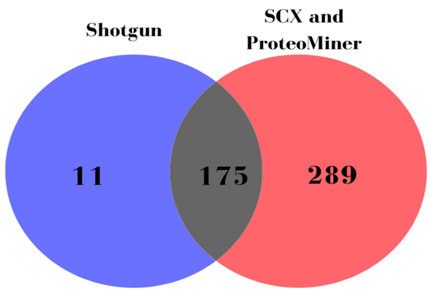
3.1. DDA-Based Proteomic Results

To generate high-quality sample-specific spectral library containing a large set of high confident peptides originating from both high and low abundant serum proteins, a pooled and ProteoMiner enriched samples as well as SCX fractions were analyzed by DDA-MS-based strategy. In this way, we identified 1405 proteins within 464 protein groups (464 master proteins), unlike 186 protein groups identified only by analyzing the pooled sample (shotgun approach without depletion and fractionation), as shown in Figure 1.

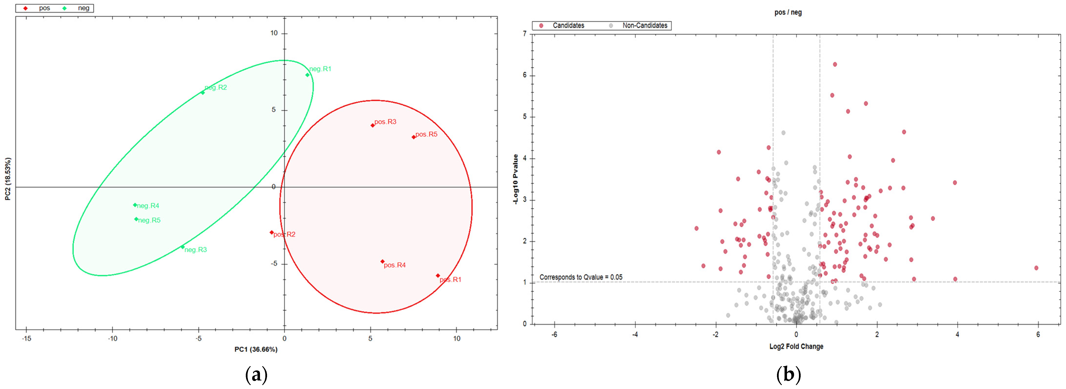
3.2. DIA-Based Proteomic Results

DIA-based proteomic analysis was employed to provide the deep insight into proteome-wide changes of host serum in canine leishmaniosis. For generating state-of-the-art libraries from both DDA data, Pulsar search integrated into Spectronaut software was used. The sample-specific spectral library (provided in Supplementary file S2) contained Canis lupus familiaris FASTA file enriched with peptides identified by DDA analysis, e.g., pooled, enriched and/or fractionated serum samples (Pulsar search). Spectral library details are shown in Figure 2.
Quantitative proteomic data of non-depleted serum samples were obtained using the SWATH-MS technology as described in Section 2.5. DIA proteomic analysis. As a result, we successfully identified a set of 554 serum proteins with a high quantitative performance. Furthermore, proteomic analysis with a wide dynamic range coverage revealed difference in the abundance of the 140 proteins with 107 being higher and 33 lower in abundance in canine leishmaniosis satisfying the cutoff criteria (q-value < 0.05 and absolute log2FC > 0.5). Belonging volcano plot is depicted in Figure 3b. The principal component analysis (PCA) plot shows differences in serum proteomes between healthy and Leishmania-infected dogs as in Figure 3a.
A shortened list containing differentially abundant proteins that change the most (the absolute value) and accompanying details, such as accession numbers and fold changes, is provided within the Table 1. The complete protein list can be found within Supplementary file S3.
Gene ontology (GO) analysis of differentially abundant proteins revealed key molecular functions and biological processes affected by Leishmania infection (Figure 4 and Figure 5). The GO term results (Figure 4) indicate that the proteins affected by Leishmania infection play roles in lipid transport (p-value 2.08 × 10⁻⁷) and cholesterol metabolism (p-value 8.3 × 10⁻³), blood coagulation (p-value 2.7 × 10⁻³), immune response (p-value 9.2 × 10⁻³), and receptor-mediated endocytosis (p-value 4.7 × 10⁻⁴), among others.

4. Discussion

Serum proteomics is a powerful tool for non-invasive monitoring of biological processes, enabling the understanding of various pathophysiological processes occurring in a living organism as well as biomarker discovery. However, although easily accessible, high abundant molecules, such as albumin (57–71%) or gamma-globulins (8–26%), as well as high dynamic range (12 orders of magnitude) molecules, have been making the serum a complex analyte for DDA bottom-up MS analysis, unlike our DIA-MS study where 554 proteins were identified in non-depleted sample. Although various depletion strategies of high abundant proteins have been introduced, serum albumin is a 66.5 kDa-protein known to act as a carrier for various endogenous and exogenous ligands [19] and other small proteins [20]. So, the removal of these large molecules often results in the loss of molecules of interest (in terms of quantity and/or quality) providing an incomplete picture. Aware of the depletion effect on the results, in our previous studies, we analyzed non-depleted serum samples in dogs with leishmaniosis for treatment monitoring using DDA tandem mass tag (TMT) label-based quantitative proteomic approach which enabled us the identification of 117 canine serum proteins among which 23 were differentially abundant [21]. However, based on the number of total identified proteins, the detection of low abundant proteins remained partially disabled, still giving the incomplete insight into the complex serum proteome. Despite DDA-MS analysis was the main choice in proteomics since its inception, DIA-based proteomics experiments are recently becoming the powerful alternative due to the fast and simplified sample preparation and significantly improved results in terms of reduced instrument time requirements and the increased number of identified proteins [22], which we also showed herein. Moreover, results addressing DIA-MS-based plasma proteomics in dogs have been reported confirming all abovementioned methodological advantages [23]. However, DIA has not yet been applied in the analysis of sera in canine leishmaniosis. Herein, we aimed to implement DIA serum proteomics workflow using next generation proteomic technology to better understand the host-specific processes during Leishmania infection in dogs.
For in-depth DIA-MS proteome analysis, we established high-quality sample specific spectral library containing extensive set of 3628 peptides and their relative retention time details obtained by spiked-in non-naturally occurring synthetic peptides for retention time normalization [7] to enable proteome profiling coverage. Having in mind that the spectral library should be sufficiently large, sample-size comparable, and that the results are library (size and fragmentation pattern) dependent [24], we chose the directDIA workflow based on in silico database search (canine FASTA files) enriched with Pulsar search results of our DDA data for the best result outputs. In this way, using SWATH methodology, we identified 554 serum proteins using 60-min gradient time, which is about 4.5 times more protein than in our previous study [8] or about 2.5 times more proteins than performing the DDA without depletion (120-min gradient time). This enabled detailed qualitative and quantitative insights into the serum proteomes of healthy and Leishmania-infected dogs, including both high and low abundant proteins with simplified sample preparation and shorter analysis time. Although the DIA database search was performed, Leishmania proteins were not found (data not shown). It is important to emphasize that some of the leishmaniosis-related serum proteins (such as prosaposin) identified herein by simple sample preparation (in-solution digestion) and SWATH-MS proteomic approach were previously identified only after laborious sample preparation (exosome isolation) and subsequent proteomic analysis [25]. Finally, GO analysis of differentially abundant proteins provided us key molecules and related molecular processes observable in serum affected by Leishmania infection in dogs. Those include lipid metabolism, hematological abnormalities, immune response, oxidative stress, etc.

4.1. Lipid Metabolism and Transport

Leishmania infantum is an intracellular parasite which relies on host lipid reservoir and synthesis mechanism for its survival [26]. Various studies related to Leishmania infection reported the multiple importance of cholesterol and cholesterol metabolic processes in Leishmania transformation process and sterol metabolism [27]. The cholesterol sequestration by Leishmania parasite begins post infection, with host cells aiming to restore initial cholesterol levels correlating with the upregulation of proteins required for cholesterol metabolism [28]. Accordingly, in our study, cholesterol metabolic processes are shown to be affected by Leishmania infection, with proteins Apo E and Apo A4 being upregulated. The elevated levels of Apo E have already been reported in serum of Leishmania-infected human patients [29]. Apo E (apolipoprotein E) is a 34 kDa glycoprotein critical for cholesterol transport and lipoprotein particle metabolism, playing an important role in lipolytic enzyme activation and immune response [30]. The study involving apolipoprotein E knockout mice infected with Leishmania donovani showed the important role of Apo E in protection against visceral leishmaniosis by displaying hypercholesterolemia, host-protective cytokines, and expansion of antileishmanial CD8 + IFN-γ + and CD8 + IFN-γ +TNF-α + T cells [31]. Apolipoprotein A4 (APOA4) is a plasma lipoprotein involved in the regulation of lipid and glucose metabolism [32], and enhances triglyceride (TG) secretion from the liver [33]. Upregulation of APOA4 protein shown herein could be related with already reported increase in triglyceride levels in patients with visceral leishmaniosis [34].
Except cholesterol, the studies have been published indicating that Leishmania sp. scavenges host sphingolipids (e.g., sphingomyelin, which is not synthesized by Leishmania but is abundant in mammals) needed for amastigote proliferation, virulence and biosynthesis of inositol phosphorylceramide (IPC, major sphingolipid in the genus Leishmania not present in mammals) [35]. In our study, we showed the upregulation of prosaposin, PSAP, which is a large precursor protein that is proteolytically cleaved into four sphingolipid activator proteins that assist in the lysosomal hydrolysis of sphingolipids into ceramide needed for the synthesis of IPC [36,37]. Our results are in accordance with existing data showing the Leishmania infection induces the host ceramide synthesis pathway [38].

4.2. Hematological Abnormalities

The progression of the canine leishmaniosis has the effect on hematological parameters, mostly caused by anemia and/or platelet aggregation abnormalities [39]. Our results are supporting these findings. Anemia is present in the majority of symptomatic dogs with leishmaniosis [39]. In our study, we detected the decreased amount of carbonic anhydrase 1 (CA1). CA1 is the most abundant protein in erythrocytes after hemoglobin. Its expression increases in various forms of anemia. However, its expression decreases in hemolytic anemia and for that reason CA1 is used as biomarker for this disease [40].
Coagulation factors, being a part of coagulation cascade, are essential for normal blood clotting. Herein, we determined the relative coagulation factor (F11, F13A1, F13B) deficiency, as well as histidine-rich glycoprotein (HRG) deficiency in Leishmania-infected dogs compared to healthy dogs that is related to decreased coagulation factor activities and fibrinolysis as reported in human patients [41,42,43]. Furthermore, we also found platelet-activating factor (PAF) acetylhydrolase (PLA-AH2) to be upregulated in Leishmania-infected dogs. Platelet-activating factor is a phospholipid that is involved in the activation of thrombotic cascades, induces leukopenia and thrombocytopenia (which are the most common hematological symptoms in leishmaniosis), as well as increases vascular permeability [44]. It is also involved in inflammatory reactions. PAF acetylhydrolase 2, also known as lipoprotein-associated phospholipase A2 (Lp-PLA2), inactivates the PAF and PAF-like oxidized phospholipids found in oxidized LDL by hydrolysis, controlling their actions. The increase in PLA-AH2 activity is positively correlated with activation of inflammatory cells. PAF was reported essential for the control of Leishmania infection. [45].

4.3. Immune Response

The immune response is a coordinated response of variety of cells and molecules on exposure to foreign antigens. Acute phase proteins (APPs) are the markers of the immune system response whose concentration in serum significantly changes during inflammation, which was also found in our study. The APP profiles and concentrations in combination with animal clinical condition monitored at physical examination may provide a valuable tool for the characterization, management and treatment monitoring of canine leishmaniosis [46]. Canine leishmaniosis is related with the increase of haptoglobin (HB) and C-reactive protein (CRP, or pentraxin) concentration that is measured in clinical practice [47]. Except increase in concentration of HB, CRP, and alpha 2 macroglobulin (A2M) in serum of Leishmania-infected dogs, we also detected negative APPs, transthyretin and retinol-binding protein, showing the decrease in abundance in Leishmania-infected dogs compared to healthy dogs. Interestingly, alpha 1 antitrypsin (ATT), a positive APP, was found to be lowered in Leishmania-infected dogs. Alpha-1 antitrypsin is a protease inhibitor synthesized mainly in the liver. Diffusing into the lung tissue and alveolar fluid, it inactivates neutrophil elastase and protects the lung tissue from the damage. The reduction of circulating levels of alpha-1 antitrypsin result in the increased risk of lung disease [48]. Accordingly, the pulmonary alterations and the inflammatory process defined as interstitial pneumonitis were reported in Leishmania-infected dogs, as well as the presence of amastigotes in canine lungs, which could support our findings [49].
Except the APPS, within the immune response, lysosomal cysteine cathepsin family of proteases, such as cathepsin S (CTSS), play a key role. CTSS, involved in TLR9 processing [50], is involved in dendritic cells activation and resolution of Leishmania major infection in mice [51]. Also, cathepsin S deficient mice are slightly more susceptible against Leishmania major infection [52]. According to those facts, animals with elevated cathepsin S in serum should be resistant against infection with Leishmania, which is not the case in our work. In contrast, in our survey, the amount of cathepsin S was three-fold elevated in positive dogs. As elevated cathepsin S is proven in many human diseases [53,54], a more detailed investigation should be made to show if the cathepsin S is a good marker for the disease diagnostics and prognosis.
Although the DIA-SWATH-MS strategy applied herein generated a long list of deregulated proteins, some of which present biomarker potential, further investigation using a larger population of healthy and Leishmania-infected canine patients is needed to determine the diagnostic potential of a single biomarker or biomarker panels, correlating the clinical symptoms and protein concentration. Additionally, it would be interesting to use a DIA-MS approach for canine leishmaniosis treatment monitoring.

5. Conclusions

Our study demonstrates the advantages of next generation DIA-MS proteomic analysis of serum, enabling deeper insights into altered expression levels of both high and low abundant proteins without enrichment in shorter analysis times. This approach was shown to be extremely useful and informative in the identification of disease-related proteins, their functions, and molecular pathways in canine leishmaniosis. Additionally, our open access canine serum spectral libraries could be useful for in silico generated combined spectral libraries to be applied in various canine serum studies. Finally, this approach may be employed to decipher the pathological processes of various diseases in serum, as well as serum biomarker development.

Supplementary Materials

The following supporting information can be downloaded at: https://www.mdpi.com/article/10.3390/metabo13030365/s1, Supplementary file S1. Diagnostic methods for determination of Leishmania infection in selected canine serum samples. The infection of Leishmania infantum was diagnosed using three serological tests: indirect fluorescence antibody test (IFAT), enzyme-linked immunosorbent assay (ELISA) and kinesin-related conserved recombinant antigen (rK39 rapid immunochromatographic test evaluating the presence of anti-Leishmania antibody, Supplementary file S2. Spectral libraries generated by Spectronaut 16 Pulsar database search and sample-specific library generated by Proteome Discoverer import in Spectronaut 16. Supplementary file S3. Differentially abundant proteins detected in serum of Leishmania-infected dogs compared to healthy dogs by employing DIA-MS.

Author Contributions

Conceptualization A.H.; methodology, F.M., M.P., O.S. and A.H.; software: A.H. formal analysis, F.M., A.H., O.S. and M.P.; investigation, F.M., A.H. and O.S.; resources, V.M., P.D.E. and F.M.; data curation, A.H. and P.D.E.; writing—original draft preparation, A.H.; writing—review and editing, all; visualization, A.H.; supervision, A.H.; project administration, V.M. and P.D.E.; funding acquisition, V.M., F.M. and P.D.E. All authors have read and agreed to the published version of the manuscript.

Funding

V.M., P.D.E. and A.H. were funded by European Commission FP7 “VetMedZg” project (Grant Number 621394) and European Regional Development Fund (Grant Agreement KK.01.1.1.04.0086).

Institutional Review Board Statement

The use of the archive samples (previously used in routine leishmaniosis diagnostics) was approved by the Ethical Committee at the Faculty of Veterinary Medicine, University of Zagreb, Croatia (No. 640-01/23-17/22; Record No. 251-61-12/384-23-01, Date: 07/02/2023.). All procedures performed in accordance with the Directive 2010/63/EU of the European Parliament and of the Council on the protection of animals used for scientific purposes.

Informed Consent Statement

Not applicable.

Data Availability Statement

The DDA mass spectrometry proteomics data have been deposited to the Consortium via the PRIDE partner repository with the dataset PXD039763. The DIA-MS data files have been deposited to the ProteomeXchange Consortium via the PRIDE partner repository with the dataset PXD039765.

Conflicts of Interest

The authors declare no conflict of interest.

List of Abbreviations

A2Malpha 2 macroglobulin
ACNacetonitrile
AGCautomatic gain control
Apo A4apolipoprotein A4
Apo E apolipoprotein E
APPacute phase protein
ATTalpha 1 antitrypsin
BCAbicinchoninic acid
BPbiological process
CA1carbonic anhydrase 1
CD8+ T cellscytotoxic T lymphocytes
CRPC-reactive protein, or pentraxin
CTSScathepsin S
DDAdata-dependent acquisition
DIA-MSdata-independent acquisition mass spectrometry
DTTdithiothreitol
ELISAenzyme-linked immunosorbent assay
FCfold change
FDRfalse discovery rate
GOgene ontology
HBhaptoglobin
HCDhigher energy collisional dissociation
HRGhistidine-rich glycoprotein
IAAiodoacetamide
IFATindirect fluorescence antibody test
IFN-γ interferon gamma
IPCinositol phosphorylceramide
iRTindexed retention time
LC-MS/MSliquid chromatography tandem mass spectrometry
LDLlow density lipoprotein
Lp-PLA2lipoprotein-associated phospholipase A2
MFmolecular function
MWCOmoleculat weight cut off
NCEnormalized collision energy
PAFplatelet-activating factor
PCAprincipal component analysis
PSAPprosaposin
rK39kinesin-related conserved recombinant antigen
SCXstrong cation exchange
SWATHsequential window acquisition of all theoretical mass spectra
TGtriglyceride
TLR9toll-like receptor 9
TMTtandem mass tag
TNF-αtumor necrosis factor alpha

References

  1. Geyer, P.E.; Holdt, L.M.; Teupser, D.; Mann, M. Revisiting biomarker discovery by plasma proteomics. Mol. Syst. Biol. 2017, 13, 942. [Google Scholar] [CrossRef]
  2. Fürsch, J.; Kammer, K.-M.; Kreft, S.G.; Beck, M.; Stengel, F. Proteome-wide structural probing of low-abundant protein interactions by crosslinking mass spectrometry. Anal. Chem. 2020, 92, 4016–4022. [Google Scholar] [CrossRef]
  3. Anderson, N.L.; Anderson, N.G. The human plasma proteome: History, character, and diagnostic prospects. Mol. Cell. Proteom. 2002, 1, 845–867. [Google Scholar] [CrossRef] [Green Version]
  4. Paul, J.; Veenstra, T.D. Separation of Serum and Plasma Proteins for In-Depth Proteomic Analysis. Separations 2022, 9, 89. [Google Scholar] [CrossRef]
  5. Košiček, M.; Gudelj, I.; Horvatić, A.; Jović, T.; Vučković, F.; Lauc, G.; Hećimović, S.; Kosicek, M.; Gudelj, I.; Horvatic, A.; et al. N-glycome of the lysosomal glycocalyx is altered in Niemann-Pick Type C disease model cells. Mol. Cell. Proteom. 2018, 17, 631–642. [Google Scholar] [CrossRef] [PubMed] [Green Version]
  6. Gillet, L.C.; Navarro, P.; Tate, S.; Röst, H.; Selevsek, N.; Reiter, L.; Bonner, R.; Aebersold, R. Targeted data extraction of the MS/MS spectra generated by data-independent acquisition: A new concept for consistent and accurate proteome analysis. Mol. Cell. Proteom. 2012, 11, O111.016717. [Google Scholar] [CrossRef] [Green Version]
  7. Escher, C.; Reiter, L.; MacLean, B.; Ossola, R.; Herzog, F.; Chilton, J.; MacCoss, M.J.; Rinner, O. Using iRT, a normalized retention time for more targeted measurement of peptides. Proteom. 2012, 12, 1111–1121. [Google Scholar] [CrossRef] [PubMed] [Green Version]
  8. Yang, Y.; Liu, X.; Shen, C.; Lin, Y.; Yang, P.; Qiao, L. In silico spectral libraries by deep learning facilitate data-independent acquisition proteomics. Nat. Commun. 2020, 11, 146. [Google Scholar] [CrossRef] [Green Version]
  9. Zhu, T.; Zhu, Y.; Xuan, Y.; Gao, H.; Cai, X.; Piersma, S.R.; Pham, T.V.; Schelfhorst, T.; Haas, R.R.G.D.; Bijnsdorp, I.V.; et al. DPHL: A DIA Pan-human Protein Mass Spectrometry Library for Robust Biomarker Discovery. Genom. Proteom. Bioinform. 2020, 18, 104–119. [Google Scholar] [CrossRef]
  10. Scarpini, S.; Dondi, A.; Totaro, C.; Biagi, C.; Melchionda, F.; Zama, D.; Pierantoni, L.; Gennari, M.; Campagna, C.; Prete, A.; et al. Visceral Leishmaniasis: Epidemiology, Diagnosis, and Treatment Regimens in Different Geographical Areas with a Focus on Pediatrics. Microorganisms 2022, 10, 1887. [Google Scholar] [CrossRef]
  11. Bosnić, S.; Gradoni, L.; Khoury, C.; Maroli, M. A review of leishmaniasis in Dalmatia (Croatia) and results from recent surveys on phlebotomine sandflies in three southern counties. Acta Trop. 2006, 99, 42–49. [Google Scholar] [CrossRef]
  12. SUNDAR, S.; SINGH, A. Chemotherapeutics of visceral leishmaniasis: Present and future developments. Parasitology 2018, 145, 481–489. [Google Scholar] [CrossRef]
  13. Abass, E.; Kang, C.; Martinkovic, F.; Semião-Santos, S.J.; Sundar, S.; Walden, P.; Piarroux, R.; el Harith, A.; Lohoff, M.; Steinhoff, U. Heterogeneity of Leishmania donovani Parasites Complicates Diagnosis of Visceral Leishmaniasis: Comparison of Different Serological Tests in Three Endemic Regions. PLoS ONE 2015, 10, e0116408. [Google Scholar] [CrossRef] [PubMed] [Green Version]
  14. Horvatic, A.; Guillemin, N.; Kaab, H.; McKeegan, D.; O’Reilly, E.; Bain, M.; Kules, J.; Eckersall, P.D. Quantitative proteomics using tandem mass tags in relation to the acute phase protein response in chicken challenged with Escherichia coli lipopolysaccharide endotoxin. J. Proteom. 2019, 192, 64–77. [Google Scholar] [CrossRef] [PubMed]
  15. Rappsilber, J.; Mann, M.; Ishihama, Y. Protocol for micro-purification, enrichment, pre-fractionation and storage of peptides for proteomics using StageTips. Nat. Protoc. 2007, 2, 1896–1906. [Google Scholar] [CrossRef] [PubMed]
  16. Li, L. Dynamic Range Compression with ProteoMinerTM: Principles and Examples BT. In Proteomic Profiling: Methods and Protocols; Posch, A., Ed.; Springer: New York, NY, USA, 2015; pp. 99–107. ISBN 978-1-4939-2550-6. [Google Scholar]
  17. Perez-Riverol, Y.; Csordas, A.; Bai, J.; Bernal-Llinares, M.; Hewapathirana, S.; Kundu, D.J.; Inuganti, A.; Griss, J.; Mayer, G.; Eisenacher, M.; et al. The PRIDE database and related tools and resources in 2019: Improving support for quantification data. Nucleic Acids Res. 2019, 47, D442–D450. [Google Scholar] [CrossRef] [PubMed]
  18. Bruderer, R.; Bernhardt, O.M.; Gandhi, T.; Miladinović, S.M.; Cheng, L.-Y.; Messner, S.; Ehrenberger, T.; Zanotelli, V.; Butscheid, Y.; Escher, C.; et al. Extending the limits of quantitative proteome profiling with data-independent acquisition and application to acetaminophen-treated three-dimensional liver microtissues. Mol. Cell. Proteom. 2015, 14, 1400–1410. [Google Scholar] [CrossRef] [Green Version]
  19. Belinskaia, D.A.; Voronina, P.A.; Shmurak, V.I.; Jenkins, R.O.; Goncharov, N. V Serum Albumin in Health and Disease: Esterase, Antioxidant, Transporting and Signaling Properties. Int. J. Mol. Sci. 2021, 22, 10318. [Google Scholar] [CrossRef]
  20. Merrell, K.; Southwick, K.; Graves, S.W.; Esplin, M.S.; Lewis, N.E.; Thulin, C.D. Analysis of low-abundance, low-molecular-weight serum proteins using mass spectrometry. J. Biomol. Tech. 2004, 15, 238–248. [Google Scholar] [PubMed]
  21. Martinez-Subiela, S.; Horvatic, A.; Escribano, D.; Pardo-Marin, L.; Kocaturk, M.; Mrljak, V.; Burchmore, R.; Ceron, J.J.; Yilmaz, Z. Identification of novel biomarkers for treatment monitoring in canine leishmaniosis by high-resolution quantitative proteomic analysis. Vet. Immunol. Immunopathol. 2017, 191, 60–67. [Google Scholar] [CrossRef]
  22. Lin, L.; Zheng, J.; Yu, Q.; Chen, W.; Xing, J.; Chen, C.; Tian, R. High throughput and accurate serum proteome profiling by integrated sample preparation technology and single-run data independent mass spectrometry analysis. J. Proteom. 2018, 174, 9–16. [Google Scholar] [CrossRef] [PubMed]
  23. Ravuri, H.G.; Noor, Z.; Mills, P.C.; Satake, N.; Sadowski, P. Data-Independent Acquisition Enables Robust Quantification of 400 Proteins in Non-Depleted Canine Plasma. Proteomes 2022, 10, 9. [Google Scholar] [CrossRef]
  24. Siyal, A.A.; Chen, E.S.-W.; Chan, H.-J.; Kitata, R.B.; Yang, J.-C.; Tu, H.-L.; Chen, Y.-J. Sample Size-Comparable Spectral Library Enhances Data-Independent Acquisition-Based Proteome Coverage of Low-Input Cells. Anal. Chem. 2021, 93, 17003–17011. [Google Scholar] [CrossRef]
  25. Keller, M.M.; Kelly, P.H.; Pope, R.M.; Thery, F.; Wilson, M.E. Proteomic analysis of exosomes released from Leishmania infected macrophages. J. Immunol. 2020, 204, 156.19. [Google Scholar] [CrossRef]
  26. Martínez, C.R.; Ruiz, C.J. Alterations in Host Lipid Metabolism Produced During Visceral Leishmaniasis Infections. Curr. Trop. Med. Rep. 2019, 6, 250–255. [Google Scholar] [CrossRef]
  27. Bouazizi-Ben Messaoud, H.; Guichard, M.; Lawton, P.; Delton, I.; Azzouz-Maache, S. Changes in Lipid and Fatty Acid Composition During Intramacrophagic Transformation of Leishmania donovani Complex Promastigotes into Amastigotes. Lipids 2017, 52, 433–441. [Google Scholar] [CrossRef] [PubMed] [Green Version]
  28. Semini, G.; Paape, D.; Paterou, A.; Schroeder, J.; Barrios-Llerena, M.; Aebischer, T. Changes to cholesterol trafficking in macrophages by Leishmania parasites infection. Microbiologyopen 2017, 6, e00469. [Google Scholar] [CrossRef]
  29. Liberopoulos, E.N.; Apostolou, F.; Gazi, I.F.; Kostara, C.; Bairaktari, E.T.; Tselepis, A.D.; Elisaf, M. Visceral leishmaniasis is associated with marked changes in serum lipid profile. Eur. J. Clin. Investig. 2014, 44, 719–727. [Google Scholar] [CrossRef]
  30. Woods, A.G.; Sokolowska, I.; Taurines, R.; Gerlach, M.; Dudley, E.; Thome, J.; Darie, C.C. Potential biomarkers in psychiatry: Focus on the cholesterol system. J. Cell. Mol. Med. 2012, 16, 1184–1195. [Google Scholar] [CrossRef]
  31. Ghosh, J.; Das, S.; Guha, R.; Ghosh, D.; Naskar, K.; Das, A.; Roy, S. Hyperlipidemia offers protection against Leishmania donovani infection: Role of membrane cholesterol. J. Lipid Res. 2012, 53, 2560–2572. [Google Scholar] [CrossRef] [Green Version]
  32. Wang, Z.; Wang, L.; Zhang, Z.; Feng, L.; Song, X.; Wu, J. Apolipoprotein A-IV involves in glucose and lipid metabolism of rat. Nutr. Metab. 2019, 16, 41. [Google Scholar] [CrossRef] [PubMed]
  33. VerHague, M.A.; Cheng, D.; Weinberg, R.B.; Shelness, G.S. Apolipoprotein A-IV expression in mouse liver enhances triglyceride secretion and reduces hepatic lipid content by promoting very low density lipoprotein particle expansion. Arterioscler. Thromb. Vasc. Biol. 2013, 33, 2501–2508. [Google Scholar] [CrossRef] [Green Version]
  34. Tsimihodimos, V.; Kei, A.; Apostolou, F.; Elisaf, M. Diagnostic lipid changes in patients with visceral leishmaniasis. Hosp. Pract. 2018, 46, 229–232. [Google Scholar] [CrossRef] [PubMed]
  35. Zhang, O.; Wilson, M.C.; Xu, W.; Hsu, F.-F.; Turk, J.; Kuhlmann, F.M.; Wang, Y.; Soong, L.; Key, P.; Beverley, S.M.; et al. Degradation of host sphingomyelin is essential for Leishmania virulence. PLoS Pathog. 2009, 5, e1000692. [Google Scholar] [CrossRef] [PubMed] [Green Version]
  36. Doering, T.; Holleran, W.M.; Potratz, A.; Vielhaber, G.; Elias, P.M.; Suzuki, K.; Sandhoff, K. Sphingolipid Activator Proteins Are Required for Epidermal Permeability Barrier Formation. J. Biol. Chem. 1999, 274, 11038–11045. [Google Scholar] [CrossRef] [Green Version]
  37. Kagan, T.; Stoyanova, G.; Lockshin, R.A.; Zakeri, Z. Ceramide from sphingomyelin hydrolysis induces neuronal differentiation, whereas de novo ceramide synthesis and sphingomyelin hydrolysis initiate apoptosis after NGF withdrawal in PC12 Cells. Cell Commun. Signal. 2022, 20, 15. [Google Scholar] [CrossRef]
  38. Ghosh, S.; Bhattacharyya, S.; Das, S.; Raha, S.; Maulik, N.; Das, D.K.; Roy, S.; Majumdar, S. Generation of ceramide in murine macrophages infected with Leishmania donovani alters macrophage signaling events and aids intracellular parasitic survival. Mol. Cell. Biochem. 2001, 223, 47–60. [Google Scholar] [CrossRef]
  39. Almeida, V.; Lima, I.; Fraga, D.; Carrillo, E.; Moreno, J.; Dos-Santos, W.L.C. Hematological Changes in Dogs with Visceral Leishmaniasis Are Associated with Increased IFN-γ and TNF Gene Expression Levels in the Bone Marrow. Microorganisms 2021, 9, 1618. [Google Scholar] [CrossRef]
  40. Menteşe, A.; Erkut, N.; Sümer, A.; Us Altay, D.; Alver, A.; Sönmez, M. Anti-carbonic anhydrase antibodies in iron deficiency anemia. Hematology 2015, 20, 363–367. [Google Scholar] [CrossRef]
  41. Shiferaw, E.; Murad, F.; Tigabie, M.; Abebaw, M.; Alemu, T.; Abate, S.; Mohammed, R.; Yeshanew, A.; Tajebe, F. Hematological profiles of visceral leishmaniasis patients before and after treatment of anti-leishmanial drugs at University of Gondar Hospital; Leishmania Research and Treatment Center Northwest, Ethiopia. BMC Infect. Dis. 2021, 21, 1005. [Google Scholar] [CrossRef]
  42. Wakabayashi Takehiko, S.K. Histidine-Rich Glycoprotein: A Possible Modulator of Coagulation and Fibrinolysis. Semin. Thromb. Hemost. 2011, 37, 389–394. [Google Scholar] [CrossRef] [PubMed]
  43. Lekic, N.; Tadic, B.; Djordjevic, V.; Basaric, D.; Micev, M.; Vucelic, D.; Mitrovic, M.; Grubor, N. Splenectomy for Visceral Leishmaniasis Out of an Endemic Region: A Case Report and Literature Review. Medicina (B. Aires) 2022, 58, 184. [Google Scholar] [CrossRef] [PubMed]
  44. Stafforini, D.M.; McIntyre, T.M.; Prescott, S.M. Platelet-activating factor acetylhydrolase from human plasma. In Arachidonate Related Lipid Mediators; Murphy, R.C., Fitzpatrick, F.A.B.T.-M., Eds.; Academic Press: Cambridge, MA, USA, 1990; Volume 187, pp. 344–357. ISBN 0076-6879. [Google Scholar]
  45. Lonardoni, M.V.; Russo, M.; Jancar, S. Essential role of platelet-activating factor in control of Leishmania (Leishmania) amazonensis infection. Infect. Immun. 2000, 68, 6355–6361. [Google Scholar] [CrossRef] [PubMed]
  46. Ceron, J.J.; Pardo-Marin, L.; Caldin, M.; Furlanello, T.; Solano-Gallego, L.; Tecles, F.; Bernal, L.; Baneth, G.; Martinez-Subiela, S. Use of acute phase proteins for the clinical assessment and management of canine leishmaniosis: General recommendations. BMC Vet. Res. 2018, 14, 196. [Google Scholar] [CrossRef]
  47. Martínez-Subiela, S.; Tecles, F.; Eckersall, P.D.; Cerón, J.J. Serum concentrations of acute phase proteins in dogs with leishmaniasis. Vet. Rec. 2002, 150, 241–244. [Google Scholar] [CrossRef]
  48. Brode, S.K.; Ling, S.C.; Chapman, K.R. Alpha-1 antitrypsin deficiency: A commonly overlooked cause of lung disease. C. Can. Med. Assoc. J. 2012, 184, 1365–1371. [Google Scholar] [CrossRef] [Green Version]
  49. Kavarnos, I.; Pardali, D.; Brellou, G.D.; Papadopoulos, E.; Kritsepi-Konstantinou, M.; Adamama-Moraitou, K.K. Bronchoscopy and Lung Fine-Needle Aspiration for Antemortem Evaluation of Pulmonary Involvement in Dogs with Naturally Occurring Canine Leishmaniosis. Pathogens 2022, 11, 365. [Google Scholar] [CrossRef] [PubMed]
  50. Sepulveda, F.E.; Maschalidi, S.; Colisson, R.; Heslop, L.; Ghirelli, C.; Sakka, E.; Lennon-Duménil, A.-M.; Amigorena, S.; Cabanie, L.; Manoury, B. Critical role for asparagine endopeptidase in endocytic Toll-like receptor signaling in dendritic cells. Immunity 2009, 31, 737–748. [Google Scholar] [CrossRef] [Green Version]
  51. Abou Fakher, F.H.; Rachinel, N.; Klimczak, M.; Louis, J.; Doyen, N. TLR9-dependent activation of dendritic cells by DNA from Leishmania major favors Th1 cell development and the resolution of lesions. J. Immunol. 2009, 182, 1386–1396. [Google Scholar] [CrossRef] [Green Version]
  52. Rasid, O.; Mériaux, V.; Khan, E.M.; Borde, C.; Ciulean, I.S.; Fitting, C.; Manoury, B.; Cavaillon, J.-M.; Doyen, N. Cathepsin B-Deficient Mice Resolve Leishmania major Inflammation Faster in a T Cell-Dependent Manner. PLoS Negl. Trop. Dis. 2016, 10, e0004716. [Google Scholar] [CrossRef] [Green Version]
  53. Wilkinson, R.D.A.; Williams, R.; Scott, C.J.; Burden, R.E. Cathepsin S: Therapeutic, diagnostic, and prognostic potential. Biol. Chem. 2015, 396, 867–882. [Google Scholar] [CrossRef] [PubMed]
  54. Liu, J.; Ma, L.; Yang, J.; Ren, A.; Sun, Z.; Yan, G.; Sun, J.; Fu, H.; Xu, W.; Hu, C.; et al. Increased serum cathepsin S in patients with atherosclerosis and diabetes. Atherosclerosis 2006, 186, 411–419. [Google Scholar] [CrossRef] [PubMed]
Figure 1. DDA-MS analysis reveals increase in number of identified serum protein groups (master proteins) after enrichment and fractionation (ProteoMiner and SCX fractions) compared to non-depleted sample without fractionation (Shotgun).
Metabolites 13 00365 g001
Figure 2. Spectral libraries generated by Spectronaut 16 Pulsar database search (in orange) and sample-specific library generated by Proteome Discoverer import (in blue). Both spectral libraries are provided through the Supplementary file S2.
Metabolites 13 00365 g002
Figure 3. (a) Principal component analysis (PCA) score plot of the second and first principal components (PC1 vs PC2) reveals the differences in serum proteomes from healthy (neg, in green) and Leishmania-infected dogs (pos, in red). (b) Volcano plot of DIA data depicted with the fold change of each differentially abundant protein and belonging q-value. Significantly differentially abundant proteins (q < 0.05) are highlighted in red (marked as Candidates).
Metabolites 13 00365 g003
Figure 4. Ridgeline diagram of enriched GO (database PANTHER: BP) showing the biological processes and log2FC of belonging deregulated proteins (red dots—up; blue dots—down) in serum of dogs with leishmaniosis. The shade of blue represents p-values as depicted. BP in grey is not significant. The GO diagram is created using ExpressAnalyst (https://www.expressanalyst.ca/ (accessed on 30 November 2022)) based on Welch’s t-test.
Metabolites 13 00365 g004
Figure 5. Bipartite network showing the results of enrichment analysis of deregulated genes (GO:MF, based on adjusted p-values ranked by Welch’s test) in serum of Leishmania-infected dogs compared to healthy ones performed using ExpressAnalyst (https://www.expressanalyst.ca/ (accessed on 30 November 2022)). Blue dots represent proteins, while yellow/red circles mark molecular functions affected by canine leishmaniosis.
Metabolites 13 00365 g005
Table 1. The shortened list of differentially abundant proteins in Leishmania-infected vs healthy dogs obtained by directDIA MS analysis in Spectronaut 16 software.
Uniprot Accession IDProtein NameGene NameLog2FCq-Value *
F1PP29Uncharacterized proteinND **−2.600.00423
A0A5F4CGT3;F1PWW0Filamin AFLNA−2.420.02513
Q6JDI3Plasminogen (Fragment)PLG−1.900.00002
F1PBK6Carbonic anhydraseCA1−1.880.01058
E2R5U8TransthyretinTTR−1.560.03280
E2R1N7Prenylcysteine oxidase 1PCYOX1−1.510.00106
F1PCG4Peroxiredoxin 2PRDX2−1.490.00102
E2RT38;F1PAQ3Maltase-glucoamylaseMGAM−1.470.00048
A0A5F4D5S2;J9PAD1C4a anaphylatoxinLOC481722−1.410.00000
F1PZC6Histidine rich glycoproteinHRG−1.140.00139
A0A5F4C5S3;F1PNR5Coagulation factor XIF11−1.050.00722
E2RA67C-C motif chemokineCCL14−0.960.00516
F1PD34Secreted phosphoprotein 2SPP2−0.930.00371
A1ILJ0Alpha 1 antitrypsinSERPINA1−0.920.01290
A0A5F4CEP4Fetuin BFETUB−0.900.00197
A0A5F4D9Z3;F1PB85Serpin family A member 7SERPINA7−0.880.03724
D2K841;J9P346C-X-C motif chemokine;PPBP−0.870.00888
E2RPW3ParaoxonasePON3−0.850.00029
A0A5F4BY85;A0A5F4CHL2; E2RK02Glycosyl-phosphatidylinositol-specific phospholipase DGPLD1−0.840.00129
A0A5F4CZV0;F2Z4P4;J9P9I7 Elongation factor 1-alphaLOC102155289−0.830.02608
A0A5F4DCK5;F1PKX3Coagulation factor XIII A chainF13A1−0.780.04243
Q28275-1Isoform 1 of FibronectinFN1−0.770.00018
A0A5F4CT11;E2RE97Numb-like proteinCOQ8B−0.770.00044
A0A5F4CD78;A0A5F4DFV6;E2R3V1Fc receptor like 4FCRL4−0.740.03556
A0A5F4CED7;F1Q4D9Plasma retinol-binding proteinRBP4−0.730.01947
F1P8Z5Apolipoprotein BAPOB1.720.00011
F1PNY2Immunoglobulin kappa IGKC1.730.00066
A0A5F4D392;A0A5F4D5Q6;J9NX46Clathrin light chain ACLTA1.730.01765
Q2EG92Lubricin (Fragment)PRG41.790.00138
A0A5S7EUL7;P18649Apolipoprotein EAPOE1.790.00194
A0A5F4BSV4;A0A5F4C284;A0A5F4C8H6;A0A5F4CPB8;F1PIA3Fibrillin 1FBN11.800.02898
F1PBT3;J9P7A6Fructose-bisphosphate aldolaseALDOA1.810.00606
A0A5F4CYW8;E2R6Q7Cathepsin BCTSB1.990.00071
A0A5F4C3X0;A0A5F4CAZ5;E2QY07Actinin alpha 1ACTN12.050.04209
F1PKL6;L7N0K1Ig-like domain-containing proteinND2.440.00506
E2R5J0;T2KEN6PentaxinCRP2.450.00301
J9NT12Fibrinogen like 1FGL12.470.01586
A0A5F4D8N3;A0A5F4DDP8Lymphocyte cytosolic protein 1LCP12.500.00036
E2RJY0Potassium channel tetramerization domain containing 12KCTD122.670.01294
F1PQ52MyeloperoxidaseMPO2.720.01008
E2RHN1Granulin precursorGRN2.840.00137
A0A5F4C236;A0A5F4CUH8;A0A5F4DFI4;E2RLA5Golgi membrane protein 1GOLM12.920.00622
F1PAK0;Q8HY81Cathepsin SCTSS2.980.00043
E2QW61Lipopolysaccharide-binding proteinLBP3.090.00238
E2R8C5Immunoglobulin lambda variable 2-33 IGLV2-334.040.00158
* adjusted p-values (Benjamini–Hochberg method); ** not defined.
Disclaimer/Publisher’s Note: The statements, opinions and data contained in all publications are solely those of the individual author(s) and contributor(s) and not of MDPI and/or the editor(s). MDPI and/or the editor(s) disclaim responsibility for any injury to people or property resulting from any ideas, methods, instructions or products referred to in the content.

