Review

Significance of Shewanella Species for the Phytoavailability and Toxicity of Arsenic—A Review

1
Institute of Environment and Sustainable Development in Agriculture, Chinese Academy of Agricultural Sciences, Beijing 100081, China
2
Department of Biological Sciences, Faculty of Life Science, Bayero University, Kano 700006, Nigeria
3
International Faculty of Applied Technology, Yibin University, Yibin 644600, China
4
College of Agronomy and Biotechnology, China Agricultural University, Beijing 100094, China
5
Institute of Biology, Pedagogical University of Krakow, Podchorążych 2 St., 30-084 Krakow, Poland
6
Department of Life Sciences, Institute for Plant Biology, Technical University of Braunschweig, 38106 Braunschweig, Germany
*
Authors to whom correspondence should be addressed.
Biology 2022, 11(3), 472; https://doi.org/10.3390/biology11030472
Submission received: 18 February 2022 / Revised: 14 March 2022 / Accepted: 16 March 2022 / Published: 18 March 2022
(This article belongs to the Special Issue Metals in Biology)

Abstract

:

Simple Summary

The availability of some toxic heavy metals, such as arsenic (As), is related to increased human and natural activities. This type of metal availability in the environment is associated with various health and environmental issues. Such problems may arise due to direct contact with or consumption of plant products containing this metal in some of their parts. A microbial approach that employs a group of bacteria (Shewanella species) is proposed to reduce the negative consequences of the availability of this metal (As) in the environment. This innovative strategy can reduce As mobility, its spread, and uptake by plants in the environment. The benefits of this approach include its low cost and the possibility of not exposing other components of the environment to unfavourable consequences.

Abstract

The distribution of arsenic continues due to natural and anthropogenic activities, with varying degrees of impact on plants, animals, and the entire ecosystem. Interactions between iron (Fe) oxides, bacteria, and arsenic are significantly linked to changes in the mobility, toxicity, and availability of arsenic species in aquatic and terrestrial habitats. As a result of these changes, toxic As species become available, posing a range of threats to the entire ecosystem. This review elaborates on arsenic toxicity, the mechanisms of its bioavailability, and selected remediation strategies. The article further describes how the detoxification and methylation mechanisms used by Shewanella species could serve as a potential tool for decreasing phytoavailable As and lessening its contamination in the environment. If taken into account, this approach will provide a globally sustainable and cost-effective strategy for As remediation and more information to the literature on the unique role of this bacterial species in As remediation as opposed to conventional perception of its role as a mobiliser of As.

Highlights

  • Dissolution of As-bearing minerals by dissimilatory arsenic-reducing bacteria (DARB) releases As into the environment.
  • Arsenic toxicity has a wide range of effects on plants and other environmental components.
  • Shewanella spec.-mediated As detoxification could be a valuable tool for As remediation.
  • Shewanella spec.-mediated methylation could limit the availability of toxic arsenicals in the environment.
  • Flavins secreted by Shewanella oneidensis MR-1 facilitate As sequestration and detoxification.

1. Introduction

The wide distribution and occurrence of arsenic (As) throughout the earth’s crust arise due to its vast natural and anthropogenic sources. It is mainly found in trace amounts with an average crustal concentration of 5.5 mg/kg [1]. Rock phosphates have As concentrations in the same range, with averages of 3.33 mg/kg for igneous and 5.59 mg/kg for sedimentary phosphates [2]. Thus, fertilising with mineral phosphate fertilisers in plant production contributes significantly to the As load in agricultural soils, which is estimated for Germany alone to be 40–73 t/yr [3]. After 70 years of intensive agriculture, fertiliser-derived As accounts for up to 1.4% of average background concentrations of As in soils [3].
Being among the most toxic heavy metals [4,5,6], its related pollution has become a global environmental concern for groundwater and agricultural soils, causing detrimental effects to plants and other living organisms [7,8]. Natural and anthropogenic processes are the leading causes of As mobilisation and release into the environment [9]. The natural rise and release of As in groundwater and sediment are always associated with the bio-reduction of As-bearing minerals [10,11,12]. In a natural setting such as underground water, sediments, and soil, As can be found in association with oxides of iron (Fe) and microbial consortia, which could influence its speciation, mobility, toxicity, and availability.
Fe is the fourth most ubiquitous element in the Earth’s crust and can be found in almost any sediment, soil, surface, or subsurface water [13,14,15]. Although it has a variable range of oxidation states (−2 to +6) [16], in natural settings, it exists as either ferrous iron (Fe(II)) or ferric iron (Fe(III)). Because Fe(II) is notably more soluble than Fe(III), it is comparatively more abundant in bioavailable forms [13,17]. The weathering of rocks [18], reduction of numerous Fe(III) ligand complexes [19], and reduction of Fe(III)-bearing minerals such as ferrihydrite, goethite, hematite, magnetite, and lepidocrocite by dissimilatory iron(III)-reducing bacteria (DIRB) [20,21,22,23,24,25,26,27,28] are some of the processes that could result in the formation of Fe(II) in the environment. Interestingly, the geochemical cycling of Fe and Fe-bearing mineral dissolution affects the availability, mobility, sequestration, decomposition, and remediation of the associated metals such as cadmium (Cd), chromium (Cr), and As [21,29,30,31,32,33,34,35,36,37,38].
Interactions between some microorganisms and Fe-bearing minerals in the environment result in the change of speciation and toxicity of some heavy metals. For instance, dissimilatory reductive dissolution of Fe and As result in a significant increase in the availability of Fe(II) and As(V) in the solid and aqueous phases [39,40]. This process could lead to the interconversion and mobilisation of inorganic arsenic, i.e., As(III) and As(V), and methylated arsenicals such as monomethylated arsenic (MMA), dimethylated arsenic (DMA), and trimethylated arsenic (TMA) [41,42]. Similarly, reductive dissolution of accessible As(V) by dissimilatory arsenic-reducing bacteria (DARB) facilitates the mobilisation of toxic As(III) [9,43]. Notably, reduction of As-containing Fe minerals does not always result in toxic As species mobilisation, but rather in the availability of As(V), which, despite being less toxic than As(III), is still toxic to some organisms [21,24]. However, depending on biological and chemical conditions, the stability of As(V) may be disrupted, making As(V) and As(III) interchangeable, thus, allowing As(III) to be freely available in the environment, resulting in As toxicity in and around the affected environment.
Shewanella oneidensis MR-1, a member of the Shewanella genus, is one of the most important bacterial strains that facilitates Fe(II) mobilisation by reductive dissolution of Fe(III) [25,30,44,45]. This bacterium is common in aquatic habitats and can be found in various ecological niches [46]. This ecological distribution has increased its ability to withstand As toxicity through diverse physiological mechanisms such as methylation [42,47] and detoxification [48,49]. Additionally, several of this bacterium’s cellular secretions, such as flavin, which primarily serves as electron shuttles for Fe(III) reduction to Fe(II) [50,51,52], have contributed to organo-arsenical detoxification [53] and production of iron plaques, which have a high affinity to arsenic and thus contribute to the immobilisation of the surrounding As [38]. Even though the primary function of MR-1 is to promote the reduction of Fe(III) to Fe(II), emerging research suggests that the bacterium may play a role in both the sequential mobilisation and detoxification of arsenic, indicating its importance in resolving issues related to As mobility and contamination. Recently, the catalytic methylation of As by some species of bacteria using methyltransferase (arsM) resulted in a decline of toxic arsenicals in the environment [54].
Presently, there is limited information on how the interplay between electron shuttles, flavin secretion, and the reduction of Fe in As-bearing minerals by the Shewanella genus, specifically MR-1, contributes to the design of in situ As remediation strategies.
This article explores arsenic toxicity and major remediation approaches with their drawbacks considering increases in information from the past decades. Given the role of Fe as a key component of the environment, as well as the numerous processes and mechanisms that drive its biogeochemical cycling, its impact, as well as the influence of MR-1, as a model dissimilatory Fe-reducing bacterium, which resulted in the availability and mobility of Fe and As in the environment, is discussed. Finally, the potential of MR-1 as a tool for As toxicity management rather than only Fe-mobilising bacteria is highlighted, together with specific gaps and future directions that will lead to achieving such a breakthrough.

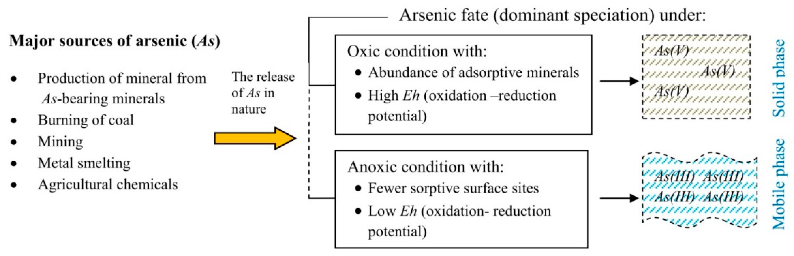
2. Arsenic Species and Their Behaviour in the Environment

Arsenic is a member of group Va of the periodic table and is classified as a metalloid that readily reacts with numerous elements [24]. It exists in the environment in different oxidation states as Arsine (As(III)), Arsenic (As(0)), Arsenate (As(V)), and Arsenite (As(III)) [24,55].
The inorganic oxyanions of As(III) or As(V) are the most typical soluble arsenic species that exist mainly in natural water [56]. By contrast, dimethyl arsenic acid (DMAA) and monomethyl arsenic acid (MMAA) mainly comprise the organic arsenic species [42,55]. The species of As(III) include AsO33−, HAsO32−, H2AsO3, and As(OH)3, while H3AsO4, H2AsO4, HAsO42−, and AsO43− constitute the As(V) species [57]. With respect to complex formation, As(V) can form complexes with sulfides and a ternary complex with polyvalent cations, whereas As(III) assists in complex formation with some metals and other natural organic materials in the environment [58,59]. Depending on the prevailing conditions, the availability of the inorganic As species differs (Figure 1). As (III) dominates under reducing anaerobic conditions while As(V) is dominant under oxygen-rich aerobic conditions and is often more associated with the solid phase [12,23,24]. However, environmental factors such as the redox potential (Eh), organic matter, and pH, among others, influence As speciation, mobility, and toxicity because studies have already shown variation in the behaviour of these inorganic As species in response to changes in some of these ecological variables [23,24,60,61,62].

Influence of Environmental Factors on the Mobilisation of As

The mobility of As species and their overall availability is strongly linked to environmental factors. These factors, as described in several research studies include but are not limited to the redox potential (Eh), pH, microbes, and natural organic matter [63,64]. In most natural settings, As is associated with mineral compounds such as realgar (As4S4), arsenopyrite (FeAsS), and orpiment (As2S3) [65] and can be closely associated with metals such as Fe, Pb, Cd, and Ni, among others [64,66]. Additionally, some bacteria, particularly IRB, utilise the As transformation process for energy generation and or as a precursor of phosphorus (P) for the synthesis of adenosine deoxyribonucleic acid (DNA) triphosphate (ATP), ribonucleic acid (RNA), and phospholipids [64,67]. Therefore, variation or alteration of the environmental factors affects the speciation and mobility of As and often influences the activity of the associated microbes, which in turn affect the solubility, availability, and toxicity of As in the environment [63]. The detailed information on the influence of environmental variables on As mobilisation has been explained by [64].

3. Arsenic Toxicity and Its Consequences

Animals and plants are susceptible to As’s toxic and carcinogenic effects [24]. Generally, organic As species are less harmful to living organisms than inorganic As forms [25,68,69,70]. The risk assessments of this metalloid in food and drinking water were comprehensively reported and documented in an article by Rasheed et al. [71] and Hirano [42]. Equally interesting is that, according to historical evidence, As species toxicity persists because it can be methylated, and its oxidation state changes during metabolism [42].
The most important and well-known toxic environmental species are As(V) and As(III) [71,72,73]. These As species are recognised to cause skin disorders, lesions in peripheral blood, and cancers in animals, particularly humans [71]. Other categories of cancers associated with chronic exposure to these species through drinking contaminated water include urinary bladder, lung, skin (primarily squamous cells), and possibly liver, kidney, and prostate cancers [42,74].
From another perspective, As toxicity in plants causes cytotoxicity and the production of reactive oxygen species (ROS), which include the hydroxyl radical, superoxide anion, and hydrogen peroxide, which cause genomic instability and inhibit the activities of some cellular enzymes [75]. Additionally, an increase in As contents in plants is associated with decreased plant growth and yield [34,76]. Moreover, other physiological and several morphological changes such as a decrease in the germination rate, reduction in the number of leaves, plasmolysis of the root cells, necrosis of leaf tips, leaf wilting, disruption of the cellular membrane structure, inhibition of photosynthesis, and disruption of growth patterns are commonly associated with As toxicities in plants [76,77]. Furthermore, when absorbed by plants, As may become a part of the food chain, posing a threat to food safety [78,79].

4. Mitigation and Remediation Strategies for Arsenic

Arsenic is mainly released into the environment due to volcanic and industrial activity. Mining, non-ferrous metal smelting, and fossil fuel combustion are major anthropogenic activities that result in As contamination of water, air, and soil [9,80,81,82]. Several reports have also proved that using As to preserve wood has resulted in As-related environmental contamination [83]. As previously stated, As contamination causes numerous health and environmental problems. Thus, it is crucial to understand and devise remediation methods to immobilise and/or reduce its availability in the environment. Several approaches were developed and evaluated for the remediation of As contamination in the environment. These strategies are grouped as physical, chemical or biological methods [55]. Furthermore, new and emerging treatments technologies have been thoroughly assessed and are now being evaluated. Some of these strategies were further studied in recent studies [84,85]. Primarily, the broad and basic strategy in controlling As contamination is the transformation of toxic As to the less harmful As species. Further, depending on the geochemistry of the contamination, the physical and chemical conversion in this approach involves the use of strong oxidants, such as hydrogen peroxide, potassium permanganate, or ozone as a strategy [9]. In this conventional approach, coagulation, filtration, inverse osmosis, and ion exchange are extensively involved, which are expensive, labour intensive, and are capable of further instigating undesirable consequences such as environmental pollution and so on due to the chemicals involved [86,87,88]. For this and other reasons, biological methods focused on plant- and microbial-based approaches were given more attention because they are safer, better, and economically viable solutions for the environment.
Phytoremediation plays an essential role in environmental pollution mitigation and management. This is because plants are both inexpensive and environmentally friendly for heavy metal decontamination of soil [34,88,89,90]. The plants usually involved in this process are called hyperaccumulators, as they can absorb and accumulate the metals in their upper parts [90,91]. For example, Epilobium fragilis was reported to accumulate As from contaminated soil [92]. Also, in a study using native plants, Yildirim and Sasmaz [90] observed that Anchusa arvensis, Phlomis sp., Glaucium flavum, and Verbascum thapsus demonstrated the capacity of cleaning and remediating soil affected by As pollution. Recently, Patel et al. [77] provided an account and list of various tolerant plants and potential contributors for As phytoremediation. Despite the widespread use of several plants in the control and management of As toxicity, the method is time-consuming and requires more effort in screening suitable prospects, as not all plants can accumulate heavy metals [93,94]. Furthermore, wider consideration is essential regarding the ion uptake mechanism of the prospective plants relative to their morphological, physiological, anatomical, and genetic traits [90,95]. Therefore, alternative strategies mediated by microbes (fungi and bacteria) serve as an additional helpful approach to mitigate and control heavy metal pollution in soil and water [21,96,97].
Heavy metal remediation by microbes, especially bacteria, provides some cushion effects from some of the challenges of other remediation approaches. This is because bacterial remediation is relatively inexpensive, effective, and eco-friendly [98]. DIRB are among the many bacteria used in this strategy because of their unique ability to use a wide range of electron acceptors in their respiration process [99,100]. This form of remediation is typically employed ex-situ (in the areas where contamination occurs) or in situ, for example, in storage tanks [100]. Because of differences in the oxidation state, solubility, and availability of each heavy metal and their varying behaviours in relation to prevailing environmental factors, bacterial heavy metal remediation is not uniform across all heavy metals [99,100].
For example, some Shewanella species have demonstrated the capacity to lessen the mobility of uranium (U) in groundwater as a result of the transformation of U(VI) to insoluble U(IV), leading to a decrease in the spread of U contamination from groundwater [100]. Bencheikh-Latmani et al. [101] reported how the cleanup effort for Cr has been greatly assisted by some Shewanella species through the formation of solid oxides during the reduction of soluble Cr(VI) to Cr(III). Conversely, methylmercury, a mobile bioaccumulative environmental toxin, has been reported to be formed as a result of S. oneidensis MR-1 reducing ionic mercury [Hg(II)] to elemental mercury [Hg(0)] [99,102]. Furthermore, catalytic reduction of As(V) to a mobile and more toxic As(III) facilitated by some Shewanella species commonly results in the mobilisation of As species that are toxic in the environment [64,103].
Having described the key role of Shewanella species in the facilitation of direct or indirect mobilisation of environmental As species, its potential remediation role could be best perceived from its contribution to the detoxification of some As species and/or sequestration of the mobilised As through flavin-mediated iron plaque formation [104,105,106]. These various processes are explained in detail in the upcoming sections of this article.

5. Iron and Its Impact on the Fate of Arsenic

Iron (Fe) is the fourth most available element on earth [107] and the lithosphere’s second most abundant transition metal element [108]. Its oxides comprise Fe and oxygen, including ferrihydrite, lepidocrocite, goethite, and magnetite [33,109,110,111]. These oxides are abundantly found in rocks, soil, sediments, water, and various minerals formed under different conditions [112]. Fe primarily exists in two redox states in aquatic settings, i.e., Fe(III) and Fe(II). At neutral pH and in the absence of complexing ligands, Fe(III) exists in the Fe(III) (hydr)oxide solid-state [37,113]. However, in a situation where a terminal electron acceptor and molecular oxygen (O2) are lacking, Fe(III) (hydr)oxides contribute to microbial anaerobic respiration where they serve as terminal electron acceptors [113,114].
Generally, under oxic conditions, toxic metals such as lead (Pb), Cd, Cr, and As are integrated and incorporated into amorphous or crystalline Fe(III) mineral phases [29,115,116,117,118]. Secondary Fe(III)- and Fe(II)-bearing minerals such as hematite and magnetite are closely associated with these metals [116,118,119]. Many studies have long established that, among these toxic heavy metals, As is adsorbed rather strongly to Fe-oxides [96,120,121], making it the primary reservoir and transporter of As in soil and aquifers [116,122].
It has been reported that the reductive dissolution of hydrous Fe-oxides and the subsequent release of related As can occasionally result in high As concentrations in subsurface waters [24,27]. Similarly, Fe(II) released from As-bearing minerals is an important catalyst linked with the mobilisation of As in the environment [123,124]. Despite the complexity of the process, many bacterial strains interact and dissolve the surface layers or structural regions of these minerals (Figure 2), resulting in Fe(II) generation [112,125,126]. This reduction of Fe(III) and subsequent Fe(II) release are strongly associated with As(V) dislodgement and its availability in the soil solution and the environment [13,118,123,127,128,129].

Effect of Dissimilatory Iron-Reducing Bacteria’s Transformation of Iron on the Fate of Arsenic Soils

In general, dissimilatory metal-reducing bacteria (DMRB) oxidise organic matter, including organic contaminants and hydrogen gas (H2) in anoxic environments, and then transmit the released electrons to solid-phase minerals containing oxidised metal ions, such as manganese (Mn(IV)) and ferric iron (Fe(III)) for anaerobic respiration [38]. These microorganisms are commonly involved in the degradation/corrosion of organic matter [130] and metals [131,132], e.g., Pb, Cd, and As, in surface and sub-surface environments such as sediments of rivers, soils, lakes, and oceans. Additionally, the bacteria involved have the potential to dramatically alter the geochemistry of the surrounding Fe mineral-containing As in groundwater and sediments, resulting in As contamination of drinking water sources, diseases, poisoning, and human health disruption [39,133,134]. Numerous bacterial strains were identified to be involved in the reduction and transformation of Fe, particularly in the As(V)-bearing Fe(III) mineral assemblage [104,135,136]. These bacteria include dissimilatory iron-reducing bacteria (DIRB) and dissimilatory arsenic-reducing bacteria (DARB), comprising Shewanella oneidensis MR-1 and Geobacter metallireducens GS-15 and Geobacter sp. OR-1 & Bacillus sp. M17-15, respectively.
Based on published evidence, Geobacter metallireducens GS-15 and Shewanella oneidensis MR-1 are among the early DIRB strains that promote Fe(III) reduction [64,107]. The role of Shewanella oneidensis MR-1 in this phenomenon was discovered when it was isolated from the silt of a freshwater lake; since then, its activity has been linked to intracellular oxidation of organic materials to extracellular Fe(III)(hydr)oxide reduction [137,138]. Similarly, numerous researchers have reported the influence of this strain on the geochemical cycling of organic matter and other metals, including Fe, As, etc. [137,139]. From the moment of such discovery to date, evidence from literature has consistently established the role of Shewanella oneidensis MR-1 as a typical DIRB that promotes the redox transformation of Fe(III)-oxides either directly or indirectly [49,123,140,141,142,143]. Therefore, it is assumed that the fate and biogeochemical transformations of Fe and associated metals, e.g., As, are strongly linked [118,144]. Nevertheless, in most situations, the transformation of Fe(III) to Fe(II) by DIRB only serves as a source of As(V) in the environment but does not guarantee As(V) solubilisation or subsequent As(III) mobilisation. This peculiarity was confirmed by Cummings et al. [145] in an effort to evaluate the role of Shewanella alga BrY in the reductive dissolution of scorodite. The study results revealed the presence of As(V), but As(III) was below the detection limit. Lopez-Adams et al. [124] recently reported the absence of As(III) in a supernatant during extracellular reduction of Fe(III)-(oxyhydr)oxide by Shewanella sp. cultures through nanoscale secondary ion mass spectrometry (NanoSIMS) imaging. These findings indicated that Fe(III) reductions by Shewanella sp. can cause As contamination but may not always result in As(III) mobilisation. Hence, the process can be considered an essential route for As(V) mobilisation into the environment.
At circumneutral pH and under oxic conditions, insoluble Fe(II) (hydr)oxides are produced from Fe(III) reduction [113]. An increase in the Fe(II) concentration in the environment is commonly interpreted as an indicator of reductive mineral dissolution by DIRB. Thus, in a complex environment where As is sorbed to Fe-bearing minerals, DIRB dissolution often promotes As mobilisation, resulting in As contamination of such an environment. Notably, readsorption of As often occurs because of the recrystallisation of more stable Fe(II)-oxides [113,127] or aggregation of Fe(II) to secondary minerals such as vivianite and magnetite, which subsequently result in arsenic re-sequestration from the environment [146,147,148].

6. The Impact of Bacteria on Arsenic Transformation and Cycling

Microorganisms have evolved diverse strategies to defend themselves against As through time. Fungi, for example, were known to protect themselves through a methylation process in which MMA or DMA was used. However, bacteria and the archaeal domain often produce volatile methylated arsines, facilitating the elimination of As in the environment [48,149]. In the process of tolerating and enduring metal toxicity, some bacteria precipitate, sequester, or even alter these metals’ oxidation state, and these processes often result in mobilisation or remediation of the metal in the environment [109]. Although As is generally toxic to life, many bacterial strains occasionally derive their energy for growth through As metabolisation [39,150], and this is achieved through the development of complex mechanisms of arsenate reduction, arsenite-oxidation, and/or As(V) resistance, a process which lessens the quantity of As entering the microbial cell [9,151,152]. Others can also use As compounds as donors or acceptors of electrons through As detoxification mechanisms [144,153]. Furthermore, metabolic and detoxification processes such as reduction, oxidation, methylation, and demethylation [154] cause the dissolution of As reservoirs, resulting in As mobilisation into the surrounding environment. Examples of such bacteria include the predominant DARB, heterotrophic arsenite-oxidising bacteria (HAO), chemoautotrophic arsenite-oxidising bacteria (CAO), and arsenate resistant bacteria [39,119,155].

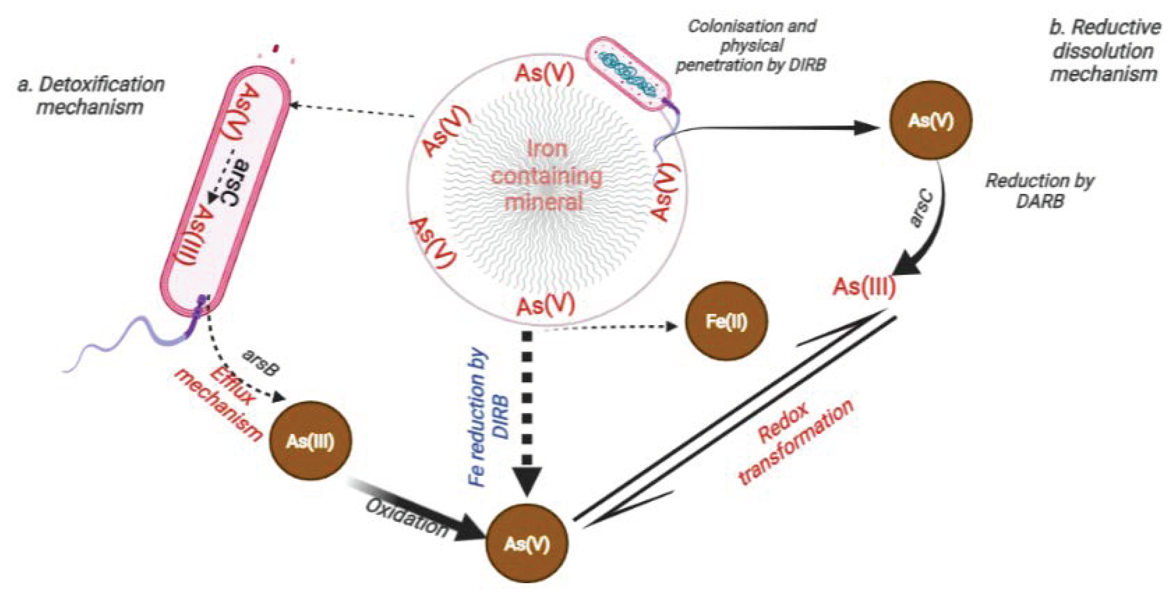
6.1. Influence of DARB on the Transformation and Mobilisation of Arsenic in the Environment

Bacteria, especially DARB, interact with less toxic As(V), and by this process, the bacteria make As(III) available via two steps (Figure 2). Firstly, by reducing As(V) to As(III) using organic material as a prime source of energy for growth and secondly, through the detoxification mechanism that facilitates the expulsion of the As(III) from the microbial cell via phosphate transporters [39,156]. Consequently, through these complex mechanisms of redox transformation that cycle between As(V) and As(III) oxidation states [157], the dynamics of As in the environment are affected and often lead to the mobilisation of As into the surrounding. It is important to note that the prevailing conditions, such as the pH, ionic strength, energy source, substrate nature, availability of Fe(III)-oxides, and type of bacteria involved, are critical factors in the entire transformation process.

6.1.1. Uptake of Nutrients from As-Bearing Minerals by DARB

Generally, microorganisms can dissolve and use different minerals as a source of nutrients and energy. Certain microbes often use minerals as their sole source of inorganic substrates such as carbon, nitrogen, and phosphorus for energy generation [9]. Others utilise sulphate (SO42−), O2, Fe(III), and organic material as terminal electron acceptors. However, some sulfide minerals serve as a reservoir of enzyme cofactors, for example, copper (Cu), zinc (Zn), molybdenum (Mo), magnesium (Mg), and apatite often serve as the source phosphorus required for deoxyribonucleic acid (DNA), ribonucleic acid (RNA), adenosine triphosphate (ATP), and phospholipid synthesis [158].
Several scholars deliberated on the mechanism evolved by microorganisms to absorb nutrients from minerals. These comprise the primary colonisation process, which includes the mechanical infiltration of the rock surface, leading to mineral disintegration [159] and the subsequent dissolution of the mineral facilitated by organic agents produced by the cells (Figure 2) [9]. To accomplish this, bacteria typically secrete exopolysaccharides (EPS), which play an essential role in mineral colonisation and protect the bacteria from environmental stresses such as heavy metal toxicity. Drewniak and Sklodowska [9] stated that the physical disruption of the minerals occurs due to the production of EPS and its binding to the exposed mineral surface. Similarly, Welch et al. [159] previously highlighted the role of the EPS as a substrate for other (heterotrophic) bacteria producing organic agents, which in turn stimulate the chemical dissolution of the underlying minerals. However, some bacteria use an indirect approach of As(V) dissolution [160], which is achieved through the biosynthesis of a protein called a siderophore. This protein usually secretes small organic metal chelators and captures insoluble Fe(III).
Essentially, some bacteria use siderophores to survive in harsh environmental conditions or as organic ligands and/or acids that may facilitate the release of nutrients in the surrounding environment when essential nutrients for growth are limited or absent [9,161]. Interestingly, these organic ligands can establish strong complexes with metal cations and bind to mineral surface atoms. For these reasons, siderophores have been among the most common natural bacterial agents involved in mineral dissolution. Furthermore, these compounds have the power to enhance the transfer of an electron to Fe(III); hence, when they form strong complexes with Fe(III), this increases the solubility, resulting in the mobilisation of the bound metals [15,162].
Siderophores are typically secreted by DARB strains outside of their bacterial cell envelopes. Notably, these secretions have a high affinity for metal ions and are capable of chelating not only As but also Fe, Mn, Mg, Zn, Cr, uranium (U), gallium (Ga), nickel (Ni), Pb, and Cd [163,164,165]. As a result, these siderophores may help to reduce As bioavailability and subsequent removal from contaminated environments [166,167]. In another study by Lukasz et al. [8], it was established that siderophore production could enhance the dissolution of secondary As-bearing minerals. This observation was based on a biochemical test performed on DARB samples from the ancient Zloty Stok gold mine, which revealed that under iron-limiting conditions, all of the sampled DARB isolates developed siderophores known as hydroxamates, which were actively involved in the dissolution of As-containing minerals [8].

6.1.2. Dissolution of As-Bearing Minerals by DARB

The reductive dissolution of the parent As-bearing minerals usually results in the release of As into the environment. Under reducing conditions, arsenate adsorbed to the surface of Fe minerals (e.g., scorodite or ferrihydrite) is used mainly by bacteria as a terminal electron acceptor in As reduction [9,31,43,168,169]. As a result of this process, dislodgment of attached arsenate and its subsequent mobilisation into the environments from secondary minerals occur (Figure 2). However, it is worth mentioning that, occasionally, chemoautotrophic arsenite-oxidising bacteria (CAO) makes use of oxygen and sometimes nitrogen as their terminal electron acceptor during the fixation of inorganic carbon (CO2) into cell material to oxidise aqueous As(III) to As(V).
In essence, the reduction of As(V) to As(III) is regarded as an efficient resistance and coping mechanism demonstrated by most DARB against the toxic effect and high arsenic concentration in the surroundings. In the same way, the process serves as the most accessible route through which DARB mobilise aqueous As(III).
To date, a wide range of bacterial species that play a key role in the redox transformations and cycling of As in the environment have been reported [64]. However, because of the differences in genomic contents, substrate types, modes of interactions with prevailing environmental conditions, and relationships with organic and inorganic fractions, including various (hydro)oxides [36,82], the action mechanism of different bacterial strains differ. For example, Desulfuribacillus alkaliarsenatis is a microbial strain with a broad dissimilatory metabolic capacity, which, apart from arsenate, can also grow using H2 pyruvate, formate, or lactate as an electron donor [170]. Furthermore, the Desulfurispirillum indicum S5 strain was reported to utilise multiple reductive pathways for arsenate and other elements such as selenate (SeO42−), selenite (SeO32−), nitrate (NO3), and nitrite (NO2) [171]. Another important bacterial strain that plays a vital role in the mobilisation of arsenic through dissolutions of As-bearing minerals is Shewanella oneidensis MR-1 [40,64,118,172,173,174,175,176].

6.2. Heterotrophic Arsenite-Oxidising Bacteria (HAO)

It is a fact that some bacteria are recognised for their ability to oxidise organic carbon using available oxygen as a terminal electron acceptor for energy generation. Using a similar process, some of these bacteria obtain their energy through As(III) to As(V) oxidation. Hence, they are regarded as HAO [177]. Notably, some of these bacteria are mixotrophic, meaning they obtain energy from organic carbon oxidation and As(III) to As(V) oxidation [177,178]. Interestingly, this process helps to detoxify As in the environment [179,180] by reducing the bioavailability of toxic As to the surrounding environment and plants [181].

6.3. Chemoautotrophic Arsenite-Oxidising Bacteria (CAO)

While some bacteria obtain their energy for metabolism through the oxidation of organic molecules, other bacterial species derive their energy from the oxidation of inorganic energy sources such as carbon monoxide, hydrogen, nitrogen and sulfur compounds, or divalent cations (e.g., Mn2+ and Fe2+) [182,183]. Similarly, some bacteria employ the same mechanism to obtain their energy during As(III) oxidation autotrophically. Hence, they are regarded as CAO [179,180]. A good example is the Thermus strain HR13, which has been shown to oxidise As(III) to As(V) under aerobic conditions while using arsenate as the terminal electron acceptor under anaerobic conditions [184]. It is worth noting that, in contrast to HAO which require the support of organic matter for their growth, the growth of CAO can be sustained through the oxidation of As(III) [185]. For this reason, most of the As(III) oxidation by HAO is widely considered a detoxification mechanism [179,181,185].
In addition to lessening the toxic effects of As, the presence of these As(III)-oxidising bacteria is believed to promote the activity and growth of some indigenous microbial communities in arsenic-contaminated soils, which subsequently enhances the As uptake efficiency. This attribute is demonstrated by Ensifer sp. M14, an As(III)-oxidising bacteria when added to As-contaminated soil [181]. According to the findings, exposure to bacteria promotes As uptake by the plant Medicago sativa (alfalfa) and reinforces the activities and growth of the microbiota [181,186].

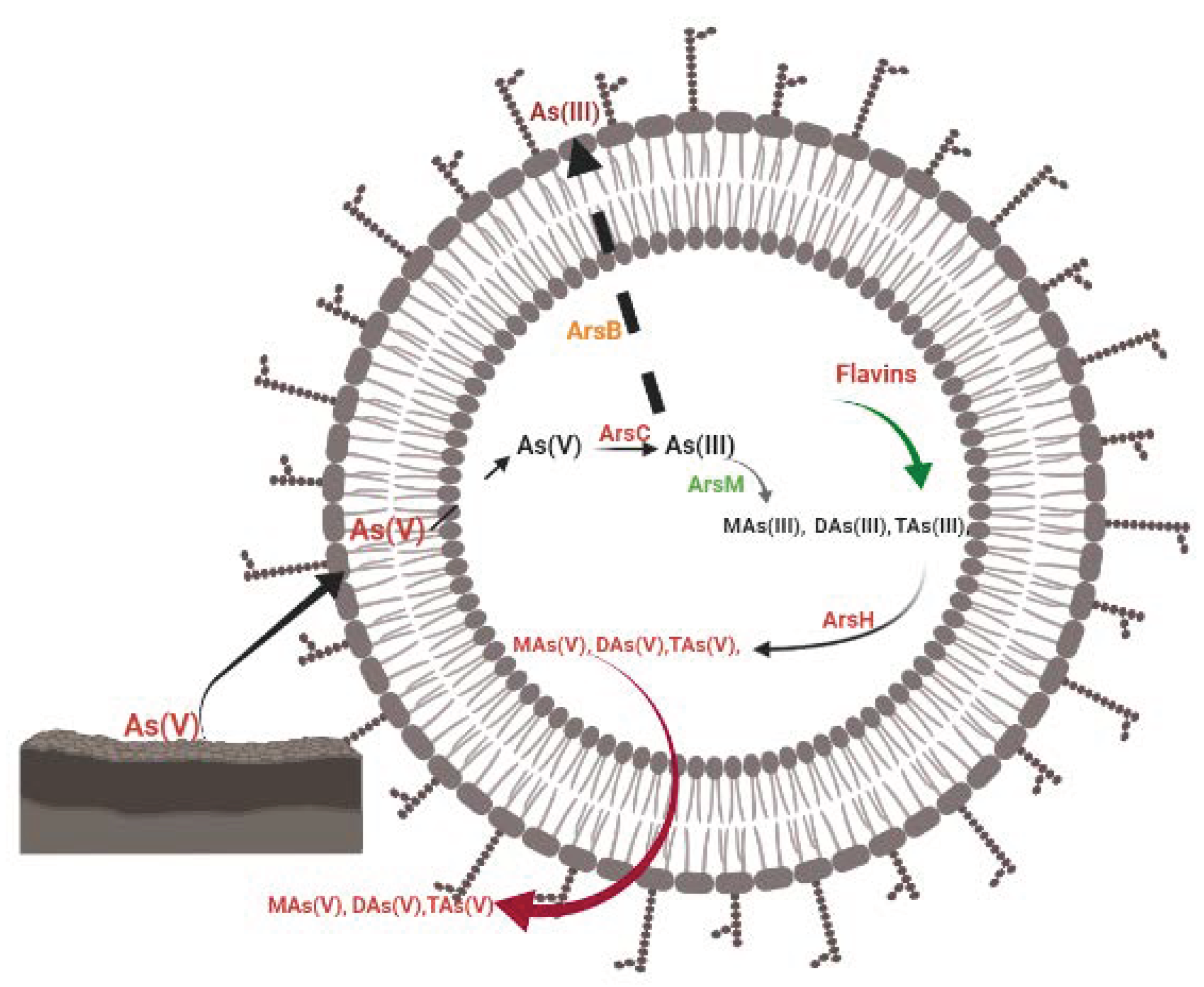
7. Removal and Detoxification of Transformed Arsenic

As pointed out earlier, bacteria have a different approach for eliminating As from the environment. DARB utilise a protein domain called arsenate reductase protein “ArsC” to enzymatically remove As in the environment [22]. ArsC is a small protein (13–16 kilodaltons) located in the cytoplasm of the bacteria facilitating the reduction of As(V) to As(III) [105,187]. In the process, glutaredoxin, glutathione or thioredoxin serve as an energy source for this transformation process [22,188]. At the end of this process, a flux of toxic As(III) is observed, mediated by an ATP-dependent efflux pump, arsB (Figure 3). Because of the integral role of ATP in this process as an essential energy source, under an anaerobic state, DARB acquires energy through redox transformation of As(V), where it is used as an electron acceptor instead of oxygen [22]. Notably, among many groups of bacteria, ε-Proteobacteria comprising Sulfurospirillum arsenophilum and Sulfurospirillum barnesii were the first DARB identified in the mid-1990s, and their representatives have since been recognised in other phylogenetic groupings, including δ- and γ-Proteobacteria and the low-GC “Gram-positive” bacteria, Crenoarchaea, and thermophilic eubacteria. Understanding the mechanism by which these bacteria detoxify and eliminate transformed As from their cells is critical to understanding the role of DARB in As transformation, toxicity, and dynamics in different settings.

7.1. Mechanism of Arsenic Detoxification from DARB Cells

As a consequence of the transformation of As(V) to As(III) by DARB and subsequent availability of As(III) in the bacterial cell, some key biochemical processes may be obstructed in the organisms [48,106]. This is because trivalent arsenic (As(III)) is more toxic than As(V) [189], primarily due to its ability to react readily with sulfhydryl and thiol (R-SH) groups in protein residues of the cell [106,189]. To minimise the excessive toxicity of this As species, the bacterial cell has to devise a coping mechanism to either (a) eject or expel the As(III) from the cell or (b) undergo methylation of As(III) to trivalent organoarsenicals [21,96,106,189].

7.1.1. Ejection or Expulsion of As(III) from the Cell

As previously stated, in the transformation of As(V), the As(III)-specific transporter (arsB) gene mediated by arsC usually facilitates the expulsion of the transformed As(III) from the microbial cell (Figure 2 and Figure 3) [190].
Generally, the detoxification pathways employed by DARB against the toxic effect of As(III) are regarded as a resistant mechanism that is controlled by an arsenic resistance (ars) gene [106]. This ars gene is mostly encoded on the operon of the plasmid or chromosome in gram-negative or gram-positive bacteria [105,106,119,187,191]. Most DARBs’ detoxification process of inorganic arsenicals is controlled by ars operons; however, to efficiently detoxify organoarsenicals, the ars gene occasionally associates and combines with other ars-related genes.
The expulsion or extrusion of As(III) by most DARB, as their basic detoxification mechanism, is controlled by the arsB gene encoded in the ars operon [106,189]. Even though the nature of the arsB as membrane transport protein has allowed it to accomplish the role of catalytic expulsion of As(III) from the cell via membrane potential alone [192], sometimes it does coexpress with another ars operon protein arsA to form a tight membrane arsAB complex that provides a greater level of resistance (Figure 3) [106,189]. Also, because the arsA gene encodes the arsA protein as an As(III)-stimulated ATPase, the complex formed becomes an ATP-driven efflux system [189,193,194]. This arsA is distributed in the periplasm and is composed of functional homodimers with identical halves, each containing ATP binding sites, which serve as the catalytic component of the ars pump [106,195].
Other key important ars genes encoded in the ars operon that enhance the expulsion of As(III) are the arsR and arsD genes. The former detects inorganic arsenic in the environment and negatively regulates the transcription of other ars operon genes [196], while the latter functions as an As(III) metallochaperone, facilitating As(III) transfer to the metal-binding region of arsA of the ars efflux pump [197,198]. Because of the interaction of these genes, the overall ability of arsB to expel As(III) is enhanced due to the greater affinity of arsA for As(III) and the surge in ATPase activity of arsA even at lower As(III) concentrations [106,199].

7.1.2. As(III) Methylation to Trivalent Organoarsenicals

Increasing evidence suggests that the methylation process functions as an additional system that is critical in the detoxification of As(III) [104], and it can also serve in the bioremediation of As [48]. The process involves adding a methyl group to As(III) and subsequent detoxification of methylated products [75,81,105]. According to Qin et al. [48], arsenic methylation involves two pathways, one involving reductions of As(V) by reduced glutathione tablets within the microbial cell and combining it to a free methyl donor [149], while the other involves a direct combination of the arsenic ion with a methyl donor [48,200]. Thus, the methylation processes comprise the reduction of As(V) to As(III), followed by the sequential coupling of As(III) with positively charged carbon ions in free methyl donors to generate MMA, DMA, and TMA as products [47].
The methyl group is transferred to As(III) by the activity of As(III) S-adenosylmethionine methyltransferase, which is catalysed by arsM encoded in the ars operon (Figure 4) [48]. At the end of the process, arsM transform As(III) into dimethyl arsenate (DAs(V)) [201], trimethyl arsenite (TAs(III)), dimethyl arsenite (DAs(III), and methyl arsenite (MAs(III)) [48,104,106,202].
Although some organoarsenicals are harmful, the toxicity of As compounds are still reduced by methylation, which can be considered another defensive mechanism against arsenic toxicity by some bacteria [47,203]. Prospectively, this process could be a valuable strategy to minimise the availability of toxic arsenic in the environment. Bacterial strains such as Aeromonas sp., Flavobacterium sp., Proteus sp., Pseudomonas sp., Rhodopseudomonas palustris, Corynebacterium sp., Escherichia coli, Achromobacter sp., Alcaligenes sp., and Nocardia sp. have demonstrated the ability to produce methylated arsenic compounds [149]. Furthermore, recently, in an attempt to evaluate the impact of methylation on arsenic by S. oneidensis MR-1, the bacteria were exposed to 1.0 mg/L of As(V). The results indicate that the bacteria could tolerate low concentrations of inorganic As by methylating it into less toxic and organic DMA [47].

8. Contribution of Shewanella oneidensis MR-1 to As Redox Cycling and Remediation

It was highlighted that Shewanella oneidensis MR-1 is an effective bacterial strain contributing significantly to redox cycling and arsenic remediation. This role is accomplished by reducing Fe mineral-containing As and total or partial transformation of some toxic arsenicals into non-toxic equivalents. Hence, it is vital to understand this bacterial strain’s biology, genomic composition, and mechanism of arsenic transformation.

8.1. Biology and Distribution of Shewanella Genus

Shewanella was initially assigned to the Achromobacter genus but was later reclassified as prokaryotic and assigned to the Shewanella genus [204], which at present contains about 70 species and more than 30 available sequenced genomes isolated from deep-sea sediments, freshwater sediments, and other food sources [46,88,205]. Free-living strains thrive under varying temperatures, pressure, salinity, and nutrient compositions [206,207]. Some of them are associated with other organisms as epibionts, syntrophs, symbionts, and pathogens [138,208]. Interestingly, it could perfectly fit into redox-stratified environments due to its ability to adapt to and withstand different environments and associated stress [46,209,210,211]. Moreover, its physiology has supported its ability to survive in broad and different ecological niches from equatorial to polar regions, fresh water to oceans, the surface to subsurface sediment, and polluted to unpolluted environments [46,208].
In addition to the ability to occupy a wide range of environments, as pointed out earlier, the Shewanella genus has metabolic flexibility, which enables species of this genus to choose a mode of metabolism according to the immediate environmental conditions [206,212]. This genus is notable for its ability to respire using a variety of organic and inorganic materials, including oxidised metals such as Mn(III, IV) and Fe(III), metal oxides such as hematite, magnetite, elemental sulphur (So), thiosulphate (S2O32−), sulphoxide, and arsenate, among others [205]. Notably, the genus possesses a high tolerance to numerous toxic compounds, allowing it to thrive in varying degrees of polluted environments and hazardous products. As a result, most strains in this genus play a positive bioremediation role [46]. Interestingly, S. oneidensis MR-1, a representative of this species, has been consistently involved in redox cycling and has been proven to be a valuable tool in the remediation of As-contaminated systems [21,47,148,213,214], accomplished through the previously mentioned reductive dissolution and detoxification mechanisms.
Another unique feature of this Shawenella genus from other prokaryotes is its role in the oxidoreduction cycle and its ability to survive in metal-rich environments [46,215]. This role is partly related to its remarkable ability to transfer electrons from the quinol pool to the external membrane’s outside face, which is facilitated by a complex assembly of cytochromes (c-type cytochromes) via a process called extracellular electron transfer (EET) [52,174,176,215]. Studies have shown that the EET process or pathway serves as an important bridge that enables electron transfer across the outer membrane through the periplasm from the inner membrane [123,126,212]. Because the minerals associated with Mn(III/IV) or Fe(III) are insoluble at circumneutral pH [126,216], it is therefore highly unlikely for them to cross the outer membrane and the periplasm, where bacterial terminal reductases for other electron acceptors, such as SO42−, O2, and NO3, are situated.
Fe(III) reduction facilitated by DIRB is a critical process for As mobilisation in the environment [124,148], a process that contributes significantly to As contamination of various environmental components. Interestingly, electrons for the Fe(III) to Fe(II) reduction are typically derived from EET through the oxidation of quinol to quinone (QH2), which further enters cells through a phosphate transport channel mediated by QH2 dehydrogenases such as tetraheme cytochrome c (CymA) [217,218]. Lopez-Adams et al. [124] recently highlighted the significance of the EET process in the mobilisation of As(V). The study revealed how flavin from Shewanella sp. significantly facilitated the release of As(V) through the reduction of Fe(III)-(oxyhydr)oxide from the As(V)-bearing Fe(III) mineral. Moreover, the same study further demonstrated the role of these bacteria in the mobilisation of As(V), as opposed to the reduction of As(V) to As (III).

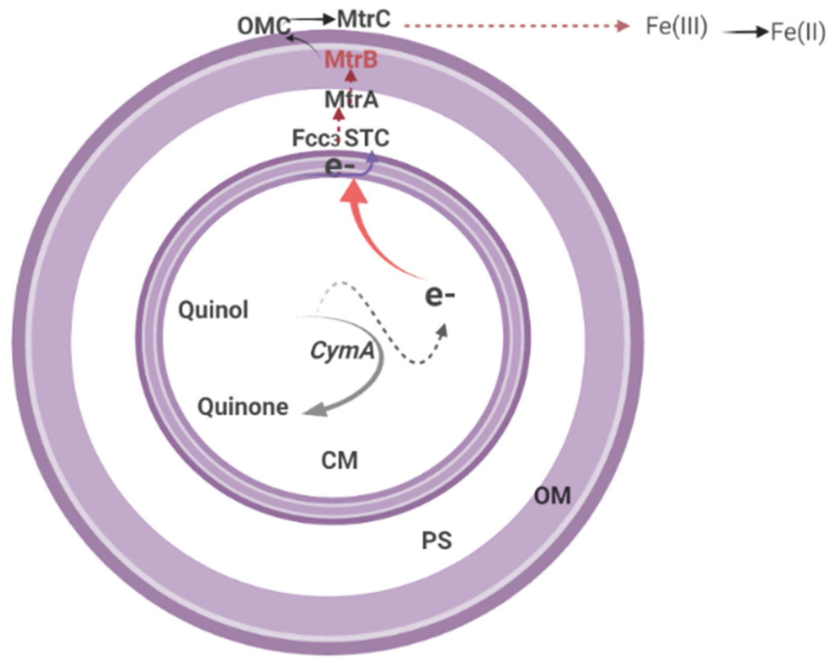
Extracellular Electron Transfer (EET)

Generally, EET is a process employed by microorganisms to exchange intracellular electrons with an extracellular electron donor/acceptor across the cell membrane [212]. This electron donor/acceptor may vary from naturally occurring metal compounds to artificial electrodes [219]. To ensure that electrons are effectively transferred from the inner to the outer membrane, organisms evolved a metal reduction pathway (Mtr), which is supported by an array of c-type cytochrome protein components such as CymA and outer membrane cytochromes OmcA, MtrC, and MtrA [126,212] as well as the porin-like trans-outer membrane protein MtrB [137,138]. The EET pathway has been described to include many interchangeable periplasmic electron carriers, such as fumarate reductase, i.e., flavocytochrome c3 (Fcc3) or the small tetrahaem cytochrome (STC), which are reduced by central quinol oxidase CymA [46,220].
In some Shewanella species, particularly Shewanella oneidensis MR-1, the EET can be assisted by two similar external membrane complexes; the metal-reducing systems MtrCAB and MtrDEF [46]. However, deletion experiments demonstrate that MtrCAB alone tends to function physiologically [212,216,221]. Broadly, each of the complexes comprises two decaheme cytochromes associated with an external membrane and a transmembrane β-barrel protein, forming a channel through the membrane’s outer layer [220].
The EET process can be carried out in one of three ways: direct, distant, or mediated contact (facilitated by external soluble electron carriers) [46,212]. Direct contact occurs when there is uninterrupted contact between the cells and the metallic acceptors, and the transfer of electrons is accomplished from the outer face of the outer membrane [222]. While in some cases, scarcity of electron acceptors causes the cell’s outer membrane to undergo dynamic structural alteration, forming an outer membrane extension called nanowires. This alteration enables connection and electron transfer between the bacterial cell and the distant electron acceptors through a distant contact process [175,222,223]. Indeed, this is important, especially in bacterial metal reduction biofilm setup, due to the barrier between upper-layer organisms and electron acceptors. In another situation, the mediated process can be achieved using secreted electron carriers such as flavin that serve in electron shuffling in the external media and as significant cofactors of the external metal-reducing systems [212,224]. However, it is important to note that most flavins (secreted electron carriers) are typically secreted in laboratory batch cultures when the concentration of the bacteria is high [46].
The importance of flavin was established based on demonstrations when it was secreted at the outer membrane of MR-1, influencing electron transfer through the electron shuttling pathway [122,216]. Although some researchers [38,107,141,225] were able to describe the detailed role of flavin in the shuttling of electrons in Fe(III) reduction in oxic settings, the mechanism by which flavin influences the reduction of Fe(III) by MR-1 has not been sufficiently explained by researchers.
On most occasions, the exterior face of the outer membrane of the Shewanella genus receives electrons from the quinol pool, which is assisted by c-type proteins [38,212]. CymA, an inner membrane dehydrogenase, oxidises quinol and indirectly or directly transfers the released electrons to MtrC through periplasm proteins to reduce insoluble Fe(III) [217,226]. MtrA, in collaboration with MtrB, facilitates the transport of electrons to MtrC and OmcA, which are situated on the bacterium’s surface [227,228]. The direct or indirect reduction of Fe(III)-containing minerals by these bacteria, more specifically MR-1, is accomplished through the action of this outer membrane c-type (MtrC and OmcA) facilitated by the extracellular secretion of the bacterial cell such as flavin, which serves as an electron shuttling system (Figure 5) [126].

8.2. EET Process in Shewanella oneidensis MR-1

Shewanella oneidensis MR-1 (previously Alteromonas putrefaciens) is a facultative aerobic bacterium capable of dissimilatory reduction of manganese and iron oxides [137,229]. The bacterium is gram-negative in structure, with its cytoplasmic or inner and outer membranes on its cell envelopes separated by periplasm [230].
So far, the genomes of more than 200 strains in the Shewanella genus, including Shewanella oneidensis MR-1, have been sequenced, and comparative genomic studies have revealed that these bacteria share genetic makeup [205]. Genomic analysis of this bacterium has also shown that it comprises 47 two-component systems and 211 one-component systems, which demonstrated the complexity of the energy conversion pathways of the organism [206]. These components serve as the foundation for understanding the bacterial system’s sensing, signal transduction, and control. The system consists of networks of receptors, transmitters, and information regulators that link intracellular and extracellular signals to cellular responses [229].
Depending on the settings and the environmental conditions, MR-1 often has different forms of respiration. For example, it can exist as a facultative anaerobe that utilises Mn(III/IV)/Fe(III) as terminal electron acceptors or molecular oxygen (O2) for aerobic respiration [126,231]. These anaerobic and aerobic forms of respiration often result in the generation of Fe(II) and hydrogen peroxide (H2O2) [135,206,229,231].
The EET process in MR-1 is similar to the Shewanella genus process described above. Quinol dehydrogenase is located on the cytoplasmic membrane of MR-1, promoting the oxidation of quinol to quinone and subsequent transmission of the liberated electron to the periplasmic STC and Fcc3 for further reduction of Fe(III) (hydr)oxides (Figure 4) [107,232]. Principally, STC and Fcc3 relay the generated electrons across the periplasm to MtrA, which forms part of the trans-outer-membrane protein complex containing MtrC and MtrB [107,113].
Electron transfer under typical anoxic conditions is accomplished through flavin secretion by MR-1 cells, which enable electron transfer for Fe(II) mobilisation from MtrC and OmcA. In a set-up comprising MR-1, the secreted flavins can either serve as electron shuttles between MtrC/OmcA and Fe(III)(hydr)oxides or as cofactors that enhance Fe(III) (hydr)oxides to receive an electron from MtrC/OmcA [38,141]. However, in a situation where there is a distance between the Fe(III) (hydr)oxides and the bacterial cells, the electron-hopping mechanism facilitated by MtrC and OmcA through the outer membrane and periplasm extension of the cells serves as an alternate channel for this species to shuttle electrons [175,222,233]. It is also worthy to note that, although the functions of MtrDEF in EET are yet unascertained, the MR-1 genes for MtrABC and OmcA are always present in the same gene cluster as MtrDEF, the homologs for MtrABC [206,228,234]. Furthermore, MR-1 genomes have two additional MtrAB homologs involved in dimethyl sulfoxide reduction (DMSO) outside the cell [235,236].
MR-1 oxidatively degrades substances, including metals, in an aerobic setting, through an advanced oxidation process (AOP) called the microbially driven Fenton reaction [38,237]. This process has a significant impact on As behaviour, particularly in the As(V)-bearing Fe(III) mineral system. In the reaction, a continuous supply of O2 and Fe(II) is required, and to compliment this requirement, this species can continuously reduce Fe(III) to Fe(II) [15]. Apart from O2, the bacterium can also utilise NO2, NO3, S2O32−, So, DMSO, and fumarate as terminal electron acceptors for respiration [140]. In Fenton’s reaction, H2O2 is produced due to the addition of O2 [238]. Subsequently, the already liberated Fe(II) reacts with the generated H2O2 to form the hydroxyl radical (HO) and hydroxyl ions (OH). The former is significant in metal decomposition and biogeochemical cycling, including As [239,240,241]. This distinguishing feature is one of the attributes allowing MR-1 to play an indispensable role in the decomposition of environmental pollutants, including heavy metals. The overview of the Fenton reaction is represented in the chemical equations below.
Fe2+ + H2O2 Fe3+ + OH + HO
Fe3+ + H2O2 → Fe2+ + HO2 + H+
H2O2 + HO → HO2 + H2O
HO2 ⇌ O2•− + H+
Fe3+ + HO2 → Fe2+ + O2 + H+
Fe3+ + O2•− Fe2+ + O2
Fe2+ + HO + H+ → Fe3+ + H2O
Fenton reaction describes the formation of Fe2+ and hydroxyl radicals due to the coexistence of hydrogen peroxide H2O2 and Fe3+ [237]. Further reaction of Fe2+ and H2O2 produces OH and more hydroxyl radicals HO [238]. The HO serves as an important component for the degradation of As and other contaminants.
Recently, the influence of Shewanella sp. was evaluated to maximise HO generation as a driver of the Fenton mechanism that promotes the elimination of environmental contaminants. The results revealed that the bacteria could aggregate the formation of HO and the electrochemical generation of H2O2 [28]. Accordingly, this hypothesis provides significant insight into the potential for other strains in the Shewanella genus, perhaps MR-1, to perform a similar role, particularly in natural settings where heavy metal contamination is prevalent.
Recently, Wang et al. [30] evaluated the impact of the Fenton reaction on As sequestration in rice grains. The study observed that all rice treated with Fenton solution had a marked reduction in DMA, As(III), and As(V) concentrations in their tissues compared with those not treated with the Fenton solutions. The outcome of this research revealed significant iron plaque formation in the soil, which resulted in a decrease in the availability of As in rice tissue due to As adsorption on the iron plaque.

9. Flavin’s Contributions to the Detoxification and Sequestration of Toxic Arsenite

As previously described in the preceding sections, MR-1 is capable of mobilising Fe(II) and As(V) to the surroundings using flavin secretion during the EET pathway, particularly in an As(V)-bearing Fe(III) mineral setting. Additionally, it was demonstrated how it could facilitate the immobilisation of As due to iron plaque formation via Fenton’s reaction. However, the contribution of flavin to As’s fate can be explored in greater depth than just those functions. A study by Pi et al. [51] indicated that riboflavin (RBFH2), a flavin derivative, could promote the extracellular oxidation of As(III) under oxic conditions. In the study, a 22% increase in As(V) was observed from the initial As(III) concentration at pH values of 5.2, 7.0, and 9.0, which indicated that RBFH2 was actively involved in the oxidation of As(III) to As(V). Furthermore, the study identified that the As(III) oxidation by RBFH2 was H2O2 driven at pH 9.0 and HO driven at pH values of 5.2 and 7.0. This finding implied that Fenton’s reaction plays a crucial role in a wide range of environmental pH values, which could serve as a promising tool for an in situ control strategy for As mobility. However, it is worth mentioning that the above findings did not occur under the influence of bacterial strains serving as the primary source of the flavin; therefore, more research is required to precisely determine the role of flavin in bacterial consortia in a natural environment.
Another study by Yan et al. [106] further elucidated the role of flavin in the oxidisation of toxic trivalent methylated arsenicals such as trimethylarsenite (TMAs(III)), dimethylarsenite (DMAs(III)), and methylarsenite (MAs(III)) to less toxic pentavalent methylated arsenicals such as trimethylarsenic oxide (TMAsO(V)), dimethylarsenate (DMAs(V)), and methylarsenate (MAs(V)) (Figure 4). The oxidation of these methylated arsenicals was speculated to be facilitated by arsH. This idea was later confirmed when arsH catalysed the oxidation of MAs(III) to less-toxic MAs(V) [242,243]. The arsH gene, as an ars operon, has previously been linked to many processes, including the reduction of quinones [244], Cr(V) to Cr(III), and Fe(III) to Fe(II) [106,245]. Similarly, an interesting study by Huijbers et al. [242] on Yersinia enterocolitica revealed that the ars operon coding arsH facilitated the oxidation of toxic trivalent aromatic arsenicals to less-toxic pentavalent species, e.g., MAs(III) to MAs(V). These findings implied that arsH could promote simultaneous oxidation-reduction reactions in bacteria. However, the most important and pressing concern is whether MR-1 can mimic the same phenomena under natural conditions, which only further research can answer.

10. Concluding Remarks

Since arsenic toxicity is still a serious issue affecting various biosphere components, an effective and eco-friendly strategy for its mitigation is required. This study explores As toxicity, availability, and how the reduction mechanism of Fe(III) to Fe(II) by model DIRB (Shewanella genus) affects its biogeocycling and redox transformation in the environment. From the articles reviewed, Shewanella oneidensis MR-1, a model Fe-reducing bacterium, has a significant influence on As mobility and behaviour as a result of As(V) desorption caused by the transformation of Fe(III) to Fe (II) facilitated by the extracellular electron transfer process (EET). Interestingly, during the EET process, the same bacteria promote flavin secretion, which could enhance the formation of iron plaques through Fenton’s reactions.
Considering the detoxification mechanism of As through methylation pathways, which reduces the amount of toxic As in the environment, the importance of flavin in the oxidation of some toxic arsenic species, and, most importantly, the affinity of As for iron plaques, the Shewanella genus and its associated species can be considered a very promising tool for reducing in situ As toxicity and mobility, rather than simply agents that promote the release of As into the environment. This unique strategy, if implemented, has the potential of lessening phytoavailable As due to its ability to detoxify and/or sequester As on iron plaques, thereby reducing the overall toxic effect of As in the ecosystem. In this regard, a thorough understanding of the molecular mechanisms underlying MR-1’s multifaceted role in Fe(III) reduction, As detoxification, methylation, and sequestration will complement efforts to reduce the toxic effects of As in the environment.

11. Future Direction

More research is needed to improve Shewanella’s ability to efficiently transform arsenic in natural settings, as the process appears to be slow based on data from relevant literature. Integrating molecular approaches will significantly deepen the understanding of how the methylation process by MR-1 enables As detoxification, particularly in different environmental conditions. In addition, studying the detailed action mechanism of ArsH in MR-1 would provide more potential for the development of As remediation strategies.
Previous research has shown that the purification and characterisation of some vital proteins in microbial EET, particularly those involved in MR-1, contribute to a general understanding of the molecular basis for microbial EET. Hence, the biochemical characterisation of newly discovered microbial proteins (e.g., MtrDEF) directly involved in EET will undoubtedly contribute to a better understanding of the molecular mechanisms of MR-1 EET.
Most flavins are secreted in laboratory batch cultures when the bacterial concentration is high. However, not much information is available in the literature on the availability and effect of flavins on the redox transformation of As in a natural setting. Thus, future studies in such a direction are strongly recommended, especially since the presence of flavin even at a sub-micro-scale could have an impact. In addition, more studies are needed to support the idea that flavin concentrations in bacterial physiological biofilms enhance the distant contact mechanisms for EET. It is also critical to understand how flavins strengthen the iron oxide mineral or electrode reduction rate, especially when Shewanella sp. is present as the principal source.
Fenton’s reaction appears to be a promising solution for decreasing As uptake and translocation in rice and other crops. Hence, more emphasis should be placed on evaluating the Fenton mechanism in the presence of MR-1 consortia in a natural environment where the required components for Fenton’s reaction are present. Most studies on the ability of Shewanella spp. to drive Fenton’s reaction for pollutant degradation, including As, are still in the experimental stage; thus, field trials involving other species of Shewanella sp., other than MR-1, should be designed.

12. Limitations

Given that the microbial transformation process, particularly in natural settings, is slow, the approach described in this article may be time-consuming, hence constituting part of the approach’s limitation. Furthermore, because most Shewanella bioremediation strategies are conducted in a laboratory, the majority of the examples reported are laboratory-based, with only a few demonstrated in a natural setting.
Because each heavy metal has a distinct behaviour, and some bear different oxidation states relative to prevailing environmental variables, the article could not address the significance of Shewanella spec. for phytoavailability and toxicity of heavy metals in general.

Author Contributions

This collaboration work was carried out among all the authors. J.Y. determined the outline; A.D. drafted the review manuscript; A.D., E.S., P.Z., J.L., K.M., A.S and J.Y. proofread, edited and revised the manuscript; J.Y., E.S., P.Z., A.D., K.M., J.L., X.X., Y.W. and A.S. improved the grammar and corrected spelling mistakes. All authors have read and agreed to the published version of the manuscript.

Funding

The work was supported by the National Natural Science Foundation of China (41877033), and the Agricultural Science and Technology Innovation Program of the Chinese Academy of Agricultural Science (2020–2022).

Institutional Review Board Statement

Not applicable.

Informed Consent Statement

Not applicable.

Data Availability Statement

No associated data marked.

Conflicts of Interest

The authors declare no conflict of interest related to this article.

References

  1. Garelick, H.; Jones, H.; Dybowska, A.; Valsami-Jones, E. Arsenic pollution sources. Rev. Environ. Contam. Toxicol. 2009, 197, 17–60. [Google Scholar] [CrossRef]
  2. Schnug, E.; Windmann, H.; Lottermoser, B.G.; Ulrich, A.E.; Bol, R.; Maekawa, M.; Haneklaus, S.H. Elemental loads with phosphate fertilizers—A constraint for soil productivity? In Soil Constraints and Productivity; Bolan, N.S., Kirkham, M.B., Eds.; CRC-Press: Boca Raton, FL, USA, 2022. [Google Scholar]
  3. Kratz, S.; Godlinski, F.; Schnug, E. Heavy metal loads to agricultural soils in Germany from the application of commercial phosphorus fertilizers and their contribution to background concentration in soils. In The New Uranium Mining Boom; Merkel, B., Schipek, M., Eds.; Springer Geology, Springer: Berlin/Heidelberg, Germany, 2011; pp. 755–762. [Google Scholar] [CrossRef]
  4. Vieira, C.; Morais, S.; Ramos, S.; Delerue-Matos, C.; Oliveira, M.B.P.P. Mercury, cadmium, lead and arsenic levels in three pelagic fish species from the Atlantic Ocean: Intra-and inter-specific variability and human health risks for consumption. Food Chem. Toxicol. 2011, 49, 923–932. [Google Scholar] [CrossRef] [PubMed] [Green Version]
  5. Cobbina, S.J.; Chen, Y.; Zhou, Z.; Wu, X.; Zhao, T.; Zhang, Z.; Feng, W.; Wang, W.; Li, Q.; Wu, X.; et al. Toxicity assessment due to sub-chronic exposure to individual and mixtures of four toxic heavy metals. J. Hazard. Mater. 2015, 294, 109–120. [Google Scholar] [CrossRef] [PubMed]
  6. Ghosh, S.; Sar, P. Microcosm based analysis of arsenic release potential of Bacillus sp. strain IIIJ3-1 under varying redox conditions. World J. Microbiol. Biotechnol. 2020, 36, 87. [Google Scholar] [CrossRef] [PubMed]
  7. Brammer, H.; Ravenscroft, P. Arsenic in groundwater: A threat to sustainable agriculture in South and South-east Asia. Environ. Int. 2009, 35, 647–654. [Google Scholar] [CrossRef] [PubMed]
  8. Lukasz, D.; Liwia, R.; Aleksandra, M.; Aleksandra, S. Dissolution of arsenic minerals mediated by dissimilatory arsenate reducing bacteria: Estimation of the physiological potential for arsenic mobilization. BioMed Res. Int. 2014, 2014, 841892. [Google Scholar] [CrossRef] [PubMed] [Green Version]
  9. Drewniak, L.; Sklodowska, A. Arsenic-transforming microbes and their role in biomining processes. Environ. Sci. Pollut. Res. Int. 2013, 20, 7728–7739. [Google Scholar] [CrossRef] [Green Version]
  10. Wu, T.L.; Cui, X.D.; Cui, P.X.; Ata-Ul-Karim, S.T.; Sun, Q.; Liu, C.; Fan, T.T.; Gong, H.; Zhou, D.M.; Wang, Y.L. Speciation and location of arsenic and antimony in rice samples around antimony mining area. Environ. Pollut. 2019, 252, 1439–1447. [Google Scholar] [CrossRef]
  11. Yamamura, S.; Kurasawa, H.; Kashiwabara, Y.; Hori, T.; Aoyagi, T.; Nakajima, N.; Amachi, S. Soil microbial communities involved in reductive dissolution of arsenic from arsenate-laden minerals with different carbon sources. Environ. Sci. Technol. 2019, 53, 12398–12406. [Google Scholar] [CrossRef]
  12. Cai, X.; Wang, P.; Li, Z.; Li, Y.; Yin, N.; Du, H.; Cui, Y. Mobilization and transformation of arsenic from ternary complex OM-Fe (III)-As (V) in the presence of As (V)-reducing bacteria. J. Hazard. Mater. 2020, 381, 120975. [Google Scholar] [CrossRef]
  13. Schwertmann, U.; Taylor, R.M. Iron oxides. In Minerals in Soil Environments; SSSA Book Series 1; Dixon, J.B., Weed, S.B., Eds.; SSSA: Madison, WI, USA, 1989; pp. 379–438. [Google Scholar]
  14. Johnson, K.S.; Gordon, R.M.; Coale, K.H. What controls dissolved iron concentrations in the world ocean. Mar. Chem. 1997, 57, 137–161. [Google Scholar] [CrossRef]
  15. Huang, J.; Jones, A.; Waite, T.D.; Chen, Y.; Huang, X.; Rosso, K.M.; Kappler, A.; Mansor, M.; Tratnyek, P.G.; Zhang, H. Fe (II) redox chemistry in the environment. Chem. Rev. 2021, 121, 8161–8233. [Google Scholar] [CrossRef] [PubMed]
  16. Taylor, K.G.; Konhauser, K.O. Iron in Earth surface systems: A major player in chemical and biological processes. Elements 2011, 7, 83–88. [Google Scholar] [CrossRef]
  17. Chuan, M.C.; Shu, G.Y.; Liu, J.C. Solubility of heavy metals in a contaminated soil: Effects of redox potential and pH. Water Air Soil Pollut. 1996, 90, 543–556. [Google Scholar] [CrossRef]
  18. Burns, R.G. Rates and mechanisms of chemical weathering of ferromagnesian silicate minerals on Mars. Geochimi. Cosmochim. Acta 1993, 57, 4555–4574. [Google Scholar] [CrossRef]
  19. Salmon, T.P.; Rose, A.L.; Neilan, B.A.; Waite, T.D. The FeL model of iron acquisition: Nondissociative reduction of ferric complexes in the marine environment. Limnol. Oceanogr. 2006, 51, 1744–1754. [Google Scholar] [CrossRef]
  20. Caccavo Jr, F.; Blakemore, R.P.; Lovley, D.R. A hydrogen-oxidizing, Fe (III)-reducing microorganism from the Great Bay Estuary, New Hampshire. Appl. Environ. Microbiol. 1992, 58, 3211–3216. [Google Scholar] [CrossRef] [PubMed] [Green Version]
  21. Turpeinen, R.; Pantsar-Kallio, M.; Häggblom, M.; Kairesalo, T. Influence of microbes on the mobilization, toxicity and biomethylation of arsenic in soil. Sci. Total Environ. 1999, 236, 173–180. [Google Scholar] [CrossRef]
  22. Oremland, R.S.; Hoeft, S.E.; Santini, J.M.; Bano, N.; Hollibaugh, R.A.; Hollibaugh, J.T. Anaerobic oxidation of arsenite in Mono Lake water and by a facultative, arsenite-oxidizing chemoautotroph, strain MLHE-1. Appl. Environ. Microbiol. 2002, 68, 4795–4802. [Google Scholar] [CrossRef] [Green Version]
  23. Smedley, P.L.; Kinniburgh, D.G. A review of the source, behaviour and distribution of arsenic in natural waters. Appl. Geochem. 2002, 17, 517–568. [Google Scholar] [CrossRef] [Green Version]
  24. Sharma, V.K.; Sohn, M. Aquatic arsenic: Toxicity, speciation, transformations, and remediation. Environ. Int. 2009, 35, 743–759. [Google Scholar] [CrossRef] [PubMed]
  25. Dhar, R.K.; Zheng, Y.; Saltikov, C.W.; Radloff, K.A.; Mailloux, B.J.; Ahmed, K.M.; van Geen, A. Microbes enhance mobility of arsenic in Pleistocene aquifer sand from Bangladesh. Environ. Sci. Technol. 2011, 45, 2648–2654. [Google Scholar] [CrossRef] [PubMed] [Green Version]
  26. Ohtsuka, T.; Yamaguchi, N.; Makino, T.; Sakurai, K.; Kimura, K.; Kudo, K.; Amachi, S. Arsenic dissolution from Japanese paddy soil by a dissimilatory arsenate-reducing bacterium Geobacter sp. OR-1. Environ. Sci. Technol. 2013, 47, 6263–6271. [Google Scholar] [CrossRef] [PubMed]
  27. Verma, J.P.; Jaiswal, D.K. Book review: Advances in biodegradation and bioremediation of industrial waste. Front. Microbiol. 2016, 6, 1555. [Google Scholar] [CrossRef] [Green Version]
  28. Zhao, M.; Cui, Z.; Fu, L.; Ndayisenga, F.; Zhou, D. Shewanella drive Fe (III) reduction to promote electro-Fenton reactions and enhance Fe inner-cycle. ACS EST Water 2020, 1, 613–620. [Google Scholar] [CrossRef]
  29. Bousserrhine, N.; Gasser, U.G.; Jeanroy, E.; Berthelin, J. Bacterial and chemical reductive dissolution of Mn-, Co-, Cr-, and Al-substituted goethites. Geomicrobiol. J. 1999, 16, 245–258. [Google Scholar] [CrossRef]
  30. Wielinga, B.; Mizuba, M.M.; Hansel, C.M.; Fendorf, S. Iron promoted reduction of chromate by dissimilatory iron-reducing bacteria. Environ. Sci. Technol. 2001, 35, 522–527. [Google Scholar] [CrossRef] [PubMed]
  31. Kocar, B.D.; Herbel, M.J.; Tufano, K.J.; Fendorf, S. Contrasting effects of dissimilatory iron (III) and arsenic (V) reduction on arsenic retention and transport. Environ. Sci. Technol. 2006, 40, 6715–6721. [Google Scholar] [CrossRef]
  32. Luo, Q.; Tsukamoto, T.K.; Zamzow, K.L.; Miller, G.C. Arsenic, selenium, and sulfate removal using an ethanol-enhanced sulfate-reducing bioreactor. Mine Water Environ. 2008, 27, 100–108. [Google Scholar] [CrossRef]
  33. Huang, J.H.; Voegelin, A.; Pombo, S.A.; Lazzaro, A.; Zeyer, J.; Kretzschmar, R. Influence of arsenate adsorption to ferrihydrite, goethite, and boehmite on the kinetics of arsenate reduction by Shewanella putrefaciens strain CN-32. Environ. Sci. Technol. 2011, 45, 7701–7709. [Google Scholar] [CrossRef]
  34. Farooq, M.A.; Islam, F.; Ali, B.; Najeeb, U.; Mao, B.; Gill, R.A.; Yan, G.; Siddiqu, K.H.M.; Zhou, W. Arsenic toxicity in plants: Cellular and molecular mechanisms of its transport and metabolism. Environ. Exp. Bot. 2016, 132, 42–52. [Google Scholar] [CrossRef]
  35. Kawa, Y.K.; Wang, J.; Chen, X.; Zhu, X.; Zeng, X.C.; Wang, Y. Reductive dissolution and release of arsenic from arsenopyrite by a novel arsenate-respiring bacterium from the arsenic-contaminated soils. Int. Biodeter. Biodegr. 2019, 143, 104712. [Google Scholar] [CrossRef]
  36. Qu, C.; Chen, W.; Hu, X.; Cai, P.; Chen, C.; Yu, X.Y.; Huang, Q. Heavy metal behaviour at mineral-organo interfaces: Mechanisms, modelling and influence factors. Environ. Int. 2019, 131, 104995. [Google Scholar] [CrossRef]
  37. Xue, Q.; Ran, Y.; Tan, Y.; Peacock, C.L.; Du, H. Arsenite and arsenate binding to ferrihydrite organo-mineral coprecipitate: Implications for arsenic mobility and fate in natural environments. Chemosphere 2019, 224, 103–110. [Google Scholar] [CrossRef] [PubMed]
  38. Jiang, Z.; Shi, M.; Shi, L. Degradation of organic contaminants and steel corrosion by the dissimilatory metal-reducing microorganisms Shewanella and Geobacter spp. Int. Biodeter. Biodegr. 2020, 147, 104842. [Google Scholar] [CrossRef]
  39. Oremland, R.S.; Stolz, J.F. The ecology of arsenic. Science 2003, 300, 939–944. [Google Scholar] [CrossRef] [Green Version]
  40. Tian, H.; Shi, Q.; Jing, C. Arsenic biotransformation in solid waste residue: Comparison of contributions from bacteria with arsenate and iron reducing pathways. Environ. Sci. Technol. 2015, 49, 2140–2146. [Google Scholar] [CrossRef]
  41. Wang, S.; Mulligan, C.N. Effect of natural organic matter on arsenic release from soils and sediments into groundwater. Environ. Geochem. Health 2006, 28, 197–214. [Google Scholar] [CrossRef]
  42. Hirano, S. Biotransformation of arsenic and toxicological implication of arsenic metabolites. Arch. Toxicol. 2020, 94, 2587–2601. [Google Scholar] [CrossRef]
  43. Krafft, T.; Macy, J.M. Purification and characterization of the respiratory arsenate reductase of Chrysiogenes arsenatis. Eur. J. Biochem. 1998, 255, 647–653. [Google Scholar] [CrossRef]
  44. Zhao, J.S.; Manno, D.; Thiboutot, S.; Ampleman, G.; Hawari, J. Shewanella canadensis sp. nov. and Shewanella atlantica sp. nov., manganese dioxide-and hexahydro-1,3,5-trinitro-1,3,5-triazine-reducing, psychrophilic marine bacteria. Int. J. Syst. Evol. Microbiol. 2007, 57, 2155–2162. [Google Scholar] [CrossRef] [PubMed] [Green Version]
  45. Shi, Z.; Hu, S.; Lin, J.; Liu, T.; Li, X.; Li, F. Quantifying microbially mediated kinetics of ferrihydrite transformation and arsenic reduction: Role of the arsenate-reducing gene expression pattern. Environ. Sci. Technol. 2020, 54, 6621–6631. [Google Scholar] [CrossRef] [PubMed]
  46. Lemaire, O.N.; Méjean, V.; Iobbi-Nivol, C. The Shewanella genus: Ubiquitous organisms sustaining and preserving aquatic ecosystems. FEMS Microbiol. Rev. 2020, 44, 155–170. [Google Scholar] [CrossRef] [PubMed]
  47. Wang, J.; Wu, M.; Lu, G.; Si, Y. Biotransformation and biomethylation of arsenic by Shewanella oneidensis MR-1. Chemosphere 2016, 145, 329–335. [Google Scholar] [CrossRef] [PubMed]
  48. Qin, J.; Rosen, B.P.; Zhang, Y.; Wang, G.; Franke, S.; Rensing, C. Arsenic detoxification and evolution of trimethylarsine gas by a microbial arsenite S-adenosylmethionine methyltransferase. Proc. Natl. Acad. Sci. USA 2006, 103, 2075–2080. [Google Scholar] [CrossRef] [PubMed] [Green Version]
  49. Tufano, K.J.; Reyes, C.; Saltikov, C.W.; Fendorf, S. Reductive processes controlling arsenic retention: Revealing the relative importance of iron and arsenic reduction. Environ. Sci. Technol. 2008, 42, 8283–8289. [Google Scholar] [CrossRef]
  50. Von Canstein, H.; Ogawa, J.; Shimizu, S.; Lloyd, J.R. Secretion of flavins by Shewanella species and their role in extracellular electron transfer. Appl. Environ. Microbiol. 2008, 74, 615–623. [Google Scholar] [CrossRef] [Green Version]
  51. Pi, K.; Markelova, E.; Zhang, P.; Van Cappellen, P. Arsenic oxidation by flavin-derived reactive species under oxic and anoxic conditions: Oxidant formation and pH dependence. Environ. Sci. Technol. 2019, 53, 10897–10905. [Google Scholar] [CrossRef]
  52. Zou, L.; Huang, Y.H.; Long, Z.E.; Qiao, Y. On-going applications of Shewanella species in microbial electrochemical system for bioenergy, bioremediation and biosensing. World J. Microbiol. Biotechnol. 2019, 35, 9. [Google Scholar] [CrossRef]
  53. Andres, J.; Bertin, P.N. The microbial genomics of arsenic. FEMS Microbiol. Rev. 2016, 40, 299–322. [Google Scholar] [CrossRef] [Green Version]
  54. Hussain, M.M.; Wang, J.; Bibi, I.; Shahid, M.; Niazi, N.K.; Iqbal, J.; Mian, I.A.; Shaheen, S.M.; Bashir, S.; Shah, N.S. Arsenic speciation and biotransformation pathways in the aquatic ecosystem: The significance of algae. J. Hazard. Mater. 2021, 403, 124027. [Google Scholar] [CrossRef] [PubMed]
  55. Adeloju, S.B.; Khan, S.; Patti, A.F. Arsenic contamination of groundwater and its implications for drinking water quality and human health in under-developed countries and remote communities—A review. Appl. Sci. 2021, 11, 1926. [Google Scholar] [CrossRef]
  56. Sundman, A.; Karlsson, T.; Sjöberg, S.; Persson, P. Complexation and precipitation reactions in the ternary As (V)–Fe (III)–OM (organic matter) system. Geochim. Cosmochim. Acta 2014, 145, 297–314. [Google Scholar] [CrossRef] [Green Version]
  57. Al-Abed, S.R.; Jegadeesan, G.; Purandare, J.; Allen, D. Arsenic release from iron rich mineral processing waste: Influence of pH and redox potential. Chemosphere 2007, 66, 775–782. [Google Scholar] [CrossRef] [PubMed]
  58. Mikutta, C.; Kretzschmar, R. Spectroscopic evidence for ternary complex formation between arsenate and ferric iron complexes of humic substances. Environ. Sci. Technol. 2011, 45, 9550–9557. [Google Scholar] [CrossRef]
  59. Liu, G.; Cai, Y. Studying arsenite–humic acid complexation using size exclusion chromatography–inductively coupled plasma mass spectrometry. J. Hazard. Mater. 2013, 262, 1223–1229. [Google Scholar] [CrossRef] [Green Version]
  60. Liu, G.; Fernandez, A.; Cai, Y. Complexation of arsenite with humic acid in the presence of ferric iron. Environ. Sci. Technol. 2011, 45, 3210–3216. [Google Scholar] [CrossRef] [Green Version]
  61. Cassone, G.; Chillé, D.; Foti, C.; Giuffré, O.; Ponterio, R.C.; Sponer, J.; Saija, F. Stability of hydrolytic arsenic species in aqueous solutions: As3+ vs. As5+. Phys. Chem. Chem. Phys. 2018, 20, 23272–23280. [Google Scholar] [CrossRef]
  62. Quinodóz, F.B.; Maldonado, L.; Blarasin, M.; Matteoda, E.; Lutri, V.; Cabrera, A.; Giacobone, D. The development of a conceptual model for arsenic mobilization in a fluvio-eolian aquifer using geochemical and statistical methods. Environ. Earth Sci. 2019, 78, 206. [Google Scholar] [CrossRef]
  63. Gadd, G.M. Metals, minerals and microbes: Geomicrobiology and bioremediation. Microbiology 2010, 156, 609–643. [Google Scholar] [CrossRef]
  64. Darma, A.; Yang, J.; Bloem, E.; Możdżen, K.; Zandi, P. Arsenic biotransformation and mobilization: The role of bacterial strains and other environmental variables. Environ. Sci. Pollut. Res. Int. 2021, 29, 1763–1787. [Google Scholar] [CrossRef] [PubMed]
  65. O’Day, P.A.; Vlassopoulos, D.; Root, R.; Rivera, N. The influence of sulfur and iron on dissolved arsenic concentrations in the shallow subsurface under changing redox conditions. Proc. Natl. Acad. Sci. USA 2004, 101, 13703–13708. [Google Scholar] [CrossRef] [PubMed] [Green Version]
  66. Hanh, H.T.; Kim, J.Y.; Bang, S.; Kim, K.W. Sources and fate of As in the environment. Geosyst. Eng. 2010, 13, 35–42. [Google Scholar] [CrossRef]
  67. Smith, J.V. Geology, mineralogy, and human welfare. Proc. Natl. Acad. Sci. USA 1999, 96, 3348–3349. [Google Scholar] [CrossRef] [Green Version]
  68. Meharg, A.A. Variation in arsenic accumulation–hyperaccumulation in ferns and their allies: Rapid report. New Phytol. 2003, 157, 25–31. [Google Scholar] [CrossRef]
  69. Majumder, A.; Bhattacharyya, K.; Kole, S.C.; Ghosh, S. Efficacy of indigenous soil microbes in arsenic mitigation from contaminated alluvial soil of India. Environ. Sci. Pollut. Res. Int. 2013, 20, 5645–5653. [Google Scholar] [CrossRef]
  70. Ferreira, P.M.; Majuste, D.; Freitas, E.T.F.; Caldeira, C.L.; Dantas, M.S.S.; Ciminelli, V.S.T. Galvanic effect of pyrite on arsenic release from arsenopyrite dissolution in oxygen-depleted and oxygen-saturated circumneutral solutions. J. Hazard. Mater. 2021, 412, 125236. [Google Scholar] [CrossRef]
  71. Rasheed, H.; Slack, R.; Kay, P. Human health risk assessment for arsenic: A critical review. Crit. Rev. Environ. Sci. Technol. 2016, 46, 1529–1583. [Google Scholar] [CrossRef]
  72. Guo, H.; Liu, Z.; Ding, S.; Hao, C.; Xiu, W.; Hou, W. Arsenate reduction and mobilization in the presence of indigenous aerobic bacteria obtained from high arsenic aquifers of the Hetao basin, Inner Mongolia. Environ. Pollut. 2015, 203, 50–59. [Google Scholar] [CrossRef]
  73. Beiyuan, J.; Awad, Y.M.; Beckers, F.; Tsang, D.C.; Ok, Y.S.; Rinklebe, J. Mobility and phytoavailability of As and Pb in a contaminated soil using pine sawdust biochar under systematic change of redox conditions. Chemosphere 2017, 178, 110–118. [Google Scholar] [CrossRef]
  74. Marshall, G.; Ferreccio, C.; Yuan, Y.; Bates, M.N.; Steinmaus, C.; Selvin, S.; Liaw, J.; Smith, A.H. Fifty-year study of lung and bladder cancer mortality in Chile related to arsenic in drinking water. J. Natl. Cancer Inst. 2007, 99, 920–928. [Google Scholar] [CrossRef] [PubMed] [Green Version]
  75. Ali, W.; Zhang, H.; Junaid, M.; Mao, K.; Xu, N.; Chang, C.; Rasool, A.; Aslam, M.W.; Ali, J.; Yang, Z. Insights into the mechanisms of arsenic-selenium interactions and the associated toxicity in plants, animals, and humans: A critical review. Crit. Rev. Environ. Sci. Technol. 2021, 51, 704–750. [Google Scholar] [CrossRef]
  76. Garg, N.; Singla, P. Arsenic toxicity in crop plants: Physiological effects and tolerance mechanisms. Environ. Chem. Lett. 2011, 9, 303–321. [Google Scholar] [CrossRef]
  77. Patel, M.; Kumari, A.; Parida, A.K. Arsenic tolerance mechanisms in plants and potential role of arsenic hyperaccumulating plants for phytoremediation of arsenic-contaminated soil. In Plant Ecophysiology and Adaptation under Climate Change: Mechanisms and Perspectives II; Hasanuzzaman, M., Ed.; Springer: Singapore, 2020; pp. 137–162. [Google Scholar]
  78. Senila, M.; Levei, E.; Cadar, O.; Senila, L.R.; Roman, M.; Puskas, F.; Sima, M. Assessment of availability and human health risk posed by arsenic contaminated well waters from Timis-Bega area, Romania. J. Anal. Methods Chem. 2017, 2017, 3037651. [Google Scholar] [CrossRef] [PubMed] [Green Version]
  79. Ahmad, A.; Khan, W.U.; Shah, A.A.; Yasin, N.A.; Naz, S.; Ali, A.; Tahir, A.; Batool, A.I. Synergistic effects of nitric oxide and silicon on promoting plant growth, oxidative stress tolerance and reduction of arsenic uptake in Brassica juncea. Chemosphere 2021, 262, 128384. [Google Scholar] [CrossRef]
  80. González, A.Z.I.; Krachler, M.; Cheburkin, A.K.; Shotyk, W. Spatial distribution of natural enrichments of arsenic, selenium, and uranium in a minerotrophic peatland, Gola di Lago, Canton Ticino, Switzerland. Environ. Sci. Technol. 2006, 40, 6568–6574. [Google Scholar] [CrossRef]
  81. Kwon, J.C.; Lee, J.S.; Jung, M.C. Arsenic contamination in agricultural soils surrounding mining sites in relation to geology and mineralization types. Appl. Geochem. 2012, 27, 1020–1026. [Google Scholar] [CrossRef]
  82. Li, Y.; Guo, H.; Hao, C. Arsenic release from shallow aquifers of the Hetao basin, Inner Mongolia: Evidence from bacterial community in aquifer sediments and groundwater. Ecotoxicology 2014, 23, 1900–1914. [Google Scholar] [CrossRef]
  83. WHO (World Health Organization). Air Quality Guidelines for Europe, 2nd ed.; European Series; WHO: Copenhagen, Denmark, 2000. Available online: https://www.euro.who.int/__data/assets/pdf_file/0014/123071/AQG2ndEd_6_1_Arsenic.PDF (accessed on 10 February 2022).
  84. Weerasundara, L.; Ok, Y.S.; Bundschuh, J. Selective removal of arsenic in water: A critical review. Environ. Pollut. 2020, 268, 115668. [Google Scholar] [CrossRef]
  85. Alka, S.; Shahir, S.; Ibrahim, N.; Ndejiko, M.J.; Vo, D.V.N.; Manan, F.A. Arsenic removal technologies and future trends: A mini review. J. Clean. Product. 2021, 278, 123805. [Google Scholar] [CrossRef]
  86. Bissen, M.; Frimmel, F.H. Arsenic—A review. Part II: Oxidation of arsenic and its removal in water treatment. Acta Hydrochim. Hydrobiol. 2003, 31, 97–107. [Google Scholar] [CrossRef]
  87. Ojuederie, O.B.; Babalola, O.O. Microbial and plant-assisted bioremediation of heavy metal polluted environments: A review. Int. J. Environ. Res. Public Health 2017, 14, 1504. [Google Scholar] [CrossRef] [Green Version]
  88. Yu, G.; Liu, J.; Long, Y.; Chen, Z.; Sunahara, G.I.; Jiang, P.; You, S.; Lin, H.; Xiao, H. Phytoextraction of cadmium-contaminated soils: Comparison of plant species and low molecular weight organic acids. Int. J. Phytoremediation 2020, 22, 383–391. [Google Scholar] [CrossRef] [PubMed]
  89. Haneklaus, S.; Schnug, E. A critical evaluation of phytoextraction on uranium-contaminated agricultural soils. In Loads and Fate of Fertilizer Derived Uranium; Schnug, E., de Kok, L.J., Eds.; Margraf Publishers Scientific Books: Weikersheim, Germany, 2008; pp. 111–125. [Google Scholar]
  90. Yildirim, D.; Sasmaz, A. Phytoremediation of As, Ag, and Pb in contaminated soils using terrestrial plants grown on Gumuskoy mining area (Kutahya Turkey). J. Geochem. Explor. 2017, 182, 228–234. [Google Scholar] [CrossRef]
  91. Baker, A.J.; McGrath, S.P.; Reeves, R.D.; Smith, J.A.C. Metal hyperaccumulator plants: A review of the ecology and physiology of a biological resource for phytoremediation of metal-polluted soils. In Phytoremediation of Contaminated Soils; Terry, N., Vangronsveld, J., Banuelos, G., Eds.; CRC Press: Boca Raton, FL, USA, 2020; pp. 85–107. [Google Scholar]
  92. Malayeri, B.E.; Chehregani, A.; Mohsenzadeh, F.; Kazemeini, F.; Asgari, M. Plants growing in a mining area: Screening for metal accumulator plants possibly useful for bioremediation. Toxicol. Environ. Chem. 2013, 95, 434–444. [Google Scholar] [CrossRef]
  93. Koptsik, G.N. Problems and prospects concerning the phytoremediation of heavy metal polluted soils: A review. Eurasian Soil Sci. 2014, 47, 923–939. [Google Scholar] [CrossRef]
  94. Li, H.; Liu, Y.; Zeng, G.; Zhou, L.; Wang, X.; Wang, Y.; Wang, C.; Hu, X.; Xu, W. Enhanced efficiency of cadmium removal by Boehmeria nivea (L.) Gaud. in the presence of exogenous citric and oxalic acids. J. Environ. Sci. 2014, 26, 2508–2516. [Google Scholar] [CrossRef]
  95. Liu, D.; Islam, E.; Li, T.; Yang, X.; Jin, X.; Mahmood, Q. Comparison of synthetic chelators and low molecular weight organic acids in enhancing phytoextraction of heavy metals by two ecotypes of Sedum alfredii Hance. J. Hazard. Mater. 2008, 153, 114–122. [Google Scholar] [CrossRef] [PubMed]
  96. Dixit, R.; Malaviya, D.; Pandiyan, K.; Singh, U.B.; Sahu, A.; Shukla, R.; Singh, B.P.; Rai, J.P.; Sharma, P.K.; Lade, H.; et al. Bioremediation of heavy metals from soil and aquatic environment: An overview of principles and criteria of fundamental processes. Sustainability 2015, 7, 2189–2212. [Google Scholar] [CrossRef] [Green Version]
  97. Basu, S.; Rabara, R.C.; Negi, S.; Shukla, P. Engineering PGPMOs through gene editing and systems biology: A solution for phytoremediation? Trends Biotechnol. 2018, 36, 499–510. [Google Scholar] [CrossRef]
  98. Lovley, D.R. Cleaning up with genomics: Applying molecular biology to bioremediation. Nat. Rev. Microbiol. 2003, 1, 35–44. [Google Scholar] [CrossRef] [PubMed]
  99. Hau, H.H.; Gralnick, J.A. Ecology and biotechnology of the genus Shewanella. Annu. Rev. Microbiol. 2007, 61, 237–258. [Google Scholar] [CrossRef]
  100. Nealson, K.H.; Scott, J. Ecophysiology of the genus Shewanella. In The Prokaryotes; Dworkin, M., Falkow, S., Rosenberg, E., Schleifer, K.H., Stackebrandt, E., Eds.; Springer: New York, NY, USA, 2006; pp. 1133–1151. [Google Scholar] [CrossRef]
  101. Bencheikh-Latmani, R.; Williams, S.M.; Haucke, L.; Criddle, C.S.; Wu, L.; Zhou, J.; Tebo, B.M. Global transcriptional profiling of Shewanella oneidensis MR-1 during Cr (VI) and U (VI) reduction. Appl. Environ. Microbiol. 2005, 71, 7453–7460. [Google Scholar] [CrossRef] [PubMed] [Green Version]
  102. Wiatrowski, H.A.; Ward, P.M.; Barkay, T. Novel reduction of mercury (II) by mercury-sensitive dissimilatory metal-reducing bacteria. Environ. Sci. Technol. 2006, 40, 6690–6696. [Google Scholar] [CrossRef] [PubMed]
  103. Saltikov, C.W.; Wildman Jr, R.A.; Newman, D.K. Expression dynamics of arsenic respiration and detoxification in Shewanella sp. strain ANA-3. J. Bacteriol. 2005, 187, 7390–7396. [Google Scholar] [CrossRef] [PubMed] [Green Version]
  104. Zeng, X.C.; Yang, Y.; Shi, W.; Peng, Z.; Chen, X.; Zhu, X.; Wang, Y. Microbially mediated methylation of arsenic in the arsenic-rich soils and sediments of Jianghan plain. Front. Microbiol. 2018, 9, 1389. [Google Scholar] [CrossRef] [PubMed] [Green Version]
  105. Saltikov, C.W.; Cifuentes, A.; Venkateswaran, K.; Newman, D.K. The ars detoxification system is advantageous but not required for As (V) respiration by the genetically tractable Shewanella species strain ANA-3. Appl. Environ. Microbiol. 2003, 69, 2800–2809. [Google Scholar] [CrossRef] [Green Version]
  106. Yan, G.; Chen, X.; Du, S.; Deng, Z.; Wang, L.; Chen, S. Genetic mechanisms of arsenic detoxification and metabolism in bacteria. Curr. Genet. 2019, 65, 329–338. [Google Scholar] [CrossRef]
  107. Jiang, Y.; Shi, M.; Shi, L. Molecular underpinnings for microbial extracellular electron transfer during biogeochemical cycling of earth elements. Sci. China Life Sci. 2019, 62, 1275–1286. [Google Scholar] [CrossRef]
  108. Zhang, H.; Li, Y.; Pu, M.; Xu, P.; Liang, G.; Yu, D. Oryza sativa positive regulator of iron deficiency response 2 (OsPRI2) and OsPRI3 are involved in the maintenance of Fe homeostasis. Plant Cell Environ. 2020, 43, 261–274. [Google Scholar] [CrossRef]
  109. Kang, C.H.; Kwon, Y.J.; So, J.S. Bioremediation of heavy metals by using bacterial mixtures. Ecol. Eng. 2016, 89, 64–69. [Google Scholar] [CrossRef]
  110. Zandi, P.; Yang, J.; Xia, X.; Yu, T.; Li, Q.; Możdżeń, K.; Barabasz-Krasny, B.; Yaosheng, W. Do sulfur addition and rhizoplane iron plaque affect chromium uptake by rice (Oryza sativa L.) seedlings in culture solution? J. Hazard. Mater. 2020, 388, 121803. [Google Scholar] [CrossRef] [PubMed]
  111. Zandi, P.; Yang, J.; Xia, X.; Barabasz-Krasny, B.; Możdżeń, K.; Puła, J.; Bloem, E.; Wang, Y.; Hussain, S.; Hashemi, S.M.; et al. Sulphur nutrition and iron plaque formation on roots of rice seedlings and their consequences for immobilisation and uptake of chromium in solution culture. Plant Soil 2021, 462, 365–388. [Google Scholar] [CrossRef]
  112. Bigham, J.M.; Fitzpatrick, R.W.; Schulze, D.G. Iron oxides. In Soil Mineralogy with Environmental Applications, SSSA Book Ser. 7; Dixon, J.B., Schulze, D.G., Eds.; SSSA: Madison, WI, USA, 2002; pp. 323–366. [Google Scholar]
  113. Shi, L.; Squier, T.C.; Zachara, J.M.; Fredrickson, J.K. Respiration of metal (hydr) oxides by Shewanella and Geobacter: A key role for multihaem c-type cytochromes. Mol. Microbiol. 2007, 65, 12–20. [Google Scholar] [CrossRef] [Green Version]
  114. Weber, F.A.; Hofacker, A.F.; Voegelin, A.; Kretzschmar, R. Temperature dependence and coupling of iron and arsenic reduction and release during flooding of a contaminated soil. Environ. Sci. Technol. 2010, 44, 116–122. [Google Scholar] [CrossRef]
  115. Morin, G.; Ostergren, J.D.; Juillot, F.; Ildefonse, P.; Calas, G.; Brown, G.E. XAFS determination of the chemical form of lead in smelter-contaminated soils and mine tailings: Importance of adsorption processes. Am. Mineral. 1999, 84, 420–434. [Google Scholar] [CrossRef]
  116. Ona-Nguema, G.; Morin, G.; Wang, Y.; Menguy, N.; Juillot, F.; Olivi, L.; Aquilanti, G.; Abdelmoula, M.; Ruby, C.; Bargar, J.R.; et al. Arsenite sequestration at the surface of nano-Fe(OH)2, ferrous-carbonate hydroxide, and green-rust after bioreduction of arsenic-sorbed lepidocrocite by Shewanella putrefaciens. Geochim. Cosmochim. Acta 2009, 73, 1359–1381. [Google Scholar] [CrossRef]
  117. Hohmann, C.; Morin, G.; Ona-Nguema, G.; Guigner, J.M.; Brown Jr, G.E.; Kappler, A. Molecular-level modes of As binding to Fe (III) (oxyhydr) oxides precipitated by the anaerobic nitrate-reducing Fe (II)-oxidizing Acidovorax sp. strain BoFeN1. Geochim. Cosmochim. Acta 2011, 75, 4699–4712. [Google Scholar] [CrossRef]
  118. Dia, A.; Lauga, B.; Davranche, M.; Fahy, A.; Duran, R.; Nowack, B.; Petitjean, P.; Henin, O.; Martin, S.; Marsac, R.; et al. Bacteria-mediated reduction of As (V)-doped lepidocrocite in a flooded soil sample. Chem. Geol. 2015, 406, 34–44. [Google Scholar] [CrossRef] [Green Version]
  119. Anderson, C.R.; Cook, G.M. Isolation and characterization of arsenate-reducing bacteria from arsenic-contaminated sites in New Zealand. Curr. Microbiol. 2004, 48, 341–347. [Google Scholar] [CrossRef]
  120. Manning, B.A.; Fendorf, S.E.; Goldberg, S. Surface structures and stability of arsenic (III) on goethite: Spectroscopic evidence for inner-sphere complexes. Environ. Sci. Technol. 1998, 32, 2383–2388. [Google Scholar] [CrossRef]
  121. Raven, K.P.; Jain, A.; Loeppert, R.H. Arsenite and arsenate adsorption on ferrihydrite: Kinetics, equilibrium, and adsorption envelopes. Environ. Sci. Technol. 1998, 32, 344–349. [Google Scholar] [CrossRef]
  122. Adra, A.; Morin, G.; Ona-Nguema, G.; Brest, J. Arsenate and arsenite adsorption onto Al-containing ferrihydrites. Implications for arsenic immobilization after neutralization of acid mine drainage. Appl. Geochem. 2016, 64, 2–9. [Google Scholar] [CrossRef]
  123. Reardon, C.L.; Dohnalkova, A.C.; Nachimuthu, P.; Kennedy, D.W.; Saffarini, D.A.; Arey, B.W.; Shi, L.; Wang, Z.; Moore, D.; McLean, J.S.; et al. Role of outer-membrane cytochromes MtrC and OmcA in the biomineralization of ferrihydrite by Shewanella oneidensis MR-1. Geobiology 2010, 8, 56–68. [Google Scholar] [CrossRef] [PubMed] [Green Version]
  124. Lopez-Adams, R.; Newsome, L.; Moore, K.L.; Lyon, I.C.; Lloyd, J.R. Dissimilatory Fe (III) reduction controls on arsenic mobilization: A combined biogeochemical and NanoSIMS imaging approach. Front. Microbiol. 2021, 12, 219. [Google Scholar] [CrossRef] [PubMed]
  125. Rosso, K.M.; Smith, D.M.; Dupuis, M. An ab initio model of electron transport in hematite (α-Fe2O3) basal planes. J. Chem. Phys. 2003, 118, 6455–6466. [Google Scholar] [CrossRef]
  126. Shi, L.; Rosso, K.M.; Zachara, J.M.; Fredrickson, J.K. Mtr extracellular electron-transfer pathways in Fe (III)-reducing or Fe (II)-oxidizing bacteria: A genomic perspective. Biochem. Soc. Trans. 2012, 40, 1261–1267. [Google Scholar] [CrossRef] [PubMed]
  127. Zachara, J.M.; Fredrickson, J.K.; Li, S.M.; Kennedy, D.W.; Smith, S.C.; Gassman, P.L. Bacterial reduction of crystalline Fe (super 3+) oxides in single phase suspensions and subsurface materials. Am. Mineral. 1998, 83, 1426–1443. [Google Scholar] [CrossRef]
  128. Brown, G.E., Jr.; Henrich, V.; Casey, W.; Clark, D.; Eggleston, C.; Andrew Felmy, A.F.; Goodman, D.W.; Gratzel, M.; Maciel, G.; McCarthy, M.I.; et al. Metal oxide surfaces and their interactions with aqueous solutions and microbial organisms and microbial organisms are part of the bioresource and agricultural engineering commons. Chem. Rev. 1999, 99, 77–174. [Google Scholar] [CrossRef] [Green Version]
  129. Jönsson, J.; Sherman, D.M. Sorption of As (III) and As (V) to siderite, green rust (fougerite) and magnetite: Implications for arsenic release in anoxic groundwaters. Chem. Geol. 2008, 255, 173–181. [Google Scholar] [CrossRef]
  130. Lovley, D.R.; Lonergan, D.J. Anaerobic oxidation of toluene, phenol, and p-cresol by the dissimilatory iron-reducing organism, GS-15. Appl. Environ. Microbiol. 1990, 56, 1858–1864. [Google Scholar] [CrossRef] [PubMed] [Green Version]
  131. Dinh, H.T.; Kuever, J.; Mußmann, M.; Hassel, A.W.; Stratmann, M.; Widdel, F. Iron corrosion by novel anaerobic microorganisms. Nature 2004, 427, 829–832. [Google Scholar] [CrossRef] [PubMed]
  132. Li, Z.; Chang, W.; Cui, T.; Xu, D.; Zhang, D.; Lou, Y.; Qian, H.; Song, H.; Mol, A.; Cao, F.; et al. Adaptive bidirectional extracellular electron transfer during accelerated microbiologically influenced corrosion of stainless steel. Commun. Mater. 2021, 2, 67. [Google Scholar] [CrossRef]
  133. Lovley, D.R. Dissimilatory metal reduction. Annu. Rev. Microbiol. 1993, 47, 263–290. [Google Scholar] [CrossRef] [PubMed]
  134. Hull, E.A.; Barajas, M.; Burkart, K.A.; Fung, S.R.; Jackson, B.P.; Barrett, P.M.; Neumann, R.B.; Olden, J.D.; Gawel, J.E. Human health risk from consumption of aquatic species in arsenic-contaminated shallow urban lakes. Sci. Total Environ. 2021, 770, 145318. [Google Scholar] [CrossRef] [PubMed]
  135. Jiang, S.; Lee, J.H.; Kim, M.G.; Myung, N.V.; Fredrickson, J.K.; Sadowsky, M.J.; Hur, H.G. Biogenic formation of As-S nanotubes by diverse Shewanella strains. Appl. Environ. Microbiol. 2009, 75, 6896–6899. [Google Scholar] [CrossRef] [Green Version]
  136. Jiang, S.; Lee, J.H.; Kim, D.; Kanaly, R.A.; Kim, M.G.; Hur, H.G. Differential arsenic mobilization from As-bearing ferrihydrite by iron-respiring Shewanella strains with different arsenic-reducing activities. Environ. Sci. Technol. 2013, 47, 8616–8623. [Google Scholar] [CrossRef]
  137. Myers, C.R.; Nealson, K.H. Respiration-linked proton translocation coupled to anaerobic reduction of manganese (IV) and iron (III) in Shewanella putrefaciens MR-1. J. Bacteriol. 1990, 172, 6232–6238. [Google Scholar] [CrossRef] [Green Version]
  138. Venkateswaran, K.; Moser, D.P.; Dollhopf, M.E.; Lies, D.P.; Saffarini, D.A.; MacGregor, B.J.; Ringelberg, D.B.; White, D.C.; Nishijima, M.; Sano, H.; et al. Polyphasic taxonomy of the genus Shewanella and description of Shewanella oneidensis sp. nov. Int. J. Syst. Evol. Microbiol. 1999, 49, 705–724. [Google Scholar] [CrossRef] [Green Version]
  139. Cullen, W.; Reimer, K.J. Arsenic speciation in the environment. Chem. Rev. 1989, 89, 713–764. [Google Scholar] [CrossRef] [Green Version]
  140. Sekar, R.; DiChristina, T.J. Microbially driven Fenton reaction for degradation of the widespread environmental contaminant 1, 4-dioxane. Environ. Sci. Technol. 2014, 48, 12858–12867. [Google Scholar] [CrossRef]
  141. White, G.F.; Edwards, M.J.; Gomez-Perez, L.; Richardson, D.J.; Butt, J.N.; Clarke, T.A. Mechanisms of bacterial extracellular electron exchange. Adv. Microb. Physiol. 2016, 68, 87–138. [Google Scholar] [CrossRef] [Green Version]
  142. Liu, H.; Li, P.; Wang, H.; Qing, C.; Tan, T.; Shi, B.; Zhang, G.; Jiang, Z.; Wang, Y.; Hasan, S.Z. Arsenic mobilization affected by extracellular polymeric substances (EPS) of the dissimilatory iron reducing bacteria isolated from high arsenic groundwater. Sci. Total Environ. 2020, 735, 139501. [Google Scholar] [CrossRef] [PubMed]
  143. Yan, W.; Guo, W.; Wang, L.; Jing, C. Extracellular polymeric substances from Shewanella oneidensis MR-1 biofilms mediate the transformation of ferrihydrite. Sci. Total Environ. 2021, 784, 147245. [Google Scholar] [CrossRef] [PubMed]
  144. Peters, S.C.; Blum, J.D.; Klaue, B.; Karagas, M.R. Arsenic occurrence in New Hampshire drinking water. Environ. Sci. Technol. 1999, 33, 1328–1333. [Google Scholar] [CrossRef]
  145. Cummings, D.E.; Caccavo, F.; Fendorf, S.; Rosenzweig, R.F. Arsenic mobilization by the dissimilatory Fe (III)-reducing bacterium Shewanella alga BrY. Environ. Sci. Technol. 1999, 33, 723–729. [Google Scholar] [CrossRef]
  146. O’Loughlin, E.J. Effects of electron transfer mediators on the bioreduction of lepidocrocite (γ-FeOOH) by Shewanella putrefaciens CN32. Environ. Sci. Technol. 2008, 42, 6876–6882. [Google Scholar] [CrossRef]
  147. Muehe, E.M.; Morin, G.; Scheer, L.; Pape, P.L.; Esteve, I.; Daus, B.; Kappler, A. Arsenic (V) incorporation in vivianite during microbial reduction of arsenic (V)-bearing biogenic Fe (III)(oxyhydr) oxides. Environ. Sci. Technol. 2016, 50, 2281–2291. [Google Scholar] [CrossRef]
  148. Wang, J.; Xie, Z.; Wang, Y.; Yang, Y.; Chen, M. Synergy between indigenous bacteria and extracellular electron shuttles enhances transformation and mobilization of Fe (III)/As (V). Sci. Total Environ. 2021, 783, 147002. [Google Scholar] [CrossRef]
  149. Bentley, R.; Chasteen, T.G. Microbial methylation of metalloids: Arsenic, antimony, and bismuth. Microbiol. Mol. Biol. Rev. 2002, 66, 250–271. [Google Scholar] [CrossRef] [Green Version]
  150. Kulp, T.R. Arsenic and primordial life. Nat. Geosci. 2014, 7, 785–786. [Google Scholar] [CrossRef]
  151. Cervantes, C.; Ji, G.; Ramírez, J.L.; Silver, S. Resistance to arsenic compounds in microorganisms. FEMS Microbiol. Rev. 1994, 15, 355–367. [Google Scholar] [CrossRef] [PubMed]
  152. Mohsin, H.; Shafique, M.; Rehman, Y. Genes and biochemical pathways involved in microbial transformation of arsenic. In Arsenic Toxicity: Challenges and Solutions; Kumar, N., Ed.; Springer: Singapore, 2021; pp. 391–413. [Google Scholar]
  153. Kruger, M.C.; Bertin, P.N.; Heipieper, H.J.; Arsène-Ploetze, F. Bacterial metabolism of environmental arsenic--mechanisms and biotechnological applications. Appl. Microbiol. Biotechnol. 2013, 97, 3827–3841. [Google Scholar] [CrossRef]
  154. Sathendra, E.R.; Kumar, R.P.; Baskar, G. Microbial transformation of heavy metals. In Waste Bioremediation: Energy, Environment, and Sustainability; Varjani, S., Gnansounou, E., Gurunathan, B., Pant, D., Zakaria, Z., Eds.; Springer: Singapore, 2018; pp. 249–263. [Google Scholar] [CrossRef]
  155. Pepi, M.; Volterrani, M.; Renzi, M.; Marvasi, M.; Gasperini, S.; Franchi, E.; Focardi, S.E. Arsenic-resistant bacteria isolated from contaminated sediments of the Orbetello Lagoon, Italy, and their characterization. J. Appl. Microbiol. 2007, 103, 2299–2308. [Google Scholar] [CrossRef]
  156. Lloyd, J.R.; Oremland, R.S. Microbial transformations of arsenic in the environment: From soda lakes to aquifers. Elements 2006, 2, 85–90. [Google Scholar] [CrossRef]
  157. Cutting, R.S.; Coker, V.S.; Telling, N.D.; Kimber, R.L.; van der Laan, G.; Pattrick, R.A.D.; Vaughan, D.J.; Arenholz, E.; Lloyd, J.R. Microbial reduction of arsenic-doped schwertmannite by Geobacter sulfurreducens. Environ. Sci. Technol. 2012, 46, 12591–12599. [Google Scholar] [CrossRef]
  158. Banfield, J.F.; Barker, W.W.; Welch, S.A.; Taunton, A. Biological impact on mineral dissolution: Application of the lichen model to understanding mineral weathering in the rhizosphere. Proc. Natl. Acad. Sci. USA 1999, 96, 3404–3411. [Google Scholar] [CrossRef] [Green Version]
  159. Welch, S.A.; Barker, W.W.; Banfield, J.F. Microbial extracellular polysaccharides and plagioclase dissolution. Geochim. Cosmochim. Acta 1999, 63, 1405–1419. [Google Scholar] [CrossRef]
  160. Saha, M.; Sarkar, S.; Sarkar, B.; Sharma, B.K.; Bhattacharjee, S.; Tribedi, P. Microbial siderophores and their potential applications: A review. Environ. Sci. Pollut. Res. Int. 2016, 23, 3984–3999. [Google Scholar] [CrossRef]
  161. Raymond, K.N.; Müller, G.; Matzanke, B.F. Complexation of iron by siderophores a review of their solution and structural chemistry and biological function. In Structural Chemistry. Topics in Current Chemistry; Bayley, H., Houk, K.N., Hughes, G., Hunter, C.A., Ishihara, K., Krische, M.J., Lehn, J.M., Luque, R., Olivucci, M., Siegel, J.S., et al., Eds.; Springer: Berlin/Heidelberg, Germany, 1984; Volume 123, pp. 49–102. [Google Scholar]
  162. Nevin, K.P.; Lovley, D.R. Mechanisms for Fe (III) oxide reduction in sedimentary environments. Geomicrobiol. J. 2002, 19, 141–159. [Google Scholar] [CrossRef]
  163. Aiken, A.M.; Peyton, B.M.; Apel, W.A.; Petersen, J.N. Heavy metal-induced inhibition of Aspergillus niger nitrate reductase: Applications for rapid contaminant detection in aqueous samples. Anal. Chim. Acta 2003, 480, 131–142. [Google Scholar] [CrossRef]
  164. Yarnell, A. Nature’s tiniest geoengineers. Chem. Eng. News 2003, 81, 24–25. [Google Scholar] [CrossRef]
  165. Nair, A.; Juwarkar, A.A.; Singh, S.K. Production and characterization of siderophores and its application in arsenic removal from contaminated soil. Water Air Soil Pollut. 2007, 180, 199–212. [Google Scholar] [CrossRef]
  166. Chibuike, G.U.; Obiora, S.C. Heavy metal polluted soils: Effect on plants and bioremediation methods. Appl. Environ. Soil Sci. 2014, 2014, 752708. [Google Scholar] [CrossRef] [Green Version]
  167. Retamal-Morales, G.; Mehnert, M.; Schwabe, R.; Tischler, D.; Zapata, C.; Chávez, R.; Schlömann, M.; Levicán, G. Detection of arsenic-binding siderophores in arsenic-tolerating actinobacteria by a modified CAS assay. Ecotoxicol. Environ. Saf. 2018, 157, 176–181. [Google Scholar] [CrossRef]
  168. Laverman, A.M.; Blum, J.S.; Schaefer, J.K.; Phillips, E.; Lovley, D.R.; Oremland, R.S. Growth of strain SES-3 with arsenate and other diverse electron acceptors. Appl. Environ. Microbiol. 1995, 61, 3556–3561. [Google Scholar] [CrossRef] [Green Version]
  169. Zobrist, J.; Dowdle, P.R.; Davis, J.A.; Oremland, R.S. Mobilization of arsenite by dissimilatory reduction of adsorbed arsenate. Environ. Sci. Technol. 2000, 34, 4747–4753. [Google Scholar] [CrossRef]
  170. Sorokin, D.Y.; Tourova, T.P.; Sukhacheva, M.V.; Muyzer, G. Desulfuribacillus alkaliarsenatis gen. nov. sp. nov., a deep-lineage, obligately anaerobic, dissimilatory sulfur and arsenate-reducing, haloalkaliphilic representative of the order Bacillales from soda lakes. Extremophiles 2012, 16, 597–605. [Google Scholar] [CrossRef] [Green Version]
  171. Bini, E.; Rauschenbach, I.; Narasingarao, P.; Starovoytov, V.; Hauser, L.; Jeffries, C.D.; Land, M.; Bruce, D.; Detter, C.; Goodwin, L.; et al. Complete genome sequence of Desulfurispirillum indicum strain S5(T). Stand. Genom. Sci. 2011, 5, 371–378. [Google Scholar] [CrossRef] [Green Version]
  172. Muehe, E.M.; Scheer, L.; Daus, B.; Kappler, A. Fate of arsenic during microbial reduction of biogenic versus abiogenic As–Fe (III)–mineral coprecipitates. Environ. Sci. Technol. 2013, 47, 8297–8307. [Google Scholar] [CrossRef]
  173. Chen, G.; Ke, Z.; Liang, T.; Liu, L.; Wang, G. Shewanella oneidensis MR-1-induced Fe (III) reduction facilitates roxarsone transformation. PLoS ONE 2016, 11, e0154017. [Google Scholar] [CrossRef]
  174. Conley, B.E.; Intile, P.J.; Bond, D.R.; Gralnick, J.A. Divergent Nrf family proteins and MtrCAB homologs facilitate extracellular electron transfer in Aeromonas hydrophila. Appl. Environ. Microbiol. 2018, 84, e02134-18. [Google Scholar] [CrossRef] [PubMed] [Green Version]
  175. Subramanian, P.; Pirbadian, S.; El-Naggar, M.Y.; Jensen, G.J. Ultrastructure of Shewanella oneidensis MR-1 nanowires revealed by electron cryotomography. Proc. Natl. Acad. Sci. USA 2018, 115, 3246–3255. [Google Scholar] [CrossRef] [Green Version]
  176. Wang, H.; Zhao, H.P.; Zhu, L. Structures of nitroaromatic compounds induce Shewanella oneidensis MR-1 to adopt different electron transport pathways to reduce the contaminants. J. Hazard. Mater. 2020, 384, 121495. [Google Scholar] [CrossRef]
  177. Silver, S.; Phung, L.T.; Rosen, B.P. Arsenic metabolism: Resistance, reduction, and oxidation. In Environmental Chemistry of Arsenic; Frankenberger, W.T., Jr., Ed.; Marcel Dekker: New York, NY, USA, 2002; pp. 247–272. [Google Scholar]
  178. Ehrlich, H.L. Inorganic energy sources for chemolithotrophic and mixotrophic bacteria. Geomicrobiol. J. 1978, 1, 65–83. [Google Scholar] [CrossRef]
  179. Oremland, R.S.; Stolz, J.F. Arsenic, microbes and contaminated aquifers. Trends Microbiol. 2005, 13, 45–49. [Google Scholar] [CrossRef]
  180. Biswas, R.; Vivekanand, V.; Saha, A.; Ghosh, A.; Sarkar, A. Arsenite oxidation by a facultative chemolithotrophic Delftia spp. BAs29 for its potential application in groundwater arsenic bioremediation. Int. Biodeter. Biodegrad. 2019, 136, 55–62. [Google Scholar] [CrossRef]
  181. Debiec-Andrzejewska, K.; Krucon, T.; Piatkowska, K.; Drewniak, L. Enhancing the plant’s growth and arsenic uptake from soil using arsenite-oxidising bacteria. Environ. Pollut. 2020, 264, 114692. [Google Scholar] [CrossRef]
  182. Butcher, B.G.; Deane, S.M.; Rawlings, D.E. The chromosomal arsenic resistance genes of Thiobacillus ferrooxidans have an unusual arrangement and confer increased arsenic and antimony resistance to Escherichia coli. Appl. Environ. Microbiol. 2000, 66, 1826–1833. [Google Scholar] [CrossRef] [Green Version]
  183. Schäfer, G. Membrane-associated energy transduction in bacteria and archaea, 2nd ed. In Encyclopedia of Biological Chemistry; Lennarz, W.J., Lane, M.D., Eds.; Academic Press: New York, NY, USA, 2013; pp. 28–35. [Google Scholar]
  184. Gihring, T.M.; Banfield, J.F. Arsenite oxidation and arsenate respiration by a new Thermus isolate. FEMS Microbiol. Lett. 2001, 204, 335–340. [Google Scholar] [CrossRef]
  185. Santini, J.M.; Sly, L.I.; Schnagl, R.D.; Macy, J.M. A new chemolithoautotrophic arsenite-oxidising bacterium isolated from a gold mine: Phylogenetic, physiological, and preliminary biochemical studies. Appl. Environ. Microbiol. 2000, 66, 92–97. [Google Scholar] [CrossRef] [PubMed] [Green Version]
  186. Franchi, E.; Cosmina, P.; Pedron, F.; Rosellini, I.; Barbafieri, M.; Petruzzelli, G.; Vocciante, M. Improved arsenic phytoextraction by combined use of mobilizing chemicals and autochthonous soil bacteria. Sci. Total Environ. 2019, 655, 328–336. [Google Scholar] [CrossRef]
  187. Páez-Espino, D.; Tamames, J.; de Lorenzo, V.; Cánovas, D. Microbial responses to environmental arsenic. Biometals 2009, 22, 117–130. [Google Scholar] [CrossRef] [PubMed]
  188. Silver, S.; Phung, L.T. Genes and enzymes involved in bacterial oxidation and reduction of inorganic arsenic. Appl. Environ. Microbiol. 2005, 71, 599–608. [Google Scholar] [CrossRef] [Green Version]
  189. Rosen, B.P. Biochemistry of arsenic detoxification. FEBS Lett. 2002, 529, 86–92. [Google Scholar] [CrossRef] [Green Version]
  190. Jackson, C.R.; Dugas, S.L. Phylogenetic analysis of bacterial and archaeal arsC gene sequences suggests an ancient, common origin for arsenate reductase. BMC Evol. Biol. 2003, 3, 18. [Google Scholar] [CrossRef] [Green Version]
  191. Krumova, K.; Nikolovska, M.; Groudeva, V. Characterization of arsenic-transforming bacteria from arsenic contaminated sites in Bulgaria. Biotechnol. Biotechnol. Equip. 2008, 22, 729–735. [Google Scholar] [CrossRef] [Green Version]
  192. Dey, S.; Rosen, B.P. Dual mode of energy coupling by the oxyanion-translocating ArsB protein. J. Bacteriol. 1995, 177, 385–389. [Google Scholar] [CrossRef] [Green Version]
  193. Tisa, L.S.; Rosen, B.P. Molecular characterization of an anion pump. The ArsB protein is the membrane anchor for the ArsA protein. J. Biol. Chem. 1990, 265, 190–194. [Google Scholar] [CrossRef]
  194. Kuroda, M.; Dey, S.; Sanders, O.I.; Rosen, B.P. Alternate energy coupling of ArsB, the membrane subunit of the Ars anion-translocating ATPase. J. Biol. Chem. 1997, 272, 326–331. [Google Scholar] [CrossRef] [Green Version]
  195. Walker, J.E.; Saraste, M.; Runswick, M.J.; Gay, N.J. Distantly related sequences in the alpha-and beta-subunits of ATP synthase, myosin, kinases and other ATP-requiring enzymes and a common nucleotide binding fold. EMBO J. 1982, 1, 945–951. [Google Scholar] [CrossRef] [PubMed]
  196. Wu, J.H.; Rosen, B.P. The ArsR protein is a trans-acting regulatory protein. Mol. Microbiol. 1991, 5, 1331–1336. [Google Scholar] [CrossRef] [PubMed]
  197. Lin, Y.F.; Walmsley, A.R.; Rosen, B.P. An arsenic metallochaperone for an arsenic detoxification pump. Proc. Natl. Acad. Sci. USA 2006, 103, 15617–15622. [Google Scholar] [CrossRef] [PubMed] [Green Version]
  198. Yang, J.; Rawat, S.; Stemmler, T.L.; Rosen, B.P. Arsenic binding and transfer by the ArsD As (III) metallochaperone. Biochemistry 2010, 49, 3658–3666. [Google Scholar] [CrossRef] [PubMed] [Green Version]
  199. Lin, Y.F.; Yang, J.; Rosen, B.P. ArsD: An As (III) metallochaperone for the ArsAB As (III)-translocating ATPase. J. Bioenerg. Biomembr. 2007, 39, 453–458. [Google Scholar] [CrossRef]
  200. Hayakawa, T.; Kobayashi, Y.; Cui, X.; Hirano, S. A new metabolic pathway of arsenite: Arsenic–glutathione complexes are substrates for human arsenic methyltransferase Cyt19. Arch. Toxicol. 2005, 79, 183–191. [Google Scholar] [CrossRef] [PubMed]
  201. Stolz, J.F.; Basu, P.; Santini, J.M.; Oremland, R.S. Arsenic and selenium in microbial metabolism. Annu. Rev. Microbiol. 2006, 60, 107–130. [Google Scholar] [CrossRef] [PubMed] [Green Version]
  202. Chen, Q.Y.; Costa, M. Arsenic: A global environmental challenge. Annu. Rev. Pharmacol. Toxicol. 2021, 61, 47–63. [Google Scholar] [CrossRef]
  203. Hamon, R.E.; Lombi, E.; Fortunati, P.; Nolan, A.L.; McLaughlin, M.J. Coupling speciation and isotope dilution techniques to study arsenic mobilization in the environment. Environ. Sci. Technol. 2004, 38, 1794–1798. [Google Scholar] [CrossRef]
  204. MacDonell, M.T.; Colwell, R.R. Phylogeny of the Vibrionaceae, and recommendation for two new genera, Listonella and Shewanella. Syst. Appl. Microbiol. 1985, 6, 171–182. [Google Scholar] [CrossRef]
  205. Ikeda, S.; Takamatsu, Y.; Tsuchiya, M.; Suga, K.; Tanaka, Y.; Kouzuma, A.; Watanabe, K. Shewanella oneidensis MR-1 as a bacterial platform for electro-biotechnology. Essays Biochem. 2021, 65, 355–364. [Google Scholar] [CrossRef]
  206. Fredrickson, J.K.; Romine, M.F.; Beliaev, A.S.; Auchtung, J.M.; Driscoll, M.E.; Gardner, T.S.; Nealson, K.H.; Osterman, A.L.; Pinchuk, G.; Reed, J.L.; et al. Towards environmental systems biology of Shewanella. Nat. Rev. Microbiol. 2008, 6, 592–603. [Google Scholar] [CrossRef]
  207. Lemaire, O.N.; Honoré, F.A.; Tempel, S.; Fortier, E.M.; Leimkühler, S.; Méjean, V.; Méjean, V.; Iobbi-Nivol, C. Insights into chromate resistance of Shewanella decolorationis LDS1. Appl. Environ. Microbiol. 2019, 85, e00777-19. [Google Scholar] [CrossRef] [Green Version]
  208. Brutinel, E.D.; Gralnick, J.A. Shuttling happens: Soluble flavin mediators of extracellular electron transfer in Shewanella. Appl. Microbiol. Biotechnol. 2012, 93, 41–48. [Google Scholar] [CrossRef] [PubMed]
  209. Euzéby, J.P. List of bacterial names with standing in nomenclature: A folder available on the Internet. Int. J. Syst. Bacteriol. 1997, 47, 590–592. [Google Scholar] [CrossRef] [Green Version]
  210. Parte, A.C. LPSN—List of prokaryotic names with standing in nomenclature. Nucleic Acid Res. 2014, 42, D613–D616. [Google Scholar] [CrossRef] [PubMed] [Green Version]
  211. Parte, A.C. LPSN—List of prokaryotic names with standing in nomenclature (bacterio. net), 20 years on. Int. J. Syst. Evol. Microbiol. 2018, 68, 1825–1829. [Google Scholar] [CrossRef] [PubMed]
  212. Beblawy, S.; Bursac, T.; Paquete, C.; Louro, R.; Clarke, T.A.; Gescher, J. Extracellular reduction of solid electron acceptors by Shewanella oneidensis. Mol. Microbiol. 2018, 109, 571–583. [Google Scholar] [CrossRef] [PubMed] [Green Version]
  213. Besold, J.; Biswas, A.; Suess, E.; Scheinost, A.C.; Rossberg, A.; Mikutta, C.; Kretzschmar, R.; Gustafsson, J.P.; Planer-Friedrich, B. Monothioarsenate transformation kinetics determining arsenic sequestration by sulfhydryl groups of peat. Environ. Sci. Technol. 2018, 52, 7317–7326. [Google Scholar] [CrossRef] [PubMed]
  214. Yam, H.M.; Leong, S.K.W.; Qiu, X.; Zaiden, N. Bioremediation of Arsenic-contaminated water through application of bioengineered Shewanella oneidensis. In IRC-SET: Proceedings of the 6th IRC Conference on Science, Engineering and Technology; Guo, H., Ren, H., Kim, N., Eds.; Springer Nature: Singapore, 2021; pp. 559–574. [Google Scholar] [CrossRef]
  215. Laso-Pérez, R.; Hahn, C.; van Vliet, D.M.; Tegetmeyer, H.E.; Schubotz, F.; Smit, N.T.; Pape, T.; Sahling, H.; Bohrmann, G.; Boetius, A.; et al. Anaerobic degradation of non-methane alkanes by “Candidatus Methanoliparia” in hydrocarbon seeps of the Gulf of Mexico. mBio 2019, 10, e01814-19. [Google Scholar] [CrossRef] [Green Version]
  216. Coursolle, D.; Gralnick, J.A. Modularity of the Mtr respiratory pathway of Shewanella oneidensis strain MR-1. Mol. Microbiol. 2010, 77, 995–1008. [Google Scholar] [CrossRef]
  217. Marritt, S.J.; Lowe, T.G.; Bye, J.; McMillan, D.G.; Shi, L.; Fredrickson, J.; Zachara, J.; Richardson, D.J.; Cheesman, M.R.; Jeuken, L.J.C.; et al. A functional description of CymA, an electron-transfer hub supporting anaerobic respiratory flexibility in Shewanella. Biochem. J. 2012, 444, 465–474. [Google Scholar] [CrossRef] [Green Version]
  218. Rahman, M.A.; Hasegawa, H.; Lim, R.P. Bioaccumulation, biotransformation and trophic transfer of arsenic in the aquatic food chain. Environ. Res. 2012, 116, 118–135. [Google Scholar] [CrossRef] [Green Version]
  219. Tanaka, K.; Yokoe, S.; Igarashi, K.; Takashino, M.; Ishikawa, M.; Hori, K.; Kato, S. Extracellular electron transfer via outer membrane cytochromes in a methanotrophic bacterium Methylococcus capsulatus (Bath). Front. Microbiol. 2018, 9, 2905. [Google Scholar] [CrossRef] [Green Version]
  220. White, G.F.; Shi, Z.; Shi, L.; Wang, Z.; Dohnalkova, A.C.; Marshall, M.J.; Fredrickson, J.K.; Zachara, J.M.; Butt, J.N.; Richardson, D.J.; et al. Rapid electron exchange between surface-exposed bacterial cytochromes and Fe (III) minerals. Proc. Natl. Acad. Sci. USA 2013, 110, 6346–6351. [Google Scholar] [CrossRef] [Green Version]
  221. Schicklberger, M.; Sturm, G.; Gescher, J. Genomic plasticity enables a secondary electron transport pathway in Shewanella oneidensis. Appl. Environ. Microbiol. 2013, 79, 1150–1159. [Google Scholar] [CrossRef] [Green Version]
  222. Chong, G.W.; Pirbadian, S.; El-Naggar, M.Y. Surface-induced formation and redox-dependent staining of outer membrane extensions in Shewanella oneidensis MR-1. Front. Energy Res. 2019, 7, 87. [Google Scholar] [CrossRef] [Green Version]
  223. Creasey, R.C.; Mostert, A.B.; Nguyen, T.A.; Virdis, B.; Freguia, S.; Laycock, B. Microbial nanowires–electron transport and the role of synthetic analogues. Acta Biomater. 2018, 69, 1–30. [Google Scholar] [CrossRef]
  224. Edwards, M.J.; White, G.F.; Norman, M.; Tome-Fernandez, A.; Ainsworth, E.; Shi, L.; Fredrickson, J.K.; Zachara, J.M.; Butt, J.N.; Richardson, D.J.; et al. Redox linked flavin sites in extracellular decaheme proteins involved in microbe-mineral electron transfer. Sci. Rep. 2015, 5, 11677. [Google Scholar] [CrossRef]
  225. Reguera, G.; Kashefi, K. The electrifying physiology of Geobacter bacteria, 30 years on. Adv. Microb. Physiol. 2019, 74, 1–96. [Google Scholar] [CrossRef]
  226. Field, S.J.; Dobbin, P.S.; Cheesman, M.R.; Watmough, N.J.; Thomson, A.J.; Richardson, D.J. Purification and magneto-optical spectroscopic characterization of cytoplasmic membrane and outer membrane multiheme c-type cytochromes from Shewanella frigidimarina NCIMB400. J. Biol. Chem. 2000, 275, 8515–8522. [Google Scholar] [CrossRef] [Green Version]
  227. Pitts, K.E.; Dobbin, P.S.; Reyes-Ramirez, F.; Thomson, A.J.; Richardson, D.J.; Seward, H.E. Characterization of the Shewanella oneidensis MR-1 decaheme cytochrome MtrA: Expression in Escherichia coli confers the ability to reduce soluble Fe (III) chelates. J. Biol. Chem. 2003, 278, 27758–27765. [Google Scholar] [CrossRef] [Green Version]
  228. Hartshorne, R.S.; Reardon, C.L.; Ross, D.; Nuester, J.; Clarke, T.A.; Gates, A.J.; Mills, P.C.; Fredrickson, J.K.; Zachara, J.M.; Shi, L.; et al. Characterization of an electron conduit between bacteria and the extracellular environment. Proc. Natl. Acad. Sci. USA 2009, 106, 22169–22174. [Google Scholar] [CrossRef] [Green Version]
  229. Heidelberg, J.F.; Paulsen, I.T.; Nelson, K.E.; Gaidos, E.J.; Nelson, W.C.; Read, T.D.; Eisen, J.A.; Seshadri, R.; Ward, N.; Methe, B.; et al. Genome sequence of the dissimilatory metal ion–reducing bacterium Shewanella oneidensis. Nat. Biotechnol. 2002, 20, 1118–1123. [Google Scholar] [CrossRef]
  230. Albers, S.V.; Meyer, B.H. The archaeal cell envelope. Nat. Rev. Microbiol. 2011, 9, 414–426. [Google Scholar] [CrossRef]
  231. Shi, M.; Xia, K.; Peng, Z.; Jiang, Y.; Dong, Y.; Shi, L. Differential degradation of BDE-3 and BDE-209 by the Shewanella oneidensis MR-1-mediated Fenton reaction. Int. Biodeter. Biodegrad. 2021, 158, 105165. [Google Scholar] [CrossRef]
  232. Shi, L.; Dong, H.; Reguera, G.; Beyenal, H.; Lu, A.; Liu, J.; Yu, H.Q.; Fredrickson, J.K. Extracellular electron transfer mechanisms between microorganisms and minerals. Nat. Rev. Microbiol. 2016, 14, 651–662. [Google Scholar] [CrossRef]
  233. Gorby, Y.; McLean, J.; Korenevsky, A.; Rosso, K.; EL-Naggar, M.Y.; Beveridge, T.J. Redox-reactive membrane vesicles produced by Shewanella. Geobiology 2008, 6, 232–241. [Google Scholar] [CrossRef]
  234. Wang, F.; Wang, J.; Jian, H.; Zhang, B.; Li, S.; Wang, F.; Zeng, X.; Gao, L.; Bartlett, D.H.; Yu, J.; et al. Environmental adaptation: Genomic analysis of the piezotolerant and psychrotolerant deep-sea iron reducing bacterium Shewanella piezotolerans WP3. PLoS ONE 2008, 3, e1937. [Google Scholar] [CrossRef]
  235. Gralnick, J.A.; Vali, H.; Lies, D.P.; Newman, D.K. Extracellular respiration of dimethyl sulfoxide by Shewanella oneidensis strain MR-1. Proc. Natl. Acad. Sci. USA 2006, 103, 4669–4674. [Google Scholar] [CrossRef] [Green Version]
  236. Jiao, Y.; Newman, D.K. The pio operon is essential for phototrophic Fe (II) oxidation in Rhodopseudomonas palustris TIE-1. J. Bacteriol. 2007, 189, 1765–1773. [Google Scholar] [CrossRef] [Green Version]
  237. Neyens, E.; Baeyens, J. A review of classic Fenton’s peroxidation as an advanced oxidation technique. J. Hazard. Mater. 2003, 98, 33–50. [Google Scholar] [CrossRef]
  238. El-Bahr, S.M. Biochemistry of free radicals and oxidative stress. Sci. Int. 2013, 1, 111–117. [Google Scholar] [CrossRef] [Green Version]
  239. Liu, W.J.; Zhu, Y.G.; Smith, F.A.; Smith, S.E. Do iron plaque and genotypes affect arsenate uptake and translocation by rice seedlings (Oryza sativa L.) grown in solution culture? J. Exp. Bot. 2004, 55, 1707–1713. [Google Scholar] [CrossRef] [Green Version]
  240. Liu, W.J.; Zhu, Y.G.; Hu, Y.; Williams, P.N.; Gault, A.G.; Meharg, A.A.; Charnock, J.M.; Smith, F.A. Arsenic sequestration in iron plaque, its accumulation and speciation in mature rice plants (Oryza sativa L.). Environ. Sci. Technol. 2006, 40, 5730–5736. [Google Scholar] [CrossRef]
  241. Wang, S.; Li, Q.; Stubbings, W.A.; Li, L.; Qin, J.; Li, H. The effect of Fenton reaction using H2O2 and water control on the distribution and accumulation of As speciation within the soil-rice system. Chemosphere 2021, 274, 129633. [Google Scholar] [CrossRef]
  242. Huijbers, M.M.; Montersino, S.; Westphal, A.H.; Tischler, D.; van Berkel, W.J. Flavin dependent monooxygenases. Arch. Biochem. Biophys. 2014, 544, 2–17. [Google Scholar] [CrossRef]
  243. Chen, J.; Bhattacharjee, H.; Rosen, B.P. ArsH is an organoarsenical oxidase that confers resistance to trivalent forms of the herbicide monosodium methylarsenate and the poultry growth promoter roxarsone. Mol. Microbiol. 2015, 96, 1042–1052. [Google Scholar] [CrossRef] [Green Version]
  244. Hervás, M.; Lόpez-Maury, L.; Leon, P.; Sanchez-Riego, A.M.; Florencio, F.J.; Navarro, J.A. ArsH from the cyanobacterium Synechocystis sp. PCC 6803 is an efficient NADPH-dependent quinone reductase. Biochemistry 2012, 51, 1178–1187. [Google Scholar] [CrossRef] [Green Version]
  245. Xue, X.M.; Yan, Y.; Xu, H.J.; Wang, N.; Zhang, X.; Ye, J. ArsH from Synechocystis sp. PCC 6803 reduces chromate and ferric iron. FEMS Microbiol. Lett. 2014, 356, 105–112. [Google Scholar] [CrossRef] [Green Version]
Figure 1. Behaviour of arsenic (As) from different sources under oxic and anoxic conditions. In oxic sub-surface systems with a high Eh (oxidation-reduction potential) and abundance of adsorptive minerals, As(V) tends to be immobilised in the solid phase. Under anoxic conditions (low Eh), As(III) slightly binds to fewer sorptive surface sites and therefore, distributes into the aqueous phase and is thus more mobile (and notably more toxic) than As(V). Courtesy of word diagram: Aminu Darma and Peiman Zandi.
Biology 11 00472 g001
Figure 2. Conceptual model explaining (a) Detoxification pathway by which bacteria protect their cells from the toxic effect of arsenic metal from the environment by blocking the membrane channels through which toxic substances enter the cell. The bacteria transform As(V) to As(III) in the cell before its active exclusion from the cell via a membrane pump, which is controlled by the arsB gene (b) Reductive dissolution involves colonisation and physical penetration of primary minerals by As/Fe-reducing bacteria, resulting in the release of As(V) and Fe(II). Further action of dissimilatory arsenic-reducing bacteria through a cytoplasmic reductive gene (arsC) facilitates the reduction of As(V) to As(III). Oxidisation of released As(III) usually occurs in a different phase, leading to an increase in the abundance of As(V) in the surroundings. Courtesy of conceptual model: Aminu Darma.
Biology 11 00472 g002
Figure 3. Conceptual diagram describing the reductive dissolution of As(V) to As(III) under the influence of the arsenic reductase enzyme, which facilitates the release of As(III). On the other side, chemoautotrophic and heterotrophic oxidising bacteria oxidise the release of As(III) into As(V). Dissimilatory arsenate-reducing bacteria (DARB) reduce available As(V) into As(III). Pst and Pit are the respective high-affinity (low-capacity) and low-affinity (high-capacity) phosphate (PO43−) transporter systems responsible for As(V) intake. Courtesy of conceptual diagram: Aminu Darma and Peiman Zandi.
Biology 11 00472 g003
Figure 4. The description of how As(V) from the environment enters bacterial cells. Within the bacterial cell, As(V) is reduced to As(III) under the influence of ArsC. The ArsB gene then facilitates the expulsion of the available As(III) from the cell, while the action of ArsM aids in the methylation of some toxic inorganic As(III) into methylated organoarsenicals. Further action of the ArsH gene within the bacterial cell leads to the formation of less harmful methylated organoarsenicals, which can be eventually ejected from the bacterial cell. Courtesy of proposed model: Aminu Darma.
Biology 11 00472 g004
Figure 5. The suggested extracellular electron transfer (EET) for Shewanella oneidensis MR1. In the cytoplasmic membrane (CM) of the system, electrons are released through the oxidation of quinol to quinone. These electrons are then passed across the periplasm (PS) via flavocytochrome c3 (Fcc3) and tetraheme cytochrome (STC) periplasmic proteins. Through the action of electron shuffling systems such as flavin, MtrAB aids electron transfer across the outer membrane decahaem c-Cyts, i.e., MtrC and OmcA on the outer membrane (OM) of the bacteria from where it stretches to the extracellular Fe(III) to yield Fe(II). Courtesy of proposed model: Aminu Darma.
Biology 11 00472 g005
Publisher’s Note: MDPI stays neutral with regard to jurisdictional claims in published maps and institutional affiliations.

