1. Introduction
Polyurethanes (PUs) are very important for the chemical industry because they are light, resistant, practical, versatile, durable, and relatively inexpensive materials [
1,
2,
3,
4]. Because of their durability, PUs are typically resistant to biodegradation in landfills, with no signs of deterioration after decades in the soil, occupying large areas, contributing to plastic accumulation and polluting the environment [
2,
5,
6].
PUs are materials that are widely used around the world, but their biodegradation is little explored [
7]. Since these materials are biodegradable, in addition to being environmentally friendly materials, they can also be used to develop new materials for various applications [
8]. Based on this, the preliminary study of biodegradation in PU soil is important. The polyurethanes were obtained from linseed and passion fruit oils. This study can make possible further studies contributing to a reduction in residues.
The American Society for Testing and Materials (ASTM) defines degradation as an irreversible process for a material that has undergone a significant change in its structure, typically characterized by loss of properties (integrity, molar mass, structural, mechanical, or chemical properties) [
9,
10]. Degradation is affected by specific environmental conditions, with changes that can be measured by appropriate standard methods, comprising one or several steps [
10,
11,
12,
13].
Scientifically, a biodegradable polymer is defined as one in which degradation primarily results from the actions of naturally occurring microorganisms, including bacteria or fungi [
9,
13]. The biodegradation process produces CO
2, methane, and water, occurring through the combination of several mechanisms (photolytic, thermal, mechanical, hydrolytic, oxidative, biological) [
11,
12,
13,
14,
15]. Initially, the materials are fragmented by biotic and abiotic factors, and then the macromolecules are cleaved into monomers and oligomers by enzymatic hydrolysis and/or oxidation. Subsequently, these molecules are assimilated and mineralized by microorganisms, promoting microbial growth [
16].
Therefore, biodegradation is related to a substance that can be converted by means of microorganisms to simpler substances in that medium [
15,
17]. In the case of polymers, the process involves two main steps. The first step, known as primary degradation, consists of oxidation and physical forces, and is denoted as abiotic. The secondary degradation, promoted by the enzymatic attack of microorganisms, is denoted as biotic [
11,
12,
13,
15,
17,
18,
19,
20,
21,
22,
23,
24].
Many factors contribute to the biodegradation resistance of PUs, such as the chemical properties of the polymer, structure, crosslink density, crystallinity, and the fact that most plastics are xenobiotics. Despite this, studies show that the deterioration of PUs induced by microorganisms is influenced by the type of polyol used as the starting material, in contrast to the polyester hydrolysis of ester bonds observed during microbial degradation [
12,
15,
19,
25,
26,
27].
Biodegradation by microorganisms can occur by a microbial community or a single strain [
16]. Microorganisms can form biofilms on the surface of the polymer by adhesion [
28]. Once colonized, the material constitutes a source of carbon and nitrogen, promoting microbial growth [
16].
The main systems for studying PU biodegradation are composting and biodegradation in soil [
16]. Studies show that in soil biodegradation, filamentous fungi are the microorganisms that are most susceptible to attack the PU structure [
12,
15,
18,
24,
26,
29].
PU can undergo hydrolysis in the initial stage of biodegradation, catalyzed by certain enzymes, such as esterases (catalyze the hydrolysis of ester bonds), proteases (catalyze the hydrolysis of amine peptide bonds), and ureases (catalyze the hydrolysis of urea functional groups). The microbiological attack on the PU surface is attributed to some bacteria, but filamentous fungi (
Aspergillus,
Pestalotiopsis) are the main microorganisms responsible for the modification of the surface of the PU [
16,
30,
31].
The tight packing of polymer chains results in the formation of crystalline regions, which limit the accessibility of the microorganisms to such chains. Thus, the PU biodegradation occurs selectively, with amorphous regions undergoing degradation more easily than crystalline regions. Heterogeneous polymer chains are relatively more susceptible to enzymatic attack than homogeneous chains. It has been proposed that the chains that contain polar and hydrolyzable groups can undergo a more significant molar mass decrease [
12,
15,
16].
Different studies that focused on developing the biodegradability of polyurethanes have been conducted. Trhlíková et al. [
7] studied the microbial in vitro degradation and abiotic degradation of a fully aliphatic polyurethane foam by the fungus
Fusarium solani and bacterium
Pseudomonas sp. The authors verified that abiotic hydrolysis showed a complete degradation of soft polyol segments and partial cleavage of hard isocyanate-derived segments. Microbial degradations of the PUR foam showed a much higher activity of the fungus
Fusarium solani than the bacterium
Pseudomonas sp. The aerobic biodegradation of the PUR foam in soil showed the final mineralization (43%) reached in the soil. The results demonstrated that biodegradable PUR foams could be successfully designed and prepared for applications, where the materials could be released into the open environment. Wu et al. [
32] studied the biodegradation of the aliphatic polycarbonate-based polyurethanes with oxalate units (AOPCUs) using the fungi,
Aspergillus sp. and
Fusarium sp. The results showed that the biodegradation analysis the oxalate unit played an important role in their biodegradability and that it could be designed by adjusting the amount of oxalate units. Szpiłyk et al. [
33] investigated the biodegradation of cellulose-derived polyol and polyurethane using a soil ecosystem digesting both polyol and PU. According to the test the polyol was totally biodegraded in soil within 28 days. The polyurethane foam obtained from this polyol was 70–80% biodegraded in soil in the same conditions. Urgun-Demirtas et al. [
34], among other investigations, studied the biodegradation of polyurethanes in soil. Their results show that the tested PUs are resistant to biodegradation and are unlikely to be considered biodegradable under anaerobic conditions.
The present work aims to evaluate PUs obtained from linseed (Linum usitatissimun L.) (LO) and passion fruit (Passiflora edulis Sims f. flavicarpa Degener) (PFO) oils when submitted to soil degradation. These materials were analyzed by scanning electron microscopy (SEM), stereomicroscope, thermogravimetry (TG), derivative of thermogravimetry (DTG), and Fourier transform infrared spectroscopy (FTIR).
2. Materials and Methods
2.1. Materials
LO was obtained from OlvepinTM (Industry of Vegetable Oils Pindorama from Panambi-RS, Brazil), and PFO was purchased from Brazilian company Naturais da AmazôniaTM, both with 99% purity. Formic acid (85%) was supplied by Brazilian company Isofar; hydrogen peroxide (30% aqueous solution) by Brazilian company Dinâmica; and sodium bisulphite, ethyl ether, sodium carbonate, anhydrous sodium sulphate, phenolphthalein, potassium hydroxide, barium hydroxide, calcium hydroxide, potassium biphthalate, pyridine, acetic anhydride, butanol, silicone oil and triethanolamine were purchased from Brazilian Vetec, all of analytical grade. In addition, 4,4’-diphenylmethane diisocyanate (MDI) was procured from Dow Chemical Brazil.
2.2. Methods
2.2.1. Polyols Synthesis
The synthesis of the polyols from LO and PFO was adapted from a procedure described in the literature [
5,
25]. In this work, 3.0 g (21.7 mmol) of degummed LO was mixed with 3.7 g (65.2 mmol) of formic acid (CH
2O
2), and 4.7 g of H
2O
2 solution was slowly added to the mixture at room temperature over the course of 30 min under strong mechanical stirring. After addition of hydrogen peroxide, the mixture was heated to 65 °C for 5 h. The heating was then removed, and 10% wt/vol of sodium bisulphite was added. The organic layer was isolated and washed with 10% wt/vol of sodium carbonate until neutralization. Anhydrous sodium sulfate was used to dry the final product solution, and the solvent was removed under vacuum. The hydroxyl index of the polyol was determined by ASTM D 1957-86 [
30]. The same methodology was used to obtain the polyol from PFO, using the following amounts: 3.8 g (20.8 mmol) of PFO, 3.4 g (62.4 mmol) of formic acid, and 4.4 g of the hydrogen peroxide solution.
2.2.2. Preparation of PU
The preparation of the PUs with [NCO]/[OH] molar ratios of 0.8 and 1.2 was adapted from a procedure described in the literature [
35]. The PUs were prepared by the proportion in grams and were mixed with 100 parts of polyol, 2.5 parts of water (expansion agent), 2.0 parts of silicone oil (surfactant) and 1.0 parts of triethanolamine (catalyst), with intense mechanical agitation for 2 min. Approximately 5 g of MDI was added, calculated based on the free isocyanate and molar ratios already mentioned under continuous agitation for 40 s [
35].
The mixtures were stirred at 3000 rpm for 5 min and then poured into Teflon
TM molds, followed by heating at 100 °C for 24 h in an oven. The free isocyanate (NCO) content present in the MDI was determined by titration according to ASTM D 5155-96 [
31], the index obtained was 30.83%. The PUs from LO and PFO were named polyurethanes from linseed oil (PULO) and polyurethanes from passion fruit oil (PUPFO), respectively. The [NCO]/[OH] molar ratios of 0.8 and 1.2 were indicated by “A” and “B”, respectively. The final PUs were named polyurethane from a linseed oil molar ratio of 0.8 (PULOA) and polyurethane from a linseed oil molar ratio of 1.2 (PULOB); polyurethanes from passion fruit oil molar ratio of 0.8 (PUPFOA) and polyurethanes from passion fruit oil molar ratio of 1.2 (PUPFOB).
2.3. Soil Degradation Test
The soil degradation tests were carried out at the Biotechnology Laboratory of the University of the Region of Joinville (Univille), according to ASTM G160 – 03, as described below [
10].
2.3.1. Soil Preparation
The soil was prepared with equal parts (wt) of fertile soil (with low clay content), horse manure, and beach sand with granulometry of 42 mesh. The total weight of dry land was 15 kg. After mixing, the sand was sieved in a 4-mesh sieve and then aged for 3 months and reexamined twice per month to ensure a pH of 6.5–7.5 (through addition of either limestone or sulfur) and a moisture content of 20–30%. After 3 months, soil viability control was performed, which consisted of burying a cotton cloth (400–475 g·m−3) and measure its tensile strength after 5 days. The soil was considered propitious to use in the experiment when there was 50% loss in tensile strength. After this period, the earth was conditioned in beakers of 1 L, resulting in a soil height of approximately 17 cm.
2.3.2. Soil Conditioning
Soil moisture was maintained between 20 and 30% based on dry soil mass. The water lost during the experiment, due to evaporation, was replaced without deforming the soil. The beakers were conditioned in an air-conditioned room, with temperature and humidity controls, able to maintain a temperature in 30 ± 2 °C and humidity between 85 and 95%.
2.3.3. Sample Preparation
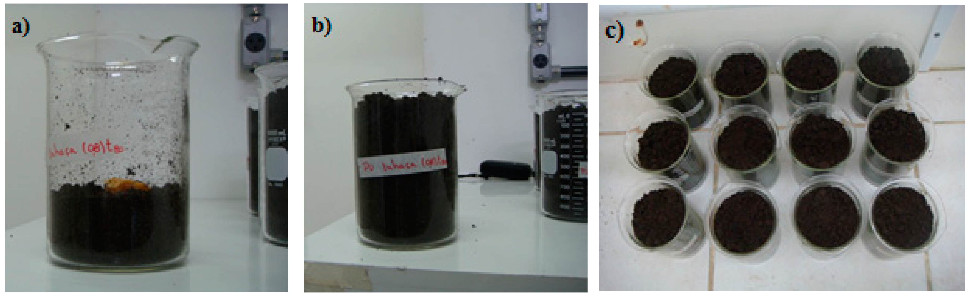
The samples were dried under vacuum for 24 h at 28 °C and, according to the standard, should be cut into 5 × 5 cm specimens. However, they were divided into three parts of equal sizes, as shown in
Figure 1 (the shape of the plate on which the PUs were cured was circular, with diameter of approximately 6 cm). The samples were placed in the soil (
Figure 2a), and the flasks were filled with soil (
Figure 2b) and incubated at 25 °C and 85% humidity (
Figure 2c).
Samples buried in the soil (one in each beaker) were removed after 40, 80, and 120 days. The samples were carefully washed with distilled water and dried in a vacuum oven at 28 °C for 24 h.
2.4. PU Characterization
Morphological analyses of the surface of the PUs obtained were performed using a scanning electron microscope, Zeiss, model DSM 962 (Jena, Germany). Sample preparation consisted of the cryogenic fracture of the material in liquid N2 and subsequent fixing of the stubs using super glue and ribbon bonder. The samples were gold-coated in a sputter Emitech (model K550) (Montigny-le-Bretonneux, France) and recorded at 10 kV. The amplified images of the PUs were acquired in a Leica stereomicroscope, model EZ4D (Wetzlar, Germany), with digital image capture system by magnifying glass of 8×.
The thermogravimetry (TG) and derivative thermogravimetry (DTG) were performed in a thermobalance Shimadzu TGA-50 (Kyoto, Japan), using a platinum crucible. The temperature was scanned up to 650 °C, with a heating rate of 10 °C·min−1, under helium atmosphere (50 mL·min−1). The sample mass was 6.0 ± 0.5 mg. The temperature of decomposition (Td) was ascertained by DTG.
The FT-IR spectra were taken with a Michelson Bomem Hartmann & Braun, Serial B, MB-100 FT-IR spectrometer (Quebec City, QC, Canada), in transmission mode. PU samples were analyzed in KBr tablets. FT-IR spectra were recorded in the 400–4000 cm−1 range.
3. Results and Discussion
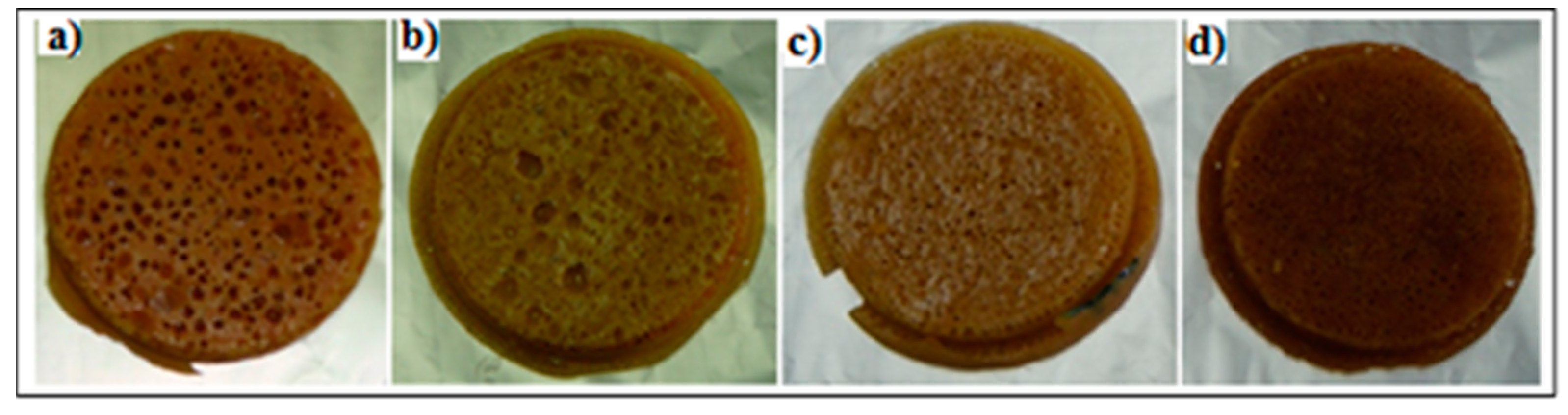
Figure 3 shows the digital images of the PU before the soil degradation tests. Digital images of materials (PU) submitted to soil degradation tests up to 120 days and samples taken periodically (40, 80, and 120 days) for analyses and consequent degradation monitoring are presented in
Figure 4.
It can be visually observed that the samples submitted to soil degradation for 40 days exhibit a less advanced stage of degradation compared to the samples exposed to same environment for 120 days. The difference in the extent of degradation is more pronounced for samples with 0.8 [NCO]/[OH] molar ratio, indicating that PUs with a [NCO]/[OH] molar ratio of 1.2 are more resistant to the action of microorganisms, with moderate deterioration. This is in agreement with the literature that indicates that the degradation is greater in the flexible segments of PUs. It is important to note that the degradation process seems to be uniform throughout the surface of the PU, especially in the PUs with 0.8 [NCO]/[OH] molar ratio, suggesting, once again, that these PUs are more susceptible to microbiological degradation.
Microbiological degradation is a selective process attributed to the smaller order of packing of the amorphous regions, which allow easy access for the enzymes to polymer chains [
36]. Based on this and because of the greater propagation of microorganisms in the PU with 0.8 [NCO]/[OH] molar ratio, it is possible to predict that these PUs are present more amorphous regions than the PU with a [NCO]/[OH] molar ratio of 1.2.
During the soil degradation process, polymers undergo hydrolysis, leading to smaller molecular units, such as oligomers and monomers. This process is known as the [
36,
37,
38] abiotic phase, which occurs without the interference of microorganisms in the first days of degradation. The biotic phase corresponds to the beginning of the microbial degradation, by the adhesion and propagation of microorganisms along the polymer chains [
39]. Based on the appearance of the PUs with a [NCO]/[OH] molar ratio of 0.8 (
Figure 4), it can be inferred that a biotic phase developed during the first 40 days of soil degradation. After 120 days in the soil, all samples appeared to develop a biotic phase, although the samples with a [NCO]/[OH] molar ratio of 1.2 PU showed areas where fungi had not yet settled.
3.1. Scanning Electron Microscopy (SEM) and Stereomicroscope
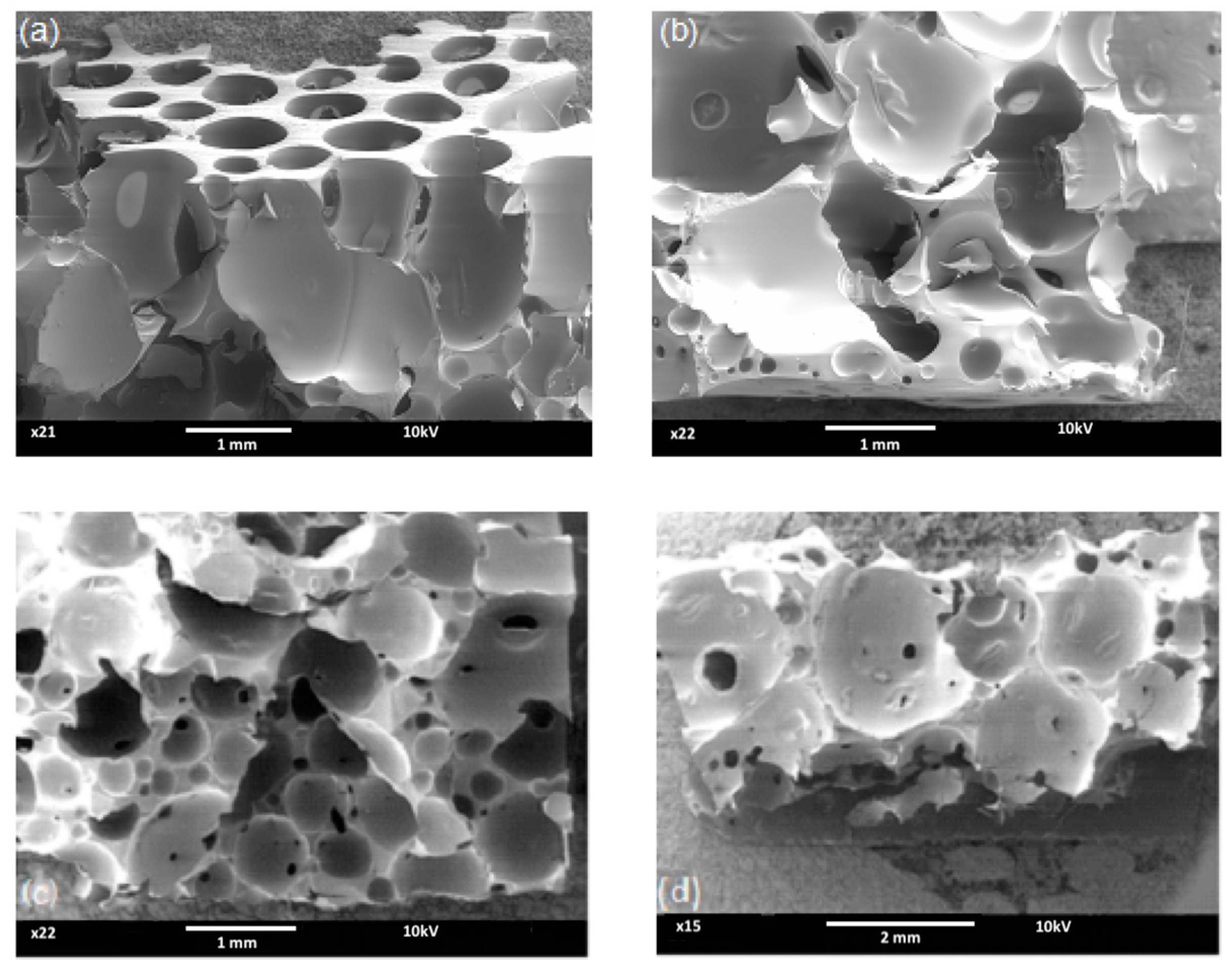
SEM micrographs obtained of PULO, with magnifications ranging from 21× to 40×, and of PUPFO, with magnifications ranging from 15× to 22×, before soil degradation, are presented in
Figure 5. SEM micrographs of PULOA and PULOB revealed the existence of closed cells and the non-homogeneity of porosity. The same was observed for PUPFOA and PUPFOB.
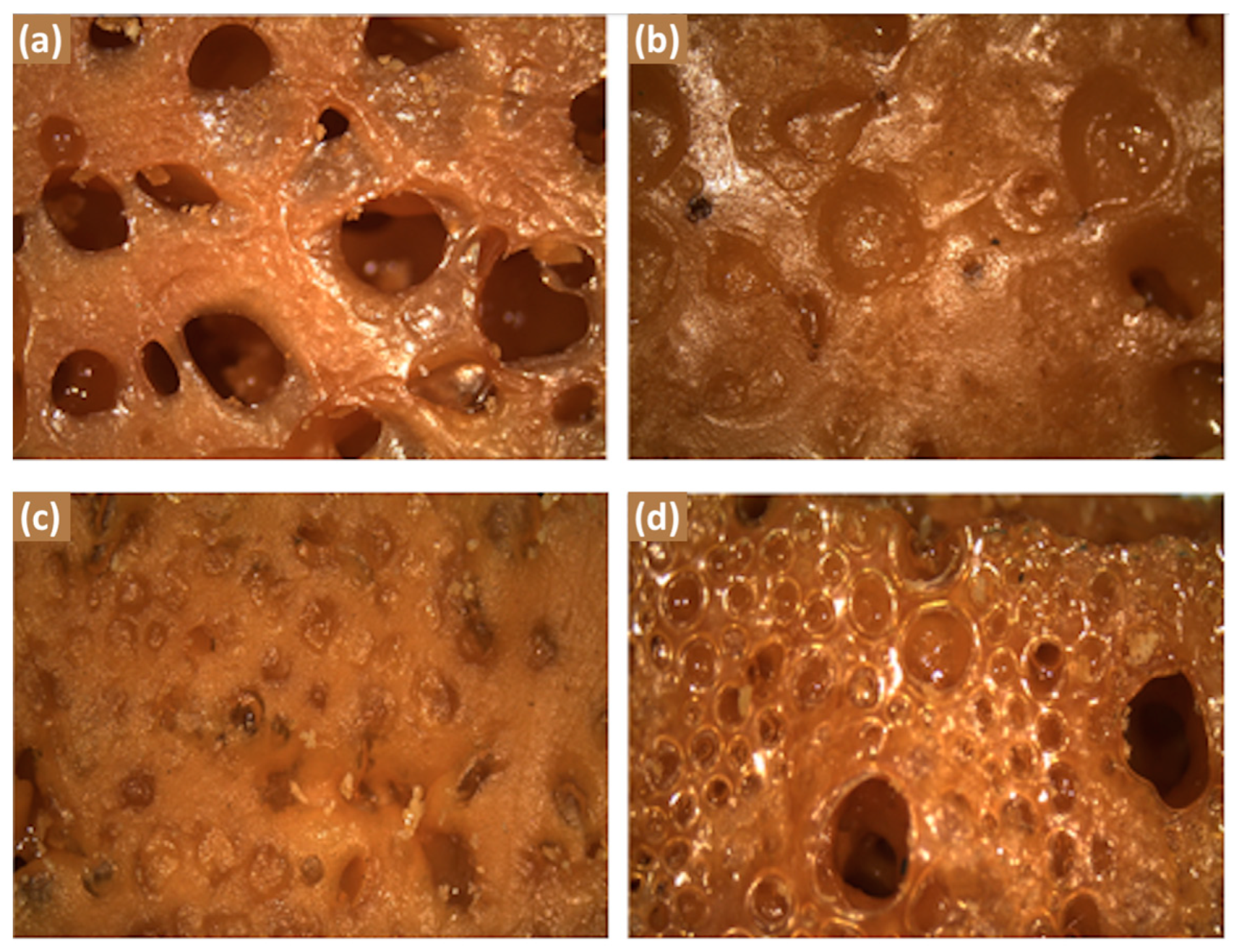
Considering that the cryogenic fracture performed on the samples to obtain images by SEM causes damage to the pores of the PU, stereomicroscope images were made for a better visualization of the pores (
Figure 6). The stereomicroscope images show the surface of the PU enlarged and in its original colors and forms. With the acquisition of approximately 30 images of each PU, the minimum, medium, and maximum pore diameters were estimated by the Axiovision se64 software (Carl Zeiss Microscopy, LLC - Jena, Germany).
Table 1 shows the results of the diameters obtained and their dependence on the molar ratio and the oil used. It is worth mentioning that the data obtained were not only performed with the images presented, but with a larger sampling, so that the analyses became representative from a statistical point of view. The data show that the molar ratio and type of oil used influences the size of the pore diameters.
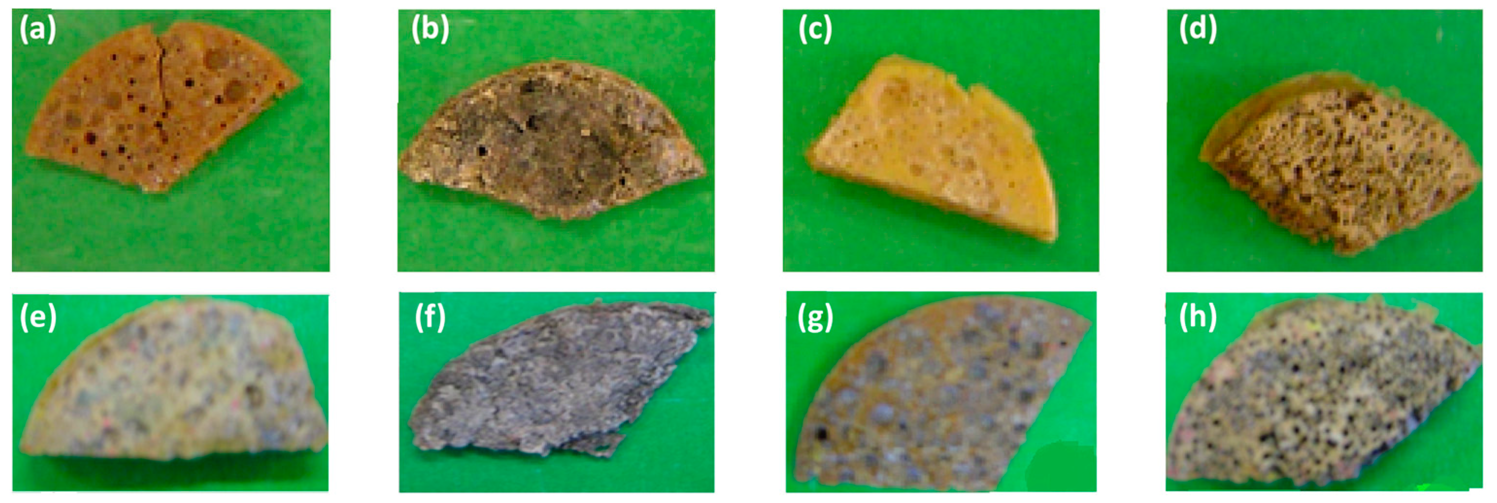
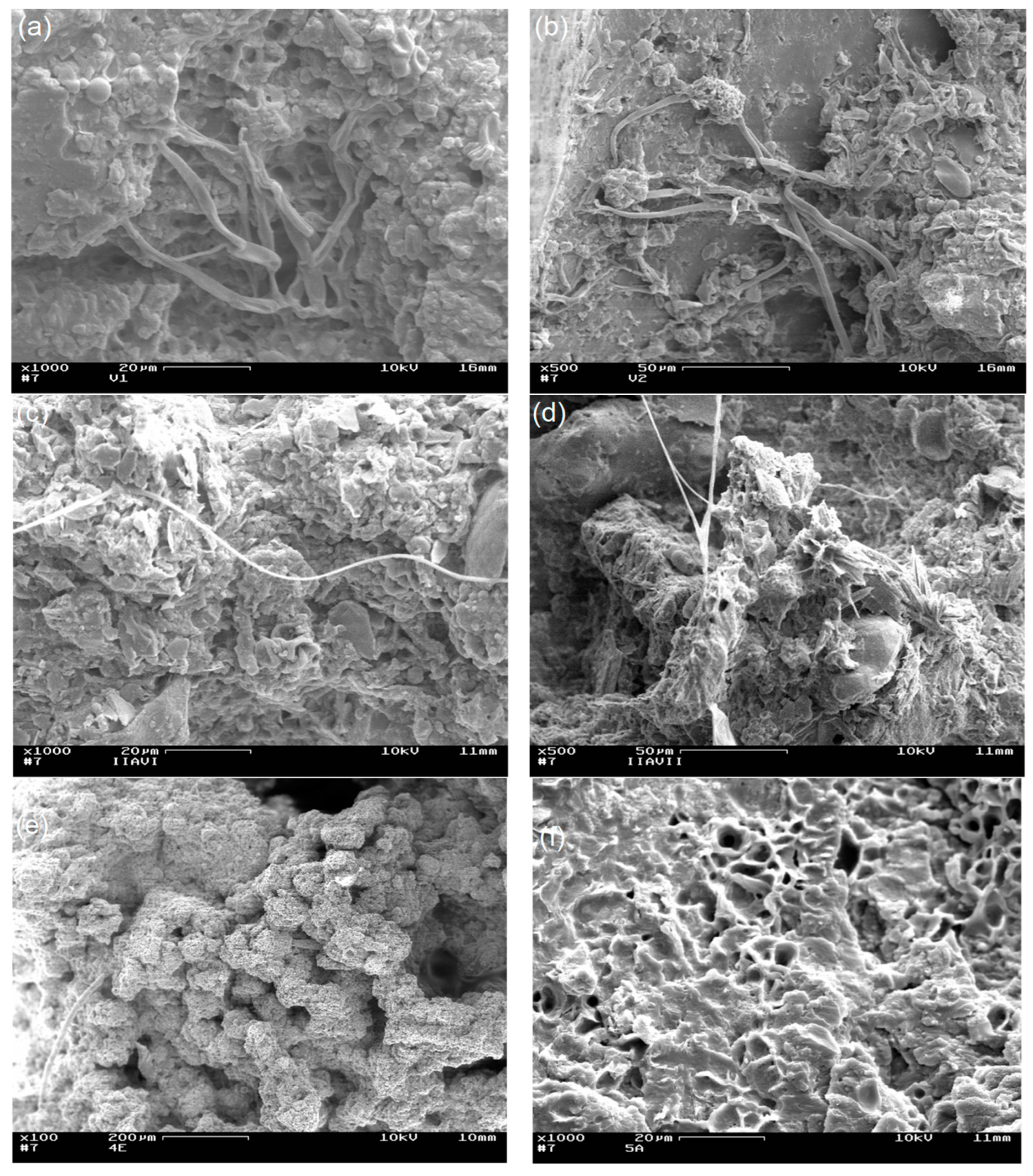
SEM micrographs of PULOA and PULOB after 40, 80, and 120 days subjected to soil degradation are shown in
Figure 7. Different magnifications were used in the micrographs of the PUs for a better visualization of the spores and hyphae. Generally, PUs are resistant to biodegradation due to the complexity of the urethane segments since such bonds are not easily hydrolysable. Studies show that PU obtained from polyethers are more resistant to microbial attack, unlike polyesters. Microbial degradation of PUs is mainly carried out by fungi because they have a higher ability to colonize solid materials than bacteria [
18,
36,
39].
SEM micrographs of PULOA and PULOB after soil degradation show the existence of erosion areas and complete coverage of their surfaces by the fungal mass, revealing the action of the microorganisms. On the surfaces of PULO, spores and hyphae can be clearly observed, although some were damaged by the removal and treatment of the materials.
Similar findings were found by Sahoo et al. [
40], who obtained PU from soybean oil with aliphatic isocyanate. They observed that, after 20 days of exposure of PU to soil burial, the formation of holes, cracks and cavities on the surface, which indicated that the PU samples was affected by the microorganisms. After 40 days of exposure deeper cracks and cavities on the surface were observed, indicating the presence of bacteria or fungi that penetrated the sample. According to the authors, this deterioration probably occurred primarily due to the presence of ester group from the soft segment, which is very sensitive to hydrolysis and microbial attack.
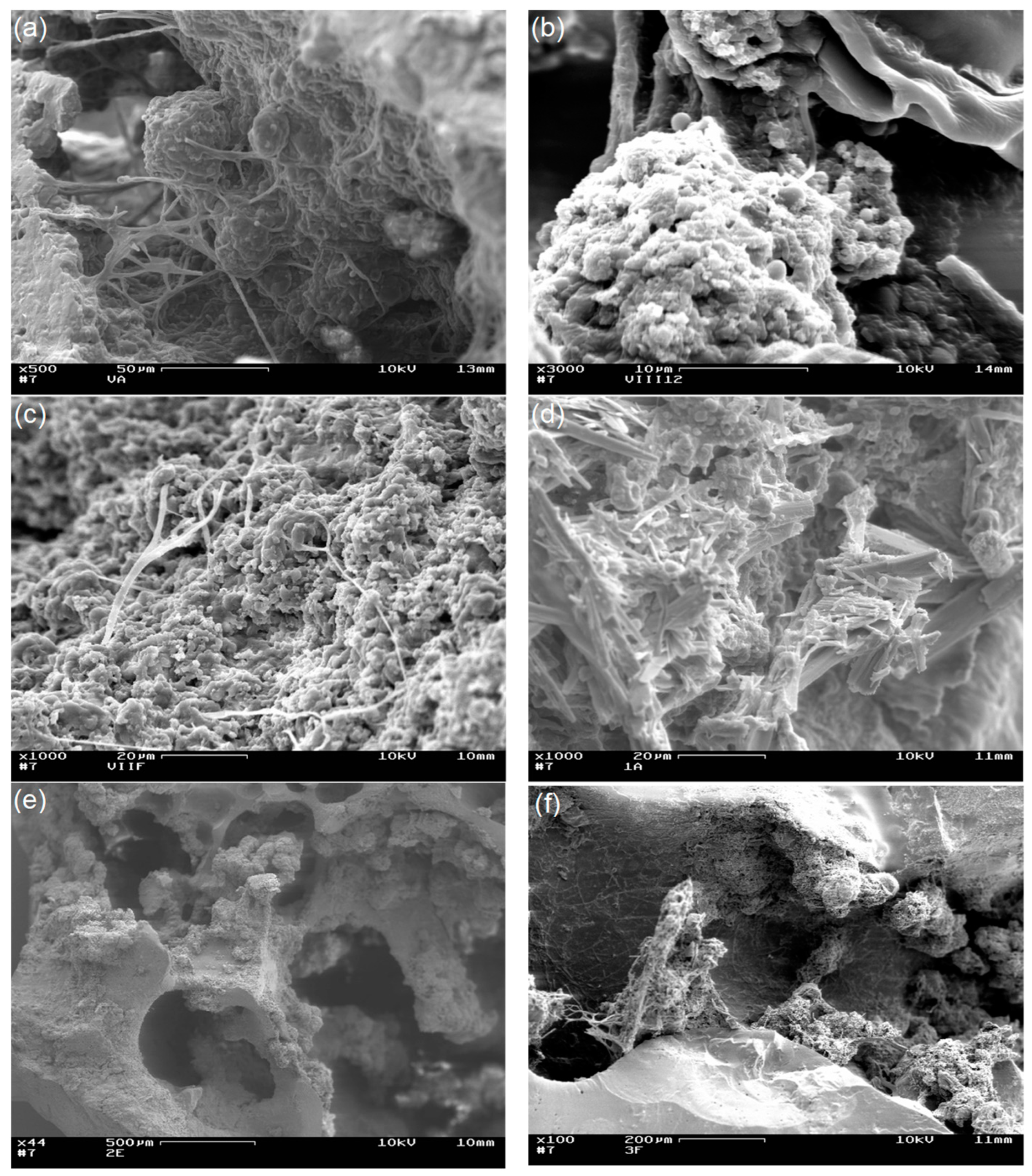
Figure 8 shows the SEM micrographs of PUPFOA and PUPFOB after 40, 80, and 120 days of soil degradation. The non-degradation of some areas of PUPFO may have occurred due to the lack of a sufficient substrate for the growth of the fungi; therefore, nutrients and the necessary additional carbon sources were missing. It can be observed that some PUPFO areas, especially those submitted to soil degradation for 120 days, exhibit a smashed appearance, indicating that, with a longer degradation time, these materials can become more fragmented. However, although the occurrence of cracks and cavities formed by microorganisms was not evident, their presence on the surface of the samples was noticeable.
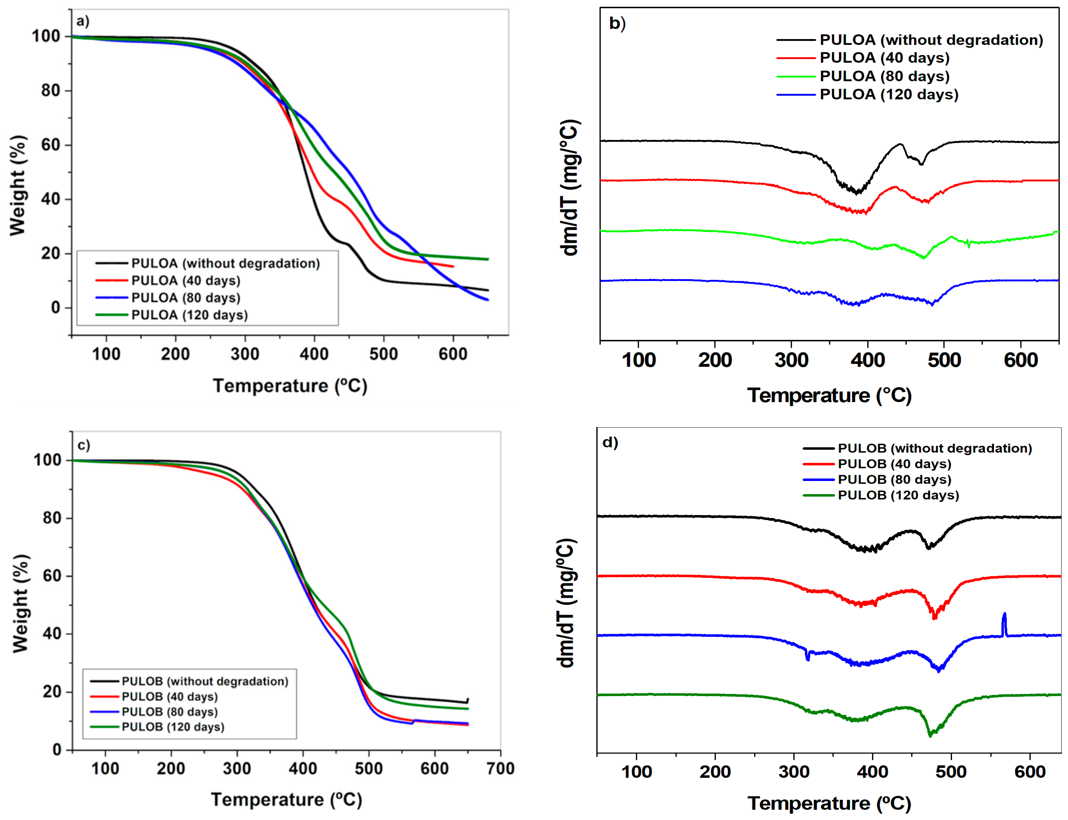
3.2. Thermal Degradation Analyses (TG/DTG)
TG/DTG analyses were used to complement the biodegradation data of the PUs studied. TG/DTG curves of PULOA and PULOB, before and after soil degradation, are shown in
Figure 9. TG/DTG curves of PULOA and PULOB exhibit differences when comparing samples before and after soil degradation. The thermal decomposition steps of the TG/DTG curves for PULOA and PULOB after soil degradation were less defined with the onset of their thermal decomposition at temperatures lower than those observed for materials before soil degradation.
After evaluating PULOA ([NCO]/[OH] molar ratio of 0.8), it was verified that after 40 days, the TG/DTG curves were similar to the PULOA curves before soil degradation, but with a thermal decomposition in lower temperature ranges (
Table 2). The DTG curve of PULOA after 80 days exhibited four successive stages of thermal decomposition. The first and second stages occurred simultaneously in the temperature range corresponding to the decomposition of the first stage of PULOA after 40 days. The third and fourth stages of PULOA decomposition (80 days) occurred, respectively, at 442–518 °C and 520–635 °C, indicating the deployment of the second stage of PULOA (40 days) (
Table 2) associated with some residues of the soil remaining in the sample. The DTG curve of PULOA after 120 days exhibited three stages of decomposition. The first two stages may be associated with the first stage of thermal decomposition of PULOA (40 days), while the third relates to the second thermal decomposition step of the same PU.
The TG/DTG curves of PULOB with a [NCO]/[OH] molar ratio of 1.2 after soil degradation for 40, 80, and 120 days, exhibited three stages of thermal decomposition, which occurred in similar ranges of temperature. It is noted that the first and second stages of thermal decomposition of this PU, for the three degradation times evaluated, occurred at close temperature ranges, corresponding to the decomposition of the first two stages of PULOB prior to soil degradation (
Table 2). It can also be observed that the third stage of decomposition for the same samples after soil degradation took place in nearby temperature ranges, as well as in the third stage of the original PULOB sample (
Table 2).
It is interesting to note that the loss of mass during the first stage of the thermal decomposition of PULOA (40 days) decreased by approximately 16%, whereas the loss of mass for the same stage of the original PULOA was 76%. After 80 and 120 days, the loss of mass of the first two stages of decomposition (45% and 48%, respectively) was attributed to a split in the first stage of PULOA (40 days), representing a considerable decrease in comparison to the loss of mass for the corresponding stage in PULOA before degradation.
Since this first stage was associated with the thermal decomposition of the urethane bonds, this means that with the increase in the residence time of the PU samples in the soil, there was a decrease in the decomposition of these bonds. It is suggested that at this stage there was no predicted thermal decomposition of the urethane bonds, having as reference the original PULOA, as a function of a type of “shielding” by the mycelium, as suggested in the literature [
13,
18,
39,
41,
42].
In the thermal decomposition stage (340–550 °C) related to the carbon chains, an increase in PULOA mass loss was observed after 40, 80, and 120 days, compared to the original PU (14%), indicating a probable fragmentation of the chains by microorganisms. A similar behavior was observed for the PULOB samples with a [NCO]/[OH] molar ratio of 1.2. Three stages of decomposition were observed, relating to the thermal decomposition of urethane bonds after 40, 80, and 120 days. As for the mass loss of the PULOB carbon decomposition stage after 40, 80, and 120 days, a small increase and a higher rate of decomposition can be observed in comparison to the original PULOB (26%).
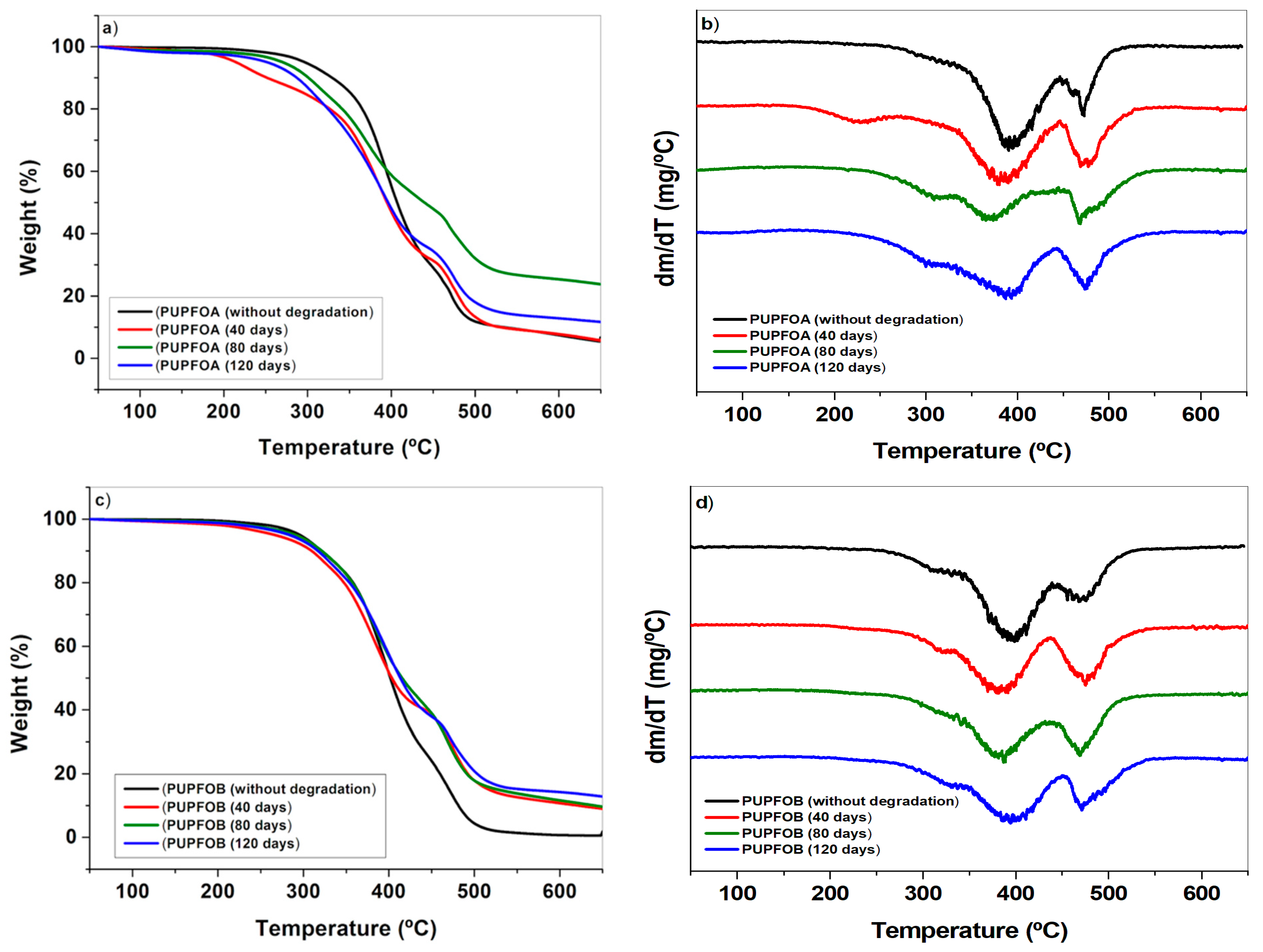
Figure 10 presents the TG/DTG curves of PUPFOA with [NCO]/[OH] molar ratio of 0.8 and PUPFOB with molar ratio of 1.2 after soil degradation for 40, 80, and 120 days. The TG/DTG curves of PUPFOA and PUPFOB after soil degradation exhibit small differences when compared to the TG/DTG curves of their original samples. The TG/DTG curves of these samples indicate a lower thermal stability after soil degradation in comparison to their respective starting samples.
DTG curves of PUPFOA exhibit three stages of thermal decomposition after soil degradation. The first and second stages occur at temperature ranges similar to the first stage of PUPFOA. Its third stage of thermal decomposition can be associated with the second stage of the original PUPFOA, due to the similarity in the temperature ranges. Similar to PUPFOB before being subjected to soil degradation, the samples subjected to soil degradation for 40, 80, and 120 days exhibited two stages of thermal decomposition.
It was verified that after soil degradation, the mass lossed for the different thermal degradation stages of the PUPFO samples were not significantly different in relation to the values for the respective starting samples. However, in general, it was noticed that the mass loss of the first stage displayed a small decrease while, in the second stage, a small increase was observed. According to Al-Atroush and Sebaey [
43], homogeneous foams have a closed-cell and nearly impermeable structure. Although the foams under study are not homogeneous in their entirety, it is possible that, during the biodegradation test, there was no water permeability in the materials’ cells. Therefore, three stages of thermal degradation in the materials were observed, similar to the starting material.
Among the factors that affect biodegradation, functional group availability (imparting hydrophobicity), complexity in structure, and the presence of weaker bonds (amide and ester linkages) stand out. Additionally, polymers that are soft degrade faster than those that are hard [
13]. Some of these factors may contribute to the partial biodegradation and thermal stability of the materials studied herein.
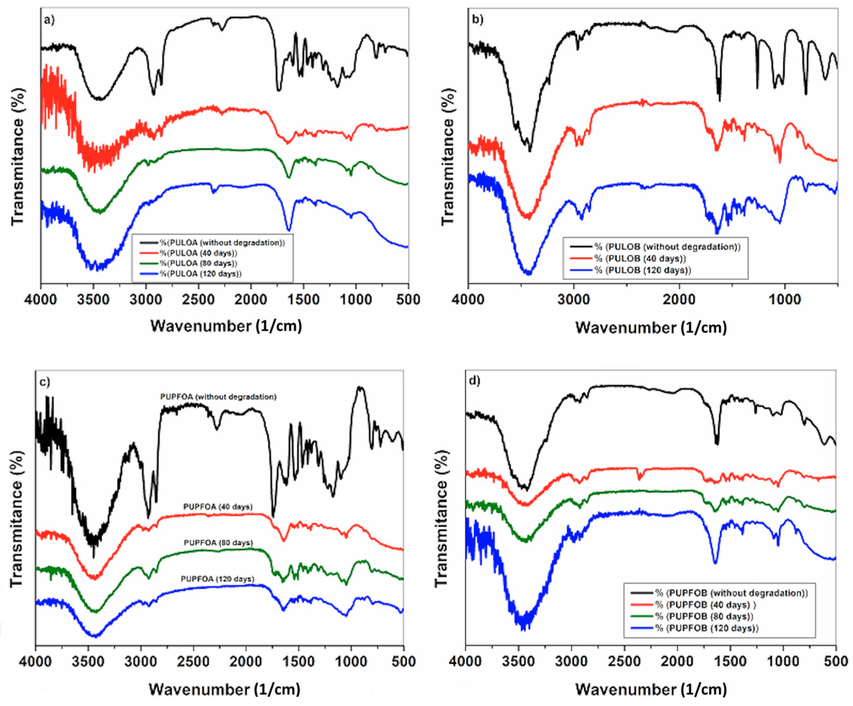
3.3. FT-IR Spectra
Figure 11 shows the FTIR spectra of PUs originated from LO and PFO after soil degradation for 40, 80, and 120 days. It can be seen that all samples have a similar FT-IR spectrum before soil degradation. In
Figure 11a, only a decrease in the signal at 2277 cm
−1 is observed and may be related to the transmittance of CO
2. The band near 3340 cm
−1, present in all spectra, is associated with N-H stretching in the urethane linkage and amino groups of the PUs because both types of groups are capable of forming hydrogen bonds, inferring the presence of hydrogen bonds in the chains of the PUs. The same urethane absorption peaks at 3340 cm
−1 found by Sahoo et al. [
40] for time of 40 days corresponding to –NH stretching in the urethane linkage. This stretch is present in all spectra, is associated with amino groups of the PUs because both types of groups are capable of forming hydrogen bonds inferring the presence of hydrogen bonds in the chains of the PUs [
16,
32,
40,
41,
43,
44].
In the FT-IR spectra of
Figure 11b–d, it is still noticeable that the OH band at ~3400 cm
−1 persists. This may be related to the preparation of the sample for analysis as it was sprayed with liquid nitrogen. It is assumed that, after the evaporation of nitrogen, water was incorporated in the samples. The absence of the band at 2260 cm
−1 was observed, which according to Sahoo et al. [
40], indicated the complete utilization of the isocyanate groups with the hydroxyl groups.
The band identified at 1646 cm
−1 was assigned to carbonyl [
44,
45,
46]. The characteristic band of the polyester (CO-O) bond occurred at 1242 cm
−1 [
9,
11,
47]. The occurrence of stretching was also observed in the 1084–1064 cm
−1 region, characteristic of the N-CO-O group of urethane. Between 900 cm
−1 and 675 cm
−1, axial deformation vibrations, characteristic of the off-plane angular deformation of the C-H bonds of aromatic rings were identified [
47].
After soil degradation, the FT-IR spectra of the PUs presented few changes with respect to the FT-IR spectra of the original samples, but there was an indication that the degradation process started, since in the spectra of the degraded PUs, characteristic bands of the PUs were still identified.
The presence of the coupled asymmetric vibration of the ester function [CC(=O)-O] at 1242 cm
−1 indicated that hydrolysis occurred in the flexible segments of the PUs during soil degradation [
39]. The presence and increase in the signal intensity at 3382 cm
−1 in some samples could be justified by the absorption of water during soil degradation. It was also observed that the occurrence of the stretch band at 1084 cm
−1 and 1064 cm
−1, characteristic of the N-CO-O group of urethanes. However, the bands at 900 cm
−1 and 675 cm
−1, related to axial strain vibrations characteristic of the out-of-plane angular deformation of C-H bonds of aromatic rings, did not appear due to the probable interference of other components present in the soil.
Comparing the spectra obtained by Sahoo et al. [
40] to the spectra of this work, there are few similarities. This may be related to the type of synthesis to obtain the polyol, the type of isocyanate used and the quality of the spectra obtained.


 ,
, 


