Article

Coralline Hydroxyapatite Coated with a Layer Biomimetic Calcium Phosphate Containing BMP-2 Induces Dose-Related Ectopic Bone Formation in Wistar Rats

1
Department of Oral Cell Biology, Academic Centre for Dentistry Amsterdam (ACTA), Gustav Mahlerlaan 3004, 1081LA Amsterdam, The Netherlands
2
School/Hospital of Stomatology, Zhejiang Chinese Medical University, Binwen Road 548, Hangzhou 310053, China
3
Stomatology Hospital, School of Stomatology, Zhejiang University School of Medicine, Clinical Research Centre for Oral Diseases of Zhejiang Province, Key Laboratory of Oral Biomedical Research of Zhejiang Province, Cancer Centre of Zhejiang University, Hangzhou 310006, China
4
Private Practice, Zutphensestraatweg 26, 6955AH Ellecom, The Netherlands
5
Department of Oral Implantology, Yantai Stomatological Hospital, No.142 Beida Street, Yantai 264001, China
*
Author to whom correspondence should be addressed.
These authors contributed equally.
Coatings 2021, 11(10), 1195; https://doi.org/10.3390/coatings11101195
Submission received: 22 July 2021 / Revised: 20 September 2021 / Accepted: 26 September 2021 / Published: 30 September 2021

Abstract

:
In order to evaluate loading methods and the dose dependency of bone morphogenetic protein 2 (BMP-2) in ectopic bone formation, an osteoinductive material consisting of commercially available coralline hydroxyapatite (CHA) was coated with a layer of biomimetic calcium phosphate (BioCaP) containing BMP-2 in different ways. Eight groups—each containing samples of 0.25 g CHA—were formed and coated with, respectively, BioCaP with internally incorporated BMP-2 in concentrations of 1, 5, 10, 20, 40 and 60 µg per sample, and the two control groups with BioCaP only and BioCaP with 20 µg of adsorbed BMP-2 per sample. The samples were implanted subcutaneously in 27 male Wistar rats. The histological results show that there is no bone formation in the group in which no BMP-2 was included. All samples with BioCaP containing BMP-2 show bone formation. The group with 20 µg of adsorbed BMP-2 per sample shows the least bone formation. Coating-incorporated BMP-2 is more efficient in inducing bone formation than adsorbed BMP-2. The group with 5 µg of coating-incorporated BMP-2 per sample shows the most bone formation. Increasing the amount of coating-incorporated BMP-2 up to 60 µg does not improve ectopic bone formation.

1. Introduction

Bone defects develop due to different causes, such as congenital disorders, infections, cysts, trauma and tumors. A bone defect that is too large to heal spontaneously is called a critical-sized bone defect [1]. The repair of a critical-sized bone defect often results in fibrous connective tissue instead of bone [1]. To prevent this unwanted effect, the use of autologous graft materials are used when treating critical-sized bone defects, which was first described in 1875 [2]. However, the use of autologous graft material implies a donor site, which, when harvesting bone from that site, is sometimes accompanied by both complications at the donor site itself and inconvenience for the patient [3,4]. To avoid these undesirable effects, other graft materials have been developed, such as allograft, xenograft and synthetic graft materials [5]. Although autologous graft material is often described as the gold standard [6], research shows that no graft biomaterial is predominant with regard to its healing capacity [7].
Marine coral is used as a natural bone graft material. Through a hydrothermal process, its calcium carbonate skeleton converses from coral to hydroxyapatite [8], thus forming coralline hydroxyapatite (CHA). CHA has been used since 1979 [9] as a bone graft material. CHA is osteoconductive and, as such, provides a biocompatible lattice for the passage and assembly of vascular, fibroblastic and osteoblastic tissues. It also provides support for surrounding osseous structures [10]. The benefits of CHA as a bone graft are predominantly its safety, biocompatibility and osteoconduction [8].
The best material for bone healing has the following properties: osteoconduction, osteoinduction and osteogenesis [6]. For surgery, primary tension-free closure, angiogenesis, space creation/maintenance and stability are prerequisites [11]. CHA has no osteoinductive properties [8]. This can, however, be achieved by incorporating an osteoinductive agent [12]. By coating CHA with our biomimetic coating and incorporating bone morphogenetic protein-2 (BMP-2), CHA could develop both osteoconductive and osteoinductive properties [13].
BMP-2 is a pleiotropic morphogen that induces a sequential cascade of events in the bone healing process, including chemotaxis, the regulation of osteoblasts, differentiation, angiogenesis and osteoclasts apoptosis [14]. In vitro and in vivo studies have demonstrated that BMPs are capable of enhancing the osteoinduction of mesenchymal stem cells (MSCs), regulating their proliferation and differentiation into osteoblasts [14,15]. BMP-2 incorporated into biomimetic calcium phosphate (BioCaP) coatings are capable of inducing ectopic bone formation in vivo [12]. BMP-2 can be bound to BioCaP by (i) adsorption, (ii) coating incorporation or (iii) internal incorporation [15].
At (i) adsorption, the BMP-2 protein is absorbed by immersing the BioCaP in a solution with BMP-2. The BMP-2 is mainly absorbed on the surface of the outside of the material. Adsorbed BMP-2 is released with a diffusion-controlled burst release [15,16]. During the production of the (ii) BioCaP coating-incorporated BMP-2, BMP-2 and BioCaP precipitate simultaneously on a calcium-based support. A BioCaP latticework with incorporated BMP-2 grows onto the support material: the BioCaP BMP-2 coating. The BMP-2 can only be released after BioCaP is dissolved [15]. If BMP-2 and BioCaP simultaneously precipitate on amorphous CaP (ACaP) to form a unit consisting exclusively of BioCaP and BMP-2, we refer to it as (iii) BioCaP with internally incorporated BMP-2 [12].
Our earlier studies [12,16] show that BMP-2 can be incorporated in BioCaP and is an effective bone formation inducer. Previous studies also show that the coating of CHA with BioCaP and BMP-2 induces bone formation [13]. However, we do not know which concentrations of BMP-2 are effective in ectopic bone formation when CHA is coated with BioCaP containing BMP-2. An animal model is often used as a method to study the efficacy of biomaterials [17]. We made samples consisting of coated CHA with BioCaP coating-incorporated BMP-2 in different concentrations, as well as BioCaP-adsorbed BMP-2. The samples were placed subcutaneously in Wistar rats to study the efficacy of the BMP-2 concentrations at ectopic bone formation.

2. Material and Methods

2.1. Material Preparation

2.1.1. Coating of CHA Granules with BioCaP

CHA granules (YHJ Bio-Osteon®, Beijing, China) with a granule size of 0.4–1.0 mm and a total weight of 0.25 g were biomimetically coated with a layer of BioCaP according to the established protocols [12,15,18]. The biomaterial is first immersed—whilst stirring at 150 r.p.m—in a fivefold concentrated simulated body fluid (684 mM NaCl; 12.5 mM CaCl2·2H2O; 21 mM NaHCO3; 5 mM Na2HPO4·2H2O) for 24 h at 37 °C under high-nucleation conditions (in the presence of 7.5 mM MgCl2·2H2O) to inhibit crystal growth. A fine, dense layer of amorphous calcium phosphate (ACaP) is formed on the surface of the CHA [15,19]. This layer serves as a seeding substratum for the deposition of a more substantial crystalline layer. The crystalline layer is produced by immersing the ACaP biomaterials in a supersaturated calcium phosphate (CaP) solution [40 mM HCl; 2 mM Na2HPO4·2H2O; 4 mM CaCl2·2H2O; 50 mM TRIS base (pH 7.4)] for 48 h at 37 °C, 60 r.p.m. whilst shaking, buffered with tris(hydroxymethyl)aminomethane (3.76 mM; pH 7.4). Through this procedure, CHA granules with an outer layer of octa calcium phosphate (OCP) are prepared [13,19]. The hydrated and carbonated calcium phosphate is now called BioCaP.

2.1.2. Preparation of BioCaP with and without Coating-Incorporated BMP-2

Adding BMP-2 (INFUSE®, Medtronic, Minneapolis, MN, USA) in the different concentrations of none, 1, 5, 10, 20, 40 and 60 µg/sample to this supersaturated CaP solution will result in co-precipitation of BMP-2 into the OCP coatings of the CHA granules, thus creating the group in which CHA was coated with BioCaP coating-incorporated BMP-2. All of the samples were freeze-dried at a temperature of around minus 30 °C. The entire procedure was performed under sterile conditions.

2.1.3. Preparation of CHA Coated with BioCaP and Adsorbed BMP-2

The samples with 0.25 g CHA coated with BioCaP and adsorbed 20 µg of BMP-2 per sample were made by merging 0.2 mL BMP-2 solution (100 µg/mL) with 0.25 g of CHA granules, after which followed centrifuge for 2 s with a relative centrifugal force (RCF) of 1019 and vortex. All of the samples were freeze-dried. The entire procedure was performed under sterile conditions.

2.1.4. Control of the Amount of Adsorbed or Incorporated BMP-2

To determine the BMP-2 incorporation, the BMP-2 release is determined, as very well described by Wu [18]. By monitoring the release kinetics of a coating-incorporated depot of protein, BMP-2 (10 μg/mL) was introduced into the supersaturated solution of calcium phosphate. Six samples were used to determine the total amount of incorporated BMP-2. These samples were immersed in 1 mL of 0.5% EDTA (pH 8.0) and vortexed twice for 5 min to ensure complete dissolution of the coatings. The supernatants were withdrawn for analysis of total loading of BMP-2. To monitor the release kinetics, six samples of CHA bearing a coating-incorporated depot of BMP-2 and six samples of CHA bearing an equivalent amount of adsorbed BMP-2 (included for the purpose of comparison) were incubated in sealed 10-mL glass tubes containing 2 mL of phosphate-buffered 0.9% saline (pH 7.4). The tubes were incubated for up to 35 days in a shaking water bath (60 agitations/min), which was maintained at 37 °C. Before each sampling, a mild centrifuge step was performed to collect all of the solution (part of which may stick to the cap and wall of the tubes due to evaporation in the 37 °C water bath). Triplicate 200-μL aliquots of the medium (containing released BMP-2) were withdrawn for analysis after 3 h, 6 h, 9 h, 1 day, 2 days, 3 days, 5 days, 7 days, 10 days, 14 days, 18 days, 23 days, 28 days and 35 days. The fluorescence density was measured in a spectrophotometer (Biotek Instruments Inc., Winooski, VT, USA; excitation wavelength: 485 nm; emission wavelength: 519 nm). The fluorescence readings were converted to amounts of protein using a standard curve, which was generated by preparing a dilution series of BMP-2 in 5 mL of phosphate-buffered 0.9% saline. The temporal release of BMP-2 was expressed as a percentage of the total amount that had been incorporated into the crystalline layer of the calcium phosphate coating or that had been adsorbed directly onto the CHA granules. The absolute amount of 20 µg of BMP-2 was directly added on the surface of 0.25 g CHA BioCaP-coated samples, which were the absorbed ones.

2.1.5. Surgical Procedure

Twenty-seven adult male Wistar rats, each weighing between 200 and 220 g and approximately 3–4 months old, were used in this study. We used male Wistar rats to avoid any differential effects due to animal gender. Every surgical intervention took place under anesthesia by administering pentobarbital sodium (50 mg/kg IP). A systematic random sampling protocol was used for the distribution of the samples over the rats. They were numbered from 1 to 27, after which, the samples out of one group were divided at random over 6 rats, so each rat received 2 different samples from different groups. The animals were fed a standard diet and had unlimited access to water. After shaving, the skin was disinfected. An incision was made, after which, two subcutaneous pockets were created in each rat: one on the right dorsal side and one on the left. According to the distribution in Table 1, one sample of 0.25 g CHA granules was directly placed into the surgically created pocket on each side in 27 rats, following a systematic random sampling protocol. The incisions were closed by suturing and thus entrapping the samples.

2.1.6. Study Design

All of the animal experiments were carried out according to the ethics laws and regulations of People’s Republic of China. The experiment was approved by the committee of the Board of Animal Experiments, traditional Chinese Medicine University, Hangzhou, China (Accreditation number: ZSLL-2016-46). Fifty-four samples, each containing 0.25 g of coralline hydroxyapatite (CHA) granules, were made. BioCaP with concentrations of 1, 5, 10, 20, 40 and 60 µg/sample of incorporated BMP-2 were made, as well as 20 µg/sample of adsorbed BMP-2. The CHA samples were coated with BioCaP, thus creating five groups containing six samples per group and three groups containing eight samples per group. Thus, eight groups were formed, as displayed in Table 1.

2.1.7. Histological Observation

Five weeks after implantation of the samples, the animals were sacrificed by an overdose of gaseous carbon dioxide. The samples with their surrounding tissues were obtained. These were dehydrated in serial steps in alcohol and xylol and embedded in methylmethacrylate (MMA, Technovit® 7200 VLC Exact®, Heraeus Kulzer Wehrheim, Germany) [20,21]. The samples were cut using a systematic random sampling protocol with a diamond saw parallel to their longitudinal axis. The distance between the slices was set at 1.0 mm. Six slices per sample were obtained, and a total of 324 slices were evaluated. Every slice was mounted separately on a plexiglass holder and polished to approximately a thickness of 100 μm. The slices were then stained with McNeal’s tetrachrome, basic fuchsine and toluidine blue [20].

2.1.8. Histomorphometric Analysis

With a Nikon-Eclipse light microscope (Nikon, Tokyo, Japan), photos were taken of all of the slices. The photos were printed in color in A4 format. The total area of interest was bounded by the external boundary of the connective tissue surrounding the samples. The volumes of bone, CHA and total area were determined per slice. The histomorphometric analysis was performed by randomly placing an appropriate grid over the prints and manually point counting according to Cruz-Orive [22].

2.1.9. Statistical Analysis

The hits that were achieved by manual point counting of newly formed bone, remaining CHA and total area were determined for each slice. Out of these hits, the means of the hits for newly formed bone, remaining CHA and total area were calculated per sample. The percentage of newly formed bone in relation to the total area, as well as the remaining CHA in relation to the total area, were calculated. These percentages were used as input for the statistical analysis, using IBM SPPS statistical software version 26.0. for Windows 10 (IBM corporation, Armonk, NY, USA). The total p-value of the one-way analysis of variance (ANOVA) was <0.01 for all three percentages, meaning that the means are statistically significant for the different groups. An LSD test was then used for pairwise comparisons between two groups within one percentage, setting p < 0.05 as statistically significant. All numerical data are presented as the average of one sample—obtained out of all the slices of that sample - per group with means and outliers.

3. Results

3.1. In Vitro Results

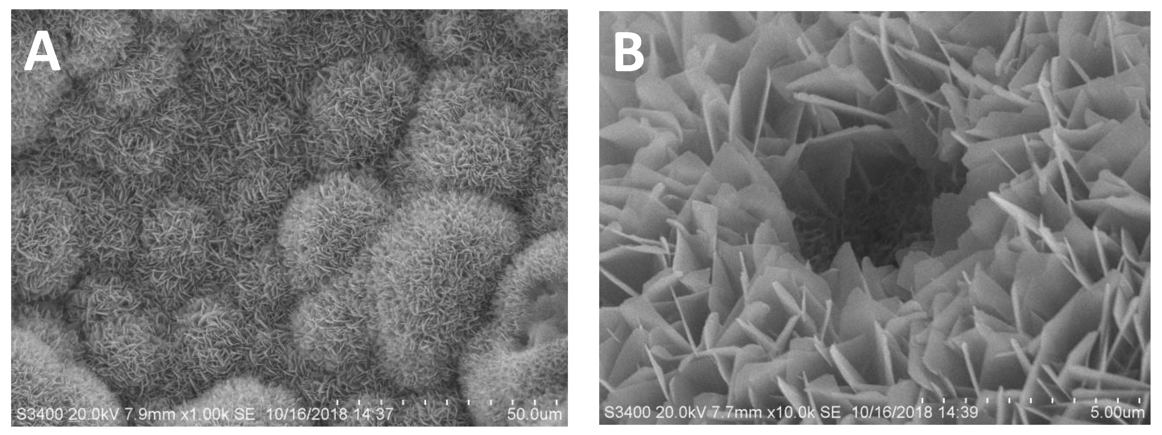
3.1.1. Material Preparation

The scanning electron microscope (SEM) images (Figure 1) showed that the structure of CHA is well interconnected. After coating the CHA with OCP containing BMP-2, an interwoven network of needle-like and plate-like crystals was formed. Our previous study [13] showed that the pore size of the obtained CHA varied from approximately 100 till 600 µm and that the OCP coating thickness was approximately 20 to 30 µm, which is approximately 3% to 30% of the CHA pore size. The trabecular structure is not changed by coating the CHA [13].

3.1.2. Amount of Encapsulated BMP-2

Using an enzyme-linked immunosorbent assay (Elisa, PreproTech EC, London, UK), as described in our earlier studies [12,23], showed that the amount of BMP-2 encapsulated in the samples was approximately 0, 1, 5, 10, 20, 40 and 60 µg of BMP-2 per sample.

3.2. Observations

After the implantation, the wounds healed well in all rats, and there were no wound infections clinically visible. We experienced no losses of rats and/or samples during the clinical phase.

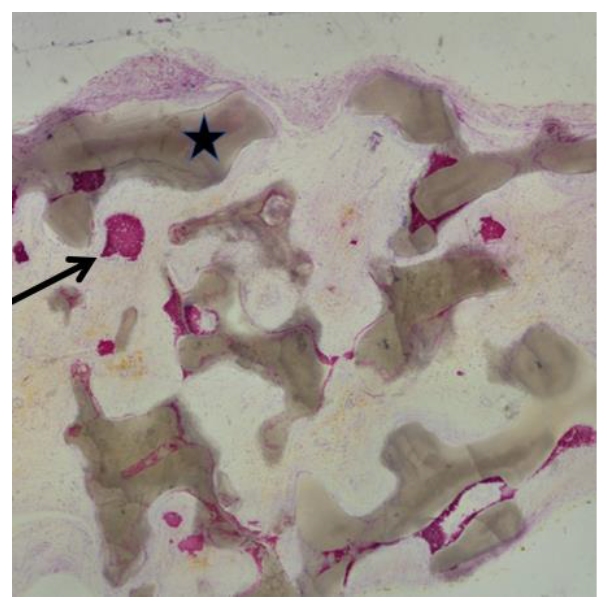
3.3. Histological Findings

All samples were completely surrounded by soft tissues, which is to be expected. In the group of 0.25 g CHA coated with BioCaP containing no BMP-2 per sample, no bone formation was found (Figure 2A). The CHA granules were surrounded by fibrous tissue. In all other groups, which all contained BMP-2, newly formed bone was present in direct contact with the BioCaP coating of the CHA particles, which is visible in Figure 2B–G. Occasionally, we saw an island of solitary newly formed bone with no direct contact to the BioCaP coating of the CHA particles (Figure 3). It seemed that newly formed bone was more located in the center of the samples than in their periphery.

3.4. Histomorphometric Analysis

The bone/total area percentage was zero in the group of 0.25 g CHA coated with BioCaP and containing no BMP-2 per sample, indicating that there was no bone formed at all. The bone/total area percentage of the group with 0.25 g CHA coated with BioCaP containing 20 µg of adsorbed BMP-2 per sample was significantly less than all other groups with BMP-2, with the exception of the group of 0.25 g CHA coated with BioCaP containing 20 µg of coating-incorporated BMP-2 per sample.
The CHA/total area percentage shows that the groups with 0.25 g CHA containing 1, 5 and 10 µg of coating-incorporated BMP-2 per sample behave differently compared to groups with 0.25 g CHA containing 0, 20, 40, 60 µg of coating-incorporated BMP-2 per sample, as well as with the group of 0.25 g CHA containing 20 µg of adsorbed BMP-2 per sample. There seems to be more remaining CHA in groups with 0.25 g CHA containing 1, 5 and 10 µg of coating-incorporated BMP-2 per sample compared to the other groups.

3.5. Statistical Findings CHA/Total Area Percentage

In Figure 4, Figure 5 and Figure 6, the whisker extending down represents the minimum value, excluding any outliers. The whisker extending up represents the maximum value, excluding any outliers. An outlier is shown as a dot (Figure 6). The bottom of the rectangle represents quartile one, and the top of the rectangle represents quartile three. The median is shown by the horizonal line in the rectangle. The mean is marked with X in the box. The tests performed, with their confidence intervals, are described in Section 2.1.9.
Figure 4 illustrates that groups of 0.25 g CHA containing 1, 5, 10 µg/sample of coating-incorporated BMP-2 and 20 µg/sample of adsorbed BMP-2 contain the most non-degraded CHA within all groups, whereby the group of 5 µg/sample of coating-incorporated BMP-2 within these groups shows the least spread in CHA/total area percentage. Two outliers show a percentage > 100, which can be caused by the cutting of the slices and the position of the grid at point counting.

3.6. Statistical Findings Bone/Total Area Percentage

The group containing 20 µg/sample of adsorbed BMP-2 induces less ectopic bone formation compared to all other groups with coating-incorporated BMP-2. In addition, the box-plot in Figure 5 shows that the group with 0.25 g CHA containing 20 µg of adsorbed BMP-2 per sample is less efficient in bone formation than the group with 0.25 g CHA containing 20 µg of coating-incorporated BMP-2 per sample; however, there is no statistically significant difference (Figure 5). Increasing the amount of incorporated BMP-2 from 5 up to 60 µg per sample does not result in more ectopic bone formation. There are statistically significant differences between all groups, whereby the group of 5 µg of coating-incorporated BMP-2 per sample shows the highest bone/total area percentage within the range of 1 to 60 µg/sample of coating-incorporated BMP-2, when the outlier of the group of 1 µg/sample of coating-incorporated BMP-2 is excluded.

3.7. Statistical Findings Bone/CHA Percentage

The group without BMP-2 has a bone/CHA percentage of 0, whereas the group of 0.25 g CHA containing 20 µg of adsorbed BMP-2 per sample shows the lowest bone/CHA percentage (Figure 6). The groups of 0.25 g CHA containing 40 and 60 µg of coating-incorporated BMP-2 per sample show the highest bone/CHA percentage. There are no statistical differences (Figure 6) between the groups of 0.25 g CHA containing 1, 5, 10 and 20 µg of coating-incorporated BMP-2 per sample.

4. Discussion

To evaluate the efficacy in bone formation of different BMP-2 concentrations in coating-incorporated CHA, in vivo research is necessary. Choosing the right animal model depends on various factors, such as significant physiological and pathophysiological analogies when compared to humans, its manageability to operate, after which it is easily possible to observe a multiplicity of study objects over time, its post-surgical period, which is preferably relatively short, the costs for animal acquisition and care, animal availability, tolerance to captivity, ease of housing and acceptability to society [17]. Male Wistar rats meet the described requirements well. However, rodent models do not show Haversian-type remodeling in the bone cortex, whereas larger animals do [17]. We opted for an ectopic ossification model because we wanted to exclude influences of any possible spontaneous bone healing.
Studies have shown that a burst release of BMP-2 alone leads to insufficient bone formation [6,24], whilst coating-incorporated BMP-2 is more effective compared to the same amount of adsorbed BMP-2 [16]. This is due to the long, slow and stable release of the coating-incorporated BMP-2 compared to the burst release of adsorbed BMP-2 [24]. However, we did not know if this is also the case with coated CHA [16]. We also did not know which concentration of BMP-2 was the most effective using coated CHA [13]. Our initial research was with five groups and a total of 15 animals. Due to the fact that we did not know which coating-incorporated amount of BMP-2 would be effective in ectopic bone formation, we started with 0 µg of BMP-2 in the control group, increasing in every next group with 20 up to 60 µg/sample per group. To minimize animal sacrifice, we made only one control group with absorbed BMP-2. This group (6) contained 20 µg of adsorbed BMP-2 per sample. For bone/total area percentages, no statistically significant differences were found (Figure 5) between groups 5, 7 and 8 (20, 40 and 60 µg of coating-incorporated BMP-2 per sample). However, the concentrations of the coating-incorporated BMP-2 per sample differ from 20 to 60 µg. A concentration above 20 µg of coating-incorporated BMP-2 per sample did not result in more ectopic bone formation. This is why we started our second research. We created a greater sample size of three groups with a total of 12 animals and a coating-incorporated BMP-2 of 1, 5 and 10 µg per sample.
Ideal bone graft material should provide an osteoconductive matrix, which allows for vascular invasion and cellular infiltration, as well as osteoinductive factors, which recruit and induce mesenchymal stem cells to differentiate [6]. CHA alone has osteoconductive characteristics, but no osteoinductive characteristics [8]. BMP-2 does have osteoinductive characteristics [12,16]. In normal male Wistar rats, no bone is present in the dorsal subcutaneous pocket we created. Therefore, this study proves, like many others [12,16,25], that BMP-2 is capable of inducing bone formation.
In our histological slices, bone was visible in direct contact with the graft material, as well as solitary, and not in direct contact with the BioCaP coating of the CHA (Figure 3). This is due to two reasons. Firstly, research shows that BMP-2 can induce bone formation at a distance of up to 340 µm from the biomimetic calcium phosphate as a carrier on a titanium disc implanted in an ectopic model [12]. Secondly, it is due to the fabrication method of the histological sections [26]. Regarding the slice level of the samples, it shows that the angle at which the sample is cut into slices, from three-dimensional objects to two-dimensional slices, determines the display image of the two-dimensional slice. For this reason, we can find bone in a slice that is not connected with the CHA surface. The bridges of newly formed bone between the two granules of CHA are based on the same phenomenon, displaying a three-dimensional structure in two dimensions.
When we exclude the control groups (1 and 6: only CHA without BMP-2 and 20 µg of adsorbed BMP-2) and look at the CHA/total area percentages (Figure 4) of our first experiment, with the groups 5, 7 and 8 (20, 40 and 60 µg of coating-incorporated BMP-2 per sample), it is striking that there are no statistically significant differences between these groups. There are also no statistically significant differences for CHA/total area percentages between the groups of our second experiment (groups 2, 3 and 4: containing 1, 5 and 10 µg of coating-incorporated BMP-2 per sample). There are, however, statistically significant differences between the groups for the CHA/total area percentage of our first and second experiment. There may have been minor differences in the material composition between the groups from our first and second experiments, although the same protocols were followed. Less CHA means a faster degradation of CHA. The amount of BMP-2 above 5 µg/sample could be considered as an ineffective burst release, causing no bone formation. However, it does cause extra blood supply [6], which can be responsible for the CHA resorption. Our previous study [27] has proven that BMP-2 can stimulate the degradation as well as the formation of bone. A higher concentration of BMP-2 does not always lead to more bone formation; it can cause a reduced bone volume, due to the fact that BMP-2 stimulates osteoblasts as well as osteoclasts. Mesenchymal stem cells (MSCs) are a prerequisite for heterotopic bone formation because they can differentiate into osteoblasts [28]. A certain concentration of BMP-2 could be enough for the differentiation of all the MSCs present, thus explaining why increasing the concentration of coating-incorporated BMP-2 above a critical value does not generate additional bone.
There are no statistically significant differences between groups 2 and 3 (containing 1 and 5 µg of coating-incorporated BMP-2 per sample) for the bone/total area percentage (Figure 5). There are statistically significant differences between groups 3 (containing 5 µg of coating-incorporated BMP-2 per sample) and all other groups, containing a higher dose of BMP-2 per sample for the bone/total area percentage (Figure 5). Group 3 (containing 5 µg of coating-incorporated BMP-2 per sample) induces more bone formation than the other groups (Figure 5). This finding would indicate that there is a maximum concentration, above which, an increase in concentration is of no use, and in this study, there is an optimal concentration of 5 µg of coating-incorporated BMP-2 per sample.
It is to be expected that there is a statistically significant difference between groups 5 and 6, whereby the coating-incorporated BMP-2 behaves as superior in bone formation compared to the same amount of adsorbed BMP-2. The spreads (Figure 4 and Figure 5), of the CHA/total area and bone/total area percentage indicate this. However, we found only a statistical difference in the bone/CHA percentage (Figure 6). This may be caused by the relatively high doses of BMP-2, as we now know that increasing the amount of incorporated BMP-2 from 5 up to 60 µg/sample does not result in more ectopic bone formation.

5. Conclusions

CHA, with BioCaP alone, is not able to generate ectopic bone formation. CHA coated with BioCaP and coating-incorporated BMP-2 is effective in ectopic bone formation and more effective than CHA coated with BioCaP and adsorbed BMP-2. In this study CHA coated with BioCaP and coating-incorporated BMP-2 in different concentrations above 5 µg per sample does not result in more ectopic bone formation.

Author Contributions

X.L. and H.J.J.U. equally contributed in study design and data analysis, completing the manuscript. Writing by H.J.J.U., L.W., X.Z. and L.D. took care of the animals, surgery, data collections and revision of the manuscript. Y.Z. and M.W. assisted with the animal care, histological preparation and data analysis. D.W. gave input the manuscript writing and its revision. Y.L., as a correspondence author, was involved in the study design, animal care, data collection and analysis, finalizing the manuscript. All authors have read and agreed to the published version of the manuscript.

Funding

This research was funded by Uijlenbroek & Partners, dentists. This project was practically supported by the Zhejiang Provincial Natural Science Foundation of China (LQ19H280008) and Shandong Taishan Scholar Program to Y.L.

Institutional Review Board Statement

The experiment was approved by the committee of the Board of Animal Experiments, traditional Chinese Medicine University, Hangzhou, China, according to Chinese law (Accreditation number: ZSLL-2016-46).

Informed Consent Statement

Not applicable.

Data Availability Statement

Not applicable.

Acknowledgments

The authors would like to thank Naichuan Su for the assistance on the statistical analysis.

Conflicts of Interest

The authors declare no conflict of interest.

References

  1. Schmitz, J.P.; Hollinger, J.O. The critical size defect as an experimental model for craniomandibulofacial nonunions. Clin. Orthop. Relat. Res. 1986, 205, 299–308. [Google Scholar] [CrossRef]
  2. Van Heest, A.; Swiontkowski, M. Bone-graft substitutes. Lancet 1999, 353 (Suppl. S1), S28–S29. [Google Scholar] [CrossRef]
  3. Goulet, J.A.; Senunas, L.E.; DeSilva, G.L.; Greenfield, M.L. Autogenous iliac crest bone graft: Complications and functional assessment. Clin. Orthop. Relat. Res. 1997, 339, 76–81. [Google Scholar] [CrossRef] [PubMed] [Green Version]
  4. Dimitriou, R.; Mataliotakis, G.I.; Angoules, A.G.; Kanakaris, N.K.; Giannoudis, P.V. Complications following autologous bone graft harvesting from the iliac crest and using the RIA: A systematic review. Injury 2011, 42 (Suppl. S2), S3–S15. [Google Scholar] [CrossRef] [PubMed]
  5. Chavda, S.; Levin, L. Human studies of vertical and horizontal alveolar ridge augmentation comparing different types of bone graft materials: A systematic review. J. Oral Implantol. 2018, 44, 74–84. [Google Scholar] [CrossRef]
  6. Miron, R.J.; Zhang, Y.F. Osteoinduction: A review of old concepts with new standards. J. Dent. Res. 2012, 91, 736–744. [Google Scholar] [CrossRef]
  7. Winkler, T.; Sass, F.A.; Duda, G.N.; Schmidt-Bleek, K. A review of biomaterials in bone defect healing, remaining shortcomings and future opportunities for bone tissue engineering: The unsolved challenge. Bone Joint Res. 2018, 7, 232–243. [Google Scholar] [CrossRef]
  8. Damien, E.; Revell, P.A. Coralline hydroxyapatite bone graft substitute: A review of experimental studies and biomedical applications. J. Appl. Biomater. Biomech. 2004, 2, 65–73. [Google Scholar]
  9. Holmes, R.E. Bone regeneration within a coralline hydroxyapatite implant. Plast. Reconstr. Surg. 1979, 63, 626–633. [Google Scholar] [CrossRef]
  10. Rahimi, F.; Maurer, B.T.; Enzweiler, M.G. Coralline hydroxyapatite: A bone graft alternative in foot and ankle surgery. J. Foot Ankle Surg. 1997, 36, 192–203. [Google Scholar] [CrossRef]
  11. Wang, H.L.; Boyapati, L. “PASS” principles for predictable bone regeneration. Implant Dent. 2006, 15, 8–17. [Google Scholar] [CrossRef] [Green Version]
  12. Liu, Y.; de Groot, K.; Hunziker, E.B. BMP-2 liberated from biomimetic implant coatings induces and sustains direct ossification in an ectopic rat model. Bone 2005, 36, 745–757. [Google Scholar] [CrossRef]
  13. Lin, X.; Hunziker, E.B.; Liu, T.; Hu, Q.; Liu, Y. Enhanced biocompatibility and improved osteogenesis of coralline hydroxyapatite modified by bone morphogenetic protein 2 incorporated into a biomimetic coating. Mater. Sci. Eng. C 2019, 96, 329–336. [Google Scholar] [CrossRef] [PubMed]
  14. Lissenberg-Thunnissen, S.N.; de Gorter, D.J.; Sier, C.F.; Schipper, I.B. Use and efficacy of bone morphogenetic proteins in fracture healing. Int. Orthop. 2011, 35, 1271. [Google Scholar] [CrossRef] [Green Version]
  15. Liu, Y.; Wu, G.; de Groot, K. Biomimetic coatings for bone tissue engineering of critical-sized defects. J. R. Soc. Interface 2010, 7 (Suppl. S5), S631–S647. [Google Scholar] [CrossRef]
  16. Liu, Y.; Huse, R.O.; de Groot, K.; Buser, D.; Hunziker, E.B. Delivery mode and efficacy of BMP-2 in association with implants. J. Dent. Res. 2007, 86, 84–89. [Google Scholar] [CrossRef] [PubMed]
  17. Li, Y.; Chen, S.K.; Li, L.; Qin, L.; Wang, X.L.; Lai, Y.X. Bone defect animal models for testing efficacy of bone substitute biomaterials. J. Orthop. Translat. 2015, 3, 95–104. [Google Scholar] [CrossRef] [PubMed] [Green Version]
  18. Wu, G.; Hunziker, E.B.; Zheng, Y.; Wismeijer, D.; Liu, Y. Functionalization of deproteinized bovine bone with a coating-incorporated depot of BMP-2 renders the material efficiently osteoinductive and suppresses foreign-body reactivity. Bone 2011, 49, 1323–1330. [Google Scholar] [CrossRef] [PubMed]
  19. Liu, Y.; Layrolle, P.; de Bruijn, J.; van Blitterswijk, C.; de Groot, K. Biomimetic coprecipitation of calcium phosphate and bovine serum albumin on titanium alloy. J. Biomed. Mater. Res. Off. J. Soc. Biomater. Jpn. Soc. Biomater. Aust. Soc. Biomater. Korean Soc. Biomater. 2001, 57, 327–335. [Google Scholar] [CrossRef]
  20. Schenk, R.K.; Olah, A.J.; Herrmann, W. Preparation of Calcified Tissues for Light Microscopy; Dickson, G.R., Ed.; Methods Calcif. Tissue Prep; Elsevier: Amsterdam, The Netherlands, 1984; pp. 1–56. [Google Scholar]
  21. Maniatopoulos, C.; Rodriguez, A.; Deporter, D.A.; Melcher, A.H. An improved method for preparing histological sections of metallic implants. Int. J. Oral Maxillofac. Implant. 1986, 1, 31–37. [Google Scholar]
  22. CRUZ-ORIVE, L.M. Precision of Cavalieri sections and slices with local errors. J. Microsc. 1999, 193, 182–198. [Google Scholar] [CrossRef] [Green Version]
  23. Wu, G.; Liu, Y.; Iizuka, T.; Hunziker, E.B. The effect of a slow mode of BMP-2 delivery on the inflammatory response provoked by bone-defect-filling polymeric scaffolds. Biomaterials 2010, 31, 7485–7493. [Google Scholar] [CrossRef] [PubMed]
  24. Liu, Y.; Schouten, C.; Boerman, O.; Wu, G.; Jansen, J.A.; Hunziker, E.B. The kinetics and mechanism of bone morphogenetic protein 2 release from calcium phosphate-based implant-coatings. J. Biomed. Mater. Res. A 2018, 106, 2363–2371. [Google Scholar] [CrossRef] [PubMed]
  25. Liu, T.; Wu, G.; Zheng, Y.; Wismeijer, D.; Everts, V.; Liu, Y. Cell-mediated BMP-2 release from a novel dual-drug delivery system promotes bone formation. Clin. Oral Implant. Res. 2014, 25, 1412–1421. [Google Scholar] [CrossRef] [PubMed]
  26. Johansson, C.; Morberg, P. Cutting directions of bone with biomaterials in situ does influence the outcome of histomorphometrical quantifications. Biomaterials 1995, 16, 1037–1039. [Google Scholar] [CrossRef]
  27. Liu, Y.; Enggist, L.; Kuffer, A.F.; Buser, D.; Hunziker, E.B. The influence of BMP-2 and its mode of delivery on the osteoconductivity of implant surfaces during the early phase of osseointegration. Biomaterials 2007, 28, 2677–2686. [Google Scholar] [CrossRef]
  28. Kenkre, J.; Bassett, J. The bone remodelling cycle. Ann. Clin. Biochem. 2018, 55, 308–327. [Google Scholar] [CrossRef]
Figure 1. Scanning electron microscope (SEM) images showing an overview of BioCaP-coated and BMP-2-functionalized coralline hydroxyapatite (CHA) granules (A). High magnification of a crystalline layer of OCP coating with an incorporated depot of BMP-2 (B).
Coatings 11 01195 g001
Figure 2. Bone formation in the groups 1 to 8. Pictures showing group 1 as A, group 2 as B, group 3 as C etcetera. All groups containing 0.25 g CHA; with no BMP-2 (A); respectively, 1, 5, 10, 20 µg coating-incorporated BMP-2 (BE); 20 µg adsorbed BMP-2 (F) and, respectively, 40, 60 µg of coating-incorporated BMP-2 (G,H). ▲—fibrous connective tissue; Coatings 11 01195 i001—newly formed bone; Coatings 11 01195 i002—CHA.
Coatings 11 01195 g002aCoatings 11 01195 g002b
Figure 3. Newly formed bone ( Coatings 11 01195 i003) not in contact with CHA ( Coatings 11 01195 i004).
Coatings 11 01195 g003
Figure 4. The CHA/total area percentage presented as a box plot with average, median and outliers. Vertical axis: percentage; horizontal axis: groups. There are statistically significant differences between the group represented by the bar and the groups represented by a number above the bar. The group of 0.25 g CHA containing 5 µg of coating-incorporated BMP-2 per sample shows the highest CHA/total area percentage.
Coatings 11 01195 g004
Figure 5. The bone/total area percentage presented as a box plot with average, median and outliers. Vertical axis: percentage; horizontal axis: groups. There are statistically significant differences between the group represented by the bar and the groups represented by a number above the bar. The group with 0.25 g CHA containing 5 µg of coating-incorporated BMP-2 shows the highest average bone/total area percentage.
Coatings 11 01195 g005
Figure 6. The bone/CHA percentage presented as a box plot with average, median and outliers. Vertical axis: percentage; horizontal axis: groups. There are statistically significant differences between the group represented by the bar and the groups represented by a number above the bar.
Coatings 11 01195 g006
Table 1. Sample distribution over the rats.
GroupNCHABMP-2BMP-2 Loaded to CHA
160.25 g--
280.25 g1 µgcoating-incorporated
380.25 g5 µgcoating-incorporated
480.25 g10 µgcoating-incorporated
560.25 g20 µgcoating-incorporated
660.25 g20 µgadsorbed
760.25 g40 µgcoating-incorporated
860.25 g60 µgcoating-incorporated
Group 1; n = 6: 0.25 g CHA containing no BMP-2 per sample. Group 2: n = 8: 0.25 g CHA containing 1 µg of coating-incorporated BMP-2 per sample. Group 3: n = 8: 0.25 g CHA containing 5 µg of coating-incorporated BMP-2 per sample. Group 4: n = 8: 0.25 g CHA containing 10 µg of coating-incorporated BMP-2 per sample. Group 5: n = 6: 0.25 g CHA containing 20 µg of coating-incorporated BMP-2 per sample. Group 6: n = 6: 0.25 g CHA containing 20 µg of adsorbed BMP-2 per sample. Group 7: n = 6: 0.25 g CHA containing 40 µg of coating-incorporated BMP-2 per sample. Group 8: n = 6: 0.25 g CHA containing 60 µg of coating-incorporated BMP-2 per sample.
Publisher’s Note: MDPI stays neutral with regard to jurisdictional claims in published maps and institutional affiliations.

