
Procalcitonin-Based Antibiotic Use for Neonatal Early-Onset Bacterial Infections: Pre- and Post-Intervention Clinical Study
 ,
,
Abstract
:1. Introduction
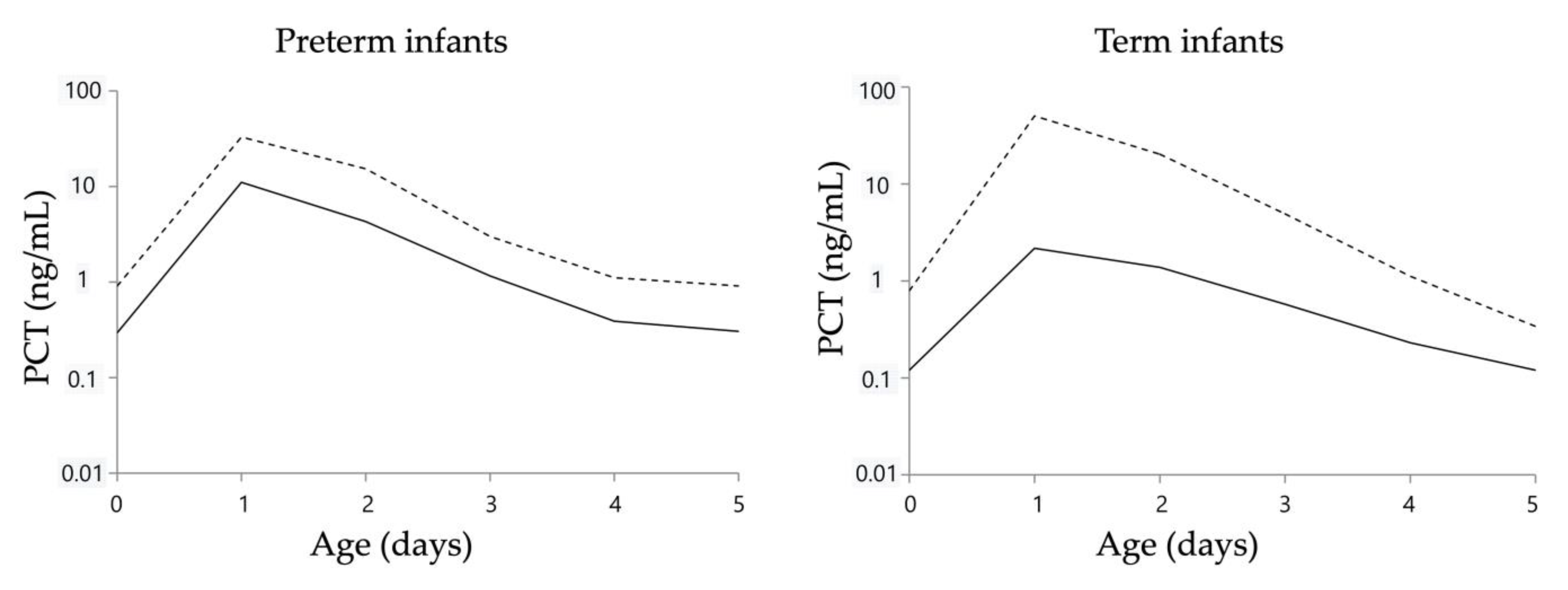
2. Results
2.1. Study Population and Baseline Characteristics
2.2. Antibiotic Therapy
2.3. Propensity Score Analysis, Multiple Regression Analysis, and Logistic Regression Analysis
3. Discussion
4. Materials and Methods
4.1. Study Design and Patients
4.2. Decision to Administer Antibiotics
4.3. Diagnosis of Bacterial Infection
4.4. Methods of Measuring Each Biomarker and Culture
4.5. Study Methods
4.5.1. Clinical Characteristics of Each Group and Causative Organisms of Early-Onset Neonatal Bacterial Infections
4.5.2. Calculation of the DOT
4.5.3. Adjustment for Background Factors
4.5.4. Statistical Analyses
5. Conclusions
Author Contributions
Funding
Institutional Review Board Statement
Informed Consent Statement
Data Availability Statement
Acknowledgments
Conflicts of Interest
References
- Tackling Drug-Resistant Infections Globally final report and recommendations. Available online: https://amr-review.org/sites/default/files/160518_Final%20paper_with%20cover.pdf (accessed on 1 September 2023).
- Silver, L. Recent advances and challenges in antibacterial drug development: Editorial. Admet Dmpk 2022, 10, 89–90. [Google Scholar] [CrossRef]
- Cotten, C.M.; McDonald, S.; Stoll, B.; Goldberg, R.N.; Poole, K.; Benjamin, D.K., Jr. The Association of Third-Generation Cephalosporin Use and Invasive Candidiasis in Extremely Low Birth-Weight Infants. Pediatrics 2006, 118, 717–722. [Google Scholar] [CrossRef]
- Murki, S.; Jonnala, S.; Mohammed, F.; Reddy, A. Restriction of cephalosporins and control of extended spectrum β-lactamase producing gram negative bacteria in a neonatal intensive care unit. Indian Pediatr. 2010, 47, 785–788. [Google Scholar] [CrossRef] [PubMed]
- Calil, R.; Marba, S.T.M.; von Nowakonski, A.; Tresoldi, A.T. Reduction in colonization and nosocomial infection by multiresistant bacteria in a neonatal unit after institution of educational measures and restriction in the use of cephalosporins. Am. J. Infect. Control 2001, 29, 133–138. [Google Scholar] [CrossRef] [PubMed]
- Tsai, M.-H.; Chu, S.-M.; Hsu, J.-F.; Lien, R.; Huang, H.-R.; Chiang, M.-C.; Fu, R.-H.; Lee, C.-W.; Huang, Y.-C. Risk Factors and Outcomes for Multidrug-Resistant Gram-Negative Bacteremia in the NICU. Pediatrics 2014, 133, e322–e329. [Google Scholar] [CrossRef]
- Cotten, C.M.; Taylor, S.; Stoll, B.; Goldberg, R.N.; Hansen, N.I.; Sánchez, P.J.; Ambalavanan, N.; Benjamin, D.K., Jr. Prolonged Duration of Initial Empirical Antibiotic Treatment Is Associated With Increased Rates of Necrotizing Enterocolitis and Death for Extremely Low Birth Weight Infants. Pediatrics 2009, 123, 58–66. [Google Scholar] [CrossRef]
- Willis, Z.; Maurice, A.d.S. Strategies to improve antibiotic use in the neonatal ICU. Curr. Opin. Pediatr. 2019, 31, 127–134. [Google Scholar] [CrossRef] [PubMed]
- Yamamoto-Hanada, K.; Yang, L.; Narita, M.; Saito, H.; Ohya, Y. Influence of antibiotic use in early childhood on asthma and allergic diseases at age 5. Ann. Allergy Asthma Immunol. 2017, 119, 54–58. [Google Scholar] [CrossRef]
- Canova, C.; Ludvigsson, J.F.; Di Domenicantonio, R.; Zanier, L.; Amidei, C.B.; Zingone, F. Perinatal and Antibiotic Exposures and the Risk of Developing Childhood-Onset Inflammatory Bowel Disease: A Nested Case-Control Study Based on a Population-Based Birth Cohort. Int. J. Environ. Res. Public Health 2020, 17, 2409. [Google Scholar] [CrossRef]
- Horton, D.B.; Scott, F.I.; Haynes, K.; Putt, M.E.; Rose, C.D.; Lewis, J.D.; Strom, B.L. Antibiotic Exposure and Juvenile Idiopathic Arthritis: A Case–Control Study. Pediatrics 2015, 136, e333–e343. [Google Scholar] [CrossRef]
- Chiesa, C.; Pacifico, L.; Osborn, J.F.; Bonci, E.; Hofer, N.; Resch, B. Early-Onset Neonatal Sepsis: Still Room for Improvement in Procalcitonin Diagnostic Accuracy Studies. Medicine 2015, 94, e1230. [Google Scholar] [CrossRef] [PubMed]
- Morioka, I.; Morikawa, S.; Miwa, A.; Minami, H.; Yoshii, K.; Kugo, M.; Kitsunezuka, Y.; Enomoto, M.; Jikimoto, T.; Nakamura, M.; et al. Culture-Proven Neonatal Sepsis in Japanese Neonatal Care Units in 2006–2008. Neonatology 2012, 102, 75–80. [Google Scholar] [CrossRef] [PubMed]
- Schrag, S.J.; Farley, M.M.; Petit, S.; Reingold, A.; Weston, E.J.; Pondo, T.; Jain, J.H.; Lynfield, R. Epidemiology of Invasive Early-Onset Neonatal Sepsis, 2005 to 2014. Pediatrics 2016, 138, e20162013. [Google Scholar] [CrossRef]
- Wynn, J.L.; Wong, H.R. Pathophysiology and Treatment of Septic Shock in Neonates. Clin. Perinatol. 2010, 37, 439–479. [Google Scholar] [CrossRef]
- Puello-Ávila, A.C.; Cataño-Villegas, A.E. Utility of C-reactive protein in early neonatal sepsis. Rev. Chil. Infectol. 2021, 38, 169–177. [Google Scholar] [CrossRef]
- Vasilcan, G.; Avasiloaiei, A.; Moscalu, M.; Dimitriu, A.G.; Stamatin, M. Procalcitonine--early marker of neonatal infection. Rev. Med. Chir. Soc. Med. Nat. Iasi 2012, 115, 1243–1250. [Google Scholar]
- Krishna, B.V.; Nadgir, S.D.; Tallur, S.S. Immunoglobulin-M estimation and C-reactive protein detection in neonatal septicemia. Indian J. Pathol. Microbiol. 2000, 43, 35–40. [Google Scholar]
- Rodwell, R.L.; Leslie, A.L.; Tudehope, D.I. Early diagnosis of neonatal sepsis using a hematologic scoring system. J. Pediatr. 1988, 112, 761–767. [Google Scholar] [CrossRef]
- Rhee, C. Using Procalcitonin to Guide Antibiotic Therapy. Open Forum Infect. Dis. 2017, 4, ofw249. [Google Scholar] [CrossRef]
- Fukuzumi, N.; Osawa, K.; Sato, I.; Iwatani, S.; Ishino, R.; Hayashi, N.; Iijima, K.; Saegusa, J.; Morioka, I. Age-specific percentile-based reference curve of serum procalcitonin concentrations in Japanese preterm infants. Sci. Rep. 2016, 6, 23871. [Google Scholar] [CrossRef]
- Chiesa, C.; Natale, F.; Pascone, R.; Osborn, J.F.; Pacifico, L.; Bonci, E.; De Curtis, M. C reactive protein and procalcitonin: Reference intervals for preterm and term newborns during the early neonatal period. Clin. Chim. Acta 2011, 412, 1053–1059. [Google Scholar] [CrossRef]
- Turner, D.; Hammerman, C.; Rudensky, B.; Schlesinger, Y.; Goia, C.; Schimmel, M.S. Procalcitonin in preterm infants during the first few days of life: Introducing an age related nomogram. Arch. Dis. Child.-Fetal Neonatal Ed. 2006, 91, F283–F286. [Google Scholar] [CrossRef] [PubMed]
- Chiesa, C.; Panero, A.; Rossi, N.; Stegagno, M.; De Giusti, M.; Osborn, J.F.; Pacifico, L. Reliability of Procalcitonin Concentrations for the Diagnosis of Sepsis in Critically III Neonates. Clin. Infect. Dis. 1998, 26, 664–672. [Google Scholar] [CrossRef] [PubMed]
- Ochi, F.; Higaki, T.; Ohta, M.; Yamauchi, T.; Tezuka, M.; Chisaka, T.; Moritani, T.; Tauchi, H.; Ishii, E. Procalcitonin as a marker of respiratory disorder in neonates. Pediatr. Int. 2015, 57, 263–268. [Google Scholar] [CrossRef] [PubMed]
- Stocker, M.; Hop, W.C.; MC van Rossum, A. Neonatal Procalcitonin Intervention Study (NeoPInS): Effect of Procalcitonin-guided decision making on Duration of antibiotic Therapy in suspected neonatal early-onset Sepsis: A multi-centre randomized superiority and non-inferiority Intervention Study. BMC Pediatr. 2010, 10, 89. [Google Scholar] [CrossRef]
- E Ballot, D.; Perovic, O.; Galpin, J.; A Cooper, P. Serum procalcitonin as an early marker of neonatal sepsis. S. Afr. Med. J. 2004, 94, 851–854. [Google Scholar]
- Bender, L.; Thaarup, J.; Varming, K.; Krarup, H.; Ellermann-Eriksen, S.; Ebbesen, F. Early and late markers for the detection of early-onset neonatal sepsis. Dan. Med. Bull. 2008, 55, 219–223. [Google Scholar]
- Resch, B.; Gusenleitner, W.; Müller, W. Procalcitonin and interleukin-6 in the diagnosis of early-onset sepsis of the neonate. Acta Paediatr. 2003, 92, 243–245. [Google Scholar] [CrossRef] [PubMed]
- Santuz, P.; Soffiati, M.; Dorizzi, R.M.; Benedetti, M.; Zaglia, F.; Biban, P. Procalcitonin for the diagnosis of early-onset neonatal sepsis: A multilevel probabilistic approach. Clin. Biochem. 2008, 41, 1150–1155. [Google Scholar] [CrossRef]
- Go, H.; Nagano, N.; Katayama, D.; Akimoto, T.; Imaizumi, T.; Aoki, R.; Hijikata, M.; Seimiya, A.; Kato, R.; Okahashi, A.; et al. Diagnostic Accuracy of Biomarkers for Early-onset Neonatal Bacterial Infections: Evaluation of Serum Procalcitonin Reference Curves. Diagnostics 2020, 10, 839. [Google Scholar] [CrossRef]
- Fluss, R.; Faraggi, D.; Reiser, B. Estimation of the Youden Index and its Associated Cutoff Point. Biom. J. 2005, 47, 458–472. [Google Scholar] [CrossRef]
- Vergnano, S.; Menson, E.; Kennea, N.; Embleton, N.; Russell, A.B.; Watts, T.; Robinson, M.J.; Collinson, A.; Heath, P.T. Neonatal infections in England: The NeonIN surveillance network. Arch. Dis. Child.-Fetal Neonatal Ed. 2010, 96, F9–F14. [Google Scholar] [CrossRef] [PubMed]
- Stoll, B.J.; Puopolo, K.M.; Hansen, N.I.; Sánchez, P.J.; Bell, E.F.; Carlo, W.A.; Cotten, C.M.; D’angio, C.T.; Kazzi, S.N.J.; Poindexter, B.B.; et al. Early-Onset Neonatal Sepsis 2015 to 2017, the Rise of Escherichia coli, and the Need for Novel Prevention Strategies. JAMA Pediatr. 2020, 174, e200593. [Google Scholar] [CrossRef] [PubMed]
- Korang, S.K.; Safi, S.; Nava, C.; Gordon, A.; Gupta, M.; Greisen, G.; Lausten-Thomsen, U.; Jakobsen, J.C. Antibiotic regimens for early-onset neonatal sepsis. Cochrane Database Syst. Rev. 2021, 2021, CD013837. [Google Scholar] [CrossRef]
- Bhat, R.; Custodio, H.; McCurley, C.; Whitehurst, R.; Gulati, R.; Jha, O.P.; Bhat, J.; Estrada, B.; Hill, A.; Eyal, F.; et al. Reducing antibiotic utilization rate in preterm infants: A quality improvement initiative. J. Perinatol. 2018, 38, 421–429. [Google Scholar] [CrossRef]
- Cantey, J.B.; Wozniak, P.S.; E Pruszynski, J.; Sánchez, P.J. Reducing unnecessary antibiotic use in the neonatal intensive care unit (SCOUT): A prospective interrupted time-series study. Lancet Infect. Dis. 2016, 16, 1178–1184. [Google Scholar] [CrossRef]
- Kitano, T.; Takagi, K.; Arai, I.; Yasuhara, H.; Ebisu, R.; Ohgitani, A.; Kitagawa, D.; Oka, M.; Masuo, K.; Minowa, H. A simple and feasible antimicrobial stewardship program in a neonatal intensive care unit of a Japanese community hospital. J. Infect. Chemother. 2019, 25, 860–865. [Google Scholar] [CrossRef]
- McCarthy, K.; Hawke, A.; Dempsey, E. Antimicrobial stewardship in the neonatal unit reduces antibiotic exposure. Acta Paediatr. 2018, 107, 1716–1721. [Google Scholar] [CrossRef] [PubMed]
- Desai, S.; Qin, H.; Rayburn, P.D.; Poon, G.; Murthy, K.; Ellsbury, D.L.; Chiruvolu, A.; Tolia, V.N. Implementation of an Automatic Stop Order and Initial Antibiotic Exposure in Very Low Birth Weight Infants. Am. J. Perinatol. 2017, 34, 105–110. [Google Scholar] [CrossRef]
- Gasparrini, A.J.; Crofts, T.S.; Gibson, M.K.; Tarr, P.I.; Warner, B.B.; Dantas, G. Antibiotic perturbation of the preterm infant gut microbiome and resistome. Gut Microbes 2016, 7, 443–449. [Google Scholar] [CrossRef]
- Li, Q.; Zhou, J.-M. The microbiota–gut–brain axis and its potential therapeutic role in autism spectrum disorder. Neuroscience 2016, 324, 131–139. [Google Scholar] [CrossRef]
- Rajar, P.; Saugstad, O.D.; Berild, D.; Dutta, A.; Greisen, G.; Lausten-Thomsen, U.; Mande, S.S.; Nangia, S.; Petersen, F.C.; Dahle, U.R.; et al. Antibiotic Stewardship in Premature Infants: A Systematic Review. Neonatology 2020, 117, 673–686. [Google Scholar] [CrossRef] [PubMed]
- Hofer, N.; Zacharias, E.; Müller, W.; Resch, B. Performance of the definitions of the systemic inflammatory response syndrome and sepsis in neonates. Jpme 2012, 40, 587–590. [Google Scholar] [CrossRef] [PubMed]
- Müller, W.; Resch, B.; Hofer, N. Definitions of SIRS and sepsis in correlation with early and late onset neonatal sepsis. J. Pediatr. Intensiv. Care 2012, 01, 017–023. [Google Scholar] [CrossRef]
- Krueger, M.; Nauck, M.S.; Sang, S.; Hentschel, R.; Wieland, H.; Berner, R. Cord Blood Levels of Interleukin-6 and Interleukin-8 for the Immediate Diagnosis of Early-Onset Infection in Premature Infants. Neonatology 2001, 80, 118–123. [Google Scholar] [CrossRef] [PubMed]
- Nishikawa, T.; Ono, K.; Hashimoto, S.; Kinoshita, H.; Watanabe, T.; Araki, H.; Otsu, K.; Sakamoto, W.; Harada, M.; Toyonaga, T.; et al. One-hour oral glucose tolerance test plasma glucose at gestational diabetes diagnosis is a common predictor of the need for insulin therapy in pregnancy and postpartum impaired glucose tolerance. J. Diabetes Investig. 2018, 9, 1370–1377. [Google Scholar] [CrossRef]
- Metzger, B.E.; Gabbe, S.G.; Persson, B.; Lowe, L.P.; Dyer, A.R.; Oats, J.J.; Buchanan, T.A. International association of diabetes and pregnancy study groups recommendations on the diagnosis and classification of hyperglycemia in pregnancy: Response to Weinert. Diabetes Care 2010, 33, e98. [Google Scholar] [CrossRef]
- Aoki, M.; Urakami, T.; Nagano, N.; Aoki, R.; Morioka, I. Association of Plasma Cortisol Levels with Gestational Age and Anthropometric Values at Birth in Preterm Infants. Int. J. Environ. Res. Public Health 2022, 19, 11448. [Google Scholar] [CrossRef]
- Watanabe, K.; Matsubara, K.; Nakamoto, O.; Ushijima, J.; Ohkuchi, A.; Koide, K.; Makino, S.; Mimura, K.; Morikawa, M.; Naruse, K.; et al. Outline of the new definition and classification of “Hypertensive Disorders of Pregnancy (HDP)”; a revised JSSHP statement of 2005. Hypertens. Res. Pregnancy 2018, 6, 33–37. [Google Scholar] [CrossRef]
- Aljerian, K. Chorioamnionitis: Establishing a correlation between clinical and histological diagnosis. Indian J. Pathol. Microbiol. 2020, 63, 44–48. [Google Scholar] [CrossRef]
- Lencki, S.G.; Maciulla, M.B.; Eglinton, G.S. Maternal and umbilical cord serum interleukin levels in preterm labor with clinical chorioamnionitis. Am. J. Obstet. Gynecol. 1994, 170, 1345–1351. [Google Scholar] [CrossRef]
- Dayal, S.; Hong, P.L. Premature Rupture of Membranes; StatPearls LLC: St. Petersburg, Russia, 2023. [Google Scholar]
- Kumari, S.; Sharma, M.; Yadav, M.; Saraf, A.; Kabra, M.; Mehra, R. Trends in neonatal outcome with low apgar scores. Indian J. Pediatr. 1993, 60, 415–422. [Google Scholar] [CrossRef]
- Giacoia, G.P. Low Apgar scores and birth asphyxia: Misconceptions that promote undeserved negligence suits. Postgrad. Med. 1988, 84, 77–82. [Google Scholar] [CrossRef] [PubMed]
- Razaz, N.; Norman, M.; Alfvén, T.; Cnattingius, S. Low Apgar score and asphyxia complications at birth and risk of longer-term cardiovascular disease: A nationwide population-based study of term infants. Lancet Reg. Health-Eur. 2023, 24, 100532. [Google Scholar] [CrossRef] [PubMed]
- Abu-Shaweesh, J.M.; Almidani, E. PDA: Does it matter? Int. J. Pediatr. Adolesc. Med. 2020, 7, 11–14. [Google Scholar] [CrossRef]
- Morris, A.M.; Brener, S.; Dresser, L.; Daneman, N.; Dellit, T.H.; Avdic, E.; Bell, C.M. Use of a Structured Panel Process to Define Quality Metrics for Antimicrobial Stewardship Programs. Infect. Control Hosp. Epidemiology 2012, 33, 500–506. [Google Scholar] [CrossRef]
- Lee, S.W. Methods for testing statistical differences between groups in medical research: Statistical standard and guideline of Life Cycle Committee. Life Cycle 2022, 2, e1. [Google Scholar] [CrossRef]


| Pre-Group n = 737 | Post-Group n = 686 | p-Value | |
|---|---|---|---|
| A. Maternal backgrounds | |||
| Gestational diabetes | 73 (10) | 83 (12) | 0.19 * |
| Hypertensive disorder of pregnancy | 78 (11) | 78 (11) | 0.63 * |
| GBS colonization | 99 (13) | 98 (14) | 0.64 * |
| Clinical chorioamnionitis | 23 (3.1) | 12 (1.8) | 0.10 * |
| PROM | 152 (21) | 75 (11) | <0.01 * |
| Delivery mode | 0.06 * | ||
| Vaginal delivery | 322 (44) | 265 (39) | |
| Cesarean section | 415 (56) | 421 (61) | |
| Precipitate delivery | 5 (0.68) | 4 (0.58) | 1.0 ** |
| Intrapartum antibiotic therapy | 483 (66) | 474 (69) | 0.15 * |
| B. Neonatal backgrounds | |||
| GA at birth, weeks | 37 (22–42) | 37 (22–42) | 0.49 *** |
| GA ≥ 37 weeks | 421 (57) | 366 (53) | 0.15 * |
| 28 ≤ GA < 37 weeks | 286 (39) | 293 (43) | |
| GA < 28 weeks | 30 (4.1) | 27 (4.0) | |
| BW, g | 2504 (356–4315) | 2448 (278–4446) | 0.21 *** |
| BW ≥ 2500 g | 372 (50) | 318 (46) | 0.12 * |
| 1500 ≤ BW < 2500 g | 279 (38) | 293 (43) | |
| 1000 ≤ BW < 1500 g | 50 (6.8) | 42 (6.1) | |
| BW < 1000 | 36 (5.0) | 33 (4.8) | |
| Apgar score | |||
| at 1 min | 8 (0–10) | 8 (0–10) | 0.87 *** |
| at 5 min | 9 (0–10) | 9 (1–10) | 0.07 *** |
| Neonatal transport | 229 (31) | 161 (23) | <0.01 * |
| Respiratory disorder | 456 (62) | 425 (62) | 0.97 * |
| Severe neonatal asphyxia | 90 (12) | 63 (9.2) | 0.11 * |
| Surgical condition | 20 (2.7) | 7 (1.0) | 0.02 * |
| Patent ductus arteriosus that required indomethacin or surgical treatment | 30 (4.1) | 34 (5.0) | 0.42 * |
| Early-onset bacterial infection | 15 (2.0) | 14 (2.0) | 0.99 * |
| Death | 0 (0) | 0 (0) | - |
| Death due to infection | 0 (0) | 0 (0) | - |
| Use of catecholamine | 62 (8.4) | 75 (11) | 0.10 * |
| Use of steroid | 11 (1.5) | 11 (1.6) | 0.87 * |
| Use of gamma globulin | 34 (4.6) | 28 (4.1) | 0.62 * |
| Age at measurement of biomarkers, days | 0 (0–3) | 0 (0–3) | 0.01 *** |
| PCT, ng/mL | - (-) | 0.17 (0.02–100) | - |
| CRP, mg/dL | 0.1 (0.1–10.64) | 0.1 (0.1–10.7) | 0.01 *** |
| WBC, /µL | 13,600 (1500–53,700) | 13,100 (2200–62,600) | 0.12 *** |
| IgM, mg/dL | 8 (2–220) | 8 (2–253) | 0.99 *** |
| No. | Detected Pathogens | Culture-Positive Samples | Diagnosis | Delivery Mode | Gestational Age at Birth |
|---|---|---|---|---|---|
| 1 | Escherichia coli, α-Streptococcus | Gastric aspiration | Clinical sepsis | Vaginal delivery | 40 weeks |
| 2 | Streptococcus agalactiae | Blood, nasopharyngeal swab, and stool | Culture-proven sepsis | Vaginal delivery | 39 weeks |
| 3 | Streptococcus agalactiae | Nasopharyngeal swab and gastric aspiration | Clinical sepsis | Vaginal delivery | 39 weeks |
| 4 | Streptococcus agalactiae | Blood, nasopharyngeal swab, and stool | Culture-proven sepsis | Cesarean section | 38 weeks |
| 5 | Staphylococcus aureus | Nasopharyngeal swab and stool | Clinical sepsis | Vaginal delivery | 37 weeks |
| 6 | Streptococcus agalactiae | Stool | Clinical sepsis | Vaginal delivery | 39 weeks |
| 7 | Enterococcus faecalis | Gastric aspiration | Clinical sepsis | Cesarean section | 41 weeks |
| 8 | Streptococcus agalactiae | Stool | Clinical sepsis | Vaginal delivery | 40 weeks |
| 9 | Streptococcus agalactiae | Gastric aspiration | Clinical sepsis | Cesarean section | 39 weeks |
| 10 | Streptococcus equisimilis | Blood, nasopharyngeal swab, and stool | Culture-proven sepsis | Vaginal delivery | 40 weeks |
| 11 | Staphylococcus aureus | Nasopharyngeal swab and stool | Clinical sepsis | Cesarean section | 40 weeks |
| 12 | Enterococcus faecalis | Nasopharyngeal swab, stool, and gastric aspiration | Clinical sepsis | Cesarean section | 33 weeks |
| 13 | Staphylococcus aureus | Nasopharyngeal swab and gastric aspiration | Clinical sepsis | Cesarean section | 25 weeks |
| 14 | Escherichia coli | Nasopharyngeal swab, stool, and gastric aspiration | Clinical sepsis | Vaginal delivery | 34 weeks |
| 15 | Streptococcus agalactiae | Urine | Clinical sepsis | Vaginal delivery | 33 weeks |
| No. | Pathogens Detected | Culture-Positive Samples | Diagnosis | Delivery Mode | Gestational Age at Birth |
|---|---|---|---|---|---|
| 1 | Escherichia coli | Blood, nasopharyngeal swab, and gastric aspiration | Culture-proven sepsis | Vaginal delivery | 38 weeks |
| 2 | Streptococcus agalactiae | Nasopharyngeal swab, stool, and gastric aspiration | Clinical sepsis | Cesarean section | 41 weeks |
| 3 | Streptococcus agalactiae | Nasopharyngeal swab, stool, and gastric aspiration | Clinical sepsis | Cesarean section | 41 weeks |
| 4 | Streptococcus agalactiae | Nasopharyngeal swab and stool | Clinical sepsis | Cesarean section | 40 weeks |
| 5 | Gardnerella vaginalis | Stool and gastric aspiration | Clinical sepsis | Cesarean section | 27 weeks |
| 6 | Streptococcus pneumoniae | Nasopharyngeal swab, stool, and gastric aspiration | Clinical sepsis | Cesarean section | 33 weeks |
| 7 | Staphylococcus aureus | Nasopharyngeal swab, stool, and urine | Clinical sepsis | Cesarean section | 33 weeks |
| 8 | Escherichia coli | Nasopharyngeal swab | Clinical sepsis | Cesarean section | 25 weeks |
| 9 | Coryneform Gram-positive rod | Gastric aspiration | Clinical sepsis | Vaginal delivery | 35 weeks |
| 10 | Escherichia coli | Nasopharyngeal swab and urine | Clinical sepsis | Cesarean section | 35 weeks |
| 11 | Prevotella bivia | Gastric aspiration | Clinical sepsis | Cesarean section | 22 weeks |
| 12 | Prevotella bivia | Gastric aspiration | Clinical sepsis | Cesarean section | 25 weeks |
| 13 | Streptococcus agalactiae | Nasopharyngeal swab and stool | Clinical sepsis | Vaginal delivery | 22 weeks |
| 14 | Gardnerella vaginalis | Nasopharyngeal swab, stool, and gastric aspiration | Clinical sepsis | Cesarean section | 30 weeks |
| Pre-Group n = 737 | Post-Group n = 686 | p-Value | |
|---|---|---|---|
| Antibiotic therapy | 280 (38) | 79 (12) | <0.01 * |
| DOT | 211.3 | 82.0 | <0.01 ** |
| Ampicillin DOT | 203.3 | 74.5 | <0.01 ** |
| Amikacin DOT | 133.2 | 62.8 | <0.01 ** |
| Cephalosporin DOT | 8.8 | 8.0 | 0.88 ** |
| Penicillin DOT | 0 | 1.4 | 0.30 ** |
| Meropenem DOT | 0 | 0.9 | 0.30 ** |
| Piperacillin-tazobactam DOT | 0 | 0.5 | 0.30 ** |
| Vancomycin DOT | 0 | 0 | - |
| Pre-Group n = 280 | Post-Group n = 79 | p-Value | |
|---|---|---|---|
| 1–3 days | 84 (30) | 3 (3.8) | <0.01 |
| 4–7 days | 196 (70) | 76 (96.2) |
| DOT | Estimate | SE | t-Value | p-Value | 95% CI | |
|---|---|---|---|---|---|---|
| Lower | Upper | |||||
| Intercept | 3.1 | 0.21 | 14.6 | <0.01 | 2.7 | 3.5 |
| Intervention, yes | −0.70 | 0.10 | −6.8 | <0.01 | −0.89 | −0.49 |
| Intervention, no (reference) | - | - | - | - | - | - |
| PS | −3.5 | 0.44 | −8.1 | <0.01 | −4.4 | −2.7 |
| Antibiotic Therapy | Odds Ratio | p-Value | 95% CI | |
|---|---|---|---|---|
| Lower | Upper | |||
| Intervention, yes | 0.26 | <0.01 | 0.20 | 0.35 |
| Intervention, no (reference) | - | - | - | - |
| PS | 0.0081 | <0.01 | 0.0026 | 0.025 |
Disclaimer/Publisher’s Note: The statements, opinions and data contained in all publications are solely those of the individual author(s) and contributor(s) and not of MDPI and/or the editor(s). MDPI and/or the editor(s) disclaim responsibility for any injury to people or property resulting from any ideas, methods, instructions or products referred to in the content. |
© 2023 by the authors. Licensee MDPI, Basel, Switzerland. This article is an open access article distributed under the terms and conditions of the Creative Commons Attribution (CC BY) license (https://creativecommons.org/licenses/by/4.0/).
Share and Cite
Go, H.; Nagano, N.; Sato, Y.; Katayama, D.; Hara, K.; Akimoto, T.; Imaizumi, T.; Aoki, R.; Hijikata, M.; Seimiya, A.; et al. Procalcitonin-Based Antibiotic Use for Neonatal Early-Onset Bacterial Infections: Pre- and Post-Intervention Clinical Study. Antibiotics 2023, 12, 1426. https://doi.org/10.3390/antibiotics12091426
Go H, Nagano N, Sato Y, Katayama D, Hara K, Akimoto T, Imaizumi T, Aoki R, Hijikata M, Seimiya A, et al. Procalcitonin-Based Antibiotic Use for Neonatal Early-Onset Bacterial Infections: Pre- and Post-Intervention Clinical Study. Antibiotics. 2023; 12(9):1426. https://doi.org/10.3390/antibiotics12091426
Chicago/Turabian StyleGo, Hidetoshi, Nobuhiko Nagano, Yuki Sato, Daichi Katayama, Koichiro Hara, Takuya Akimoto, Takayuki Imaizumi, Ryoji Aoki, Midori Hijikata, Ayako Seimiya, and et al. 2023. "Procalcitonin-Based Antibiotic Use for Neonatal Early-Onset Bacterial Infections: Pre- and Post-Intervention Clinical Study" Antibiotics 12, no. 9: 1426. https://doi.org/10.3390/antibiotics12091426