
Cranberry/Chondroitin Sulfate Co-precipitate as a New Method for Controlling Urinary Tract Infections

 ,
,  , and
, and
Abstract
:1. Introduction
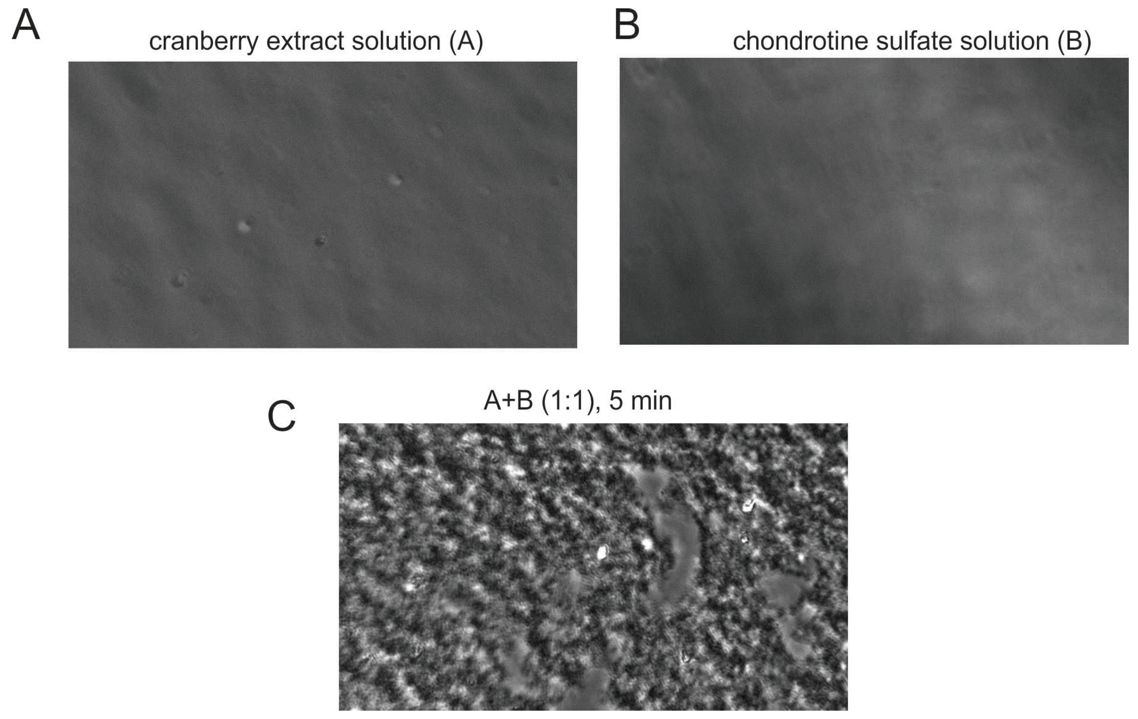
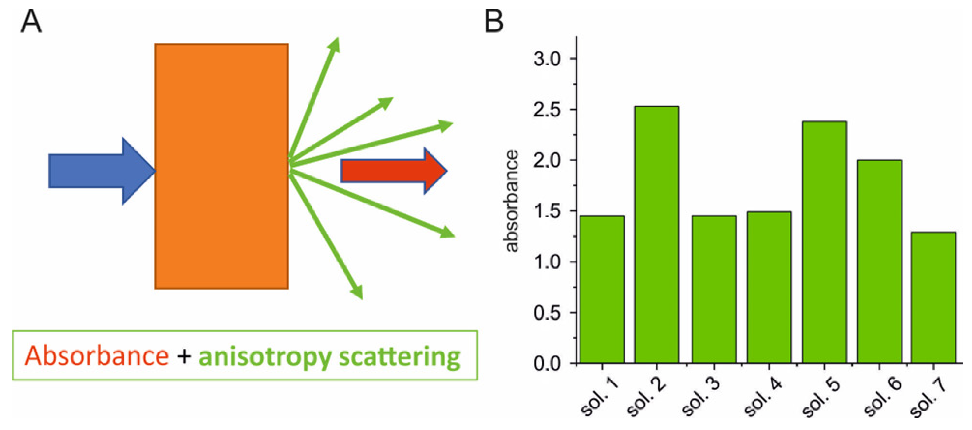
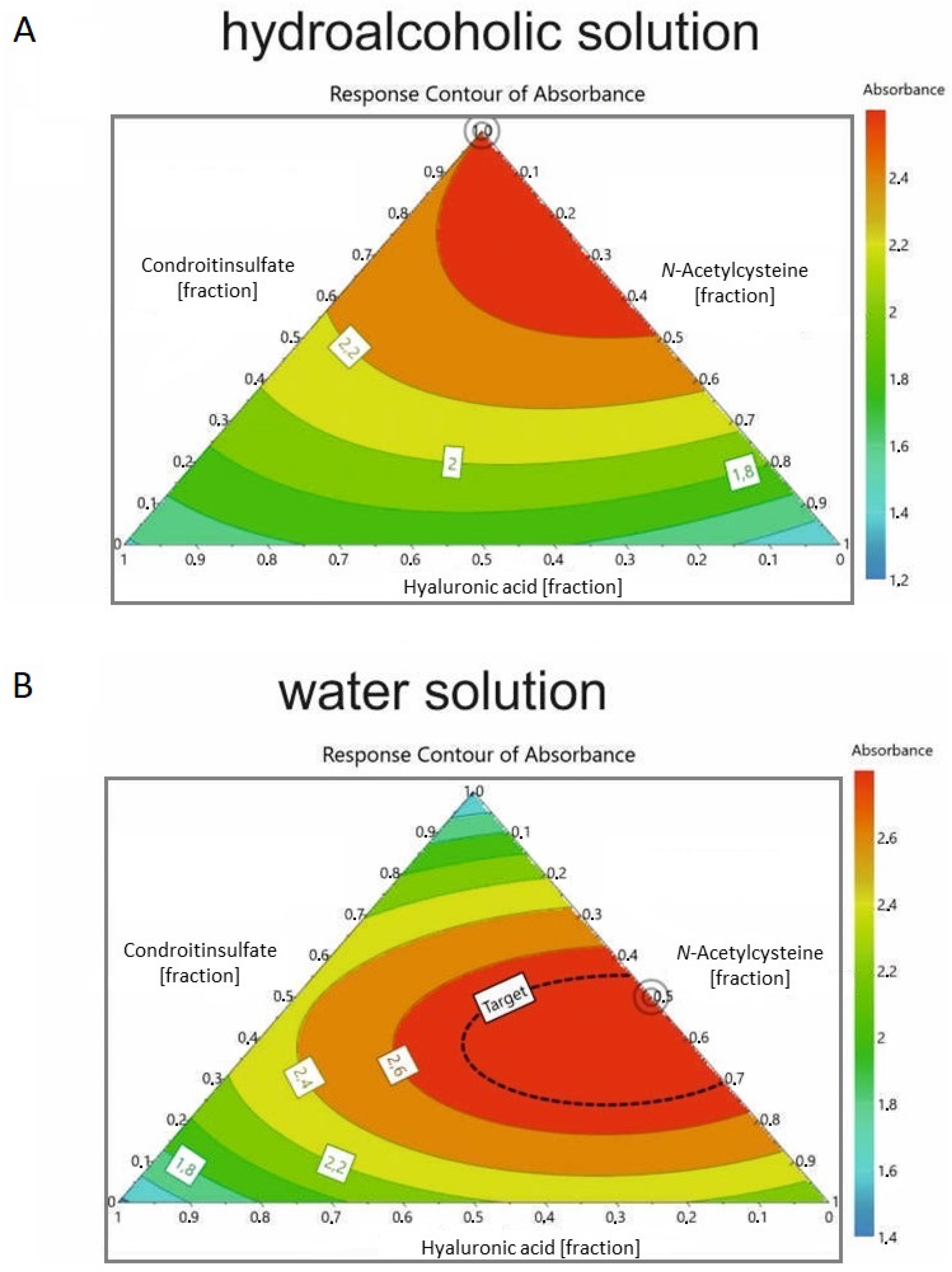
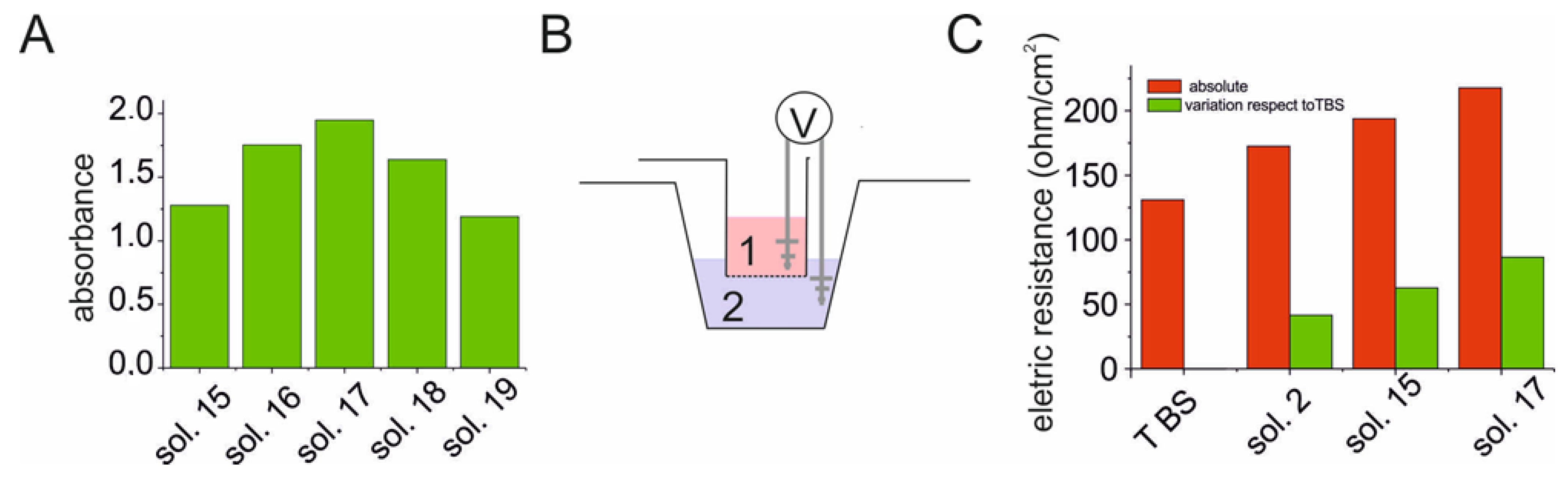
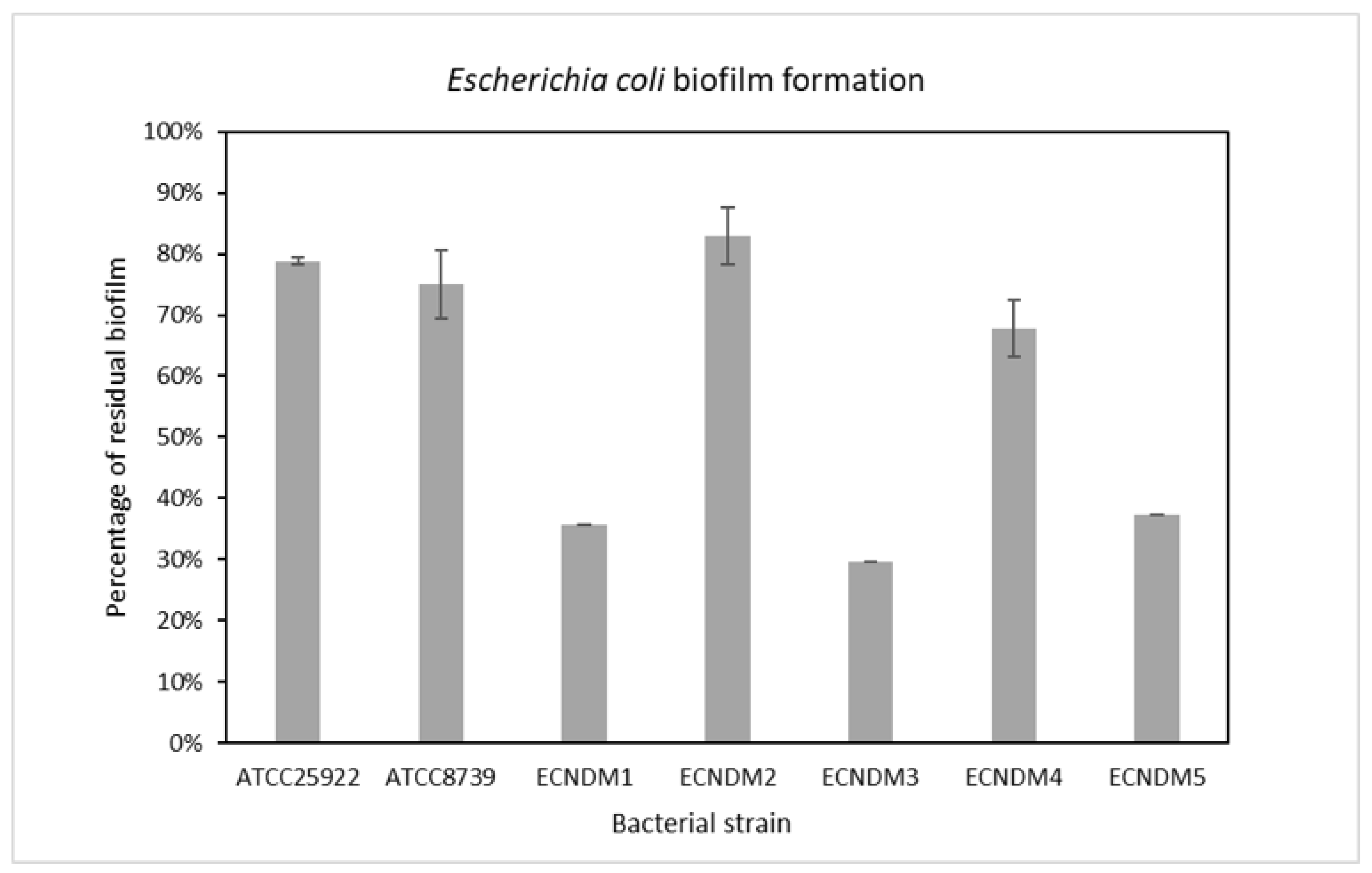
2. Results
3. Discussion
4. Materials and Methods
4.1. Bacterial Strains and Growth Conditions
4.2. Biofilm Formation
5. Conclusions
Author Contributions
Funding
Data Availability Statement
Conflicts of Interest
References
- Flores-Mireles, A.L.; Walker, J.N.; Caparon, M.; Hultgren, S.J. Urinary tract infections: Epidemiology, mechanisms of infection and treatment options. Nat. Rev. Microbiol. 2015, 13, 269–284. [Google Scholar] [CrossRef] [PubMed]
- Foxman, B. Urinary Tract Infection Syndromes: Occurrence, recurrence, bacteriology, risk factors, and disease burden. Infect. Dis. Clin. North. Am. 2014, 28, 1–13. [Google Scholar] [CrossRef]
- Guglietta, A. Recurrent urinary tract infections in women: Risk factors, etiology, pathogenesis and prophylaxis. Futur. Microbiol. 2017, 12, 239–246. [Google Scholar] [CrossRef] [PubMed]
- Lecomte, F.; Allaert, F.A. Single-dose treatment of cystitis with fosfomycin trometamol (Monuril®): An analysis of 15 comparative trials on 2,048 patients. Mdd Mal. Infect. 1996, 26, 338–343. [Google Scholar] [CrossRef]
- Nicolle, L.E. Pivmecillinam in the treatment of urinary tract infections. J. Antimicrob. Chemother. 2000, 46 (Suppl. 1), 35. [Google Scholar] [CrossRef] [Green Version]
- Huttner, A.; Verhaegh, E.M.; Harbarth, S.; Muller, A.E.; Theuretzbacher, U.; Mouton, J.W. Nitrofurantoin revisited: A systematic review and meta-analysis of controlled trials. J. Antimicrob. Chemother. 2015, 70, 2456. [Google Scholar] [CrossRef] [Green Version]
- Gupta, K.; Hooton, T.M.; Roberts, P.L.; Stamm, W.E. Short-course nitrofurantoin for the treatment of acute uncomplicated cystitis in women. Arch. Intern. Med. 2007, 167, 2207. [Google Scholar] [CrossRef] [Green Version]
- Gupta, K.; Stamm, W.E. Outcomes associated with trimethoprim/sulphamethoxazole (TMP/SMX) therapy in TMP/SMXresistant community-acquired UTI. Int. J. Antimicrob. Agents 2002, 19, 554. [Google Scholar] [CrossRef]
- Wang, R.; LaSala, C. Role of antibiotic resistance in urinary tract infection management: A cost-effectiveness analysis. Am. J. Obstet. Gynecol. 2021, 225, 550.e1–550.e10. [Google Scholar] [CrossRef]
- Klein, R.D.; Hultgren, S.J. Urinary tract infections: Microbial pathogenesis, host–pathogen interactions and new treatment strategies. Nat. Rev. Genet. 2020, 18, 211–226. [Google Scholar] [CrossRef]
- Fu, Z.; Liska, D.; Talan, D.; Chung, M. Cranberry Reduces the Risk of Urinary Tract Infection Recurrence in Otherwise Healthy Women: A Systematic Review and Meta-Analysis. J. Nutr. 2017, 147, 2282. [Google Scholar] [CrossRef] [PubMed] [Green Version]
- Wang, C.H.; Fang, C.-C.; Chen, N.-C.; Liu, S.S.-H.; Yu, P.-H.; Wu, T.-Y.; Chen, W.-T.; Lee, C.-C.; Chen, S.-C. Cranberry-containing products for prevention of urinary tract infections in susceptible populations: A systematic review and meta-analysis of randomized controlled trials. Arch. Intern. Med. 2012, 172, 988. [Google Scholar] [CrossRef] [PubMed] [Green Version]
- Lenger, S.; Bradley, M.S.; Thomas, D.A.; Bertolet, M.H.; Lowder, J.L.; Sutcliffe, S. D-mannose vs other agents for recurrent urinary tract infection prevention in adult women: A systematic review and meta-analysis. Am. J. Obstet. Gynecol. 2020, 223, 265.e1–265.e13. [Google Scholar] [CrossRef] [PubMed]
- Ray, K. Infection: Lactobacillus probiotic could prevent recurrent UTI. Nat. Rev. Urol. 2011, 8, 292. [Google Scholar] [CrossRef] [PubMed]
- Ochoa-Brust, G.J.; Fernández, A.R.; Villanueva-Ruiz, G.J.; Velasco, R.; Trujillo-Hernández, B.; Vásquez, C. Daily intake of 100 mg ascorbic acid as urinary tract infection prophylactic agent during pregnancy. Acta Obstet. Gynecol. Scand. 2007, 86, 783–787. [Google Scholar] [CrossRef]
- De Vita, D.; Giordano, S. Effectiveness of intravesical hyaluronic acid/chondroitin sulfate in recurrent bacterial cystitis: A randomized study. Int. Urogynecol J. 2012, 23, 1707. [Google Scholar] [CrossRef] [PubMed]
- Goddard, J.C.; Janssen, D.A.W. Intravesical hyaluronic acid and chondroitin sulfate for recurrent urinary tract infections: Systematic review and meta-analysis. Int. Urogynecol J. 2018, 29, 933. [Google Scholar] [CrossRef] [Green Version]
- Manoharan, A.; Ognenovska, S.; Paino, D.; Whiteley, G.; Glasbey, T.; Kriel, F.H.; Farrell, J.; Moore, K.H.; Manos, J.; Das, T. N-Acetylcysteine Protects Bladder Epithelial Cells from Bacterial Invasion and Displays Antibiofilm Activity against Urinary Tract Bacterial Pathogens. Antibiotics 2021, 10, 900. [Google Scholar] [CrossRef]
- Kranjčec, B.; Papeš, D.; Altarac, S. D-mannose powder for prophylaxis of recurrent urinary tract infections in women: A ran-domized clinical trial. World J. Urol. 2014, 32, 79–84. [Google Scholar] [CrossRef]
- Stracy, M.; Snitser, O.; Yelin, I.; Amer, Y.; Parizade, M.; Katz, R.; Rimler, G.; Wolf, T.; Herzel, E.; Koren, G.; et al. Minimizing treatment-induced emergence of antibiotic resistance in bacterial infections. Science 2022, 375, 889–894. [Google Scholar] [CrossRef]
- Martinez, J.J.; Hultgren, S.J. Requirement of Rho-family GTPases in the invasion of Type 1-piliated uropathogenic Escherichia coli. Cell. Microbiol. 2002, 4, 19–28. [Google Scholar] [CrossRef] [PubMed] [Green Version]
- Iavazzo, C.; Athanasiou, S.; Pitsouni, E.; Falagas, M.E. Hyaluronic Acid: An Effective Alternative Treatment of Interstitial Cystitis, Recurrent Urinary Tract Infections, and Hemorrhagic Cystitis? Eur. Urol. 2007, 51, 1534–1541. [Google Scholar] [CrossRef] [PubMed]
- Nickel, J.C.; Hanno, P.; Kumar, K.; Thomas, H. Second multicenter, randomized, double-blind, parallel-group evaluation of effec-tiveness and safety of intravesical sodium chondroitin sulfate compared with inactive vehicle control in subjects with interstitial cystitis/bladder pain syndrome. Urology 2012, 79, 1220–12244. [Google Scholar] [CrossRef] [PubMed]
- Cicione, A.; Cantiello, F.; Ucciero, G.; Salonia, A.; Madeo, I.; Bava, I.; Aliberti, A.; Damiano, R. Restoring the glycosaminoglycans layer in recurrent cystitis: Experimental and clinical foundations. Int. J. Urol. 2014, 21, 763–768. [Google Scholar] [CrossRef] [PubMed]
- Damiano, R.; Quarto, G.; Bava, I.; Ucciero, G.; De Domenico, R.; Palumbo, M.I.; Autorino, R. Prevention of Recurrent Urinary Tract Infections by Intravesical Administration of Hyaluronic Acid and Chondroitin Sulphate: A Placebo-Controlled Randomised Trial. Eur. Urol. 2011, 59, 645–651. [Google Scholar] [CrossRef] [PubMed]
- Palleschi, G.; Carbone, A.; Zanello, P.P.; Mele, R.; Leto, A.; Fuschi, A.; Al Salhi, Y.; Velotti, G.; Al Rawashdah, S.; Coppola, G.; et al. Prospective study to compare antibiosis versus the association of N-acetylcysteine, D-mannose and Morinda citrifolia fruit extract in preventing urinary tract infections in patients submitted to urodynamic investigation. Arch. Ital. di Urol. e Androl. 2017, 89, 45–50. [Google Scholar] [CrossRef] [Green Version]
- Cela-López, J.M.; Roldán, C.J.C.; Gómez-Lizarraga, G.; Martínez, V. A Natural Alternative Treatment for Urinary Tract Infections: Itxasol©, the Importance of the Formulation. Molecules 2021, 26, 4564. [Google Scholar] [CrossRef]
- Benderev, T.V. Acetylcysteine for Urinary Tract Mucolysis. J. Urol. 1988, 139, 353–354. [Google Scholar] [CrossRef]
- Ceccarini, M.R.; Codini, M.; Cataldi, S.; Vannini, S.; Lazzarini, A.; Floridi, A.; Moretti, M.; Villarini, M.; Fioretti, B.; Beccari, T.; et al. Acid sphingomyelinase as target of Lycium Chinense: Promising new action for cell health. Lipids Heal. Dis. 2016, 15, 183. [Google Scholar] [CrossRef] [Green Version]
- Foo, L.Y.; Lu, Y.; Howell, A.B.; Vorsa, N. A-Type Proanthocyanidin Trimers from Cranberry that Inhibit Adherence of Uropathogenic P-Fimbriated Escherichia coli. J. Nat. Prod. 2000, 63, 1225–1228. [Google Scholar] [CrossRef]
- Feliciano, R.P.; Meudt, J.J.; Shanmuganayagam, D.; Krueger, C.G.; Reed, J.D. Ratio of “A-type” to “B-type” Proanthocyanidin Interflavan Bonds Affects Extra-intestinal Pathogenic Escherichia coli Invasion of Gut Epithelial Cells. J. Agric. Food Chem. 2013, 62, 3919–3925. [Google Scholar] [CrossRef] [PubMed]
- Pappas, E.; Schaich, K.M. Phytochemicals of Cranberries and Cranberry Products: Characterization, Potential Health Effects, and Processing Stability. Crit. Rev. Food Sci. Nutr. 2009, 49, 741–781. [Google Scholar] [CrossRef] [PubMed]
- Zafriri, D.; Ofek, I.; Adar, R.; Pocino, M.; Sharon, N. Inhibitory activity of cranberry juice on adherence of type 1 and type P fimbriated Escherichia coli to eucaryotic cells. Antimicrob. Agents Chemother. 1989, 33, 92–98. [Google Scholar] [CrossRef] [Green Version]
- Howell, A.B. Bioactive compounds in cranberries and their role in prevention of urinary tract infections. Mol. Nutr. Food Res. 2007, 51, 732–737. [Google Scholar] [CrossRef]
- de Servi, B.; Ranzini, F.; Piqué, N. Effect of Utipro® (containing gelatin-xyloglucan) against Escherichia coli invasion of intestinal epithelial cells: Results of an in vitro study. Futur. Microbiol. 2016, 11, 651–658. [Google Scholar] [CrossRef] [PubMed] [Green Version]
- Iannitti, R.G.; Floridi, A.; Lazzarini, A.; Tantucci, A.; Russo, R.; Ragonese, F.; Monarca, L.; Caglioti, C.; Spogli, R.; Leonardi, L.; et al. Resveratrol Supported on Magnesium DiHydroxide (Resv@MDH) Represents an Oral Formulation of Resveratrol With Better Gastric Absorption and Bioavailability Respect to Pure Resveratrol. Front. Nutr. 2020, 7, 570047. [Google Scholar] [CrossRef]
- Arcuri, C.; Monarca, L.; Ragonese, F.; Mecca, C.; Bruscoli, S.; Giovagnoli, S.; Donato, R.; Bereshchenko, O.; Fioretti, B.; Costantino, F. Probing Internalization Effects and Biocompatibility of Ultrasmall Zirconium Metal-Organic Frameworks UiO-66 NP in U251 Glioblastoma Cancer Cells. Nanomaterials 2018, 8, 867. [Google Scholar] [CrossRef] [Green Version]
- Sklan, D.; Hurwitz, S. Movement and Absorption of Major Minerals and Water in Ovine Gastrointestinal Tract. J. Dairy. Sci. 1985, 68, 1659–1666. [Google Scholar] [CrossRef]
- Griffith, M.L.; Halloran, J. Scattering of ultraviolet radiation in turbid suspensions. J. Appl. Phys. 1997, 81, 2538–2546. [Google Scholar] [CrossRef] [Green Version]
- Sobota, A. Inhibition of Bacterial Adherence by Cranberry Juice: Potential Use For the Treatment of Urinary Tract Infections. J. Urol. 1984, 131, 1013–1016. [Google Scholar] [CrossRef]
- Howell, A.B.; Vorsa, N.; Der Marderosian, A.; Foo, L.Y. Inhibition of the Adherence of P-Fimbriated Escherichia coli to Uroep-ithelial-Cell Surfaces by Proanthocyanidin Extracts from Cranberries. N. Engl. J. Med. 1998, 339, 1085–1086. [Google Scholar] [CrossRef] [PubMed]
- Cai, T.; Konstantinidis, C.; Ward, S.A. A non-pharmacological approach to the treatment of urinary tract infections: Case reports with Utipro® Plus. Drugs Context 2021, 24, 2021–2022. [Google Scholar] [CrossRef] [PubMed]
- Berg, R.D. Bacterial translocation from the gastrointestinal tract. Trends Microbiol. 1995, 3, 149–154. [Google Scholar] [CrossRef]
- Yamamoto, S.; Tsukamoto, T.; Terai, A.; Kurazono, H.; Takeda, Y.; Yoshida, O. Genetic evidence supporting the fecal-perineal-urethral hypothesis in cystitis caused by Escherichia coli. J. Urol. 1997, 157, 1127–1129. [Google Scholar] [CrossRef] [PubMed]
- Dellino, M.; Cascardi, E.; Laganà, A.S.; Di Vagno, G.; Malvasi, A.; Zaccaro, R.; Maggipinto, K.; Cazzato, G.; Scacco, S.; Tinelli, R.; et al. Lactobacillus crispatus M247 oral administration: Is it really an effective strategy in the management of papillomavirus-infected women? Infect. Agents Cancer 2022, 17, 53. [Google Scholar] [CrossRef]
- Anghel, I.; Grumezescu, A.M.; Holban, A.M.; Ficai, A.; Anghel, A.G.; Chifiriuc, M.C. Biohybrid Nanostructured Iron Oxide Nanoparticles and Satureja hortensis to Prevent Fungal Biofilm Development. Int. J. Mol. Sci. 2013, 14, 18110–18123. [Google Scholar] [CrossRef] [PubMed] [Green Version]
- Høiby, N.; Bjarnsholt, T.; Givskov, M.; Molin, S.; Ciofu, O. Antibiotic resistance of bacterial biofilms. Int. J. Antimicrob. Agents 2010, 35, 322–332. [Google Scholar] [CrossRef] [Green Version]
- Costerton, J.W.; Stewart, P.S.; Greenberg, E.P. Bacterial Biofilms: A Common Cause of Persistent Infections. Science 1999, 284, 1318–1322. [Google Scholar] [CrossRef] [Green Version]
- Tabibian, J.H.; Gornbein, J.; Heidari, A.; Dien, S.L.; Lau, V.H.; Chahal, P.; Churchill, B.M.; Haake, D.A. Uropathogens and Host Characteristics. J. Clin. Microbiol. 2008, 46, 3980–3986. [Google Scholar] [CrossRef] [Green Version]
- Stewart, P.S. Mechanisms of antibiotic resistance in bacterial biofilms. Int. J. Med. Microbiol. 2002, 292, 107–113. [Google Scholar] [CrossRef]
- Aravamudhan, A.; Ramos, D.M.; Nada, A.A.; Kumbar, S.G. Natural polymers: Polysaccharides and their derivatives for biomedical applications. In Natural and Synthetic Biomedical Polymers; Elsevier: Amsterdam, The Netherlands, 2014; pp. 67–89. [Google Scholar] [CrossRef]
- Pedre, B.; Barayeu, U.; Ezeriņa, D.; Dick, T.P. The mechanism of action of N-acetylcysteine (NAC): The emerging role of H2S and sulfane sulfur species. Pharmacol. Ther. 2021, 228, 107916. [Google Scholar] [CrossRef] [PubMed]
- Artini, M.; Imperlini, E.; Buonocore, F.; Relucenti, M.; Porcelli, F.; Donfrancesco, O.; Assanti, V.T.G.; Fiscarelli, E.V.; Papa, R.; Selan, L. Anti-Virulence Potential of a Chionodracine-Derived Peptide against Multidrug-Resistant Pseudomonas aeruginosa Clinical Isolates from Cystic Fibrosis Patients. Int. J. Mol. Sci. 2022, 23, 13494. [Google Scholar] [CrossRef] [PubMed]
- Barbosa, J.R.; Junior, R.N.D.C. Occurrence and possible roles of polysaccharides in fungi and their influence on the development of new technologies. Carbohydr. Polym. 2020, 246, 116613. [Google Scholar] [CrossRef]
- Kozarski, M.; Klaus, A.; van Griensven, L.; Jakovljevic, D.; Todorovic, N.; Wan-Mohtar, W.A.A.Q.I.; Vunduk, J. Mushroom β-glucan and polyphenol formulations as natural immunity boosters and balancers: Nature of the application. Food Sci. Hum. Wellness 2023, 12, 378–396. [Google Scholar] [CrossRef]
- Choocheep, K.; Nathip, N. Detection of a Non-animal Source of Glycosaminoglycans from Edible Mushrooms in Northern Thailand. Chiang Mai Univ. J. Nat. Sci. 2018, 17, 213–218. [Google Scholar] [CrossRef]


| Solution | Active | Experimental Concentration Solution (mg/mL) |
|---|---|---|
| A | Cranberry | 15 |
| B | Chondroitin sulfate | 50 |
| C | N-acetyl cysteine, NAC | 100 |
| D | Hyaluronic acid | 5 |
| Solution | EtOH 90% | A | B | C | D |
|---|---|---|---|---|---|
| 1 | 200 | 100 | |||
| 2 | 100 | 100 | 100 | ||
| 3 | 100 | 100 | 100 | ||
| 4 | 100 | 100 | 100 | ||
| 5 | 100 | 100 | 50 | 50 | |
| 6 | 100 | 100 | 50 | 50 | |
| 7 | 100 | 100 | 50 | 50 | |
| 8 | 100 | 100 | 33 | 33 | 33 |
| Solutions | A | B | C | D |
|---|---|---|---|---|
| 9 | 100 | 33 | 33 | 33 |
| 10 | 100 | 40 | 30 | 30 |
| 11 | 100 | 50 | 40 | 10 |
| 12 | 100 | 50 | 30 | 20 |
| 13 | 100 | 50 | 20 | 30 |
| 14 | 100 | 50 | 10 | 40 |
| 15 | 100 | 50 | 25 | 25 |
| 16 | 80 | 50 | 25 | 25 |
| 17 | 60 | 50 | 25 | 25 |
| 18 | 40 | 50 | 25 | 25 |
| 19 | 20 | 50 | 25 | 25 |
| Penicillins | Cephalosporins | Carbapenem | Monobactams | Fluoroquinolone | Aminoglycoside | Miscellaneous | 24 h Biofilm | |||
|---|---|---|---|---|---|---|---|---|---|---|
| AMP | AMC | FOX | CRO | IM | ATM | CIP | AK | SXT | Absorbance | |
| (10 μg) | (30 μg) | (30 μg) | (30 μg) | (10 μg) | (30 μg) | (5μg) | (30 μg) | (25 μg) | (590 nm) | |
| ATCC25922 | S | S | S | S | S | S | S | S | S | 0.332 ± 0.063 |
| ATCC8739 | S | S | S | S | S | S | S | S | S | 0.230 ± 0.025 |
| ECNDM1 | R | R | R | R | S | R | R | S | S | 0.529 ± 0.236 |
| ECNDM2 | R | R | R | R | R | R | R | S | R | 0.351 ± 0.166 |
| ECNDM3 | R | R | R | R | S | R | R | S | S | 0.175 ± 0.083 |
| ECNDM4 | R | R | R | R | R | R | R | S | R | 0.177 ± 0.087 |
| ECNDM5 | R | R | R | R | R | R | R | S | R | 0.370 ± 0.218 |
Disclaimer/Publisher’s Note: The statements, opinions and data contained in all publications are solely those of the individual author(s) and contributor(s) and not of MDPI and/or the editor(s). MDPI and/or the editor(s) disclaim responsibility for any injury to people or property resulting from any ideas, methods, instructions or products referred to in the content. |
© 2023 by the authors. Licensee MDPI, Basel, Switzerland. This article is an open access article distributed under the terms and conditions of the Creative Commons Attribution (CC BY) license (https://creativecommons.org/licenses/by/4.0/).
Share and Cite
Caglioti, C.; Iannitti, R.; Ceccarelli, G.; Selan, L.; Artini, M.; Papa, R.; Malvasi, A.; Gentile, R.; Del Bianco, D.; Apone, F.; et al. Cranberry/Chondroitin Sulfate Co-precipitate as a New Method for Controlling Urinary Tract Infections. Antibiotics 2023, 12, 1053. https://doi.org/10.3390/antibiotics12061053
Caglioti C, Iannitti R, Ceccarelli G, Selan L, Artini M, Papa R, Malvasi A, Gentile R, Del Bianco D, Apone F, et al. Cranberry/Chondroitin Sulfate Co-precipitate as a New Method for Controlling Urinary Tract Infections. Antibiotics. 2023; 12(6):1053. https://doi.org/10.3390/antibiotics12061053
Chicago/Turabian StyleCaglioti, Concetta, Rossana Iannitti, Giada Ceccarelli, Laura Selan, Marco Artini, Rosanna Papa, Antonio Malvasi, Rosaria Gentile, Diletta Del Bianco, Florinda Apone, and et al. 2023. "Cranberry/Chondroitin Sulfate Co-precipitate as a New Method for Controlling Urinary Tract Infections" Antibiotics 12, no. 6: 1053. https://doi.org/10.3390/antibiotics12061053