Article

Occult Vancomycin-Resistant Enterococcus faecium ST117 Displaying a Highly Mutated vanB2 Operon

1
Department of Biomedical Science, University of Sassari, Viale San Pietro 43/b, 07100 Sassari, Italy
2
Dipartimento di Scienze della Terra, Dell’ambiente e della Vita—DISTAV—Università degli Studi di Genova, Corso Europa 26, 16132 Genova, Italy
3
Clinical-Chemical Analysis and Microbiology Laboratory, San Francesco Hospital, 08100 Nuoro, Italy
4
Microbiology Department, Da Nang University of Medical Technology and Pharmacy, Da Nang 550000, Vietnam
5
Division of Microbiology and Virology, University Hospital of Sassari, 07100 Sassari, Italy
*
Author to whom correspondence should be addressed.
Antibiotics 2023, 12(3), 476; https://doi.org/10.3390/antibiotics12030476
Submission received: 19 January 2023 / Revised: 17 February 2023 / Accepted: 23 February 2023 / Published: 27 February 2023

Abstract

:
Rare information is available on clinical Enterococcus faecium encountered in Sardinia, Italy. This study investigated the antimicrobial susceptibility profiles and genotypic characteristics of E. faecium isolated at the University Hospital of Sassari, Italy, using the Vitek2 system and PCR, MLST, or WGS. Vitek2 revealed two VanB-type vancomycin-resistant Enterococcus faecium (VREfm) isolates (MICs mg/L = 8 and ≥32) but failed to detect vancomycin resistance in one isolate (MIC mg/L ≤ 1) despite positive genotypic confirmation of vanB gene, which proved to be vancomycin resistant by additional phenotypic methods (MICs mg/L = 8). This vanB isolate was able to increase its vancomycin MIC after exposure to vancomycin, unlike the “classic” occult vanB-carrying E. faecium, becoming detectable by Vitek 2 (MICs mg/L ≥ 32). All three E. faecium had highly mutated vanB2 operons, as part of a chromosomally integrated Tn1549 transposon, with common missense mutations in VanH and VanB2 resistance proteins and specific missense mutations in the VanW accessory protein. There were additional missense mutations in VanS, VanH, and VanB proteins in the vanB2-carrying VREfm isolates compared to Vitek2. The molecular typing revealed a polyclonal hospital-associated E. faecium population from Clade A1, and that vanB2-VREfm, and nearly half of vancomycin-susceptible E. faecium (VSEfm) analyzed, belonged to ST117. Based on core genome-MLST, ST117 strains had different clonal types (CT), excluding nosocomial transmission of specific CT. Detecting vanB2-carrying VREfm isolates by Vitek2 may be problematic, and alternative methods are needed to prevent therapeutic failure and spread.

1. Introduction

Nosocomial Enterococcus faecium (E. faecium) infections are caused by a specific hospital-associated (HA) E. faecium subpopulation, capable of prolonged survival outside the human body, contributing to cross-contamination through direct contact between patients and healthcare workers. This leads to local outbreaks, as well as increased length of hospitalization, mortality rate, and healthcare costs [1,2]. HA-E. faecium infections are characterized by ampicillin resistance (AREfm) and often by multidrug resistance (MDR) [1].
In their pathway towards MDR, HA-AREfm can acquire vancomycin resistance determinants under antibiotic pressure, becoming the worrisome vancomycin-resistant E. faecium (VREfm), which is responsible for more severe infections in high-risk patients, especially from intensive care unit (ICU) and surgery wards, with limited therapeutic options [3].
The most common vancomycin resistance determinant in clinical settings is vanA, but in the last few years, vanB clones have been emerging, overtaking vanA in some European countries [4,5]. Both vanA and vanB determinants are associated with mobile genetic elements (MGEs)—the vanA gene cluster as a part of Tn3 family transposon Tn1546, and vanB as part of Tn1547 or Tn1549/Tn5382-like conjugative transposons, which play a crucial role in disseminating vancomycin resistance intra- and interspecies [6,7].
The VanA- and VanB-type glycopeptide resistance expressions are regulated by a two-component regulatory system, VanRASA and VanRBSB, respectively, composed of a membrane-associated sensor kinase (VanSA and VanSB) and a cytoplasmic response regulator (VanRA and VanRB) [8]. The cytoplasmic response regulator VanR acts as a transcriptional activator controlling the transcription of vanRASA regulatory and vanHAXYZ resistance genes by PR and PH promoters’ activation in the vanA operon, and vanRBSB and vanYBWHBBXB genes by activating the PRB and PYB promoters in the vanB operon [8]. Unlike vanA, which confers inducible resistance to high levels of both glycopeptides vancomycin and teicoplanin, the vanB operon is not induced by teicoplanin, and it encodes for a variable level of inducible vancomycin resistance, in some cases below the European Committee on Antimicrobial Susceptibility Testing (EUCAST) breakpoint of 4 mg/L. Thus, vanB-containing isolates can go undetected by phenotypic methods, as recently described for the Vitek2 instrument [9].
Further, E. faecium isolates can harbor silenced van operons resulting in vancomycin susceptible clones, thus escaping the routine antimicrobial tests [10]. These types of isolates, named Vancomycin-Variable E. faecium (VVEfm), showed various vancomycin resistance silencing mechanisms, including deletions of the vanRS regulatory genes [11], deletions or disruptions of resistance genes [12,13,14], and mutations in resistance genes [15]. During vancomycin therapy, VVEfm can revert to vancomycin-resistant phenotypes through various mechanisms, causing treatment failures, silent transmission, and outbreaks [10,11,12,13,14,15].
Genomic surveillance through whole-genome sequencing (WGS) has proved its usefulness for accurate and effective infection control practices, for detecting variants of VREfm that are missed by conventional screening techniques, and for molecular epidemiology studies also tracing intra-hospitals dissemination of nosocomial clones [16].
In this study, we aimed to characterize, through phenotypic and molecular methods, E. faecium isolated from 2013–2018 in the University Hospital of Sassari, Italy. WGS was used to characterize the vanB determinants of the first described vanB-VREfm isolates in our hospital.

2. Results

2.1. Phenotypic and Genotypic (PCR) Characterization of E. faecium Isolates

Overall, 39 E. faecium isolates collected from patients from ICU (23%), Surgery (54%), and Medicine (23%) wards of the University Hospital of Sassari were analyzed. A description of the characteristics of the isolates is given in Table 1. The antibiotic resistance pattern was similar among all isolates regardless of isolation date, source, and ward. The majority (92%) were ampicillin resistant (AMP MIC ≥ 32 mg/L), as well as resistant to high-level streptomycin (92%), high-level gentamicin (80%), imipenem (92%), and ciprofloxacin and erythromycin (100%). All isolates were susceptible to both linezolid and tigecycline. Two isolates (SSM5811 and SSM5812) showed resistance to vancomycin by the Vitek2 system exhibiting VanB phenotypes with VAN MIC = 8 mg/L and ≥32 mg/L, respectively, and TEC MIC ≤ 0.5 mg/L (Table 1). The two isolates were collected from the same patient (from a wound swab and a central vascular catheter 24hr later) who was admitted at the ICU in May 2018.
The presence of the vanB gene in the two VREfm isolates SSM5811 and SSM5812 was confirmed by PCR. A third isolate (SSM5777), isolated in 2017 from ICU, also carried the vanB gene, even displaying susceptibility to glycopeptides (VAN MIC ≤ 1 mg/L and TEC MIC ≤ 0.5 mg/L) by Vitek2 using the gram-positive susceptibility test cards P592 and P658.
The three vanB E. faecium isolates were then tested by MicroScan using gram-positive card AB33 (Table 2) and by disk diffusion; both methods were also able to detect the vancomycin resistance in the occult SSM5777 isolate (Table 2). As a result of disk diffusion for vancomycin susceptibility in the SSM5777 isolate, a fuzzy inhibition zone edge was observed in the SSM5777 isolate. Despite the zone diameter of 12 mm, colonies were found within the inhibition zone, so the isolate was reported as resistant according to the notes of the EUCAST clinical breakpoint.
A further investigation of the three vanB-VREfm isolates was conducted by placing the isolates on BHI broth with increasing concentrations of vancomycin (4, 10, 25, 50, 100 mg/L). All three isolates were able to grow at all tested concentrations at 37 °C after 24 h, including at 100 mg/L after 48 h of incubation. Following exposure to 100 mg/L vancomycin, glycopeptide susceptibility was retested, growing the isolates in the absence of vancomycin. This enabled the Vitek2 system to detect the vancomycin resistance in SSM5777 as well. Glycopeptides MICs were determined by both the Vitek2 system and MicroScan before exposure to vancomycin, and by Vitek2 following exposure to vancomycin, as shown in Table 2.

2.2. Multi Locus Sequence Typing (MLST)

Genetic analysis by MLST, which characterizes E. faecium using the sequences of internal fragments of seven housekeeping genes (atp, ddl, ghd, purK, gyd, pstS, and adk), revealed seven different sequence types (STs) among the isolates, most belonging to ST117 (n = 18) and ST78 (n = 12), followed by ST916 (n = 3), ST80 (n = 2), ST780 (n = 2), ST54 (n = 1), and ST74 (n = 1) (Table 1). Most isolates, including the vanB positive isolates, belonged to ST117 (46%) found in all wards, mainly in the ICU, whereas the ST78 E. faecium predominated in the surgery ward. The ST117 (9,1,1,1,1,1,1) was first detected in 2013, while the ST80 (9,1,1,1,12,1,1), which differs from ST117 at a single locus (gyd) by eBURSTv3 analysis, appeared in 2017 (Table 1).

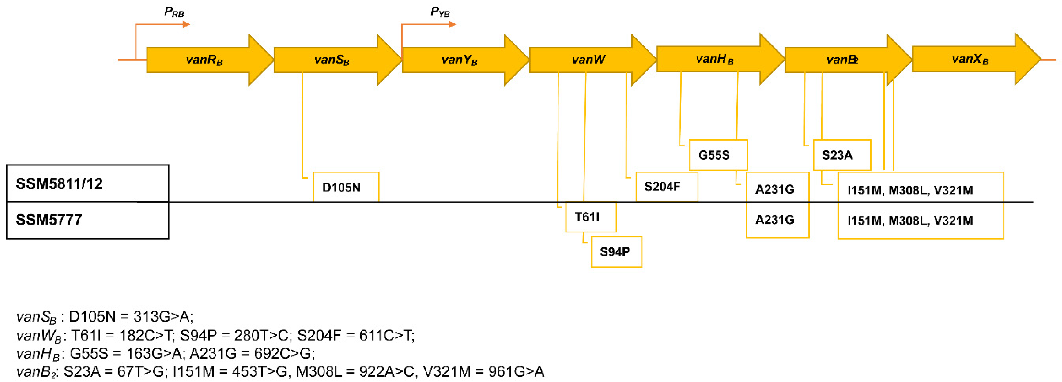
2.3. WGS In Silico Analysis

Ten genomes of E. faecium isolates were selected for WGS according to hospital associated STs. Five ST117—including the vanB-VREfm, two ST916, and one isolate from ST80, ST78, and ST780—were sequenced by an Illumina platform and assembled to the scaffold level. The accession numbers, type of data, and status of the sequences are listed in Supplementary Table S1a. The presence of the vanB2 gene was confirmed in VREfm isolates, as part of a chromosomally integrated Tn1549 transposon. The Tn1549 assembled sequence resulted in identical SSM5811 and SSM5812 isolates sharing 99.97% of sequence identity, with the Tn1549 sequence of E. faecium isolate E7654 from Utrecht, the Netherlands (Accession no. LR135324.1), and E. faecium AUS0004 (Accession no. CP003351.1). Compared to the vanB2 gene cluster of AUS0004 (VAN MIC ≥ 16 mg/L and TEC MIC = 2 mg/L) used as a reference strain, our VREfm displayed single nucleotide mutations leading to amino acid substitutions in VanS (D105N), VanW (S204F), VanH (G55S/A231G), and VanB (S23A_I151M_M308L_M321V) proteins (Figure 1). The assembled Tn1549 of the SSM5777 isolate showed 99.98% sequence identity with those of isolates SSM5811 and SSM5812, while the vanB2 operon showed a slight genetic variation (99.89%), having a wild type vanS gene and different point mutations leading to amino acid substitutions in VanW (T61I_S94P) (Figure 1).
The SSM5777 was re-sequenced and analyzed after exposure to 100 mg/L of vancomycin, showing the same operon mutations observed before vancomycin exposure.
Moreover, among the ten sequenced E. faecium, other resistance genes were detected in silico by ResFinder. The most common were genes conferring acquired resistance to aminoglycosides [ant(6)-Ia (n = 8), aph(3′)-III (n = 9), aac(6′)Ii (9), and aac(6’)-aph(2′′) (n = 5)], macrolide-lincosamide-streptogramin B [msrC (n = 10) and ermB (n = 8)], trimethoprim [dfrF (n = 2) and dfrG (n = 1)], phenicols [cat (n = 1)], and tetracycline [tet(M) (n = 2) and tet(L) (n = 1)] (Supplementary Table S1b), while fluoroquinolones resistance was associated with mutations in the quinolone resistance-determining region of topoisomerase genes leading to amino acid substitution in gyrA (Ser83-Tyr) and parC (Ser80-Ile). For these ten isolates, the phenotypic antibiotic susceptibility patterns aligned with the resistant genes identified (Supplementary Table S1b).

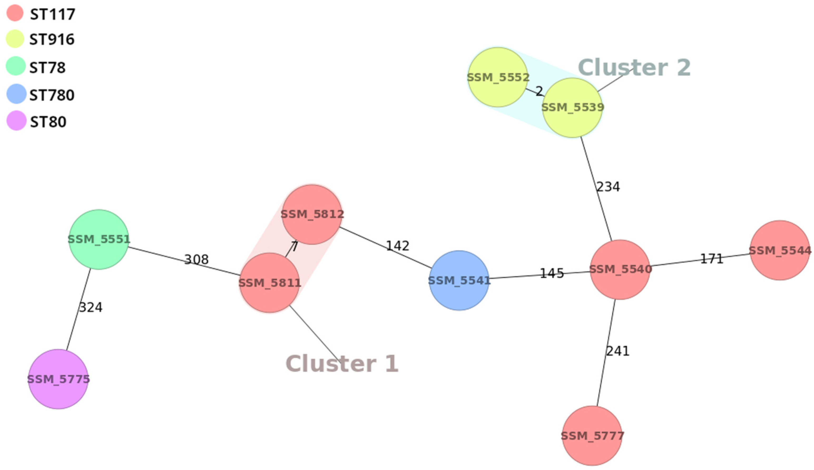
2.4. CgMLST Phylogenetic Analysis

Core genome MLST analysis revealed that the ST117 isolates belonged to four different complex types (CTs). VSEfm isolates were from CT130 and CT132, and occult vanB2-VREfm (SSM5777) was from CT798. A new CT was assigned to the vanB2-VREfm isolates detected in the same patient; they were clonal (single nucleotide polymorphism, SNP = 7) and clustered in Cluster 1 (Figure 2). A putative nosocomial transmission of VSEfm ST916 CT131 was found in the surgery ward in 2013 and clustered in Cluster 2 (Figure 2). The ST80 (9,1,1,1,12,1,1), erroneously designed as a single locus variant of ST117 (9,1,1,1,1,1,1) by eBURSTv3, was genetically more related to isolates from ST78 (15,1,1,1,1,1,1), and hence it is a double locus variant.

3. Discussion

Hospital settings provide a reservoir for MDR pathogens; thus, understanding molecular resistance mechanisms and epidemiology of circulating strains is crucial for preventing infection, including the emergence and dissemination of VREfm, which is of particular clinical concern [17].
In this study, we performed phenotypic and genotypic characterization of E. faecium isolates recovered during 2013–2018 from patients recovering in the main wards of the University Hospital of Sassari. Our findings evidenced three isolates characterized by harboring highly mutated vanB2 operons that were associated with variable vancomycin MICs representing, to our knowledge, the first description of vanB-VREfm in our hospital and in Sardinia.
Notably, one vanB2-VREfm isolate was undetected by Vitek2 (occult VREfm), as previously described by other authors [9], while it proved phenotypically vancomycin resistant by alternative methods. Interestingly, unlike the “classic” occult vanB-VREfm [9], this isolate was able to increase its vancomycin MIC after exposure to vancomycin, as previously described for vanA-VVEfm [12,13,14] and stealthy vanB2-E. faecium isolates [15].
These types of E. faecium going undetected by routine phenotypic testing are at risk of silent spread and are currently considered a developing threat because they can revert to a vancomycin-resistant phenotype upon exposure to vancomycin, causing treatment failures. This ability is typical of vanA E. faecium [12,14,18]; however, Hashimoto et al. described that Japan HA-E. faecium isolates harbor stealthy vanB2 operons producing mutated VanW and VanB2 proteins, which regained a resistant phenotype on exposure to vancomycin thanks to a mutation in the VanS sensor [15]. Our occult vanB2 isolate showed three mutations in the VanB2 protein that were the same as in the other two vanB2VREfm. Two of these mutations, V321M and M308L, were described to be involved in the decrease of vancomycin MIC in vanB2-VREfm isolates from Japan and Taiwan, respectively [15,19]. The presence of a VanS mutation (D105N) located in the same periplasmic domain [20] in reverted Japanese isolates [15] may be responsible for the higher vancomycin MIC in our VREfm compared to the occult VREfm, lacking this mutation.
Moreover, the analysis of the vanB2 operon highlighted additional mutations in VanW and VanH proteins, including a A231G missense mutation in VanH, which occurs in an amino acidic residue essential for its dehydrogenase activity [21]. Considering this, we hypothesized that our vanB2-VREfm may represent reverted-stealthy vanB2 E. faecium, and that vancomycin exposure may lead to similar mutations in the occult vanB2-VREfm, as previously demonstrated in Japanese isolates [15]. However, re-sequencing of the vanB2 operon after vancomycin exposure did not detect additional mutations, suggesting an alternative mechanism for the increased vancomycin MIC. Further studies are needed to confirm this hypothesis and to clarify the role of single mutations in the modulation of the vanB2 operon.
The Tn1549 transposons detected in different years showed high nucleotide identity with common and univocal mutations, suggesting a common ancestral source.
Moreover, most E. faecium isolates, including the VSEfm, showed a consistently multidrug-resistant phenotype and genotype over the years. In sequenced isolates, an analysis of acquired antimicrobial resistance genes revealed that aminoglycosides and macrolide resistances were associated with the carriage of the aph(3′)-IIIa and/or aac(6′)-Ii, and/or aac(6′)-Ie-aph(2′′)-Ia genes and the msr(C) and/or ermB genes, respectively.
Furthermore, despite the low number of isolates analyzed in this study, the polyclonal nature of nosocomial E. faecium STs is clearly shown.
Our results highlighted that vanB2-VREfm and nearly half of VSEfm analyzed were from the hospital-associated (HA) ST117/CC17 or CladeA1 [22]. These STs are frequently associated with nosocomial outbreaks in Europe [23,24,25] and have also been reported in Italian hospitals [26], along with the other HA-STs that were detected here (ST78, ST916, ST80, and ST780).
Finally, cgMLST subdivided ST117 into different CTs excluding the nosocomial transmission of a specific CT. However, the presence of ST117 over the years and its association to vancomycin resistance determinants compared to the other STs also capable of escaping phenotypic detection makes ST117 even more dangerous, confirming its high potential for diffusion.

4. Materials and Methods

4.1. E. faecium Isolates, Antimicrobial Susceptibility

Thirty-nine E. faecium strains isolated from patients admitted at the University Hospital of Sassari, Italy during 2013–2018, all part of the strains collection of the Microbiology Unit of the Biomedical Sciences Department of the University of Sassari, were included in the study. The isolation was performed at the Division of Microbiology and Virology of the University Hospital of Sassari, Italy using Slanetz Bartley or Blood Agar media (Microbiol UTA, Cagliari, Italy). Species identification was determined by Maldi-TOF Mass Spectrometry (Bruker, Billerica, MA, USA). After identification, antimicrobial susceptibility testing was conducted by Vitek2 using the AST cards P592 and P658 (bioMerieux, Marcy-l’Étoile, France), which are specifically for gram-positive Staphylococcus spp., Enterococcus spp., and Streptococcus agalactiae. For Enterococcus spp., among the antibiotics tested, we report results for ampicillin (AMP), high-level gentamicin (GEN), high-level streptomycin (STR), ciprofloxacin (CIP), imipenem (IMI), erythromycin (ERY), linezolid (LNZ), teicoplanin (TEC), vancomycin (VAN), and tigecycline (TGC) following the European Committee on Antimicrobial Susceptibility Testing (EUCAST) guidelines [27].
Antimicrobial susceptibility testing was also performed by MicroScan autoSCAN-4 using the AB33 Cards (Beckman Coulter Inc., CA, USA), following the EUCAST guidelines [27]. Glycopeptide susceptibility was additionally tested by the disk diffusion method [27] with E. faecalis ATCC 29212 as the control.

4.2. Molecular Characterization

4.2.1. Screening for vanA, -B, -C Genes and MLST

Bacterial DNA was extracted with the DNeasy Blood & Tissue Kit (QIAGEN, Inc., Valencia, CA, USA). The presence of vanA, vanB, vanC1, and vanC2/C3 genes was found by Multiplex-PCR as previously described [28]. MLST was carried out on all isolates with the standard primers included in the E. faecium MLST scheme [29]. In addition, purified amplicons with DNA clean and concentratorTM-5 columns (Zymo Research, Irvine, CA, USA) were sequenced at the LMU Sequencing Service (Munich, Germany). Obtained sequences were analyzed using Geneious Pro 4.8.4 (http://www.geneious.com/ accessed on 1 March 2017). Allelic profiles and STs were assigned according to the E. faecium MLST database (http://pubmlst.org/efaecium/ accessed on 16 December 2018) and included in CCs based on eBURSTv3 analysis (http://eburst.mlst.net/ accessed on 16 December 2018) used to assign isolates to genetic complexes and to evaluate the genetic relationships of the STs.

4.2.2. WGS and In Silico Analysis

WGS was performed on ten selected E. faecium isolated from the surgery (SSM5539, SSM5540, SSM5541, SSM5544, SSM5551, SSM5552, and SSM5775) and ICU (SSM5777, SSM5811, and SSM5812) ward. Chromosomal DNAs, extracted using the DNeasy Blood & Tissue Kit (QIAGEN, Inc., Valencia, CA, USA), were quantified by the NanoDrop Microvolume Spectrophotometer (ThermoFisher, MA, USA) and submitted to WGS using a HiScanSQIllumina platform (Porto Conte Ricerche Srl, Tramariglio, Italy). Preparation of the DNA libraries was performed with the Nextera XTDNA Sample Preparation Kit (Illumina Inc., San Diego, CA, USA) and sequenced using the HiScanSQ (Illumina Inc., San Diego, CA, USA) with 93bp × 2 paired-end reads.
Generated sequences were assembled de novo into contigs using the software Velvet version 1.2.10 [30]. Contigs were reordered against the reference genome of E. faecium AUS0004 (Accession no. CP003351.1) using Mauve. Whole-genome alignment was done using the Artemis Comparative Tool (ACT) and MUMmer. The NUCmer tool of the MUMmer software was used to search individual gene sequences within the genomes. All ten genomes were submitted to the RAST platform for annotation (https://rast.nmpdr.org/ accessed on on 15 May 2021. The assembled genomes were subjected to the online search tools MLST 2.0 (Achtman MLST scheme) and ResFinder 4.0 available at the Center for Genomic Epidemiology (CGE) (http://www.genomicepidemiology.org/ accessed on 30 June 2021. The vanB2 operon was resequenced using specific primers designed on the AUS0004 reference strain (CP003351) and shown in Supplementary Table S1c. Amplicons were purified, sequenced, and analyzed as described above.

4.3. Phylogenetic Analysis by cgMLST

The Ridom SeqSphere+ version 6.0.2 [31] whole-genome typing platform was used to infer phylogenetic relatedness of the isolates based on 2554 loci of the E. faecium cgMLST v1.1 and E. faecium MLSTv1.0 schemes, with cgMLST complex type/cluster-alert distance set as 20.

5. Conclusions

In this work, we highlighted a highly mutated vanB2-VREf ST117 occult isolate by Vitek2 with the ability to increase its vancomycin MIC after exposure to vancomycin. This isolate was found to have similarities to both vanA-VVEfm and stealthy vanB2-E. faecium.
The risk of non-detection of vanB2-VREfm by the Vitek2 system is of great concern, and E. faecium should always be processed using an alternative method to Vitek2 to avoid therapeutic failure and vanB2-VREfm spread.
Stricter preventive measures, such as isolation of infected/colonized patients and more accurate disinfection procedures, can reduce the risk of dissemination of these coverts and all HA-VREfm/VSEfm clones.

Supplementary Materials

The following supporting information can be downloaded at https://www.mdpi.com/2079-6382/12/3/476/s1; Supplementary Tables: Table S1a: Accession numbers of the sequences, type of data and status of Enterococcus faecium isolates from University Hospital, Sassari, Italy; Table S1b: Characteristics and antimicrobial resistant genes of the 10 sequenced Enterococcus faecium isolates; Table S1c: Primers utilized to amplify the vanB2 operon of Enterococcus faecium isolate SSM5777 after exposure to vancomycin.

Author Contributions

Conceptualization, A.S. and B.P.; methodology, A.S., M.F., H.M.H., and S.S.; investigation, formal analysis, and validation, A.S. and B.P.; software, E.T. and M.D.; data curation, A.S. and B.P.; writing—original draft preparation, A.S.; writing—review and editing, A.S., E.T., and B.P.; visualization, A.S. and B.P.; supervision, B.P. and S.R.; funding acquisition, B.P. and S.R. All authors have read and agreed to the published version of the manuscript.

Funding

This work was financially supported by the University of Sassari, FAR 2020 given to B.P. and FAR 2020 given to S.R.

Institutional Review Board Statement

Not applicable.

Informed Consent Statement

Not applicable.

Data Availability Statement

The data presented in this study are available in the article and in Supplementary Materials.

Acknowledgments

B.P. is supported by Bando Fondazione di Sardegna 2022–2023, Progetti di base dipartimentali.

Conflicts of Interest

The authors declare no conflict of interest.

References

  1. Arias, C.A.; Murray, B.E. The rise of the Enterococcus: Beyond vancomycin resistance. Nat. Rev. Microbiol. 2012, 10, 266–278. [Google Scholar] [CrossRef] [PubMed]
  2. Cheah, A.L.Y.; Spelman, T.; Liew, D.; Peel, T.; Howden, B.P.; Spelman, D.; Graysonca, M.L.; Nationa, R.L.; Konga, D.D.M. Enterococcal bacteraemia: Factors influencing mortality, length of stay and costs of hospitalization. Clin. Microbiol. Infect. 2013, 19, 384. [Google Scholar] [CrossRef] [PubMed]
  3. Cattoir, V.; Leclercq, R. Twenty-five years of shared life with vancomycin-resistant enterococci: Is it time to divorce. J. Antimicrob. Chemother. 2013, 68, 731–742. [Google Scholar] [CrossRef] [PubMed]
  4. Brinkwirth, S.; Ayobami, O.; Eckmanns, T.; Markwart, R. Hospital-acquired infections caused by enterococci: A systematic review and meta-analysis, WHO European Region, 1 january 2010 to 4 february 2020. Eurosurveillance 2021, 26, 2001628. [Google Scholar] [CrossRef] [PubMed]
  5. Correa-Martínez, C.L.; Jurke, A.; Schmitz, J.; Schaumburg, F.; Kampmeier, S.; Mellmann, A. Molecular Epidemiology of Vancomycin-Resistant Enterococci Bloodstream Infections in Germany: A Population-Based Prospective Longitudinal Study. Microorganisms 2022, 10, 130. [Google Scholar] [CrossRef]
  6. Leavis, H.L.; Willems, R.J.L.; van Wamel, W.J.B.; Schuren, F.H.; Caspers, M.P.M.; Bonten, M.J.M. Insertion sequence-driven diversification creates a globally dispersed emerging multiresistant subspecies of E. faecium. PLoS Pathog. 2007, 3, e7. [Google Scholar] [CrossRef]
  7. Hegstad, K.; Mikalsen, T.; Coque, T.M.; Werner, G.; Sundsfjord, A. Mobile genetic elements and their contribution to the emergence of antimicrobial resistant Enterococcus faecalis and Enterococcus faecium. Clin. Microbiol. Infect. 2010, 16, 541–554. [Google Scholar] [CrossRef]
  8. Lee, T.; Pang, S.; Abraham, S.; Coombs, G.W. Antimicrobial-resistant CC17 Enterococcus faecium: The past, the present and the future. J. Glob. Antimicrob. Resist. 2019, 16, 36–47. [Google Scholar] [CrossRef]
  9. Walker, S.V.; Wolke, M.; Plum, G.; Weber, R.E.; Werner, G.; Hamprecht, A. Failure of Vitek2 to reliably detect vanB-mediated vancomycin resistance in Enterococcus faecium. J. Antimicrob. Chemother. 2021, 76, 1698–1702. [Google Scholar] [CrossRef] [PubMed]
  10. Hammerum, A.M.; Justesen, U.S.; Pinholt, M.; Roer, L.; Kaya, H.; Worning, P.; Nygaard, S.; Kemp, M.; Clausen, M.E.; Nielsen, K.L.; et al. Surveillance of vancomycin-resistant enterococci reveals shift in dominating clones and national spread of a vancomycin-variable vanA Enterococcus faecium ST1421-CT1134 clone, Denmark, 2015 to March 2019. Eurosurveillance 2019, 24, 1900503. [Google Scholar] [CrossRef]
  11. Gagnon, S.; Lévesque, S.; Lefebvre, B.; Bourgault, A.-M.; Labbé, A.-C.; Roger, M. vanA-containing Enterococcus faecium susceptible to vancomycin and teicoplanin because of major nucleotide deletions in Tn1546. J. Antimicrob. Chemother. 2011, 66, 2758–2762. [Google Scholar] [CrossRef]
  12. Sivertsen, A.; Pedersen, T.; Larssen, K.W.; Bergh, K.; Rønning, T.G.; Radtke, A.; Hegstada, K. A silenced vanA gene cluster on a transferable plasmid caused an outbreak of vancomycin-variable enterococci. Antimicrob. Agents Chemother. 2016, 60, 4119–4127. [Google Scholar] [CrossRef]
  13. Wagner, T.M.; Janice, J.; Sivertsen, A.; Sjögren, I.; Sundsfjord, A.; Hegstad, K. Alternative vanHAX promoters and increased vanA-plasmid copy number resurrect silenced glycopeptide resistance in Enterococcus faecium. J. Antimicrob. Chemother. 2021, 76, 876–882. [Google Scholar] [CrossRef] [PubMed]
  14. Thaker, M.N.; Kalan, L.; Waglechner, N.; Eshaghi, A.; Patel, S.N.; Poutanen, S.; Willey, B.; Coburn, B.; McGeer, A.; Low, D.E.; et al. Vancomycin-variable enterococci can give rise to constitutive resistance during antibiotic therapy. Antimicrob. Agents Chemother. 2015, 59, 1405–1410. [Google Scholar] [CrossRef]
  15. Hashimoto, Y.; Kurushima, J.; Nomura, T.; Tanimoto, K.; Tamai, K.; Yanagisawa, H.; Shirabe, K.; Iket, Y.; Tomita, H. Dissemination and genetic analysis of the stealthy vanB gene clusters of Enterococcus faecium clinical isolates in Japan. BMC Microbiol. 2018, 18, 213. [Google Scholar] [CrossRef] [PubMed]
  16. De Been, M.; Pinholt, M.; Top, J.; Bletz, S.; Mellmann, A.; van Schaik, W.; Brouwer, E.; Rogers, M.; Kraat, Y.; Bonten, M.; et al. Core genome multilocus sequence typing scheme for high-resolution typing of Enterococcus faecium. J. Clin. Microbiol. 2015, 53, 3788–3797. [Google Scholar] [CrossRef]
  17. Guzman, P.A.M.; van Schaik, W.; Rogers, M.R.C.; Coque, T.M.; Baquero, F.; Corander, J.; Willems, R.J.L. Global Emergence and Dissemination of Enterococci as Nosocomial Pathogens: Attack of the Clones? Front. Microbiol. 2016, 7, 788. [Google Scholar] [CrossRef]
  18. Merlino, J.; Gray, T. Vancomycin variable Enterococcus (VVE), E. faecium, harbouring the vanA gene complex. Pathology 2021, 53, 680–682. [Google Scholar] [CrossRef]
  19. Lu, J.J.; Perng, C.L.; Ho, M.F.; Chiueh, T.S.; Lee, W.H. High prevalence of VanB2 vancomycin-resistant Enterococcus faecium in Taiwan. J. Clin. Microbiol. 2001, 39, 2140–2145. [Google Scholar] [CrossRef] [PubMed]
  20. Guffey, A.A.; Loll, P.J. Regulation of Resistance in Vancomycin-Resistant Enterococci: The VanRS Two-Component System. Microorganisms 2021, 9, 2026. [Google Scholar] [CrossRef]
  21. Stogios, P.J.; Savchenko, A. Molecular mechanisms of vancomycin resistance. Protein Sci. 2020, 29, 654–669. [Google Scholar] [CrossRef] [PubMed]
  22. Freitas, A.R.; Pereira, A.P.; Novais, C.; Peixe, L. Multidrug-resistant high-risk Enterococcus faecium clones: Can we really define them? Int. J. Antimicrob. Agents 2021, 57, 106227. [Google Scholar] [CrossRef] [PubMed]
  23. Weber, A.; Maechler, F.; Schwab, F.; Gastmeier, P.; Kola, A. Increase of vancomycin-resistant Enterococcus faecium strain type ST117 CT71 at Charité—Universitätsmedizin Berlin, 2008 to 2018. Antimicrob. Resist. Infect. Control 2020, 9, 109. [Google Scholar] [CrossRef]
  24. Rodríguez-Lucas, C.; Fernández, J.; Raya, C.; Bahamonde, A.; Quiroga, A.; Muñoz, R.; Rodicio, M.R. Establishment and Persistence of Glycopeptide-Resistant Enterococcus faecium ST80 and ST117 Clones within a Health Care Facility Located in a Low-Prevalence Geographical Region. Microb. Drug Resist. 2022, 28, 217–221. [Google Scholar] [CrossRef]
  25. Lisotto, P.; Couto, N.; Rosema, S.; Lokate, M.; Zhou, X.; Bathoorn, E.; Harmsen, H.J.M.; Friedrich, A.W.; Rossen, J.W.A.; Chlebowicz-Fliss, M.A. Molecular Characterisation of Vancomycin-Resistant Enterococcus faecium Isolates Belonging to the Lineage ST117/CT24 Causing Hospital Outbreaks. Front. Microbiol. 2021, 12, 728356. [Google Scholar] [CrossRef] [PubMed]
  26. Fioriti, S.; Simoni, S.; Caucci, S.; Morroni, G.; Ponzio, E.; Coccitto, S.N.; Brescini, L.; Cirioni, O.; Menzo, S.; Biavasco, F.; et al. Trend of clinical vancomycin-resistant enterococci isolated in a regional Italian hospital from 2001 to 2018. Braz. J. Microbiol. 2020, 51, 1607–1613. [Google Scholar] [CrossRef]
  27. The European Committee on Antimicrobial Susceptibility Testing. Breakpoint tables for Interpretation of MICs and Zone Diameters. Version 12.0. 2022. Available online: http://www.eucast.org (accessed on 30 March 2022).
  28. Santona, A.; Taviani, E.; Hoang, H.M.; Fiamma, M.; Deligios, M.; Ngo, T.V.Q.; Van, A.L.; Cappuccinelli, P.; Rubino, S.; Paglietti, B. Emergence of unusual vanA/vanB2 genotype in a highly mutated vanB2 -vancomycin-resistant hospital-associated E. faecium background in Vietnam. Int. J. Antimicrob. Agents 2018, 52, 586–592. [Google Scholar] [CrossRef] [PubMed]
  29. Homan, W.L.; Tribe, D.; Poznanski, S.; Li, M.; Hogg, G.; Spalburg, E.; Van Embden, J.D.; Willems, R.J. Multilocus sequence typing scheme for Enterococcus faecium. J. Clin. Microbiol. 2002, 40, 1963–1971. [Google Scholar] [CrossRef]
  30. Zerbino, D.R.; Birney, E. Velvet: Algorithms for de novo short read assembly using de Bruijn graphs. Genome Res. 2008, 18, 821–829. [Google Scholar] [CrossRef]
  31. Jünemann, S.; Sedlazeck, F.J.; Prior, K.; Albersmeier, A.; John, U.; Kalinowski, J.; Mellmann, A.; Goesmann, A.; von Haeseler, A.; Stoye, J. Updating benchtop sequencing performance comparison. Nat. Biotechnol. 2013, 31, 294–296. [Google Scholar] [CrossRef] [PubMed]
Figure 1. Schematic representation of vanB2 operon in VREfm and in occult isolate, with the amino acid substitutions framed and in line with the corresponding regulation and resistance genes. For each van gene, the amino acid replacements (D = Asp, N = Asn, T = Thr, I = Ile, S = Ser, P = Pro, F = Phe, G = Gly, A = Ala, M = Met, L = Leu, and V = Val) corresponding to nucleotide mutations are shown below on the left.
Antibiotics 12 00476 g001
Figure 2. Minimum spanning tree generated by Ridom SeqSphere+ showing the genetic relatedness among E. faecium isolates. Each circle represents an isolate, and the color of each circle corresponds to the ST. The numbers on the connecting lines indicate the core gene SNP difference between strains.
Antibiotics 12 00476 g002
Table 1. Characteristics of E. faecium isolates.
SSM StrainDateSourceWardAMPGENSTRERYCIPIMILNZTGCVAN mg/LTEC
mg/L
Van GenesST
55384 February 2013B. aspirateMedicineRRRRRRSS1≤0.5-117
55506 March 2013UrineMedicineRSRRRRSS≤0.5≤0.5-117
55634 April 2013PusSurgeryRRRRRRSS≤0.5≤0.5-78
55567 April 2013F. fluidSurgeryRRRRRRSS≤0.5≤0.5-78
554217April 2013SputumSurgerySRRRRSSS1≤0.5-78
5540 *22 April 2013CVCSurgeryRRRRRRSS≤0.5≤0.5-117
55481 June 2013WoundICURRRRRRSS1≤0.5-78
55493 June 2013BileSurgeryRRRRRRSS1≤0.5-78
5551 *6 June 2013A. fluidSurgeryRRRRRRSS1≤0.5-78
55628 June 2013UrineSurgeryRRRRRRSS≤0.51-117
55551 July 2013BloodMedicineRRRRRRSS1≤0.5-78
564612 July 2013UrineSurgeryRRRRRRSS11-117
556019 July 2013B. aspirateMedicineSSSRRSSS≤0.5≤0.5-54
5539 *4 August 2013BileSurgeryRSRRRRSS≤0.5≤0.5-916
558710 August 2013D. fluidSurgeryRRRRRRSS≤0.5≤0.5-78
558810 August 2013B. aspirateMedicineRRRRRRSS≤0.5≤0.5-916
558328 September 2013BiopsyMedicineRRRRRRSS≤0.5≤0.5-117
5552 *6 October 2013BloodSurgeryRRSRRRSS≤0.5≤0.5-916
55912 November 2013BileSurgeryRRRRRRSS≤0.5≤0.5-78
5544 *4 November 2013B. aspirateSurgeryRRRRRRSS≤0.25≤0.25-117
55924 November 2013PusSurgeryRRRRRRSS≤0.5≤0.5-78
5541 *4 December 2013F. fluidSurgeryRRRRRRSS1≤0.12-780
55749 December 2013WoundSurgeryRRRRRRSS≤0.5≤0.5-78
565228 December 2013BloodMedicineRRRRRRSS≤0.5≤0.5-780
566221 February 2014A. fluidSurgeryRRRRRRSS≤0.5≤0.5-117
567026 February 2014B. aspirateICURRRRRRSS≤0.5≤0.5-117
567320 March 014BloodICURSRRRRSS12-117
567127 March 2014BloodICURRRRRRSS≤0.5≤0.5-117
56532 April 2014BloodICURRRRRRSS≤0.5≤0.5-117
56551 May 2014BileSurgeryRRRRRRSS1≤0.5-78
56541 May 2014UrineMedicineRRRRRRSS≤0.51-117
56582 May 2014UrineMedicineRRRRRRSS≤0.52-117
56562 May 2014PusICUSSSRRSSS≤0.5≤0.5-74
5775 *20 January 2017D. fluidSurgeryRSRRRRSS≤0.51-80
5777 *25 January 2017A. fluidICURRRRRRSS≤0.5≤0.5vanB117
57799 February 2017BileSurgeryRSRRRRSS≤0.52-117
57809 February 2017BileSurgeryRSRRRRSS≤0.5≤0.5-80
5811 *2 May 2018WoundICURRRRRRSS8≤0.5vanB117
5812 *3 May 2018CVCICURRRRRRSS≥32≤0.5vanB117
* These isolates were subjected to whole genome sequencing. CVC = central venous catheter; A. fluid: abdominal fluid; D. fluid: drainage fluid; F. fluid: fistula fluid; B. aspirate: broncho aspirate. The medicine ward included pathology and pneumology. AMP: ampicillin; GEN: high-level gentamicin; STR: high-level streptomycin; ERY: erythromycin; CIP: ciprofloxacin; IMI: imipenem; LNZ: linezolid; TGC: tigecycline; VAN: vancomycin; TEC: teicoplanin.
Table 2. Features and levels of vancomycin resistance mediated by vanB2-VREfm. Glycopeptides MICs are given before (A, B) and following (C) exposure to 100mg/L of vancomycin.
SSM StrainDateSourceWardA
Vitek2
B
MicroScan
C
Vitek2
VAN mg/LTEC mg/LVAN
mg/L
TEC mg/LVAN mg/LTEC mg/L
577725/01/2017A. fluidICU≤1≤0.58≤2≥32≤0.5
5811 *02/05/2018WoundICU8≤0.5>8≤2≥32≤0.5
5812 *03/05/2018CVCICU≥32≤0.5>8≤2≥32≤0.5
* Same patient.
Disclaimer/Publisher’s Note: The statements, opinions and data contained in all publications are solely those of the individual author(s) and contributor(s) and not of MDPI and/or the editor(s). MDPI and/or the editor(s) disclaim responsibility for any injury to people or property resulting from any ideas, methods, instructions or products referred to in the content.

