Article

New Insights into Listeria monocytogenes Antimicrobial Resistance, Virulence Attributes and Their Prospective Correlation

by
Mahmoud E. Elsayed
1,
Marwa I. Abd El-Hamid
2,
Attia El-Gedawy
3,
Mahmoud M. Bendary
4,*,
Reham M. ELTarabili
1,
Majid Alhomrani
5,6,
Abdulhakeem S. Alamri
5,6,
Saleh A. Alghamdi
5,6,
Marwa Arnout
7,
Dalal N. Binjawhar
8,
Mohammad M. Al-Sanea
9 and
Amira I. Abousaty
10
1
Department of Bacteriology, Immunology, and Mycology, Faculty of Veterinary Medicine, Suez Canal University, Ismailia 41522, Egypt
2
Department of Microbiology, Faculty of Veterinary Medicine, Zagazig University, Zagazig 44511, Egypt
3
Department of Bacteriology, Animal Health Research Institute, Dokki, Giza 12618, Egypt
4
Department of Microbiology and Immunology, Faculty of Pharmacy, Port Said University, Port Said 42511, Egypt
5
Department of Clinical Laboratories Sciences, The Faculty of Applied Medical Science, Taif University, Taif 26432, Saudi Arabia
6
Centre of Biomedical Science Research (CBSR), Deanship of Scientific Research, Taif University, Taif 26432, Saudi Arabia
7
Veterinary Quarantine, Cairo Airport, Zagazig 44511, Egypt
8
Department of Chemistry, College of Science, Princess Nourah Bint Abdulrahman University, Riyadh 11671, Saudi Arabia
9
Pharmaceutical Chemistry Department, College of Pharmacy, Jouf University, Sakaka 72341, Saudi Arabia
10
Microbiology Department, Faculty of Science, Zagazig University, Zagazig 44511, Egypt
*
Author to whom correspondence should be addressed.
Antibiotics 2022, 11(10), 1447; https://doi.org/10.3390/antibiotics11101447
Submission received: 4 September 2022 / Revised: 11 October 2022 / Accepted: 19 October 2022 / Published: 21 October 2022

Abstract

:
Listeriosis is one of the most common foodborne diseases caused by Listeria monocytogenes (L. monocytogenes). A poor prognosis has been recorded for the invasive listeriosis, especially neurolisteriosis. In several countries throughout the world, foodborne infections with L. monocytogenes exceeded the legal safety limits in animal sourced foods. Therefore, we decided to investigate the variability, virulence and antimicrobial resistance profiles of this pathogen. Both phenotypic and genotypic methods were used for identifying L. monocytogenes isolates and confirming their virulence profiles. The antimicrobial resistances and their correlation analysis with the existence of virulence genes were detected. Additionally, sequencing and phylogenetic analysis based on L. monocytogenes inlA and inlB genes were undertaken. The prevalence rate (11.9%) and the resistance profiles of L. monocytogenes were shocking. The multi-drug resistance (MDR) phenotypes were common among our isolates (64.9%). Fortunately, the resistance phenotypes were always associated with low virulence arrays and the MDR strains possessed low virulence fitness. Herein, the high genotypic and phenotypic diversity of L. monocytogenes isolates and their weak clonality and adaptability highlighted the difficulty in controlling and managing this pathogen. Therefore, it is important to add more restriction guidelines from national authorities on the consumption of ready to eat foods.

1. Introduction

Listeria monocytogenes is an animal and human pathogen, which is responsible for causing intracellular foodborne diseases as well as invasive listeriosis, encephalitis, endocarditis, perinatal infections, gastroenteritis, septicemia, meningitis, ophthalmitis and abortion. Life threatening listeriosis occurs among people with weakened immune systems, neonates, the elderly, and pregnant women [1]. The fatality rate among infected individuals is low; meanwhile, it may increase in the high-risk patients [2]. Regarding the animal infections, there are several forms of listeriosis, such as encephalitic and septicemic. The encephalitic form is characterized by neurologic signs such as depression and incoordination; meanwhile, the septicemic form may include depression, listlessness, emaciation and diarrhea [3].
In recent years, foodborne pathogens have caused major public health crises around the world. The incidence of foodborne diseases continues to grow, with high mortality and morbidity rates. These types of infections pose challenges in developing countries, especially in the Middle East and North Africa regions [4]. Several pathogens such as Staphylococcus aureus, Salmonella Enteritidis, enterohaemorrhagic Escherichia coli and L. monocytogenes can be transmitted via food chains [5,6,7,8].
L. monocytogenes can infect both humans and animals such as domestic pets, rodents, livestock, fish, avian species, and rabbits. The principle route of transmission of listeriosis is contaminated food, which has been estimated to be the source of infection in about 99% of the cases [9]. Listeria monocytogenes is commonly recovered from various types of poultry and meat products and it can grow well in dry sausage and in products with high pH values [10]. For that, public health officials should be watchful and should reassess manufacturing practices for processed poultry, rabbit and beef products and milk in order to decrease the spread of listeriosis.
Of note, L. monocytogenes had many virulence factors including listeriolysine O encoded by hlyA gene, internalins encoded by internalin (inl) genes, fibronectin-binding protein (FbpA), actA protein and enzymes such as lecithinase, serine protease and zinc metal protease. Additionally, L. monocytogenes is found everywhere in food processing and distribution, and it has contaminated a wide range of foods and animal products such as raw meat products, cheese, raw milk, and salads due to poor hygienic conditions [11]. The failure in management approaches with regard to this pathogen was attributed to its ability to form biofilms and its intrinsic physiological resistance against low temperature and high salt concentrations. Additionally, the antimicrobial resistance fitness of L. monocytogenes could be increased by the acquisition of resistance genes from Enterococcus and Streptococcus species [12,13]. Therefore, it is important to perform the in vitro evaluation of the antimicrobial agent before starting the treatment [14,15], and to find new and alternatives antimicrobial agents [16,17]. Delay in treatment in addition to the wide spreading of multidrug resistant (MDR) L. monocytogenes, which harbor many virulence factors, are considered the main causes for developing the infection [18]. In light of the above mentioned threats and high case fatality rates caused by L. monocytogenes, especially in Egypt, we found that it is timely and urgent to study the pathogenicity, virulence and antimicrobial resistance of this pathogen.

2. Results

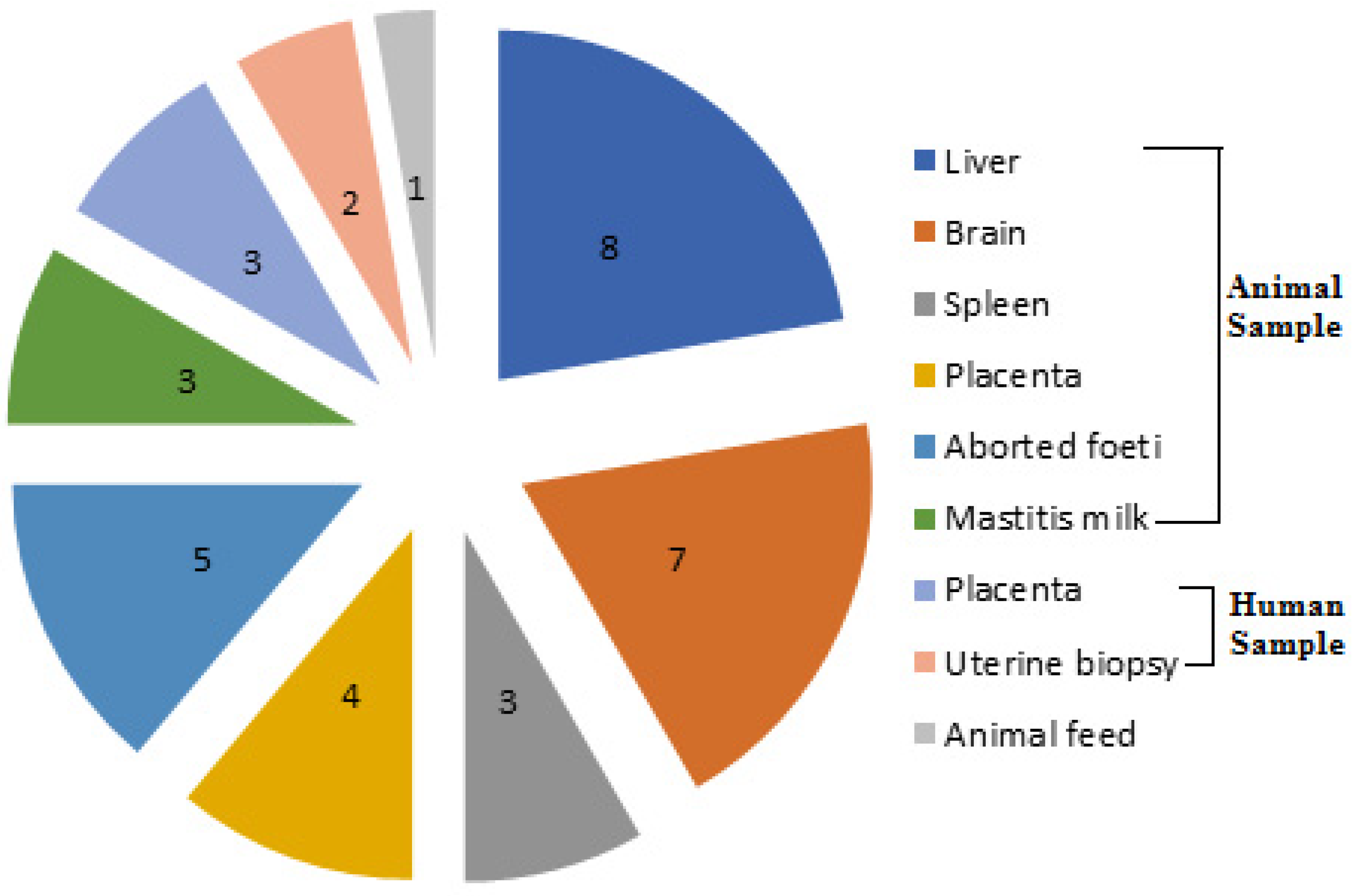
2.1. Prevalence of L. monocytogenes from Different Sources and Governorates

Out of the 310 samples screened in our study, 37 L. monocytogenes isolates were recovered with an overall prevalence rate of 11.9% comprising 13.5% (31/230), 10% (5/50) and 3.3% (1/30) in animal, human and animal feed sources, respectively. The recovered isolates were identified on the basis of their cultural characteristics, biochemical reactions, and via the Microbact™ Listeria 12L system. These isolates exhibited positive results for Anton’s eye, CAMP and hemolysis tests, and possessed the specific 16S rRNA gene. Therefore, genotypic identification was in accordance with phenotypic characterization of L. monocytogenes isolates. The distribution of L. monocytogenes from different sources is illustrated in Figure 1. Regarding the animal origin, the majority of our isolates (26.7%, 8/30) were recovered from liver samples of sheep suffering from septicemia. Moreover, the high proportion of L. monocytogenes isolates from human sources was from placental samples (12%, 3/25), and only one isolate was obtained from 30 animal feed samples (3.3%). Notably, Sharkia had the highest prevalence rate of L. monocytogenes isolates (13.8%, 18/130), followed by Giza (12.2%, 11/90) and then Qaliubia (8.9%, 8/90) Governorates.

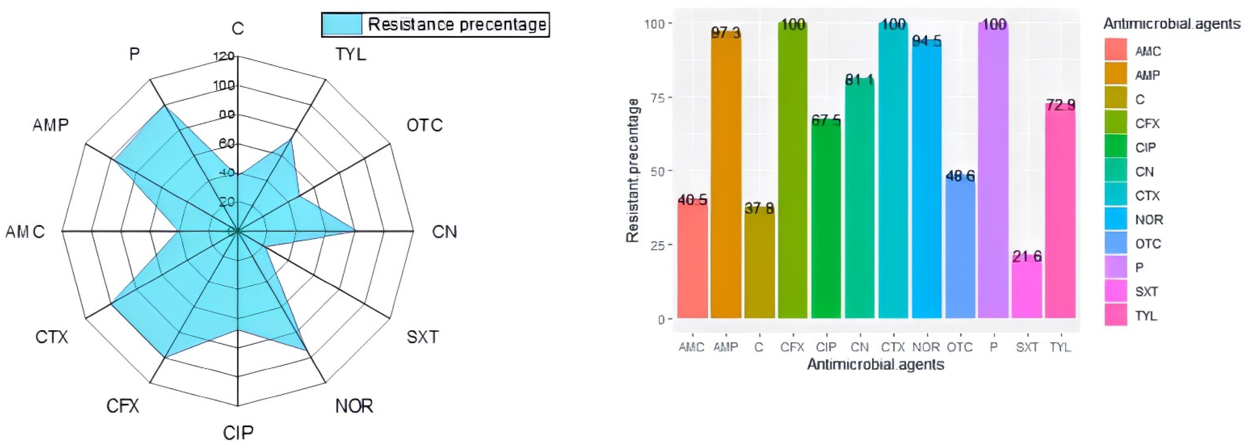
2.2. Antibiogram Typing and Virulence Gene Profiles of L. monocytogenes

According to the antimicrobial susceptibility results, L. monocytogenes isolates showed high resistance rates to penicillin, cephalexin and cefotaxime (100% each), followed by ampicillin (97.3%); meanwhile, the highest sensitivity levels were recorded to sulfamethoxazole-trimethoprim (78.4%) and amoxicillin-clavulanic acid (64.9%), as shown in Figure 2. Unfortunately, the majority of our isolates (24/37; 64.9%) showed the MDR pattern being resistant to three or more antimicrobial drugs from different classes. Moreover, MAR indices of the recovered isolates ranged from 0.17 to 0.42, indicating that they were originated from high-risk contamination. Regarding the distribution of the tested virulence genes among the recovered isolates, our results documented that all L. monocytogenes isolates possessed both inlA and inlB genes; meanwhile, 73% (27/37) and 64.9% (24/37) of the examined isolates harbored prfA and hlyA genes, respectively.

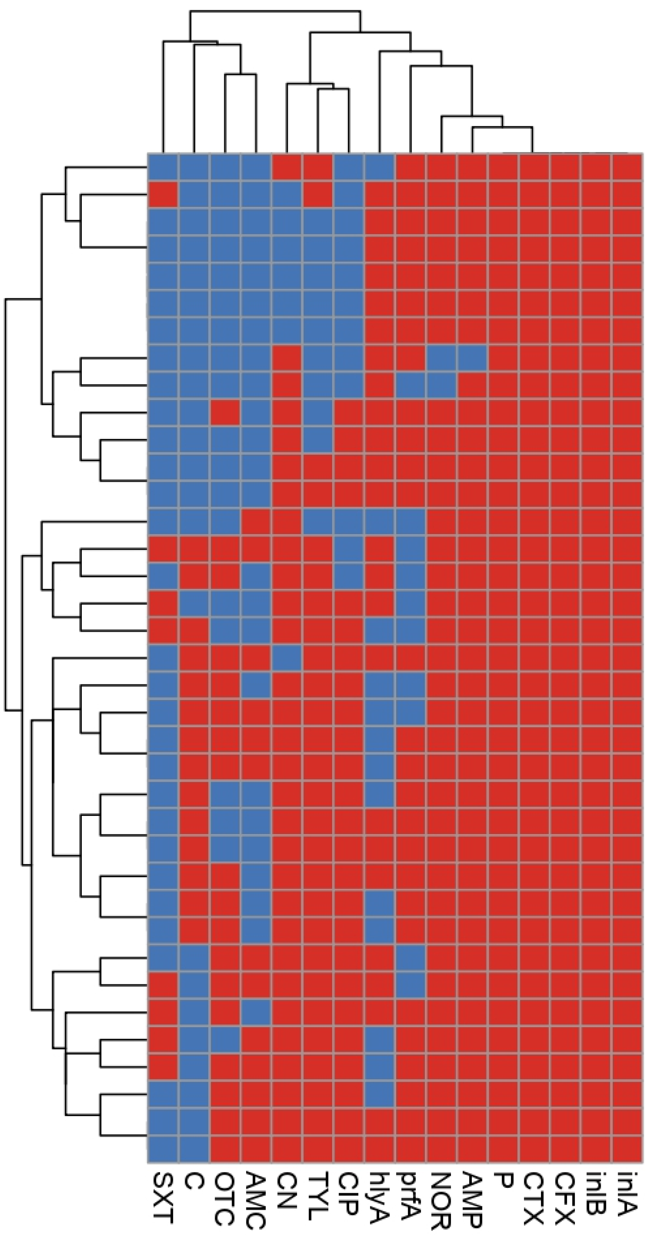
2.3. Phenotypic and Genetic Diversity of L. monocytogenes

Based on both antibiogram typing and virulence gene profiles, all of our recovered L. monocytogenes isolates were cloned into 28 clusters (Figure 3), which reflected the high degree of heterogeneity among these isolates. With the exception of six clusters, each isolate belonged to a unique lineage with a specific phenotypic and genotypic profile. The discriminatory power for combined phenotypic (antibiogram typing) and genotypic (virulence genes’ profiles) methods was 0.98, indicating the high variability of the tested isolates. Out of the six clusters, one contained five isolates; but 10 isolates belonged to the other five clusters (two isolates for each cluster) as shown in Figure 3.

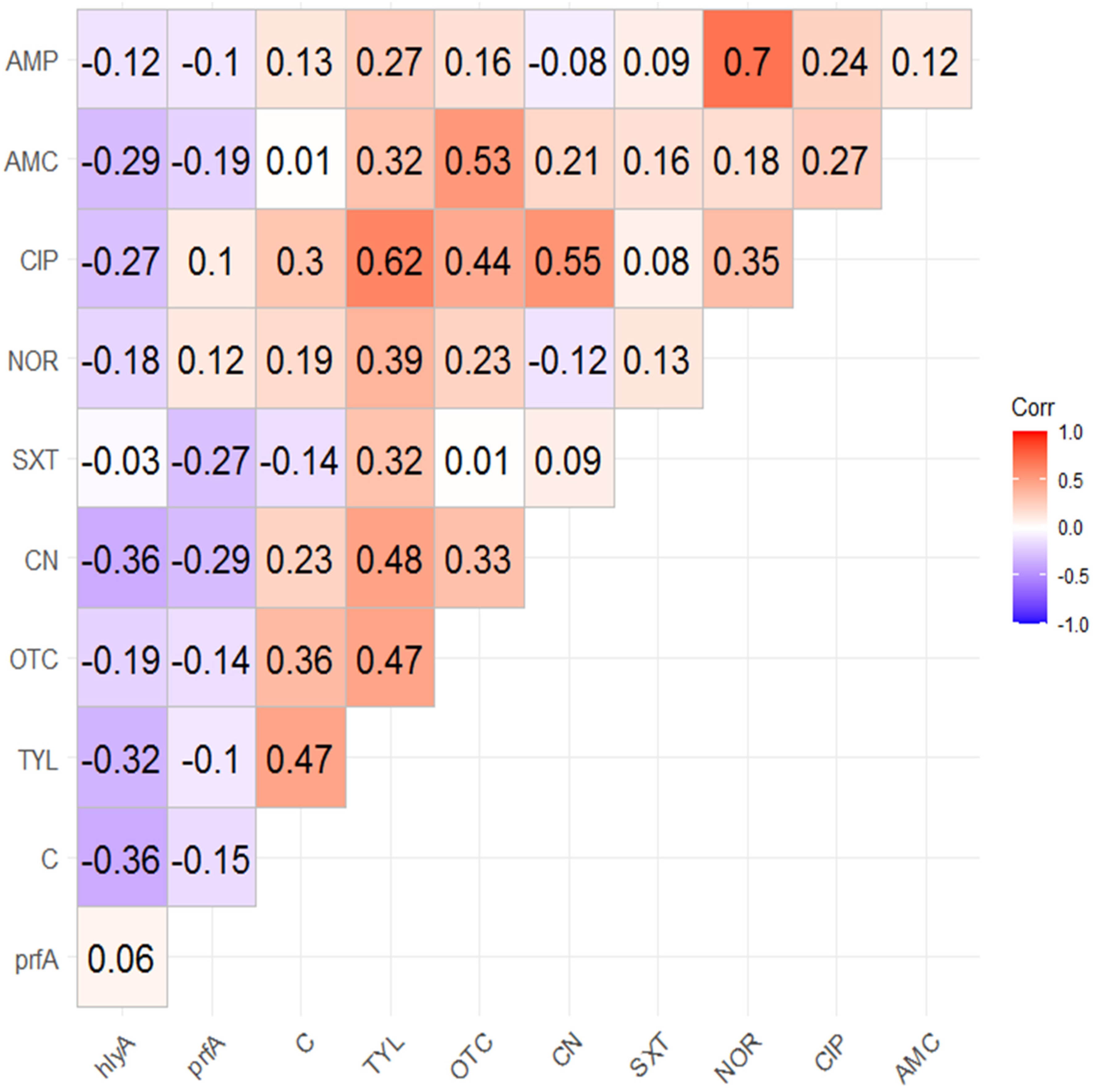
2.4. Correlation Analysis

The correlation analysis among and between resistances to the investigated antimicrobials and the presence of tested virulence genes are illustrated in Figure 4. The results revealed that there were negative correlations between the existence of prfA or hlyA gene and the resistance to all tested antimicrobial drugs, with the exception of the very weak positive correlations between the presence of prfA gene and the resistances to ciprofloxacin and norfloxacin. Of note, strong positive correlations were detected between resistances to ampicillin and norfloxacin (r-value = 0.7) and resistances to ciprofloxacin and tylosin (r-value = 0.62). On the other hand, strong negative correlations between the existence of hlyA gene and the resistances to each of chloramphenicol and gentamycin (r-value = −0.36 each) is presented in Figure 4.

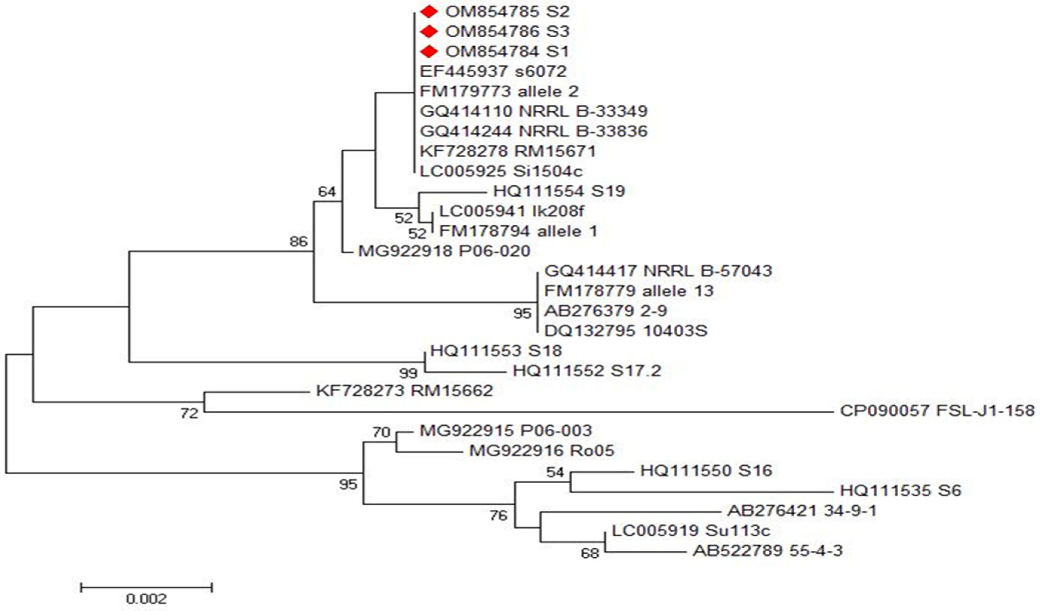
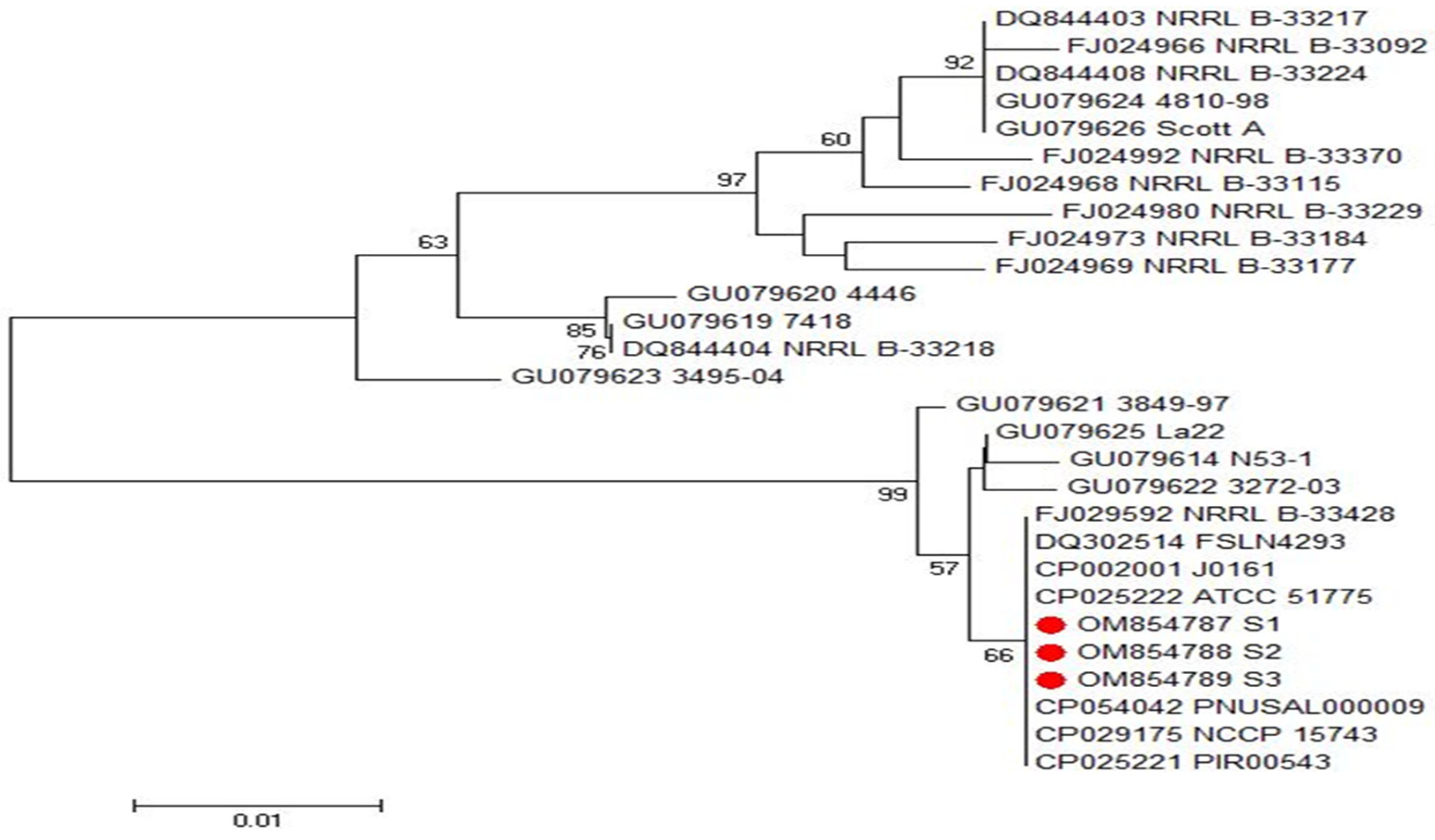
2.5. Phylogenetic Analyses of inlA and inlB Genes of L. monocytogenes

Phylogenetic and sequence analyses of inlA and inlB genes’ sequences among three representative L. monocytogenes isolates showing the highest resistance rates revealed that the examined isolates exhibited a remarkable genetic identity to other L. monocytogenes strains from different origins, which were deposited on the GenBank database, as demonstrated in Figure 5 and Figure 6. These sequences were deposited in the GenBank under the accession numbers of OM854784, OM854785 and OM854786 for inlA gene and OM854787, OM854788 and OM854789 for inlB gene. The final alignments of inlA gene consisted of 800 bp; 769 conserved and 31 variable sites; meanwhile, that of inlB gene comprised 343 bp; 304 conserved and 39 variable sites.
Based on the sequence analyses of both inlA and inlB genes, we did not find any new or unique sequences, and there were no new or unique mutations in these genes. Phylogenetic and sequence analyses of inlA genes revealed that our tested isolates (Accession No. OM854784, OM854785 and OM854786) displayed a complete identity (100%) to another food L. monocytogenes strain in Japan (Accession No. LC005925). Additionally, they showed high similarity percentages with L. monocytogenes food isolate in France (Accession No. FM178794, 99.9%), L. monocytogenes strain isolated from pig in Spain (Accession No. HQ111554, 99.7%), L. monocytogenes strain isolated from seafood in Japan (Accession No. AB276379, 99.5%), and L. monocytogenes strain isolated from watershed samples in the USA (Accession No. KF728273, 99.0%), as demonstrated in Figure 5. On the other hand, our inlB gene sequences (Accession No. OM854787, OM854788 and OM854789) displayed a remarkable genetic identity to other L. monocytogenes strains from different origins such as the food L. monocytogenes strain in South Korea (Accession No. CP029175, 100%), the L. monocytogenes strain isolated from cold smoked salmon in Denmark (Accession No. GU079625, 99.7%), the L. monocytogenes strain isolated from maternofetal interface in Denmark (Accession No. GU079621, 99.4%), the L. monocytogenes strain isolated from fish smoke house equipment in Denmark (Accession No. GU079614, 99.4%) and the L. monocytogenes strain isolated from spreadable sausage in Denmark (Accession No. GU079619, 93.6%), as demonstrated in Figure 6.

3. Discussion

Recently, infections from L. monocytogenes increased worldwide, and high death rates associated with this pathogen were recorded. Listeriosis is a serious disease that affects a wide range of animals and people, especially those with weakened immune systems, pregnant women and newborns. The prevalence rates of L. monocytogenes were variable among different sources in this study, with an overall percentage of 11.9%. The animal subjects were the most common source of L. monocytogenes isolates. This prevalence rate was higher than those recorded in previous studies conducted on different ecological niches in the same geographic area [19,20] reflecting the decreasing in awareness, carefulness and good practices in the Egyptian community. This pathogen can cause a zoonotic disease (listeriosis) in both humans and animals and it can be transmitted via several food chains such as meat, poultry and seafood products. Additionally, L. monocytogenes is widely spread and contaminates a wide range of foods and animal products such as raw milk, cheese, raw meat products and salads due to poor hygienic conditions [21]. Listeria monocytogenes can multiply in food to dangerous levels, even at refrigeration temperatures during distribution and storage [22]. Therefore, there is an urgent need to control L. monocytogenes at all stages in the food chains and manage its infection in animals. We can overcome this challenge by strengthening the good manufacturing and hygienic practices in all sectors of the food chain, avoiding the animal infections and rapid diagnosis and treatment of both animal and human infections.
Another crisis in L. monocytogenes infections is the wide spread of antimicrobial resistance. The MDR phenotypes are common among our isolates, which lead to treatment failure. The acquired resistance to antimicrobial drugs were rarely developed among L. monocytogenes isolates [23,24]; however, and in accordance with our findings, several recent reports have found increased rates of antimicrobial resistance among clinical food-borne pathogens [25,26,27]. Overall, L. monocytogenes isolates in this study exhibited MDR patterns to four classes of antibiotics: penicillins, quinolones, aminoglycoside, and macrolides. This is a great issue for public health, as it may pose a challenge in the treatment of listeriosis. Of note, L. monocytogenes exhibit varying degrees of resistances to the most common antibiotics. High levels of resistance to amoxicillin, cefotaxime, norfloxacin, tetracyclines and gentamicin were recorded in this study and in other studies [28,29]; meanwhile, the high susceptibility of our L. monocytogenes against trimethoprim/sulfamethoxazole was announced and is consistent with an earlier study [30]. The antimicrobial resistance in L. monocytogenes may be attributed to the acquisition of antibiotic resistance genes from other pathogens [27,31]. Moreover, the acquisition of conjugative transposons [32], active efflux associated genes [33], and ribosomal and chromosomal mutations [34] are the major mechanisms of L. monocytogenes resistance. Fortunately, sulfamethoxazole-trimethoprim and amoxicillin-clavulanic acid are still the alternative therapies in the case of unresponsiveness to the treatment with beta lactams’ antibiotics and intolerance to the first line therapies, as observed in our study and other reports [35,36].
Many factors affect the pathogenicity of L. monocytogenes involving numerous key virulence arrays such as internalins and haemolysin [37,38]. Notably, L. monocytogenes clinical isolates are multi-virulent; this was announced in several previous publications [39,40] as well as in our study. Previously, all clinical isolates possessed InlA and InlB proteins [41,42], which are essential for the recognition of several receptors and the invasion of different cell types. Furthermore, majority of our isolates showed hemolytic activities and harbored hlyA and prfA genes, which contribute to L. monocytogenes virulence and transmission. As expected from the correlation analysis, negative correlations between the existence of virulence genes (hlyA and prfA) and the antimicrobial resistances were detected.
Of note, the antimicrobial resistance genes were distributed between plasmid and chromosome [43]; however, the resistance genes can be acquired from the environment through plasmids and transposons [34]. Antimicrobial resistance, which enhances bacterial performance in vivo, may carry fitness costs, prompting a renewed interest in understanding the complex relationships between resistance and virulence. When both virulence and antimicrobial resistance genes are on the same mobile genetic element, a direct link can be observed [44]. The prediction of these correlations is highly challenging, as the bacterial genome is context-dependent and highly dynamic. Therefore, the correlation studies between the resistance to antimicrobials with each other, the existence of virulence genes with each other, in addition to the association between resistances to antimicrobials and the existence of virulence genes are very necessary owing to the continuous evolution in the genetic background. The costs of acquisition of antimicrobial resistance genes (increasing the resistance abilities) can be outweighed by benefits such as loss of virulence genes (decreasing in the virulence fitness) [14]. Fortunately, the high virulence arrays were associated with the sensitive phenotypes, and the MDR strains possess low virulence fitness [45]. This finding is accepted depending on the fixed genetic capacity of the bacterial genome. Moreover, the acquisition of resistance genes occurs in parallel with the loss of other genes such as virulence ones [46,47].
In this study, all inlA and inlB genes’ sequences were identical to the previous sequence deposited in the database of GeneBank considering different sources and geographic areas. We did not observe any new mutations among our genes sequences. This observation confirmed the slow evolution in the virulence genes compared to antimicrobial resistance profiles. Notably, most evolution in the virulence genes may lead to negative selection [48].
The control of listeriosis becomes more tedious due to the high degree of heterogeneity among L. monocytogenes isolates. Our report recorded the high variability level and the low host specificity among the tested L. monocytogenes isolates. In this context, several authors announced the weak clonality and adaptability of L. monocytogenes due to the high phenotypic and genotypic diversity, which is associated with different evolutionary processes within different hosts or in an ecological niche [49,50]. Therefore, specific control and prevention recommendations and more restricted isolation guidelines are urgently needed.

4. Materials and Methods

4.1. Ethics Statement

All study procedures were conducted in accordance with the guidelines of the Animal Ethics Review Committee of Suez Canal University (AERC-SCU201856), Egypt. Moreover, written informed consent was obtained from all patients.

4.2. Sampling

The study was conducted from April 2018 to November 2021. Three hundred and ten samples were collected from various sources at Sharkia (n = 130), Giza (n = 90) and Qaliubia (n = 90) Governorates, Egypt. The samples were collected from animal (n = 230), human (n = 50), and animal feed (n = 30) sources. The animal samples comprised the brain of sheep suffering from nervous manifestations (n = 30), placenta (n = 30), and aborted foeti (n= 30) of sheep suffering from abortion at the third trimester stage of pregnancy, lung (n= 30), liver (n = 30) and spleen (n = 30) of sheep suffering from septicemia, and finally mastitis milk (n = 50) of sheep suffering from mastitis. Human placental samples (n = 25) and uterine biopsies (n = 25) were collected from febrile and aborted women attending tropical hospitals. The samples were aseptically placed into sterile containers, kept in an ice box and transferred as soon as possible to the laboratory of the Bacteriology Unit at Animal Health Research Institute, Dokki, Giza for further bacteriological examination and the isolation of L. monocytogenes.

4.3. Isolation and Characterization of L. monocytogenes

Listeria monocytogenes isolates were phenotypically identified based on characteristic culture, morphological and biochemical features [51,52]. Furthermore, the recovered isolates were further identified using the Microbact™ Listeria 12L system (Oxoid, UK). The genotypic identification was used to confirm all L. monocytogenes isolates via a 16S rRNA gene-based PCR assay [53].

4.4. Phenotypic Assessment of L. monocytogenes Virulence

4.4.1. Anton’s Test

The test was performed by inserting 0.1 mL of L. monocytogenes suspension (109 colony forming units) into the conjunctiva of one eye of rabbits, and the other eye was used as a control. Anton’s test positive results were recorded as purulent conjunctivitis within 24–48 h, followed by keratitis [54].

4.4.2. CAMP (Christie–Atkins–Munch–Peterson) Test

The test was conducted, as described previously [55], using a standard beta-hemolytic Staphylococcus aureus (ATCC 25923) strain streaked in a straight line across the center of blood agar (Oxoid, UK) plates, followed by the streaking of identified L. monocytogenes strains in a direction perpendicular or vertical to the S. aureus culture without touching. The plates were subsequently incubated at 37 °C for 18–24 h and examined for the presence of hemolysis, which appeared as an arrowhead, circle, or rectangle in CAMP positive strains.

4.4.3. Hemolytic Activity

Heavy pure colonies from tryptic soy broth yeast extract agar (Oxoid, UK) plates were inoculated onto 5% sheep blood agar (Oxoid, UK) plates and incubated at 35 °C for 24–48 h to detect hemolysis [56,57].

4.5. Antibiogram Patterns of L. monocytogenes Isolates

The Kirby-Bauer disc diffusion method was used to determine the antimicrobial susceptibility profiles of the retrieved L. monocytogenes isolates on Mueller-Hinton agar (Oxoid, UK) supplemented with 5% fresh defibrinated sheep blood using twelve different antimicrobial disks (Oxoid, UK): penicillin (P), ampicillin (AMP), amoxicillin-clavulanic acid (AMC), cefotaxime (CTX), cephalexin (CFX), ciprofloxacin (CIP), norfloxacin (NOR), sulfamethoxazole-trimethoprim (SXT), gentamycin (CN), oxytetracycline (OTC), tylosin (TYL) and chloramphenicol (C). Inhibition zones were measured and interpreted adopting EUCAST guidelines for L. monocytogenes [58]. To ensure the accuracy of the disc diffusion method, the MIC values for the investigated antimicrobials were detected by broth microdilution method according to EUCAST guidelines. Briefly, selected L. monocytogenes strains were streaked onto brain heart infusion agar plates and the plates were incubated at 37 °C for 24 h. Three to five colonies were picked and incubated in brain heart infusion broth for 6 h, and this culture was adjusted to 5 × 10⁶ CFU /mL using 0.9% NaCl solution. Ten microliters of this solution were used to inoculate 96-well microtiter plates containing Mueller-Hinton broth with different antibiotic concentrations. The L. monocytogenes strain LMEGY1 was utilized as a quality control organism. The isolates exhibiting resistance to at least one agent in three or more antimicrobial classes were categorized as MDR [59]. Multiple antibiotic resistance (MAR) indices were then calculated following the previously standardized formula [60]. The chance of a high risk source of contamination is always observed with high MAR indices.

4.6. Molecular Detection of L. monocytogenes Virulence Genes

The PCR techniques were used to determine some virulence genes: prfA, inlA, inlB and hlyA in the recovered L. monocytogenes isolates [61,62,63]. The primers’ pairs used in PCR protocols and their predicted amplicons’ sizes are listed in Table 1. For genomic DNA extraction, the QIAamp DNA Mini Kit (Qiagen, Germantown, MD, USA, catalog no. 51326) was used. All PCR assays were carried out in triplicates and included positive control strains provided by the Animal Health Research Institute, Dokki, Giza, Egypt, with a DNA-free reaction as a negative control. The amplified PCR products were separated on electrophoresis gel (1.5% agarose stained with 0.5 μg/mL of ethidium bromide) and photographed.

4.7. Sequencing and Phylogenetic Analysis of L. monocytogenes inlA and inlB Genes

Direct sequencing of inlA and inlB genes among MDR L. monocytogenes isolates were carried out in both directions following purification with the QIAquick PCR Purification Kit (Qiagen, Germantown, MD, USA) using a Bigdye™ Terminator V3.1 cycle sequencing kit (Thermo Fisher Scientific, Waltham, MA, USA). The sequence similarity to GenBank accessions was determined using a Basic Local Alignment Search Tool. Phylogenetic trees were then created by the MegAlign module of Laser gene DNASTAR version 12.1 utilizing the neighbor-joining method in MEGA6.

4.8. Statistical Analysis

Hierarchical clustering and the correlation coefficient (r-value) between resistances to various antimicrobials in addition to the existence of virulence genes were evaluated using the R package corrplot, heatmaply, and GraphPad Prism (version 6; GraphPad Software Inc.; San Diego, CA, USA). Moreover, the discriminatory power of the typing methods used in the current study was assessed using Simpson’s index of diversity [64].

5. Conclusions

Several ready to eat foods, especially those of an animal source, are always contaminated with L. monocytogenes, which is associated with serious public health implications worldwide. As observed among our tested L. monocytogenes isolates, this problem is compounded by the high prevalence rate, the wide spread of antimicrobial resistance, the heterogeneity and diversity of this pathogen. In contrast to the evolution in the virulence fitness, we observed acceleration in the acquisition of antimicrobial resistance among L. monocytogenes isolates, which increase the possibility of treatment failure with the available drugs. Therefore, we are in urgent need for new and alternative therapies. Our study spotlighted and predicted the serious health problems in the near future due to the evolution in L. monocytogenes strains. Therefore, we encourage the health organizations in all countries to take more stringent actions to hinder the spread of this resistant pathogen.

Author Contributions

M.I.A.E.-H., M.E.E., A.E.-G. and M.M.B. designed, validated and supervised this work. A.E.-G., R.M.E. and M.A. (Majid Alhomrani) carried out the statistical analyses. A.S.A., S.A.A., M.A. (Marwa Arnout), D.N.B., M.M.A.-S. and A.I.A. performed antibiotyping activities and molecular analyses. All authors have read and agreed to the published version of the manuscript.

Funding

This work was funded by Zagazig, Suez Canal and Port Said Universities and Princess Nourah Bint Abdulrahman University Researchers supporting project number (PNURSP2022R155) Princess Nourah Bint Abdurahman University, Rihadh, Saudi Arabia. The APC and molecular studies were funded by Taif University Researchers Supporting Project number (TURSP 2020/257).

Institutional Review Board Statement

Not applicable for this study.

Informed Consent Statement

Informed consent was obtained from all patiants involved in the study.

Data Availability Statement

All generated data in this report are available in the submitted manuscript.

Acknowledgments

The authors would like to acknowledge Zagazig, Suez Canal and Port Said Universities and Princess Nourah Bint Abdulrahman University Researchers supporting project number (PNURSP2022R155), Princess Nourah Bint Abdurahman University, Rihadh, Saudi Arabia. Additionally, the authors would like to acknowledge Taif University Researchers Supporting Project number (TURSP 2020/257), Taif University, Saudi Arabia. Finally, Mohammad M. Al-Sanea expresses his gratitude to Korea Institute of Science and Technology (KIST) for funding this work through the grant name 2022 KIST School partnership project.

Conflicts of Interest

The authors declare that they have no conflicts of interest.

References

  1. Bintsis, T. Foodborne pathogens. AIMS Microbiol. 2017, 3, 529–563. [Google Scholar] [CrossRef]
  2. Jalali, M.; Abedi, D. Prevalence of Listeria species in food products in Isfahan, Iran. Int. J. Food Microbiol. 2007, 122, 336–340. [Google Scholar] [CrossRef]
  3. Berrang, M.E.; Meinersmann, R.J.; Frank, J.F.; Smith, D.P.; Genzlinger, L.L. Distribution of Listeria monocytogenes subtypes within a poultry further processing plant. J. Food Prot. 2005, 68, 980–985. [Google Scholar] [CrossRef] [PubMed] [Green Version]
  4. Faour-Klingbeil, D.; Todd, E. Prevention and Control of Foodborne Diseases in Middle-East North African Countries: Review of National Control Systems. Int. J. Environ. Res. Public Health 2019, 17, 70. [Google Scholar] [CrossRef] [Green Version]
  5. Abd El-Hamid, M.I.; Sewid, A.H.; Samir, M.; Hegazy, W.; Bahnass, M.M.; Mosbah, R.A.; Ghaith, D.M.; Khalifa, E.; Ramadan, H.; Alshareef, W.A.; et al. Clonal Diversity and Epidemiological Characteristics of ST239-MRSA Strains. Front. Cell. Infect. Microbiol. 2022, 12, 782045. [Google Scholar] [CrossRef] [PubMed]
  6. Mosallam, F.M.; Helmy, E.A.; Bendary, M.M.; El-Batal, A.I. Potency of a novel synthesized Ag-eugenol nanoemulsion for treating some bacterial and fungal pathogens. J. Mater. Res. 2021, 36, 1524–1537. [Google Scholar] [CrossRef]
  7. Bendary, M.M.; Abd El-Hamid, M.I.; Alhomrani, M.; Alamri, A.S.; Elshimy, R.; Mosbah, R.A.; Bahnass, M.M.; Omar, N.N.; Al-Sanea, M.M.; Elmanakhly, A.R. What Is behind the Correlation Analysis of Diarrheagenic, E. coli Pathotypes? Biology 2022, 11, 1004. [Google Scholar] [CrossRef]
  8. Ghaly, M.F.; Nasr, Z.M.; Abousaty, A.I.; Seadawy, H.G.; Shaheen, M.; Albogami, S.; Al-Sanea, M.M.; Bendary, M.M. Alternative and Complementary Therapies against Foodborne Salmonella Infections. Antibiotics 2021, 10, 1453. [Google Scholar] [CrossRef]
  9. Moretro, T.; Langsrud, S. Listeria monocytogenes: Biofilm formation and persistence in food-processing environments. Biofilms 2004, 1, 107–121. [Google Scholar] [CrossRef]
  10. Samelis, J.; Metaxopoulos, J. Incidence and principal sources of Listeria spp. and Listeria monocytogenes contamination in processed meats and a meat processing plant. Food Microbiol. 1999, 16, 465–477. [Google Scholar] [CrossRef]
  11. Feng, Y.; Wu, S.; Varma, J.K.; Klena, J.D.; Angulo, F.J.; Ran, L. Systematic review of human listeriosis in China, 1964–2010. Trop. Med. Int. Health 2013, 18, 1248–1256. [Google Scholar] [CrossRef] [PubMed]
  12. Flamm, R.K.; Hinrichs, D.J.; Thomashow, M.F. Introduction of pAM beta 1 into Listeria monocytogenes by conjugation and homology between native L. monocytogenes plasmids. Infect. Immun. 1984, 44, 157–161. [Google Scholar] [CrossRef] [PubMed] [Green Version]
  13. Pérez-Diaz, J.C.; Vicente, M.F.; Baquero, F. Plasmids in Listeria. Plasmid 1982, 8, 112–118. [Google Scholar] [CrossRef]
  14. Bendary, M.M.; Abd El-Hamid, M.I.; El-Tarabili, R.M.; Hefny, A.A.; Algendy, R.M.; Elzohairy, N.A.; Ghoneim, M.M.; Al-Sanea, M.M.; Nahari, M.H.; Moustafa, W.H. Clostridium perfringens Associated with Foodborne Infections of Animal Origins: Insights into Prevalence, Antimicrobial Resistance, Toxin Genes Profiles, and Toxinotypes. Biology 2022, 11, 551. [Google Scholar] [CrossRef] [PubMed]
  15. Ammar, A.M.; Abd El-Hamid, M.I.; El-Malt, R.M.S.; Azab, D.S.; Albogami, S.; Al-Sanea, M.M.; Soliman, W.E.; Ghoneim, M.M.; Bendary, M.M. Molecular Detection of Fluoroquinolone Resistance among Multidrug-, Extensively Drug-, and Pan-Drug-Resistant Campylobacter Species in Egypt. Antibiotics 2021, 10, 1342. [Google Scholar] [CrossRef]
  16. Elfaky, M.A.; Abdel-Hamid, M.I.; Khalifa, E.; Alshareef, W.A.; Mosbah, R.A.; Elazab, S.T.; Ghoneim, M.M.; Al-Sanea, M.M.; Bendary, M.M. Innovative next-generation therapies in combating multi-drug-resistant and multi-virulent Escherichia coli isolates: Insights from in vitro, in vivo, and molecular docking studies. Appl. Microbiol. Biotechnol. 2022, 106, 1691–1703. [Google Scholar] [CrossRef]
  17. Ghaly, M.F.; Shaheen, A.A.; Bouhy, A.M.; Bendary, M.M. Alternative therapy to manage otitis media caused by multidrug-resistant fungi. Arch. Microbiol. 2020, 202, 1231–1240. [Google Scholar] [CrossRef]
  18. Pagliano, P.; Ascione, T.; Boccia, G.; Caro, F.D.; Esposito, S. Listeria monocytogenes meningitis in the elderly: Epidemiological, clinical and therapeutic findings. Infez Med. 2016, 24, 105–111. [Google Scholar]
  19. Abdeen, E.E.; Mousa, W.S.; Harb, O.H.; Fath-Elbab, G.A.; Nooruzzaman, M.; Gaber, A.; Alsanie, W.F.; Abdeen, A. Prevalence, Antibiogram and Genetic Characterization of Listeria monocytogenes from Food Products in Egypt. Foods 2021, 10, 1381. [Google Scholar] [CrossRef]
  20. Osman, K.M.; Kappell, A.D.; Fox, E.M.; Orabi, A.; Samir, A. Prevalence, pathogenicity, virulence, antibiotic resistance, and phylogenetic analysis of biofilm-producing L. monocytogenes isolated from different ecological niches in Egypt. food, humans, animals, and environment. Pathogens 2020, 9, 5. [Google Scholar] [CrossRef] [Green Version]
  21. Gartley, S.; Anderson-Coughlin, B.; Sharma, M.; Kniel, K.E. Listeria monocytogenes in Irrigation Water: An Assessment of Outbreaks, Sources, Prevalence, and Persistence. Microorganisms 2022, 10, 1319. [Google Scholar] [CrossRef] [PubMed]
  22. Szczawiński, J.; Ewa Szczawińska, M.; Łobacz, A.; Tracz, M.; Jackowska-Tracz, A. Modelling the Growth Rate of Listeria Monocytogenes in Cooked Ham Stored at Different Temperatures. J. Vet. Res. 2017, 61, 45–51. [Google Scholar] [CrossRef] [Green Version]
  23. Aureli, P.; Ferrini, A.M.; Mannoni, V.; Hodzic, S.; Wedell-Weergaard, C.; Oliva, B. Susceptibility of Listeria monocytogenes isolated from food in Italy to antibiotics. Int. J. Food Microbiol. 2003, 83, 325–330. [Google Scholar] [CrossRef]
  24. Charpentier, E.; Gerbaud, G.; Jacquet, C.; Rocourt, J.; Courvalin, P. Incidence of antibiotic resistance in Listeria species. J. Infect. Dis. 1995, 172, 277–281. [Google Scholar] [CrossRef]
  25. Ahmed, H.A.; Tahoun, A.B.M.B.; Abou Elez, R.M.M.; Abd El-Hamid, M.I.; Abd Ellatif, S.S. Prevalence of Yersinia enterocolitica in milk and dairy products and the effects of storage temperatures on survival and virulence gene expression. Int. Dairy J. 2019, 94, 16–21. [Google Scholar] [CrossRef]
  26. Conter, M.; Paludi, D.; Zanardi, E.; Ghidini, S.; Vergara, A.; Ianieri, A. Characterization of antimicrobial resistance of foodborne Listeria monocytogenes. Int. J. Food Microbiol. 2009, 128, 497–500. [Google Scholar] [CrossRef]
  27. Ammar, A.M.; El-Naenaeey, E.-S.Y.; El-Malt, R.M.S.; El-Gedawy, A.A.; Khalifa, E.; Elnahriry, S.S.; Abd El-Hamid, M.I. Prevalence, antimicrobial susceptibility, virulence and genotyping of Campylobacter jejuni with a special reference to the anti-virulence potential of eugenol and beta-resorcylic acid on some multi-drug resistant isolates in Egypt. Animals 2021, 11, 3. [Google Scholar] [CrossRef]
  28. Hansen, J.M.; Gerner-Smidt, P.; Bruun, B. Antibiotic susceptibility of Listeria monocytogenes in Denmark 1958–2001. APMIS 2005, 113, 31–36. [Google Scholar] [CrossRef]
  29. Elsayed, M.M.; Elkenany, R.M.; Zakaria, A.I.; Badawy, B.M. Epidemiological study on Listeria monocytogenes in Egyptian dairy cattle farms’ insights into genetic diversity of multi-antibiotic-resistant strains by ERIC-PCR. Environ. Sci. Pollut. Res. 2022, 29, 54359–54377. [Google Scholar] [CrossRef]
  30. Akrami-Mohajeri, F.; Derakhshan, Z.; Ferrante, M.; Hamidiyan, N.; Soleymani, M.; Conti, G.O.; Tafti, R.D. The prevalence and antimicrobial resistance of Listeria spp in raw milk and traditional dairy products delivered in Yazd, Central Iran (2016). Food Chem. Toxicol. 2018, 114, 141–144. [Google Scholar] [CrossRef]
  31. Heger, W.; Dierich, M.P.; Allerberger, F. In vitro susceptibility of Listeria monocytogenes: Comparison of the E test with the agar dilution test. Chemotherapy 1997, 43, 303–310. [Google Scholar] [CrossRef] [PubMed]
  32. Poyart-Salmeron, C.; Trieu-Cuot, P.; Carlier, C.; Courvalin, P. Molecular characterization of two proteins involved in the excision of the conjugative transposon Tn1545: Homologies with other site-specific recombinases. EMBO J. 1989, 8, 2425–2433. [Google Scholar] [CrossRef]
  33. Godreuil, S.; Galimand, M.; Gerbaud, G.; Jacquet, C.; Courvalin, P. Efflux pump Lde is associated with fluoroquinolone resistance in Listeria monocytogenes. Antimicrob. Agents Chemother. 2003, 47, 704–708. [Google Scholar] [CrossRef] [Green Version]
  34. Charpentier, E.; Courvalin, P. Antibiotic resistance in Listeria spp. Antimicrob. Agents Chemother. 1999, 43, 2103–2108. [Google Scholar] [CrossRef] [PubMed] [Green Version]
  35. Hof, H. An update on the medical management of listeriosis. Expert Opin. Pharmacother. 2004, 5, 1727–1735. [Google Scholar] [CrossRef] [PubMed]
  36. Temple, M.E.; Nahata, M.C. Treatment of listeriosis. Ann. Pharmacother. 2000, 34, 656–661. [Google Scholar] [CrossRef]
  37. Vázquez-Boland, J.A.; Kuhn, M.; Berche, P.; Chakraborty, T.; Domínguez-Bernal, G.; Goebel, W.; González-Zorn, B.; Wehland, J.; Kreft, J. Listeria pathogenesis and molecular virulence determinants. Clin. Microbiol. Rev. 2001, 14, 584–640. [Google Scholar] [CrossRef] [Green Version]
  38. Quereda, J.J.; Morón-García, A.; Palacios-Gorba, C.; Dessaux, C.; García-Del, P.F.; Pucciarelli, M.G.; Ortega, A.D. Pathogenicity and virulence of Listeria monocytogenes: A trip from environmental to medical microbiology. Virulence 2021, 12, 2509–2545. [Google Scholar] [CrossRef]
  39. Berche, P.; Gaillard, J.L.; Sansonetti, P.J. Intracellular growth of Listeria monocytogenes as a prerequisite for in vivo induction of T cell-mediated immunity. J. Immunol. 1987, 138, 2266–2271. [Google Scholar]
  40. Busch, D.H.; Kerksiek, K.; Pamer, E.G. Processing of Listeria monocytogenes antigens and the in vivo T-cell response to bacterial infection. Immunol. Rev. 1999, 172, 163–169. [Google Scholar] [CrossRef]
  41. Braun, L.; Ghebrehiwet, B.; Cossart, P. gC1q-R/p32, a C1q-binding protein, is a receptor for the InlB invasion protein of Listeria monocytogenes. EMBO J. 2000, 19, 1458–1466. [Google Scholar] [CrossRef] [PubMed] [Green Version]
  42. Mengaud, J.; Ohayon, H.; Gounon, P.; Mege, R.M.; Cossart, P. E-Cadherin is the receptor for internalin, a surface protein required for entry of L. monocytogenes into epithelial cells. Cell 1996, 84, 923–932. [Google Scholar] [CrossRef]
  43. Morvan, A.; Moubareck, C.; Leclercq, A.; Hervé-Bazin, M.; Bremont, S.; Lecuit, M.; Courvalin, P.; Le Monnier, A. Antimicrobial resistance of Listeria monocytogenes strains isolated from humans in France. Antimicrob. Agents Chemother. 2010, 54, 2728–2731. [Google Scholar] [CrossRef] [PubMed] [Green Version]
  44. Scortti, M.; Han, L.; Alvarez, S.; Leclercq, A.; Moura, A.; Lecuit, M.; Vazquez-Boland, J. Epistatic control of intrinsic resistance by virulence genes in Listeria. PLoS Genet. 2018, 14, e1007525. [Google Scholar] [CrossRef] [PubMed] [Green Version]
  45. Beceiro, A.; Tomás, M.; Bou, G. Antimicrobial resistance and virulence: A successful or deleterious association in the bacterial world? Clin. Microbiol. Rev. 2013, 26, 185–230. [Google Scholar] [CrossRef] [Green Version]
  46. Bendary, M.M.; Solyman, S.M.; Azab, M.M.; Mahmoud, N.F.; Hanora, A.M. Genetic diversity of multidrug resistant Staphylococcus aureus isolated from clinical and non-clinical samples in Egypt. Cell. Mol. Biol. 2016, 62, 55–61. [Google Scholar]
  47. Bendary, M.M.; Solyman, S.M.; Azab, M.M.; Mahmoud, N.F.; Hanora, A.M. Characterization of Methicillin Resistant Staphylococcus aureus isolated from human and animal samples in Egypt. Cell. Mol. Biol. 2016, 62, 94–100. [Google Scholar]
  48. Diard, M.; Hardt, W.D. Evolution of bacterial virulence. FEMS Microbiol. Rev. 2017, 41, 679–697. [Google Scholar] [CrossRef] [Green Version]
  49. Chen, Y.; Chen, M.; Wang, J.; Wu, Q.; Cheng, J.; Zhang, J.; Sun, Q.; Xue, L.; Zeng, H.; Lei, T.; et al. Heterogeneity, Characteristics, and Public Health Implications of Listeria monocytogenes in Ready-to-Eat Foods and Pasteurized Milk in China. Front. Microbiol. 2020, 15, 642. [Google Scholar] [CrossRef] [Green Version]
  50. Nyarko, E.B.; Donnelly, C.W. Listeria monocytogenes: Strain Heterogeneity, Methods, and Challenges of Subtyping. J. Food Sci. 2015, 80, 2868–2878. [Google Scholar] [CrossRef]
  51. Hitchins, A.D.; Whiting, R.C. Food-borne Listeria monocytogenes risk assessment. Food Addit. Contam. 2001, 18, 1108–1117. [Google Scholar] [CrossRef] [PubMed]
  52. Markey, B.K.; Leonard, F.C.; Archambault, M.; Cullinane, A.; Maguire, D. Clinical Veterinary Microbiology, 2nd ed.; MOSBY. Elsevier Ltd.: Edinburgh, UK; London, UK; New York, NY, USA; Oxford, UK; Philadelphia, PA, USA; St. Louis, MI, USA; Sydney, Australia; Toronto, ON, Canada, 2013. [Google Scholar]
  53. Kumar, A.; Grover, S.; Batish, V.K. Exploring specific primers targeted against different genes for a multiplex PCR for detection of Listeria monocytogenes. 3 Biotech 2015, 5, 261–269. [Google Scholar] [CrossRef] [PubMed] [Green Version]
  54. Quinn, P.J.; Markey, B.K.; Carter, M.E.; Donnelly, W.J.C.; Leonard, F.C.; Maguire, D. Veterinary Microbiology and Microbial Disease; Blackwell Science: Oxford, UK, 2002; pp. 84–96. [Google Scholar]
  55. ISO 11290-1:1996; Microbiology of Food and Animal Feeding Stuffs–Horizontal Method for the Detection and Enumeration of Listeria Monocytogenes–Part 1: Detection Method. 1st ed. ISO: Geneva, Switzerland, 1996.
  56. Abd El-Hamid, M.I.; Ibrahim, S.M.; Eldemery, F.; El-Mandrawy, S.A.M.; Metwally, A.S.; Khalifa, E.; Elnahriry, S.S.; Ibrahim, D. Dietary cinnamaldehyde nanoemulsion boosts growth and transcriptomes of antioxidant and immune related genes to fight Streptococcus agalactiae infection in Nile tilapia (Oreochromis niloticus). Fish Shellfish Immunol. 2021, 113, 96–105. [Google Scholar] [CrossRef] [PubMed]
  57. Fujisawa, T.; Mori, M. Evaluation of media for determining hemolytic activity and that of API Listeria system for identifying strains of Listeria monocytogenes. J. Clin. Microbiol. 1994, 32, 1127–1129. [Google Scholar] [CrossRef]
  58. The European Committee on Antimicrobial Susceptibility Testing (ECoAS). Breakpoint Tables for Interpretation of MICs and Zone Diameters. 2018, Version 8.0. 2018. Available online: http://www.sciepub.com/reference/368572 (accessed on 1 October 2022).
  59. Magiorakos, A.P.; Srinivasan, A.; Carey, R.B.; Carmeli, Y.; Falagas, M.E.; Giske, C.G.; Harbarth, S.; Hindler, J.F.; Kahlmeter, G.; Olsson-Liljequist, B.; et al. Multidrug-resistant, extensively drug-resistant and pandrug-resistant bacteria: An international expert proposal for interim standard definitions for acquired resistance. Clin. Microbiol. Infect. 2012, 18, 268–281. [Google Scholar] [CrossRef] [PubMed] [Green Version]
  60. Krumperman, P.H. Multiple antibiotic resistance indexing of Escherichia coli to identify high-risk sources of fecal contamination of foods. Appl. Environ. Microbiol. 1983, 46, 165–170. [Google Scholar] [CrossRef] [Green Version]
  61. Dickinson, J.H.; Kroll, R.G.; Grant, K.A. The direct application of the polymerase chain reaction to DNA extracted from foods. Lett. Appl. Microbiol. 1995, 20, 212–216. [Google Scholar] [CrossRef]
  62. Liu, D.; Lawrence, M.L.; Austin, F.W.; Ainsworth, A.J. A multiplex PCR for species- and virulence-specific determination of Listeria monocytogenes. J. Microbiol. Methods 2007, 71, 133–140. [Google Scholar] [CrossRef]
  63. Deneer, H.G.; Boychuk, I. Species-Specific Detection of Listeria monocytogenes by DNA Amplification. Appl. Environ. Microbiol. 1991, 57, 606–609. [Google Scholar] [CrossRef] [Green Version]
  64. Hunter, P.R.; Gaston, M.A. Numerical index of the discriminatory ability of typing systems: An application of Simpson’s index of diversity. J. Clin. Microbiol. 1988, 26, 2465–2466. [Google Scholar] [CrossRef]
Figure 1. Number of L. monocytogenes isolates recovered from different sample types of animal, human and animal feed sources.
Antibiotics 11 01447 g001
Figure 2. Resistance rates of L. monocytogenes isolates to the tested antimicrobial drugs. NOR: norfloxacin, CIP: ciprofloxacin, P: penicillin, AMP: ampicillin, CFX: cephalexin, CTX: cefotaxime, CN: gentamycin, C: chloramphenicol, TYL: tylosin, OTC: oxytetracycline and SXT: sulfamethoxazole-trimethoprim and AMC: amoxicillin-clavulanic acid.
Antibiotics 11 01447 g002
Figure 3. Heatmap showing the distribution of 37 L. monocytogenes isolates based on both virulence genes’ profiles and antimicrobial resistance patterns. The absence and presence of a particular virulence gene or sensitivity and resistance to a certain antimicrobial are indicated by blue and red colors, respectively. The text labels on the bottom specify the tested virulence genes and antimicrobial agents. Each row denotes one investigated isolate. The tree on the left displays the relatedness of the examined isolates according to their virulence genes’ profiles and the resistance they confer. AMP: Ampicillin, NOR: norfloxacin, P: penicillin, CFX: cephalexin, CTX: cefotaxime, CIP: ciprofloxacin, TYL: tylosin, CN: gentamycin, AMC: amoxicillin-clavulanic acid, OTC: oxytetracycline, C: chloramphenicol, SXT: sulfamethoxazole-trimethoprim, inlA: internalin A, inlB: internalin A, prfA: positive regulatory factor and hlyA: haemolysin.
Antibiotics 11 01447 g003
Figure 4. Heatmap illustrating the correlation coefficient (r) among and between resistances to the investigated antimicrobials and the presence of the tested virulence genes. The deep blue color reflects strong negative correlations; meanwhile, the deep red color indicates strong positive correlations. CIP: ciprofloxacin, TYL: tylosin, CN: gentamycin, C: chloramphenicol, AMC: amoxicillin-clavulanic acid, OTC: oxytetracycline, AMP: Ampicillin, NOR: norfloxacin, SXT: sulfamethoxazole-trimethoprim, prfA: positive regulatory factor and hlyA: haemolysin.
Antibiotics 11 01447 g004
Figure 5. Phylogenetic analyses of inlA gene of the three investigated L. monocytogenes isolates (OM854784; S1, OM854785; S2 and OM854786; S3), illustrating the genetic relationships between the investigated isolates and those have been deposited in the GenBank database, as shown in the phylogenetic tree.
Antibiotics 11 01447 g005
Figure 6. Phylogenetic analysis of inlB gene of the three investigated L. monocytogenes isolates (OM854787; S1, OM854788; S2 and OM854789; S3) illustrating the genetic relationships between the investigated isolates and those have been deposited in the GenBank database, as shown in the phylogenetic tree.
Antibiotics 11 01447 g006
Table 1. Target virulence genes, oligonucleotide primer sequences and fragments’ sizes used in PCR protocols for L. monocytogenes.
Target GeneOligonucleotide Primer Sequence (5′-3′)Amplified Product (bp)Reference
16S rRNAF: GGACCGGGGCTAATACCGAATGAT AA
R: TTC ATGTAGGCGAGTTGCAGC CTA
1200[53]
prfAF: TCTCCGAGCAACCTCGGAACC
R: TGGATTGACAAAATGGAACA
1052[61]
inlAF: ACG AGT AAC GGG ACA AAT GC
R: CCC GAC AGT GGT GCT AGA TT
800[62]
inlBF: CTGGAAAGTTTGTATTTGGGAAA
R: TTTCATAATCGCCATCATCACT
343[62]
hlyAF: GCATCTGCATTCAATAAAGA
R: TGTCACTGCATCTCCGTGGT
174[63]
Publisher’s Note: MDPI stays neutral with regard to jurisdictional claims in published maps and institutional affiliations.

