Article

Flavocytochrome b2-Mediated Electroactive Nanoparticles for Developing Amperometric L-Lactate Biosensors

1
Department of Analytical Biotechnology, Institute of Cell Biology National Academy of Sciences of Ukraine (ICB NASU), 14/16, Dragomanova Str., 79005 Lviv, Ukraine
2
NanoTechnas—Center of Nanotechnology and Materials Science, Institute of Chemistry, Faculty of Chemistry and Geosciences, Vilnius University, Naugarduko 24, LT-03225 Vilnius, Lithuania
3
Department of Chemical Engineering, Ariel University, Kyriat-ha-Mada, Ariel 4070000, Israel
*
Authors to whom correspondence should be addressed.
Biosensors 2023, 13(6), 587; https://doi.org/10.3390/bios13060587
Submission received: 20 April 2023 / Revised: 17 May 2023 / Accepted: 26 May 2023 / Published: 28 May 2023

Abstract

:
L-Lactate is an indicator of food quality, so its monitoring is essential. Enzymes of L-Lactate metabolism are promising tools for this aim. We describe here some highly sensitive biosensors for L-Lactate determination which were developed using flavocytochrome b2 (Fcb2) as a bio-recognition element, and electroactive nanoparticles (NPs) for enzyme immobilization. The enzyme was isolated from cells of the thermotolerant yeast Ogataea polymorpha. The possibility of direct electron transfer from the reduced form of Fcb2 to graphite electrodes has been confirmed, and the amplification of the electrochemical communication between the immobilized Fcb2 and the electrode surface was demonstrated to be achieved using redox nanomediators, both bound and freely diffusing. The fabricated biosensors exhibited high sensitivity (up to 1436 A·M−1·m−2), fast responses, and low limits of detection. One of the most effective biosensors, which contained co-immobilized Fcb2 and the hexacyanoferrate of gold, having a sensitivity of 253 A·M−1·m−2 without freely diffusing redox mediators, was used for L-Lactate analysis in samples of yogurts. A high correlation was observed between the values of analyte content determined using the biosensor and referenced enzymatic-chemical photometric methods. The developed biosensors based on Fcb2-mediated electroactive nanoparticles can be promising for applications in laboratories of food control.

1. Introduction

A healthy lifestyle is a popular trend in the modern world, which has prompted scientists working in the fields of medicine and the food industry to research ways to expand the range of healthy and functional foods. While they are not medicines, they can affect the person’s psychological or physiological state [1,2,3,4]. A special place in this category is occupied by dairy products: yogurt, cottage cheese, butter, buttermilk, kefir, koumiss, and others. These products have high nutritional value, being dietary and tasty, and are usually used to prevent and treat various gastrointestinal and other diseases. Dairy products are obtained from the milk of different animals via the action of lactic acid bacteria or other microorganisms that ferment carbohydrates, lactose in particular, into lactic acid [4,5,6].
The lactic acid anion L-Lactate (from now on—Lact), a nontoxic probiotic metabolite produced by lactic acid bacteria, plays an essential role in maintaining intestinal homeostasis and normal functioning, including providing human colon cells with metabolic energy sources [4,5,6,7]. Thus, eating foods containing significant amounts of Lact, such as yogurt, can substantially impact health by improving the composition of the microbiota [5]. That is why Lact is used in a wide range of foods and feeds, drinks, consumer goods, and products for healthcare. As an essential probiotic metabolite, Lact is an important analyte that must be monitored to assess the quality of foods and beverages [8,9,10].
For Lact analysis, many optic and electrochemical methods have been proposed [8,9,10,11,12,13,14,15,16,17]. Most of these methods are nonselective, time-consuming, costly, need pretreatment, and require expensive equipment. Enzymatic test systems and biosensors are promising instruments for Lact determination in industry and healthcare analytical laboratories. The available classic enzymatic methods of Lact analysis are generally using NAD+-dependent lactate dehydrogenase (LDH) from animal organs and bacterial lactate oxidase (LOX) [9,10,17,18,19,20,21,22]. Although LOX-based ABSs have excellent characteristics, especially if the enzyme is immobilized on the surface of 3D nano/microcarriers, their application is limited by the need for a second enzyme (peroxidase), native or artificial [21,22].
The yeast L-Lactate-cytochrome c-oxidoreductase (EC 1.1.2.3; flavocytochrome b2, Fcb2) is a promising biocatalyst in analytical methods for Lact determination, including biosensors [23,24,25,26,27,28,29,30]. Fcb2 from the baker’s yeast Saccharomyces cerevisiae is a homotetramer that contains two noncovalently bound cofactors, FMN and heme, per subunit [25]. Each subunit of Fcb2 is composed of two domains linked by a short hinge peptide: a C-terminal flavin-binding domain (or FMN L-Lactate dehydrogenase), which includes the active site for lactate oxidation and an N-terminal b2-cytochrome domain (or heme-containing domain), required for efficient cytochrome c reduction [25,26]. In experiments with the isolated, purified enzyme, the O2 molecules, instead of cytochrome c, become the subjects of reduction [26,31]. The enzyme exhibits absolute specificity to Lact, but the wide-scale application of Fcb2 from baker’s yeast in bio-analytics is limited by its instability and difficulties in enzyme purification [25,30,31,32,33,34].
The first reported Fcb2-based ABSs were developed using cells or cell lysates, from the yeasts S. cerevisiae and Hansenula anomala as the parental cells of this enzyme [30,31]. Many Lact-sensitive ABSs based on the recombinant cells have been proposed, especially in the last several years [32,33,34,35,36,37].
The highly purified thermostable Fcb2, isolated by Mykhailo Gonchar et al. twenty years ago from the cells of the thermotolerant methylotrophic yeast Ogataea polymorpha [38,39], was successfully used for the development of ABSs [27,28,39,40,41,42] and enzymatic–chemical methods for Lact determination [28]. The proposed analytical methods were successfully tested on real samples of food products, beverages, and biological liquids; these data are summarized in our earlier reviews [28,39].
The first ABS with Fcb2 of O. polymorpha was reported in 2005, and the possibility of direct electron transfer (DET) from the reduced form of this enzyme to a graphite electrode (GE) with a sensitivity of 1.1 A·M−1·m−2 was demonstrated [40]. The main disadvantage of this ABS was low sensitivity; even in the presence of the most electroactive mediator, phenazine methosulfate, the sensitivity value was 40 A·M−1·m−2 [40]. As efficient ways to improve the biosensors for Lact analysis, the methods of genetic engineering and/or nanotechnology were used [27,28]. Additionally, freely diffusing mediators were found to be effective amplifiers of a current signal in an ABS, due to efficient Fcb2-catalyzed oxidation of Lact [27,28].
Nanomaterials in the bio-recognition layer of the ABS serve as a matrix for enzyme immobilization and mediate electron transfer from the enzyme to the electrode surface. Nanoparticles (NPs) of metal oxides, metals, semiconductors, and composite NPs or nanohybrids perform various functions in electrochemical conversion circuits; in particular, much attention is paid to their mediator properties [10,11,12,13,14,15,16,17]. To improve electronic exchange between the surface of the working electrode and the enzyme in the planned ABS, a search for optimal nanomediators was performed.
The aims of our study were to develop highly sensitive ABSs for Lact determination using the thermostable yeast Fcb2 as a bio-recognition element, co-immobilized with electroactive nanomaterials and to demonstrate the applicability of the most sensitive reagentless ABS for Lact analysis in samples of commercial yogurts.

2. Materials and Methods

2.1. Reagents and Enzyme

Salts of transitional and noble metals, the sodium salt of L-Lactic acid, ascorbic acid, K3(Fe(CN)6), phenazine methosulfate (PMS), the Nafion solution, and all other reagents and solvents used in this work were purchased from Sigma-Aldrich (Steinheim, Germany). All reagents were of analytical grade and were used without additional purification. All solutions were prepared using ultrapure water.
L-Lactate-cytochrome c oxidoreductase (EC 1.1.2.3; flavocytochrome b2, Fcb2) was isolated from a cell-free extract (CE) of the thermotolerant methylotrophic yeast Ogataea (Hansenula) polymorpha 356, as described in detail earlier [39]. Briefly, yeast cells from the archived collection of microbial strains (ICB NASU, Lviv, Ukraine) were cultivated in flasks at 30 °C under intensive aeration in a mineral medium that contained 1% glucose, 0.2% sodium L-Lactate and 0.05% yeast extract. Freshly grown cells were collected via centrifugation, washed twice with water, suspended in a working buffer (50 mM phosphate buffer, pH 8.0), lyophilized, and kept at –20 °C until used. The dried cells were lysed with 10% n-butanol in a working buffer for 2 h at +4 °C. After the removal of cell debris via centrifugation (5000× g, 20 min, +4 °C), the supernatant was used as the CE for the isolation of Fcb2. The CE was put through a column with the anion exchange sorbent DEAE-Toyopearl 650 M (TSK-Gel, Kanagawa, Japan). Unbound proteins were washed with a buffer, and the Fcb2 was eluted with 15% ammonium sulfate (saturation at 0 °C) in a working buffer. Activity and protein concentration in each fraction were monitored. Fractions with the highest specific activity (16 U mg–1 of protein), for enzyme stabilization and concentration, were supplemented with ammonium sulfate (up to 70% saturation at 0 °C) and stored at −10 °C. L-Lactate standard solutions were prepared using the working buffer.

2.2. Synthesis of NPs

Bimetallic and trimetallic NPs were synthesized using the chemical reduction method [41]. Briefly, to obtain NiPtPdNPs (further—NiPtPd), solution 1 (H2PtCl6 and PdCl3) and solution 2 (NiSO4) were independently reduced by NaBH4. Then, solutions were mixed and supplemented with NaOH. PtZnNPs (further—PtZn) were synthesized from 10 mM H2PtCl6 using the Zn bath deposition method [42] with our modification [43].
To synthesize the hexacyanoferrates (HCFs) of Pt, Pd, or Au, 2 mL of 1 mM H2PtCl6, PdCl3, or HAuCl4 solutions were first reduced by the addition of 0.2 mL of 100 mM ascorbic acid. After heating at 100 °C for 10 min under stirring, 8 mL of a 50 mM K4Fe(CN)6 solution was added and incubated for three days without stirring [44]. AgHCF and “green” gCu(II)HCF were synthesized as described earlier in [41,44], respectively.
The synthesized NPs were concentrated via centrifugation, washed with water, tested for pseudo-peroxidase activities, and stored at 4 °C [41].
Morphological analyses of the synthesized NPs were performed via scanning electron microscopy (SEM) [44].

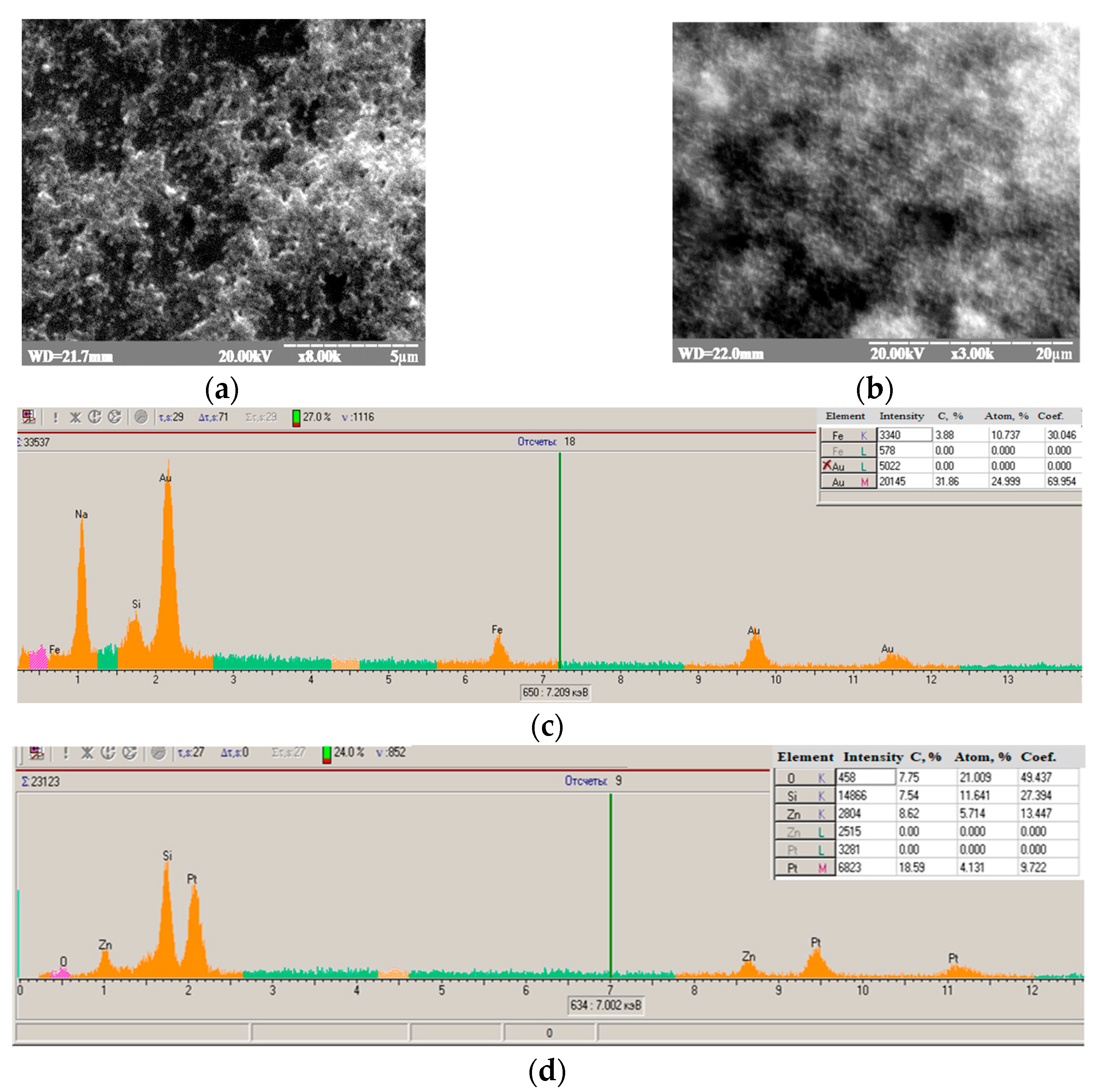
2.3. Modification of Graphite Electrodes with Nanoparticles and Their Characterization

A graphite rod (GE, 3.05 mm diameter) was used as a working electrode. Modification of GEs with NPs and a study of their electrochemical properties were carried out as described earlier [41,44].

2.4. Development, Characterization, and Application of Bioelectrodes

To develop the Fcb2/NPs/GE, 5–10 μL of the Fcb2 solution was dropped onto the surface of the NPs/GE, air-dried, covered with Nafion, and stored as described in Section 2.3. The resulting bioelectrodes were studied in more detail as biosensors on Lact.
The most effective Fcb2/AuHCF-based ABS was used to determine Lact in the real samples of commercial yogurts.
The samples were tested using the graphical calibration method in a variant of the standard addition test (SAT) [41].
Each assay was performed in triplicate for two dilutions of the following yogurts: Lactel (“Lactalis”, Mykolaiv, Ukraine), Choodo (“Vimm-Bill-Dann”, Vyshneve, Ukraine), and Activia (“Danone”, Kherson, Ukraine).

2.5. Reference Method for Lact Determination in Real Sample

As a reference, the Prussian blue-based enzymatic–chemical method was used [28,43]. The principle of this method is the Fcb2-catalyzed oxidation of Lact in the presence of K3Fe(CN)6 (the enzymatic reaction). In this case, [Fe(CN)6]3− is reduced to [Fe(CN)6]4−, which, when FeCl3 is added, forms a precipitate of Prussian blue (PB) (the chemical reaction). After the solubilization of the sediment, the concentration of the color product is detected via photometry or evaluated visually. The formation of a colloidal solution of PB indicates the presence of Lact, and the brightness of the color correlates with the Lact concentration.
Lact was analyzed in protein-free extracts of yogurts, described in Section 2.4. To obtain a protein-free extract, a 50% trichloroacetic acid solution was added to an aliquot of yogurt up to a final concentration of 10%; the solution was mixed, incubated for 30 min on ice, and centrifuged for 5 min at 10,000 rpm. The supernatant (Sn) neutralized by NaOH was used for the determination of Lact with the described Fcb2/PB method.
The protocol is as follows: 30 μL of the Sn sample, first diluted 5-fold with water, was incubated for 30 min at 37 °C with 270 μL of the reaction mixture that contained 0.04 units/mL of Fcb2 and 3 mM K3Fe(CN)6 in a 50 mM phosphate buffer, at pH 8.0. Then, 100 μL of a 0.2 M FeCl3 solution in 30 mM HCl was added, and the formed precipitate was dissolved by adding 560 μL of 0.9 M oxalic acid. Optical density was determined with a SHIMADZU UV-1650 PC spectrophotometer at 680 nm, using the standard software “UV Probe 2.20” against a blank sample containing the phosphate buffer instead of Lact.

3. Results

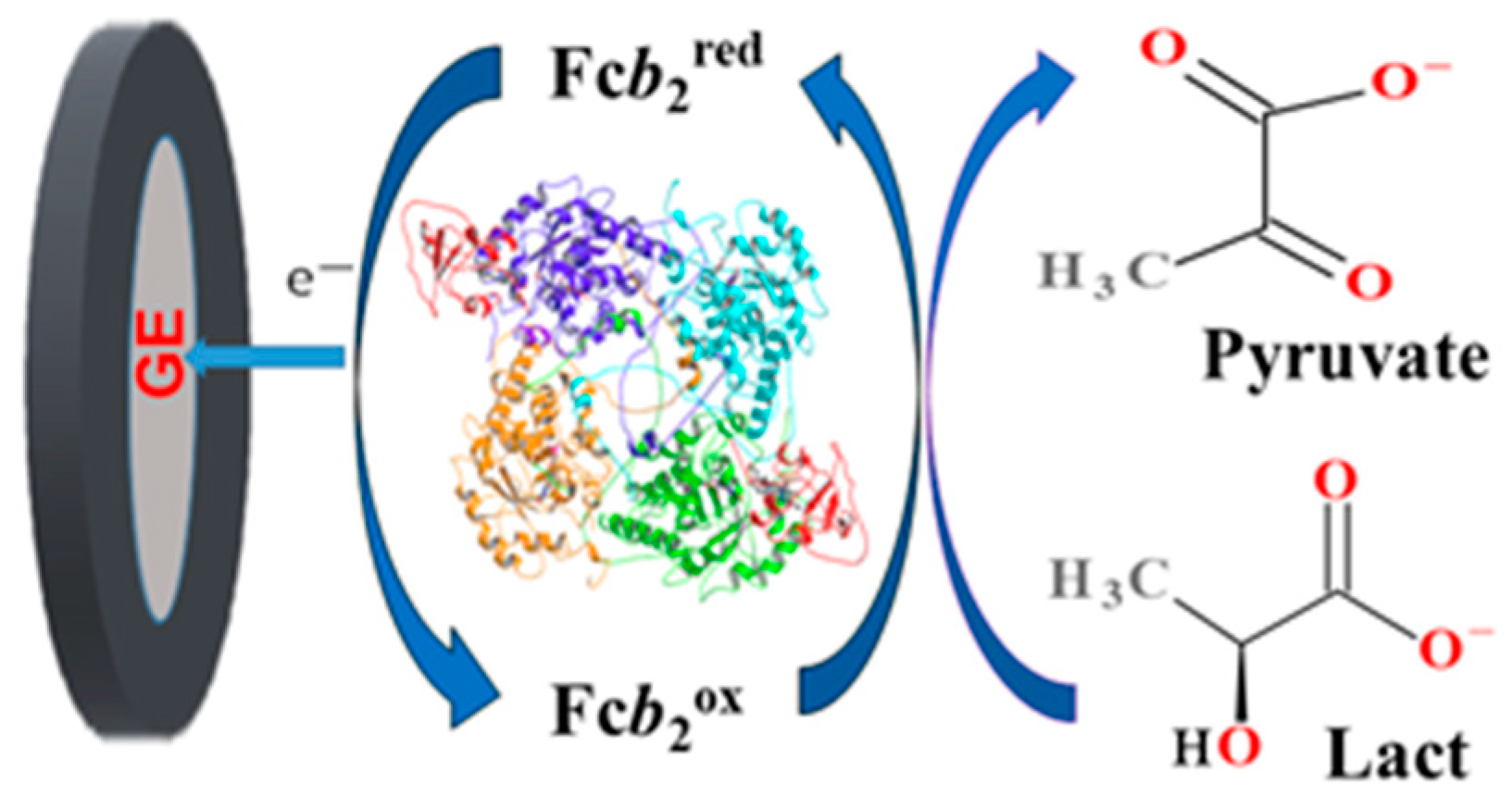
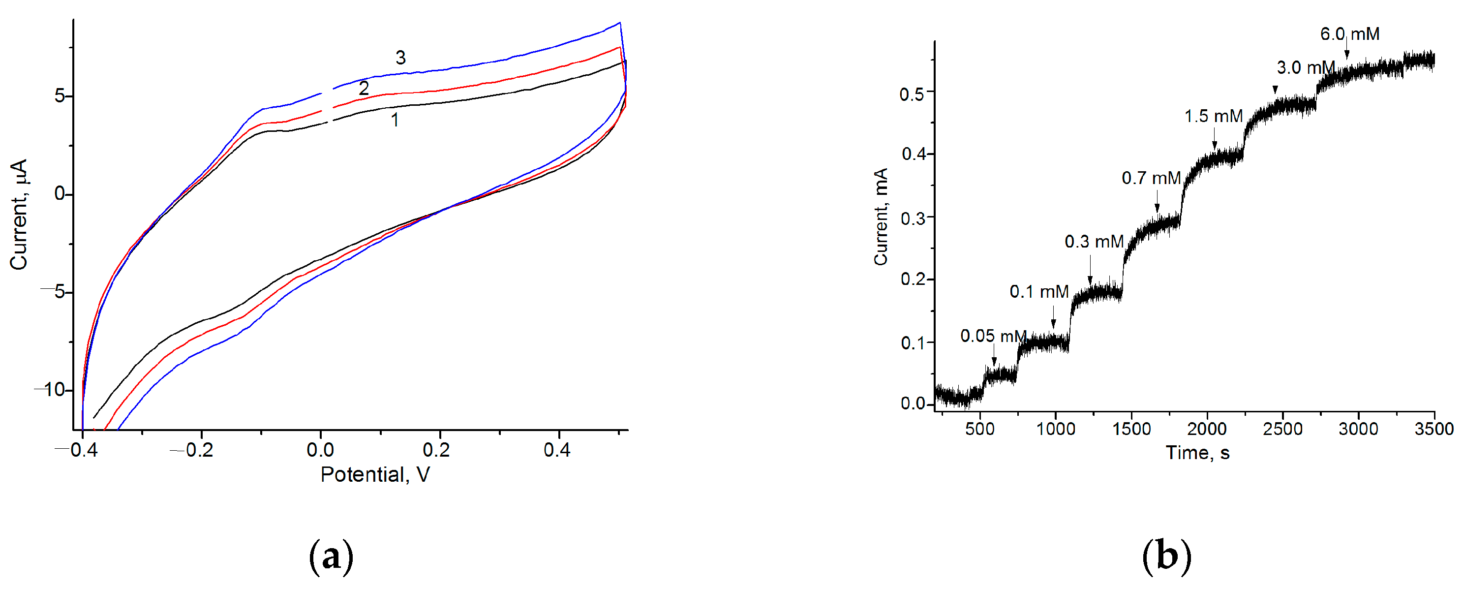
3.1. Development of Fcb2-Based Amperometric Biosensors

Fcb2 is a complex tetramer molecule containing a total of eight domains. Each domain is composed of heme and FMN, so the direct transfer of electrons (DET) is complicated, but it is possible. The ability of Fcb2 isolated from the cells of methylotrophic yeast O. polymorpha to achieve DET in an ABS was first demonstrated by our group [40]. It was concluded that the DET from the reduced form of Fcb2 to a graphite electrode (GE) takes place only for the molecules of the enzyme monolayer that are in direct contact with the surface of the GE. At the same time, the heme group must be correctly oriented at a specific distance from the electrode to achieve DET [40]. The scheme of DET in the ABS during the conversion of Lact in Fcb2-mediated catalysis is presented in Figure 1.
To select the optimal working potential for the Fcb2-based ABS, the CV profiles for the Fcb2/GE in the presence and absence of Lact were compared. Figure 2a demonstrates increased oxidation and reduction peaks due to the addition of an analyte to the ABS.
According to the CV results, the peak of oxidation, as an output upon Lact addition, appeared in the range of −(150–50) mV. For further experiments, including a chronometric study, the potential of –75 mV was chosen as the optimal working potential.

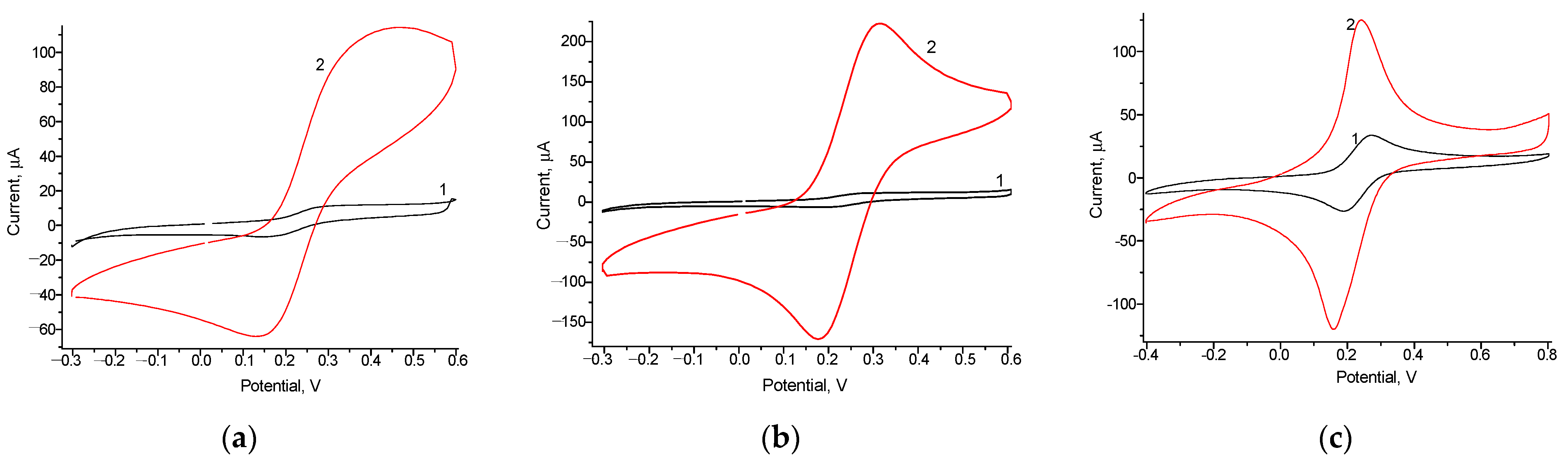
3.2. Selection of the Optimal Redox Nanomediators and Their Properties

To enhance the effectiveness of electron transfer (ET) between the GE and Fcb2, the surface of the GE was modified with redox-active NPs as carriers for enzyme immobilization. These compounds, namely, NPs and hexacyanoferrates (HCFs) of noble and transition metals, were synthesized (see Section 2.2). Some of the previously obtained redox NPs, including the HCFs of Ag, Au, Pd, Pt, as well as NiPtPd, were characterized using CV in our previous papers [41,44,45] and are not described here.
Some NPs were used as artificial peroxidases (PO) or PO-like nanozymes in an arginine oxidase-based ABS [41] and as nanomediators for a laccase-based ABS [44]. Although the sizes of some studied materials did not satisfy the nanoscale criterion in all three dimensions, in our previous papers and here, we identify as NPs those materials whose nanoscale was confirmed using physical methods for at least one dimension [46].
Additionally, we searched for new NPs with high redox activity to use as prospective platforms for enzyme immobilization in the subsequent Fcb2-based ABS. For the AuHCF and the PtZn, detailed structural and morphological characteristics were analyzed (Figure A1) using the SEM-XRM approach. Redox properties of the GEs modified by NPs, and the control, were tested using CV (Figure 3) under the experimentally chosen optimal conditions [44].
According to the results of the CV study, the tested NPs were electroactive; the NPs/GEs had higher peaks of oxidation and reduction than those of the control GE (see Figure 3). All the studied NPs showed significantly increased electron transfer efficiency, making them very promising electroactive mediators for ABSs. Among them, the AuHCF/GE (Figure 3a) and Pt/GE (Figure 3b) were the most promising ones, having the highest redox activities.

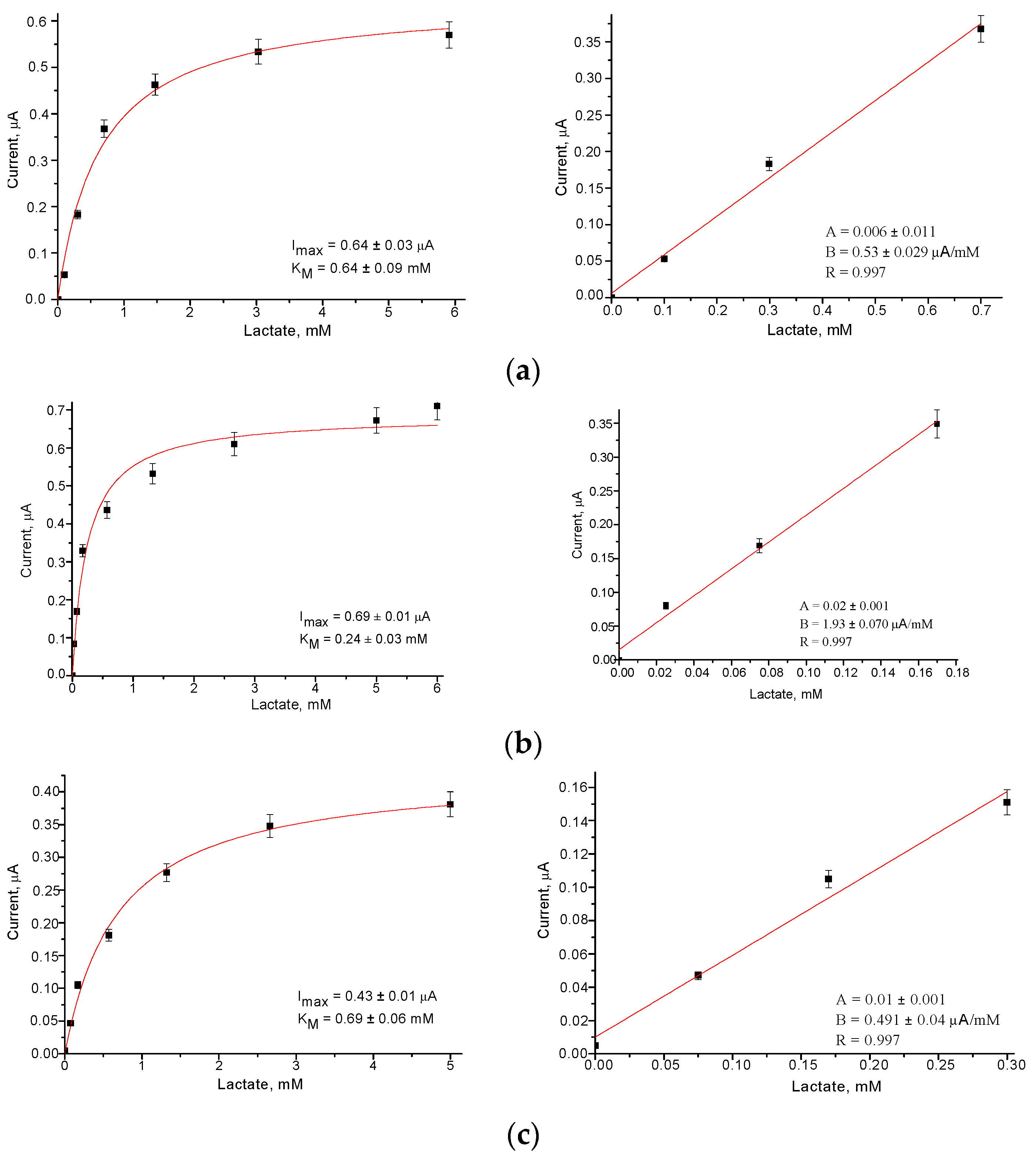
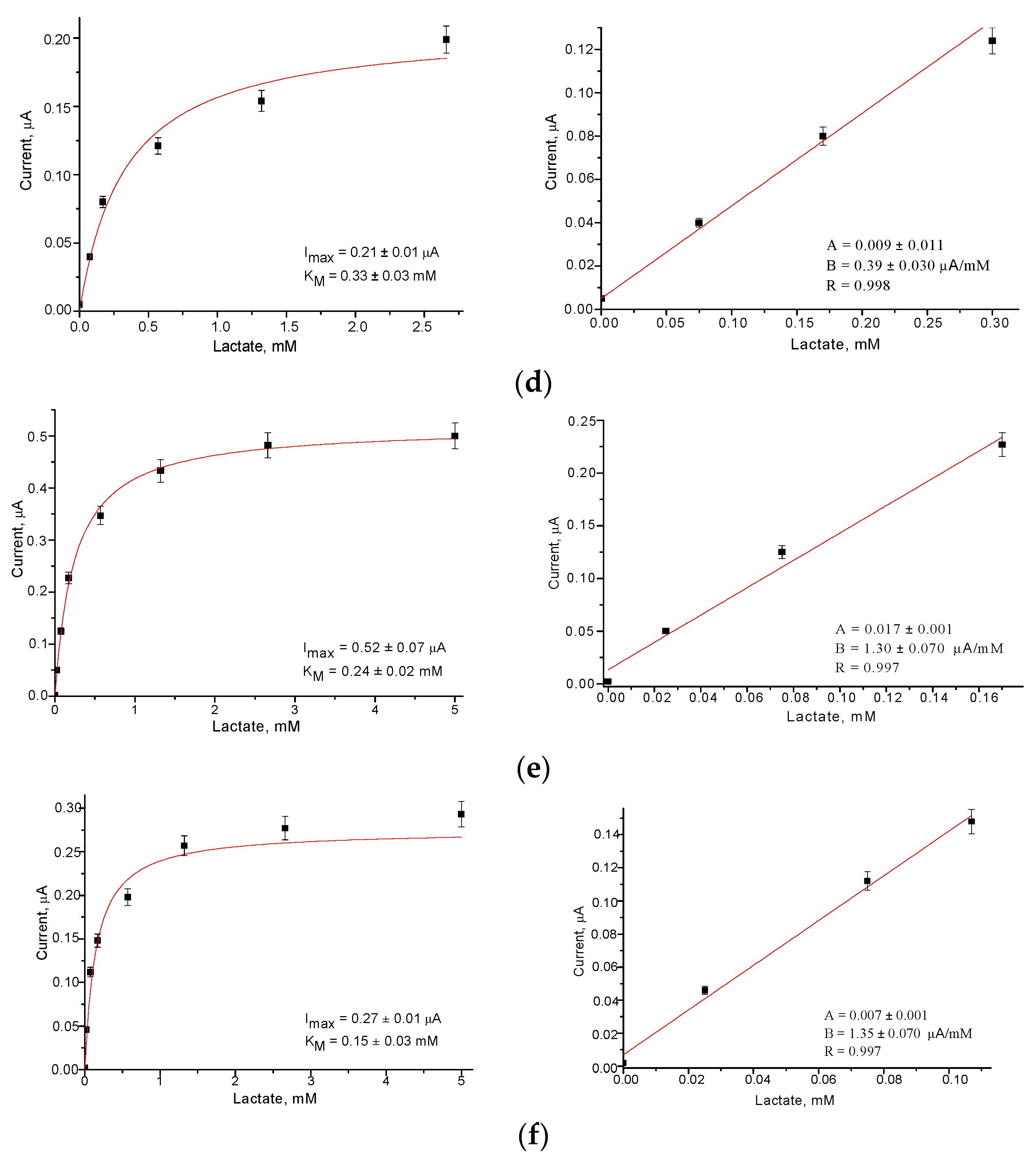
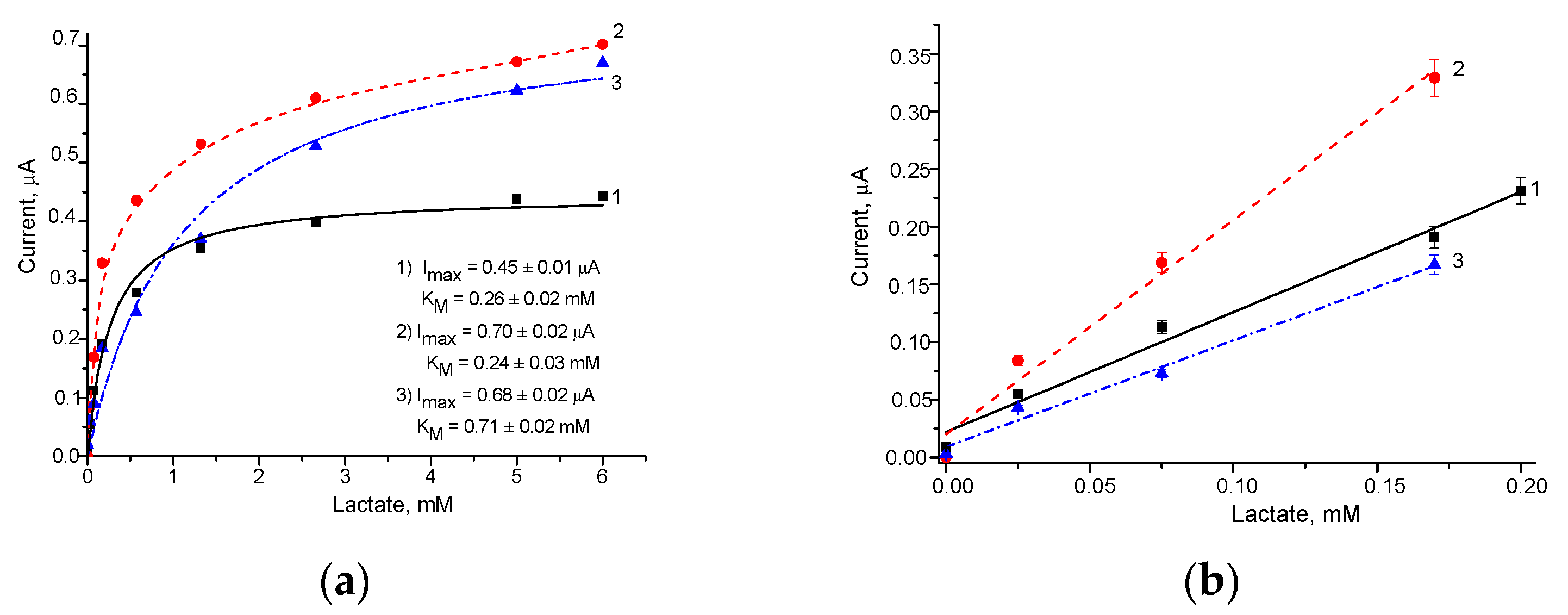
3.3. Development of Fcb2/NPs-Based Amperometric Biosensors

The most active redox mediators coupled with Fcb2 were used to construct ABSs to ensure the MET between the enzyme and GE. According to the CV results for the Fcb2/GEs (Figure 2a), the peak of oxidation, as an output upon Lact addition, appeared in the range of −(150–50) mV. The optimal working potential for the study of the Fcb2/GEs and of the other proposed Fcb2/NPs/GEs was selected as –75 mV.
The calibration of the developed Fcb2/NPs/GEs was performed via a stepwise addition of the Lact solution, and the detailed chronoamperometric experiments are omitted here. The calibration graphs resulting from the corresponding chronoamperograms for the studied ABSs are presented in Figure 4.
The main operational characteristics of the ABSs, determined automatically from the calibration graphs (Figure 4) as described in our recent papers [41,44,45], are summarized in Table 1. Sensitivity was calculated as the ratio of the slope B value (from the linear regression graph) to the square of the active GE surface (7.3 mm2).
As can be seen from the presented results (Figure 4 and Table 1), the modification of the GE surface with PtZn, NiPtPd, and AuHCF led to an improvement in the operating parameters of the ABSs, namely, to an increasing sensitivity (2.5, 2.6 and 3.5-fold, respectively) and to a decreasing LOD (2.7, 4.3 and 2.7-fold, respectively), compared to the control Fcb2/GE. These ABSs may be useful for the analysis of Lact in food products, biological liquids, pharmaceuticals, and other real samples.
It is worth mentioning that NiPtPd is a PO mimetic and also demonstrates laccase-like activity [45]. Such properties of NPs may complicate our study and our understanding of MET processes. Therefore, for the detailed investigation of the influence of electroactive nanomediators on the effectivity of MET in the Fcb2/NPs/based ABSs, we selected the NPs of AuHCF and PtZn.
We concluded that the increased electron transfer in the Fcb2/NPs/GE may have been achieved thanks to NPs of a special shape and size. As Fcb2 is a large multidomain enzyme, it needs a matrix with small-size particles for immobilization and effective MET. SEM images proved that NPs of AuHCF and PtZn are really such materials (Figure A1). As a result, the Fcb2/AuHCF/GE and Fcb2/PtZn/GE demonstrated better operational characteristics in comparison with those of Fcb2/GE, due to MET without additional manipulation; that is, without using freely diffusing mediators.

3.4. Optimization of Lact Sensing for the Fcb2/AuHCF/GE

To improve the effectiveness of Lact sensing, the optimal quantities of Fcb2 placed on the surface of the AuHCF/GEs were experimentally chosen. The analytical properties of the resulting bioelectrodes were deduced from the graphs in Figure 5 and are summarized in Table 2.
According to the data presented in Table 2, the highest sensitivity (253 A·M−1·m−2) was achieved with 25 mU of Fcb2 on the GE surface in ABS-2. This sensitivity was 3.5-fold higher in comparison to that of the control GE without NPs in ABS-5, and 2-fold higher than that in ABS-3 and ABS-4, which contained 2-fold and 10-fold elevated quantities of Fcb2, respectively.
To characterize the Fcb2/AuHCF/GE in more detail, ABS-2 was chosen (see Table 2); the results are presented in Figure A3. To study the selectivity, ABS-2 was tested for its ability to respond to several individual natural substrates, namely, organic acids and glucose (Figure A2a). The stability outcomes of ABS-2 and ABS-5 kept under similar conditions (at +4 °C, over vapors of a working buffer) were compared (Figure A2b). According to the presented results, Fcb2/AuHCF/GE is highly selective (Figure A2a), and rather stable (Figure A2b). It is worth mentioning that co-immobilization of Fcb2 with AuHCF resulted in the enhanced stability of ABS-2, in comparison with ABS-5 without NPs.

3.5. Application of the Fcb2/AuHCF/GE for Lact Determination in the Real Samples

To demonstrate the applicability of the developed Fcb2/AuHCF/ABS for the determination of Lact, real samples of commercial yogurts were tested.
Lact is generated from lactose through a chain of enzymatic reactions. Cow’s milk contains 4 or 5% lactose. Lactose, being water-soluble, is associated with the whey portion of dairy foods. In the process of yogurt production, about 20% of the lactose present in milk is converted into lactic acid, and the Lact content in yogurt is about 0.9% or 100 mM [47]. Other fermented milk products, such as kefir, contain up to 2% or 220 mM Lact [47,48].
We carried out a determination of the Lact contents in strawberry yogurts. The results of the biosensor analysis using the graphical calibration method in a SAT variant [41,44] are presented in Figure A3 and summarized in Table 3. The numbering of the tested samples in Table 3 is the same as that in Figure A3.
The estimated average contents of Lact in yogurts corresponded to 60–95 mM, and such values are similar to the reported data (0.6–1.1%) [47,49]. The results of the quantitative analysis of Lact in the samples of yogurts using the Fcb2/AuHCF/ABS were compared with the data obtained using the enzymatic–chemical reference Fcb2/PB method (Table 3).
The results of testing the effectiveness of the Fcb2/AuHCF-based ABS were shown to be within the permissible error limits (±10.0%). The results of Lact determination using both methods showed a reliably linear character with a strong correlation (R = 0.99), with differences of less than 5% (Figure A4). Thus, we demonstrated the applicability of the developed Fcb2/AuHCF/ABS for Lact analysis in yogurts.

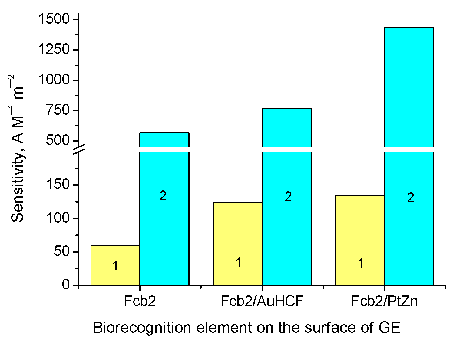
3.6. Ways to Enhance the Sensitivity of the Fcb2-Based ABS

ABS sensitivity can be improved by increasing the rate/efficiency of DET and MET from the enzyme to the electrode. To satisfy this demand, close contact of the enzyme with the electrode surface must be ensured. The well-known general way to enhance the effectiveness of ET is the application of electroactive mediators—freely diffusing or/and immobilized ones coupled with the enzyme on the surface of the electrode [50].
Another way to enhance the sensitivity of an ABS is by selecting the optimal amount of the enzyme. Actually, according to the data of Table 2 and Table A1, the increased enzyme quantity on the surface of the Fcb2/AuHCF/ABS (more than 25 mU) caused decreasing ABS sensitivity. According to Table A1, a two-fold increase in enzyme quantity (from 250 to 500 mU) in Fcb2/ABSs led to a 2.9-fold decrease in sensitivity to Lact. Such results proved the importance of using optimal but not maximal quantities of the enzyme [44]. In other words, using the highest amount of the enzyme does not result in higher ABS sensitivity; this is probably related to the worsening diffusion process in the protein-enriched recognition layer.
Additionally, we studied the impact of 1 mM PMS, one of the most electroactive freely diffusing mediators, on the analytical properties of the developed ABSs (Table A1 and Figure 6). Figure 6 demonstrates that the presence of PMS contributed to the improved sensitivity of the ABSs containing 250 mU of the enzyme.
As can be seen from the presented results (Table A1 and Figure 6), the addition of 1 mM PMS in the electrochemical cell caused increasing sensitivity in the Fcb2/GE, Fcb2/AuHCF/GE, and Fcb2/PtZn/GE (9.4-, 6.2- and 10.6-fold, respectively) compared to that of the corresponding bioelectrodes that were tested without PMS. The simultaneous impact of freely diffusing and co-immobilized mediators in NPs with the enzyme resulted in significantly enhanced sensitivity, due to the highly effective MET from the enzyme to the surface of the electrode. For example, the sensitivity of the Fcb2/PtZn/GE in the presence of PMS was 24-fold higher, in comparison to that of the Fcb2/GE without any mediator (Table A1).

4. Discussion

Fcb2 is a large ferrum-containing enzyme with a complex structure; its study is limited by protein instability. The Fcb2 successfully isolated by us from the thermotolerant yeast O. polymorpha was much more stable than the corresponding enzymes from the yeasts S. cerevisiae and H. anomala. The Fcb2 of O. polymorpha is stable enough to isolate, purify, characterize, lyophilize, and store for a sufficiently long time for use in developing analytical methods, including biosensors [28,39].
The possibility of DET from the reduced form of O. polymorpha Fcb2 to a GE was demonstrated earlier, as mentioned. It was reported that an optimal enzyme quantity is necessary to ensure its monolayer placement on the electrode’s surface and the appropriate orientation of hemes on the GE [40]. In a higher quantity, Fcb2 may form a multilayer structure on the GE surface, thus causing the limitation of DET and decreased sensitivity of the ABS. Additionally, generated pyruvate, a product of catalytic Lact oxidation gathered under a Nafion film, may inhibit Fcb2 as well [51,52].
We report here the fabrication and characterization of Lact-sensitive biosensors based on Fcb2 and electroactive NPs. All the proposed Fcb2/NPs/ABSs demonstrated improved operational parameters, compared to the control Fcb2/ABS. Specifically, the Fcb2/AuHCF/GE was 3.5-fold more sensitive to Lact in comparison to the control Fcb2/GE.
The analytical characteristics of other reported Lact-sensitive ABSs, compared to the ABSs proposed by us, are presented in Table A2.
It is worth mentioning that all the NPs reported here, and in our recent papers, are not only electroactive but catalytically active, too. They all possess pseudo-PO activity that allows them to decompose H2O2 in-solution and on amperometric electrodes. Being co-immobilized in a sensing layer with different oxidases, PO-like nanozymes positively impacted the analytical characteristics, especially the sensitivity of the developed ABSs, of catechol [44,51], ethanol, glucose, and arginine [41,51].
The following questions arise: What is the reason for the significant positive influence of PO-like nanozymes on the increased sensitivity of the Fcb2-based ABS? Is the electroactivity of the best NPs perhaps the only cause of such an effect? Unfortunately, the detailed mechanisms of heme-containing Fcb2 activity are still unknown [25,51,52,53,54,55,56,57,58,59,60]. Additionally, the structures and kinetic characteristics of Fcb2 from the yeasts S. cerevisiae and H. anomala have some differences [55,56,57], and the structure of Fcb2 from the yeast O. polymorpha is not yet reported. Perhaps this gap in enzymology is due to the lack of an enzyme available as a commercial product.
It can be concluded that the developed ABSs may be useful in the food industry and in other areas where precision analysis of Lact is necessary. It should be noted that the application of electroactive metallic NPs immobilized on the surface of the electrode can improve the sensitivity and stability of the Fcb2-based ABS.

5. Conclusions

In the current paper, a number of novel Lact-sensitive amperometric bioelectrodes based on oxidoreductase Fcb2 of thermotolerant methylotrophic yeast O. polymorpha, and the most effective nanomediators were developed and characterized. We investigated the enzyme-to-electrode ET process and showed that both DET and MET processes could be achieved by using bound NPs and low-molecular freely diffusing redox mediators. We found that the MET led to improved operational characteristics of Fcb2-based ABSs, especially to increased sensitivity. The main advantages of the proposed mono-enzyme ABSs are the simple architecture of the sensing layer and the ability to operate at low potentials in an oxygen-independent manner. The latter property of the developed Fcb2-based ABS is very valuable to ensure interference-free measurements and avoid the limitations inherent in LOX-based biosensors. The fabricated ABSs exhibited high sensitivities, fast responses, and low limits of detection. One of the most effective biosensors, Fcb2/AuHCF/GE, was used for Lact determination in commercial yogurts. A high correlation was observed between the values of Lact content determined using the biosensor and the reference methods.

Author Contributions

Conceptualization, O.D. and G.G.; methodology, O.D. and N.S.; software, A.M. and R.S.; validation, O.D. and R.S.; formal analysis, G.G. and A.K.-M.; investigation, O.D., A.M., R.S. and N.S.; resources, M.N. and A.K.-M.; data curation, A.K.-M. and M.G.; writing—original draft preparation, A.M. and G.G.; writing—review and editing, G.G. and M.N.; visualization, G.G.; supervision, M.G.; project administration, M.G.; funding acquisition, M.N. All authors have read and agreed to the published version of the manuscript.

Funding

This research was partially funded by the National Research Foundation of Ukraine (project no. 2020.02/0100) and Ukrainian Short-Term Fellowships 2022 (FEBS FELLOWSHIP) for Demkiv O. and Stasyuk N.

Institutional Review Board Statement

Not applicable.

Informed Consent Statement

Not applicable.

Data Availability Statement

The data are included within the present article.

Acknowledgments

We acknowledge the Research Authority of Ariel University, Ariel, Israel, for supporting this research. The authors thank M. Horecha (Institute of Cell Biology NAS of Ukraine) for her assistance in the experiments.

Conflicts of Interest

The authors declare no conflict of interest.

Appendix A

Appendix A presents the structural and morphological characteristics of the most effective nanoparticles (Figure A1), properties of Fcb/AuHCF/GE, namely, the selectivity and stability tests (Figure A2), Lact determination in the samples of yogurts using a biosensor (Figure A3), comparison of the Lact contents in yogurts analyzed with different methods (Figure A4), the effect of PMS on the main operational parameters of the ABSs (Table A1), and a comparison of the most effective Lact-sensitive ABSs (Table A2).
Figure A1. Structural and morphological characterizations of the AuHCF/NPs (a,c) and PtZn/NPs (b,d); the SEM images (a,b) with the correspondent XRM spectra (c,d).
Biosensors 13 00587 g0a1
Figure A2. Properties of the Fcb2-based ABSs. The selectivity test for AuHCF/GE (ABS-2); current outputs in relative units (%) on the added compounds, up to 2 mM concentration. As a 100% signal, the highest current response on L-Lact was taken (a). Stabilities of the NPs-based ABS-2, and control ABS-5 without NPs (b).
Biosensors 13 00587 g0a2
Figure A3. Assay of Lact in the samples of different yogurts (1–3) via graph SAT method (ac, respectively), using Fcb2/AuHCF/GE (25 mU Fcb2 per ABS).
Biosensors 13 00587 g0a3
Figure A4. Correlation between the results of the determination of Lact content (mM) in drinking yogurts via reference enzymatic–chemical (Fcb2/PB) and biosensor methods.
Biosensors 13 00587 g0a4
Table A1. The dependence of the main operational parameters of the ABSs on the presence of PMS.
ABSSensitivity,
A·M−1·m−2
Linear Range,
up to, mM
Imax, µAKMapp, mMTotal Fcb2, m-Units
Fcb2/GE600.160.230.44250
Fcb2/GE + PMS5650.081.700.30250
Fcb2/GE + PMS1950.200.900.39500
Fcb2/AuHCF/GE1240.350.480.10250
Fcb2/AuHCF/GE + PMS7690.071.650.33250
Fcb2/PtZn/GE1350.440.750.43250
Fcb2/PtZn/GE + PMS14360.122.740.18250
Table A2. Comparison of the most effective Lact-sensitive ABSs.
EnzymeWorking Potential, VSensitivity,
A M−1m−2
Linear Range,
up to, mM
LOD, mMStability
(days)
Reference
LOX−0.152330.140.0015[22]
0.1579740.10.0034.5[21]
0.345030.05* NR[20]
0.522800.60.001NR[61]
0.81561.20.01214[62]
0.64000.350.032NR[63]
0.15NR10.75 × 10−3NR[64]
0.443000.70.02230[65]
** rFcb0.2548.62.70.3NR[28]
rFcb20.2510626NR[28]
rLDH0287.58100.3[37]
0.1638.36140.9
LDH0.6830.12NR7[66]
0.134.5550.001210[67]
034.6100.1NR[68]
Fcb2−0.755650.080.0155This paper
7690.070.0107
14360.120.0107
* NR—not reported; ** r—recombinant enzyme.

References

  1. Średnicka, P.; Juszczuk-Kubiak, E.; Wójcicki, M.; Akimowicz, M.; Roszko, M.Ł. Probiotics as a biological detoxification tool of food chemical contamination: A review. Food Chem. Toxicol. 2021, 153, 112306. [Google Scholar] [CrossRef]
  2. López-Varela, S.; González-Gross, M.; Marcos, A. Functional foods and the immune system: A review. Eur. J. Clin. Nutr. 2002, 56, 29–33. [Google Scholar] [CrossRef] [PubMed]
  3. Granato, D.; Barba, F.J.; Kovačević, D.B.; Lorenzo, J.M.; Cruz, A.G.; Putnik, P. Functional Foods: Product Development, Technological Trends, Efficacy Testing, and Safety. Annu. Rev. Food Sci. Technol. 2020, 11, 93–118. [Google Scholar] [CrossRef]
  4. Kerry, R.G.; Patra, J.K.; Sushanto Gouda, S.; Park, Y.; Shin, H.-S.; Das, G. Benefaction of probiotics for human health: A review. J. Food Drug Anal. 2018, 26, 927–939. [Google Scholar] [CrossRef] [PubMed]
  5. Garcia-Albiach, R.; Pozuelo de Felipe, M.J.; Angulo, S.; Morosini, M.-I.; Bravo, D.; Baquero, F.; del Campo, R. Molecular analysis of yogurt containing Lactobacillus delbrueckii subsp. bulgaricus and Streptococcus thermophilus in human intestinal microbiota. Am. J. Clin. Nutr. 2008, 87, 91–96. [Google Scholar] [CrossRef] [PubMed]
  6. Roselli, M.; Natella, F.; Zinno, P.; Guantario, B.; Canali, R.; Schifano, E.; De Angelis, M.; Nikoloudaki, O.; Gobbetti, M.; Perozzi, G.; et al. Colonization Ability and Impact on Human Gut Microbiota of Foodborne Microbes From Traditional or Probiotic-Added Fermented Foods: A Systematic Review. Front. Nutr. 2021, 8, 689084. [Google Scholar] [CrossRef]
  7. Markowiak-Kopeć, P.; Śliżewska, K. The Effect of Probiotics on the Production of Short-Chain Fatty Acids by Human Intestinal Microbiome. Nutrients 2020, 12, 1107. [Google Scholar] [CrossRef]
  8. Bakker, J.; Postelnicu, R.; Mukherjee, V. Lactate: Where Are We Now? Crit. Care. Clin. 2020, 36, 115–124. [Google Scholar] [CrossRef]
  9. García-Guzmán, J.J.; Sierra-Padilla, A.; Palacios-Santander, J.M.; Fernández-Alba, J.J.; Macías, C.G.; Cubillana-Aguilera, L. What Is Left for Real-Life Lactate Monitoring? Current Advances in Electrochemical Lactate (Bio)Sensors for Agrifood and Biomedical Applications. Biosensors 2022, 12, 919. [Google Scholar] [CrossRef]
  10. Pundir, C.S.; Narwal, V.; Batra, B. Determination of lactic acid with special emphasis on biosensing methods: A review. Biosens. Bioelectron. 2016, 86, 777–790. [Google Scholar] [CrossRef]
  11. Rattu, G.; Murali Krishna, P. Enzyme-Free Colorimetric Nanosensor for the Rapid Detection of Lactic Acid in Food Quality Analysis. J. Agric. Food Res. 2022, 7, 100268. [Google Scholar] [CrossRef]
  12. Pranveer, S. Chapter 1—Electrochemical Biosensing: Progress and Perspectives. In Electrochemical Biosensors; Singh, P., Ed.; Academic Press: Cambridge, MA, USA, 2022; pp. 1–31. ISBN 9780323906326. [Google Scholar] [CrossRef]
  13. Ragavan, K.V.; Neethirajan, S. Chapter 7–Nanoparticles as Biosensors for Food Quality and Safety Assessment. In Nanomaterials for Food Applications; Rubio, A.L., Rovira, M.J.F., Sanz, M.M., Gomez-Mascaraque, L.G., Eds.; Micro and Nano Technologies; Elsevier: Amsterdam, The Netherlands, 2019; pp. 147–202. ISBN 9780128141304. [Google Scholar] [CrossRef]
  14. Villalonga, A.; Sánchez, A.; Mayol, B.; Reviejo, J.; Villalonga, R. Electrochemical biosensors for food bioprocess monitoring. Curr. Opin. Food Sci. 2022, 43, 18–26. [Google Scholar] [CrossRef]
  15. Escalona-Villalpando, R.A.; Viveros-Palma, K.; Espinosa-Lagunes, F.I.; Rodríguez-Morales, J.A.; Arriaga, L.G.; Macazo, F.C.; Minteer, S.D.; Ledesma-García, J. Comparative Colorimetric Sensor Based on Bi-Phase γ-/α-Fe2O3 and γ-/α-Fe2O3/ZnO Nanoparticles for Lactate Detection. Biosensors 2022, 12, 1025. [Google Scholar] [CrossRef] [PubMed]
  16. Kim, H.J.; Park, I.; Pack, S.P.; Lee, G.; Hong, Y. Colorimetric Sensing of Lactate in Human Sweat Using Polyaniline Nanoparticles-Based Sensor Platform and Colorimeter. Biosensors 2022, 12, 248. [Google Scholar] [CrossRef] [PubMed]
  17. Rathee, K.; Dhull, V.; Dhull, R.; Singh, S. Biosensors Based on Electrochemical Lactate Detection: A Comprehensive Review. Biochem. Biophys. Rep. 2016, 5, 35–54. [Google Scholar] [CrossRef] [PubMed]
  18. Hiraka, K.; Kojima, K.; Tsugawa, W.; Asano, R.; Ikebukuro, K.; Sode, K. Rational engineering of Aerococcus viridans L-lactate oxidase for the mediator modification to achieve quasi-direct electron transfer type lactate sensor. Biosens. Bioelectron. 2020, 151, 111974. [Google Scholar] [CrossRef]
  19. Ma, Y.; Wang, Y.; Liu, Y.; Shi, L.; Yang, D. A cascade-triggered ratiometric fluorescent sensor based on nanocomposite for lactate determination. Sens. Actuators B Chem. 2022, 355, 131295. [Google Scholar] [CrossRef]
  20. Hickey, D.P.; Reid, R.C.; Milton, R.D.; Minteer, S.D. A self-powered amperometric lactate biosensor based on lactate oxidase immobilized in dimethylferrocene-modified LPEI. Biosens. Bioelectron. 2016, 77, 26–31. [Google Scholar] [CrossRef]
  21. Bollella, P.; Sharma, S.; Cass, A.E.G.; Antiochia, R. Minimally-invasive microneedle-based biosensor array for simultaneous lactate and glucose monitoring in artificial interstitial fluid. Electroanalysis 2019, 31, 374–382. [Google Scholar] [CrossRef]
  22. Smutok, O.; Kavetskyy, T.; Prokopiv, T.; Serkiz, R.; Šauša, O.; Novák, I.; Švajdlenková, H.; Maťko, I.; Gonchar, M.; Katz, E. Biosensor Based on Peroxidase-Mimetic Nanozyme and Lactate Oxidase for Accurate L-Lactate Analysis in Beverages. Biosensors 2022, 12, 1042. [Google Scholar] [CrossRef]
  23. Haas, E.; Horecker, B.L.; Hogness, T.R. Cytochrome b2. Science 1942, 95, 406. [Google Scholar] [CrossRef]
  24. Bach, S.J.; Dixon, M.; Zerfas, L.G. Yeast lactic dehydrogenase and cytochrome b2. Biochem J. 1946, 40, 229–239. [Google Scholar] [CrossRef]
  25. Lederer, F. Flavocytochrome b2. In Chemistry and Biochemistry of Flavoenzymes, 1st ed.; CRC Press: Boca Raton, FL, USA, 1991; p. 90. ISBN 9781351070584. [Google Scholar] [CrossRef]
  26. Lederer, F. On the First Steps of Lactate Oxidation by Bakers’ Yeast L-(+)-Lactate Dehydrogenase (Cytochrome b2). Eur. J. Biochem. 1974, 46, 393. [Google Scholar] [CrossRef] [PubMed]
  27. Smutok, O.; Kavetskyy, T.; Gonchar, M.; Katz, E. Microbial L- and D-Lactate Selective Oxidoreductases as a Very Prospective but Still Uncommon Tool in Commercial Biosensors. ChemElectroChem 2021, 8, 4725–4731. [Google Scholar] [CrossRef]
  28. Smutok, O.; Dmytruk, K.; Kavetskyy, T.; Sibirny, A.; Gonchar, M. Flavocytochrome b2 of the Methylotrophic Yeast Ogataea polymorpha: Construction of Overproducers, Purification, and Bioanalytical Application. In Flavin and Flavoproteins; Methods Molecular Biology; Springer: New York, NY, USA, 2021; pp. 249–260. ISBN 978-1-0716-1285-9. [Google Scholar]
  29. Schachinger, F.; Chang, H.; Scheiblbrandner, S.; Ludwig, R. Amperometric Biosensors Based on Direct Electron Transfer Enzymes. Molecules 2021, 26, 4525. [Google Scholar] [CrossRef]
  30. Garjonyte, R.; Malinauskas, A. Investigation of Saccharomyces cerevisiae- and mediator-based carbon paste electrodes as amperometric biosensors for lactic acid. Sens. Actuat. B Chem. 2003, 96, 509–515. [Google Scholar] [CrossRef]
  31. Staškevičiene, S.L.; Čenas, N.K.; Kulys, J.J. Reagentless lactate electrodes based on electrocatalytic oxidation of flavocytochrome b2. Anal. Chim. Acta 1991, 243, 167–171. [Google Scholar] [CrossRef]
  32. Brunt, C.E.; Cox, M.C.; Thurgood, A.G.P.; Moore, G.R.; Reid, G.A.; Chapman, S.K. Isolation and characterization of the cytochrome domain of flavocytochrome b2 expressed independently in Escherichia coli. Biochem. J. 1992, 283, 87–90. [Google Scholar] [CrossRef]
  33. Silvestrini, M.C.; Tegoni, M.; Célerier, J.; Desbois, A.; Gervais, M. Expression in Escherichia coli of the flavin and the haem domains of Hansenula anomala flavocytochrome b2 (flavodehydrogenase and b2 core) and characterization of the recombinant proteins. Biochem. J. 1993, 295 Pt 2, 501–508. [Google Scholar] [CrossRef]
  34. Hiraka, K.; Tsugawa, W.; Asano, R.; Yokus, M.A.; Ikebukuro, K.; Daniele, M.A.; Sode, K. Rational design of direct electron transfer type l-lactate dehydrogenase for the development of multiplexed biosensor. Biosens. Bioelectron. 2021, 176, 112933. [Google Scholar] [CrossRef]
  35. Tsvik, L.; Steiner, B.; Herzog, P.; Haltrich, D.; Sützl, L. Flavin Mononucleotide-Dependent L-Lactate Dehydrogenases: Expanding the Toolbox of Enzymes for L-Lactate Biosensors. ACS Omega 2022, 7, 41480–41492. [Google Scholar] [CrossRef] [PubMed]
  36. Boyarski, A.; Shlush, N.; Paz, S.; Eichler, J.; Alfonta, L. Electrochemical characterization of a dual cytochrome-containing lactate dehydrogenase. Bioelectrochemistry 2023, 152, 108406. [Google Scholar] [CrossRef] [PubMed]
  37. Cohen, R.; Herzallh, N.S.; Meirovich, M.M.; Bachar, O.; Frech, L.; Cohen, Y.; Yehezkeli, O. An Oxygen-Insensitive Biosensor and a Biofuel Cell Device Based on FMN L-Lactate Dehydrogenase. Bioelectrochemistry 2023, 149, 108316. [Google Scholar] [CrossRef]
  38. Gaida, G.; Stel’mashchuk, S.; Smutok, O.; Gonchar, M. A new method of visualization of the enzymatic activity of flavocytochrome b2 in electrophoretograms. Appl. Biochem. Microbiol. 2003, 39, 221–223. [Google Scholar] [CrossRef]
  39. Gayda, G.; Demkiv, O.; Klepach, H.; Gonchar, M.; Nisnevitch, M. Effective Technologies for Isolating Yeast Oxido-Reductases of Analytical Importance. In Non-Conventional Yeasts: From Basic Research to Application; Andriy, S., Ed.; Springer Nature Switzerland AG: Cham, Switzerland, 2019; Volume 5, pp. 119–151. ISBN 978-3-030-21110-3. [Google Scholar] [CrossRef]
  40. Smutok, O.; Gayda, G.; Gonchar, M.; Schuhmann, W. A novel L-lactate-selective biosensor based on the use of flavocytochrome b2 from methylotrophic yeast Hansenula polymorpha. Biosens. Bioelectron. 2005, 20, 1285–1290. [Google Scholar] [CrossRef]
  41. Stasyuk, N.; Gayda, G.; Demkiv, O.; Darmohray, L.; Gonchar, M.; Nisnevitch, M. Amperometric Biosensors for L-Arginine Determination Based on L-Arginine Oxidase and Peroxidase-Like Nanozymes. Appl. Sci. 2021, 11, 7024. [Google Scholar] [CrossRef]
  42. Mugle, D.; Jadhav, G. Short review on chemical bath deposition of thin film and characterization. AIP Conf. Proc. 2016, 1728, 020597. [Google Scholar] [CrossRef]
  43. Stasyuk, N.; Gayda, G.; Kavetskyy, T.; Gonchar, M. Nanozymes with reductase-like activities: Electrochemical and antioxidant properties. RSC Adv. 2022, 12, 2026–2035. [Google Scholar] [CrossRef]
  44. Demkiv, O.; Gayda, G.; Stasyuk, N.; Brahinetz, O.; Gonchar, M.; Nisnevitch, M. Nanomaterials as Redox Mediators in Laccase-Based Amperometric Biosensors for Catechol Assay. Biosensors 2022, 12, 741. [Google Scholar] [CrossRef]
  45. Demkiv, O.; Stasyuk, N.; Gayda, G.; Gonchar, M. Highly sensitive amperometric sensor based on laccase-mimicking metal-based hybrid nanozymes for adrenaline analysis in pharmaceuticals. Catalysts 2021, 11, 1510. [Google Scholar] [CrossRef]
  46. European Union. Commission Recommendation of 10 June 2022 on the definition of nanomaterial (Text with EEA relevance). Off. J. 2022, 229, 1–5. Available online: https://eur-lex.europa.eu/legal-content/EN/TXT/?uri=CELEX:32022H0614(01) (accessed on 16 May 2023).
  47. Cheng, H. Volatile flavor compounds in yogurt: A review. Crit. Rev. Food Sci. Nutr. 2010, 50, 938–950. [Google Scholar] [CrossRef] [PubMed]
  48. Londero, A.; Hamet, M.F.; De Antoni, G.L.; Garrote, G.L.; Abraham, A.G. Kefir grains as a starter for whey fermentation at different temperatures: Chemical and microbiological characterisation. J. Dairy Res. 2012, 79, 262–271. [Google Scholar] [CrossRef] [PubMed]
  49. Deeth, H.C.; Tamime, A.Y. Yogurt: Nutritive and Therapeutic Aspects. J. Food Prot. 1981, 44, 78–86. [Google Scholar] [CrossRef]
  50. Warren, J.J.; Gray, H.B. 8.02—Electron Transfer Proteins. In Comprehensive Coordination Chemistry III; Constable, E.C., Parkin, G., Que, L., Jr., Eds.; Elsevier: Amsterdam, The Netherlands, 2021; pp. 3–18. ISBN 9780081026892. [Google Scholar] [CrossRef]
  51. Stasyuk, N.; Demkiv, O.; Gayda, G.; Zakalskiy, A.; Klepach, H.; Bisko, N.; Gonchar, M.; Nisnevitch, M. Highly Porous 3D Gold Enhances Sensitivity of Amperometric Biosensors Based on Oxidases and CuCe Nanoparticles. Biosensors 2022, 12, 472. [Google Scholar] [CrossRef]
  52. Tegoni, M.; Janot, J.M.; Labeyrie, F. Inhibition of L-lactate: Cytochrome-c reductase (flavocytochrome b2) by product binding to the semiquinone transient. Loss of reactivity towards monoelectronic acceptors. Eur. J. Biochem. 1990, 190, 329–342. [Google Scholar] [CrossRef]
  53. Blumberger, J. Electron transfer and transport through multi-heme proteins: Recent progress and future directions. Curr. Opin. Chem. Biol. 2018, 47, 24–31. [Google Scholar] [CrossRef]
  54. Bertrand, P.; Janot, J.M.; Benosman, H.; Gayda, J.P.; Labeyrie, F. An EPR study of the interactions between heme and flavin in yeast flavocytochrome b2. Eur. Biophys. J. 1987, 14, 273–278. [Google Scholar] [CrossRef]
  55. White, P.; Manson, F.D.; Brunt, C.E.; Chapman, S.K.; Reid, G.A. The importance of the interdomain hinge in intramolecular electron transfer in flavocytochrome b2. Biochem. J. 1993, 291 Pt 1, 89–94. [Google Scholar] [CrossRef]
  56. Black, M.T.; Gunn, F.J.; Chapman, S.K.; Reid, G.A. Structural basis for the kinetic differences between flavocytochromes b2 from the yeasts Hansenula anomala and Saccharomyces cerevisiae. Biochem. J. 1989, 263, 973–976. [Google Scholar] [CrossRef]
  57. Capeillère-Blandin, C. Electron-transfer steps involved in the reactivity of Hansenula anomala flavocytochrome b2 as deduced from deuterium isotope effects and simulation studies. Biochem. J. 1991, 274 Pt 1, 207–217. [Google Scholar] [CrossRef]
  58. Lederer, F. Another look at the interaction between mitochondrial cytochrome c and flavocytochrome b2. Eur. Biophys. J. 2011, 40, 1283–1299. [Google Scholar] [CrossRef] [PubMed]
  59. Boubacar, O.A.K.; Pethe, S.; Mahy, J.-P.; Lederer, F. Flavocytochrome b2: Reactivity of Its Flavin with Molecular Oxygen. Biochemistry 2007, 46, 13080–13088. [Google Scholar] [CrossRef] [PubMed]
  60. Urban, P.; Lederer, F.; Alliel, P.M. On the Transhydrogenase Activity of Baker’s Yeast Flavocytochrome b2. Eur. J. Biochem. 1983, 134, 275–281. [Google Scholar] [CrossRef] [PubMed]
  61. Lei, Y.; Luo, N.; Yan, X.; Zhao, Y.; Zhang, G.; Zhang, Y. A highly sensitive electrochemical biosensor based on zinc oxide nanotetrapods for L-lactic acid detection. Nanoscale 2012, 4, 3438–3443. [Google Scholar] [CrossRef] [PubMed]
  62. Zhao, Y.; Yan, X.; Fang Kang, Z.; Fang, X.; Zheng, X.; Zhao, L.; Du, H.; Zhang, Y. Zinc oxide nanowires-based electrochemical biosensor for L-lactic acid amperometric detection. J. Nanoparticle Res. 2014, 16, 2398. [Google Scholar] [CrossRef]
  63. Ozoglu, O.; Uzunoglu, A.; Unal, M.A.; Gumustas, M.; Ozkan, S.A.; l Korukluoglu, M.; Altuntas, E.G. Electrochemical detection of lactate produced by foodborne presumptive lactic acid bacteria. JBB 2023, 135, 313–320. [Google Scholar] [CrossRef]
  64. Cunha-Silva, H.; Arcos-Martinez, M.J. Dual range lactate oxidase-based screen printed amperometric biosensor for analysis of lactate in diversified samples. Talanta 2018, 188, 779–787. [Google Scholar] [CrossRef]
  65. Zanini, V.P.; de Mishima, B.L.; Solís, V. An amperometric biosensor based on lactate oxidase immobilized in laponite–chitosan hydrogel on a glassy carbon electrode. Application to the analysis of L-lactate in food samples. Sens. Actuat. 2011, 155, 75–80. [Google Scholar] [CrossRef]
  66. Tsai, Y.-C.; Chen, S.-Y.; Liaw, H.-W. Immobilization of lactate dehydrogenase within multiwalled carbon nanotube-chitosan nanocomposite for application to lactate biosensors. Sens. Actuat. B Chem. 2007, 125, 474–481. [Google Scholar] [CrossRef]
  67. Narwal, V.; Sharma, M.; Rani, S.; Pundir, C.S. An ultrasensitive amperometric determination of lactate by lactate dehydrogenase nanoparticles immobilized onto Au electrode. Int. J. Biol. Macromol. 2018, 115, 767–775. [Google Scholar] [CrossRef] [PubMed]
  68. Pereira, A.C.; Aguiar, M.R.; Kisner, A.; Macedo, D.V.; Kubota, L.T. Amperometric biosensor for lactate based on lactate dehydrogenase and Meldola Blue co-immobilized on multi-wall carbon-nanotube, Sens. Actuat. B Chem. 2007, 124, 269–276. [Google Scholar] [CrossRef]
Figure 1. The principal scheme of Lact determination using the Fcb2-based ABS.
Biosensors 13 00587 g001
Figure 2. Amperometric characteristics of the Fcb2/GE as current responses to the increasing concentrations of Lact. (a) CV profiles at Lact concentrations: 0 mM (1, black), 2 mM (2, red), and 5 mM (3, blue); (b) chronoamperogram at the working potential of −75 mV.
Biosensors 13 00587 g002
Figure 3. CV profiles of the control GE (ac, line 1) and the modified electrodes: AuHCF/GE (a, 2), PtZn/GE (b, 2) and gCuHCF/GE (c, 2). Conditions: 10 mM K3Fe(CN)6, 100 mM KCl in 50 mM phosphate buffer, pH 6.5, and 20 °C.
Biosensors 13 00587 g003
Figure 4. Calibration curves for Lact determination in wide (left) and linear (right) ranges using the Fcb2/NPs/GEs: (a)control, without NPs; (bf) samples with AuHCF (b), PdHCF (c), PtHCF (d), PtZn (e), and NiPtPd (f). Each bioelectrode contained 25 mU of Fcb2 and 1 µg of NPs on the surface of the GE. Conditions: 50 mM phosphate buffer, pH 8.0, and working potential of −75 mV.
Biosensors 13 00587 g004aBiosensors 13 00587 g004b
Figure 5. Calibration curves of the Fcb2/AuHCF/GEs in the broad (a) and linear (b) ranges of the dependence of enzyme quantity on the surface of GE; lines 1, 2, 3 correspond to 13, 25 and 50 mU of Fcb2, respectively. The working potential is −75 mV.
Biosensors 13 00587 g005
Figure 6. Impact of electroactive mediators, both immobilized (AuHCF and PtZn) and freely diffusing (PMS), on the sensitivities of the ABSs to Lact. ABSs with compositions Fcb2/GE, Fcb2/AuHCF/GE, and Fcb2/PtZn/GE were tested without PMS (1, yellow columns) and in the presence of PMS (2, cyan columns) in a 50 mM phosphate buffer, at pH 8.0, and at a working potential of −75 mV.
Biosensors 13 00587 g006
Table 1. The main operational parameters of the constructed Fcb2-based ABSs.
NoNanomediatorSensitivity,
A·M−1·m−2
Linear Range, up to mMLOD, mMImax, µAKMapp, mM
1720.700.050.640.64
2PtHCF530.300.050.210.33
3PdHCF700.300.020.430.70
4gCuHCF800.300.010.841.90
5PtZn1780.180.020.520.24
6NiPtPd1850.110.010.270.15
7AuHCF2530.180.010.690.24
Table 2. Analytical properties of the Fcb2-based ABSs with different compositions.
NoABS Total Fcb2,
mU
Sensitivity,
A·M−1·m−2
Linear Range,
up to mM
Imax, µA KMapp,
mM
1Fcb2/AuHCF/GE131420.200.450.26
2Fcb2/AuHCF/GE252530.170.700.24
3Fcb2/AuHCF/GE501260.170.680.71
4Fcb2/AuHCF/GE2501240.350.480.10
5Fcb2/GE25720.700.640.64
6Fcb2/GE250600.160.230.44
Table 3. Lact content (mM) in the samples of yogurts estimated using two methods.
NoYogurtABS MethodCV, %Reference MethodCV, %
1Lactel61 ± 4.57.463 ± 4.06.3
2Activia72 ± 1.01.471 ± 5.47.6
3Choodo94.5 ± 2.52.695 ± 7.07.3
Disclaimer/Publisher’s Note: The statements, opinions and data contained in all publications are solely those of the individual author(s) and contributor(s) and not of MDPI and/or the editor(s). MDPI and/or the editor(s) disclaim responsibility for any injury to people or property resulting from any ideas, methods, instructions or products referred to in the content.

