Recent Development of Neural Microelectrodes with Dual-Mode Detection

 , and
, and
Abstract
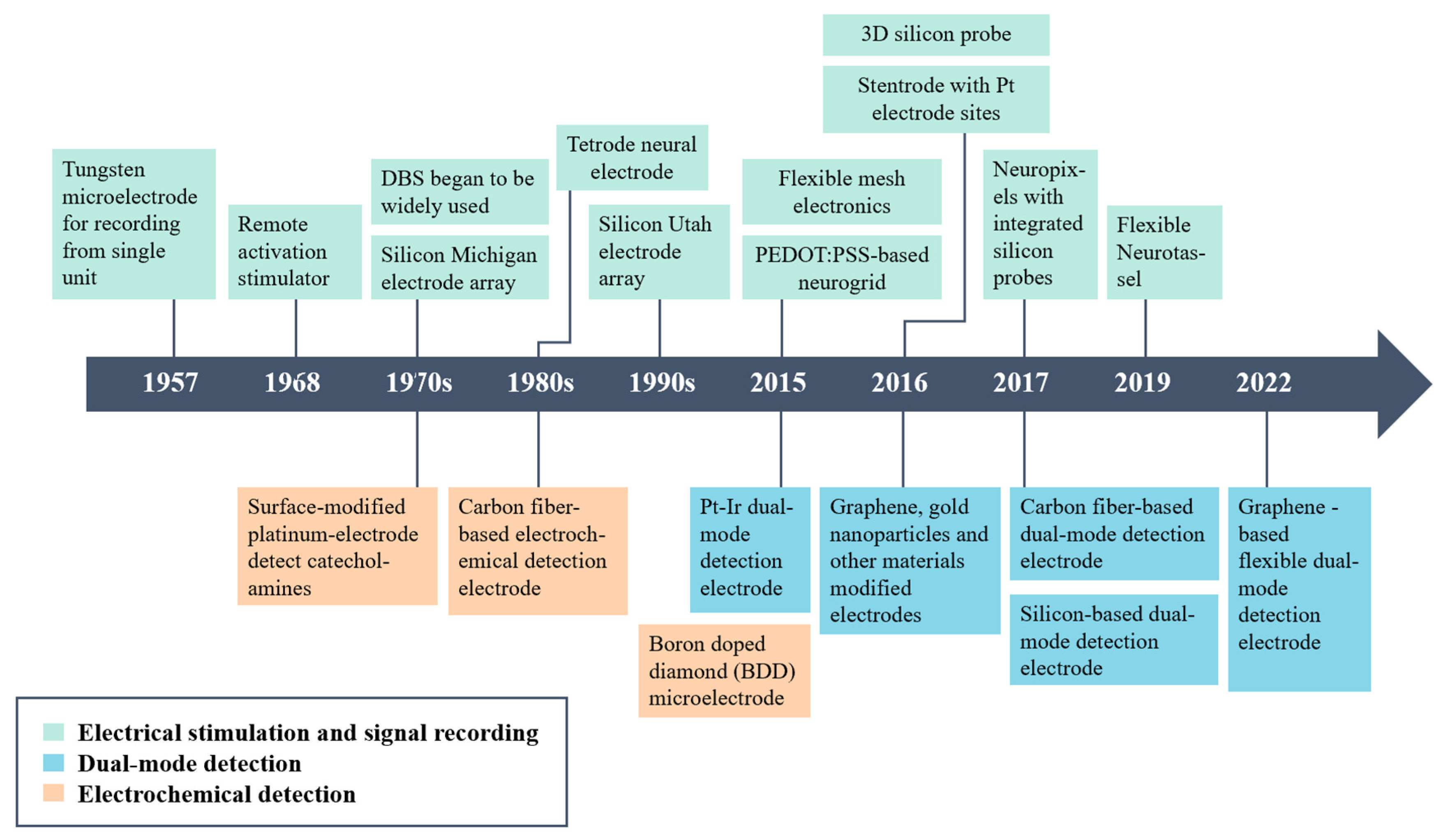
:1. Introduction
2. Electrochemical Detection
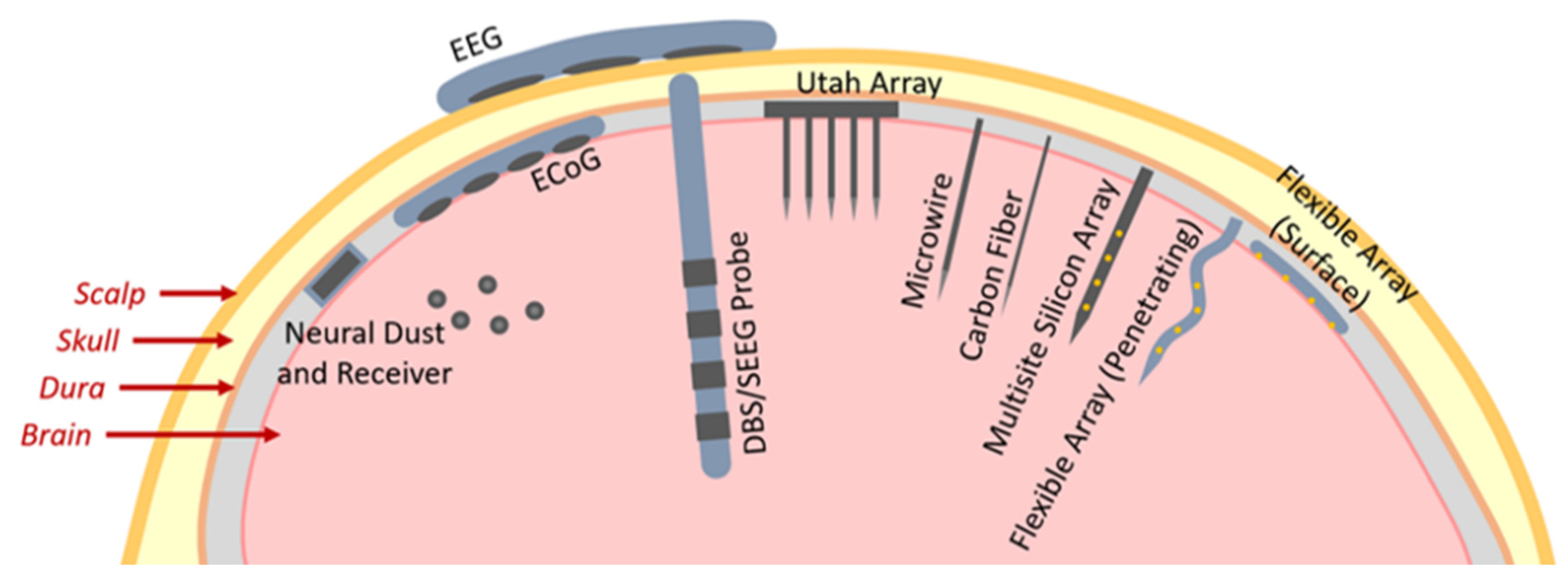
3. Electrophysiological Signal Detection
4. Dual-Mode Neural Microelectrodes
4.1. Carbon-Based Neural Microelectrodes
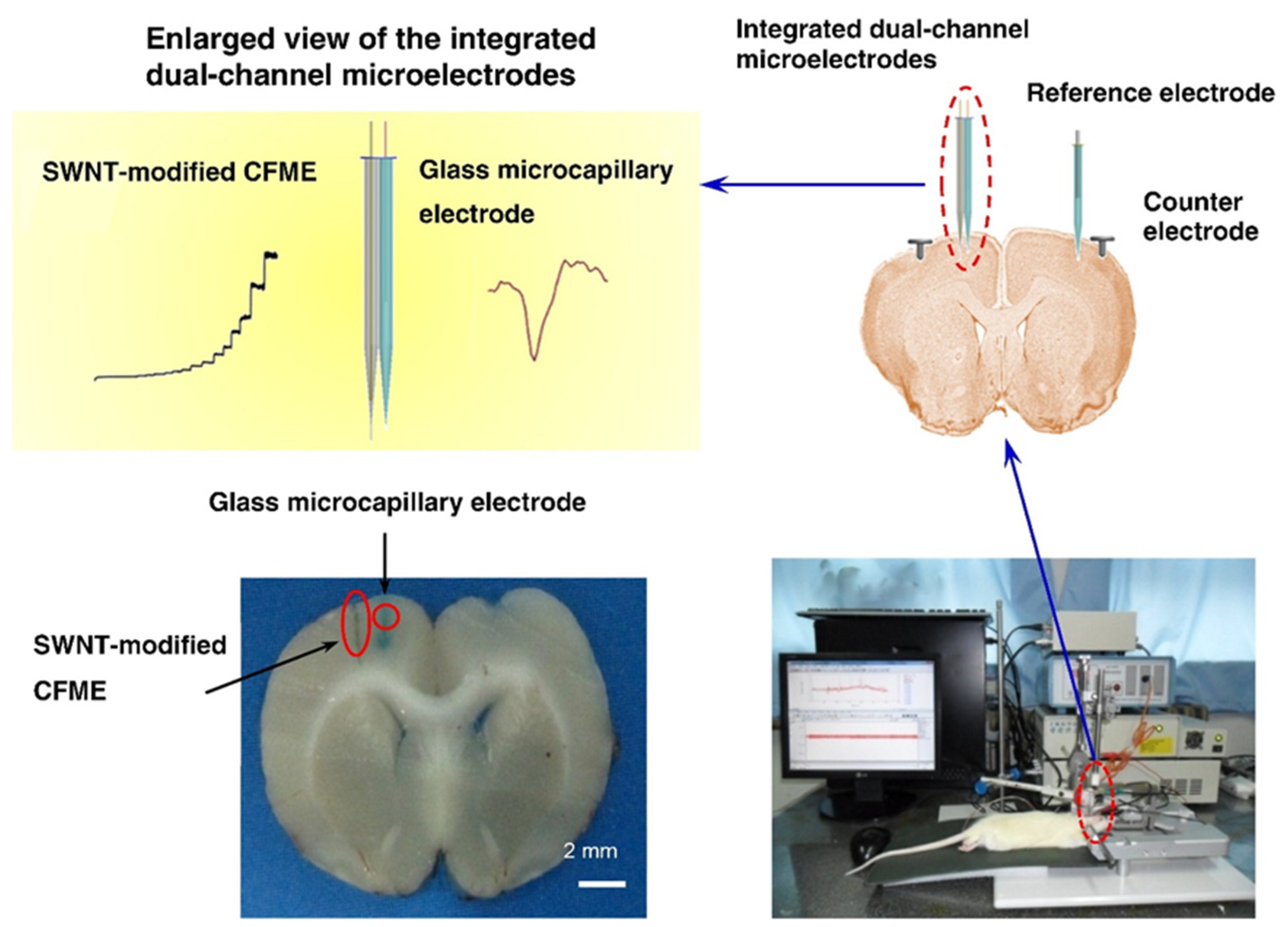
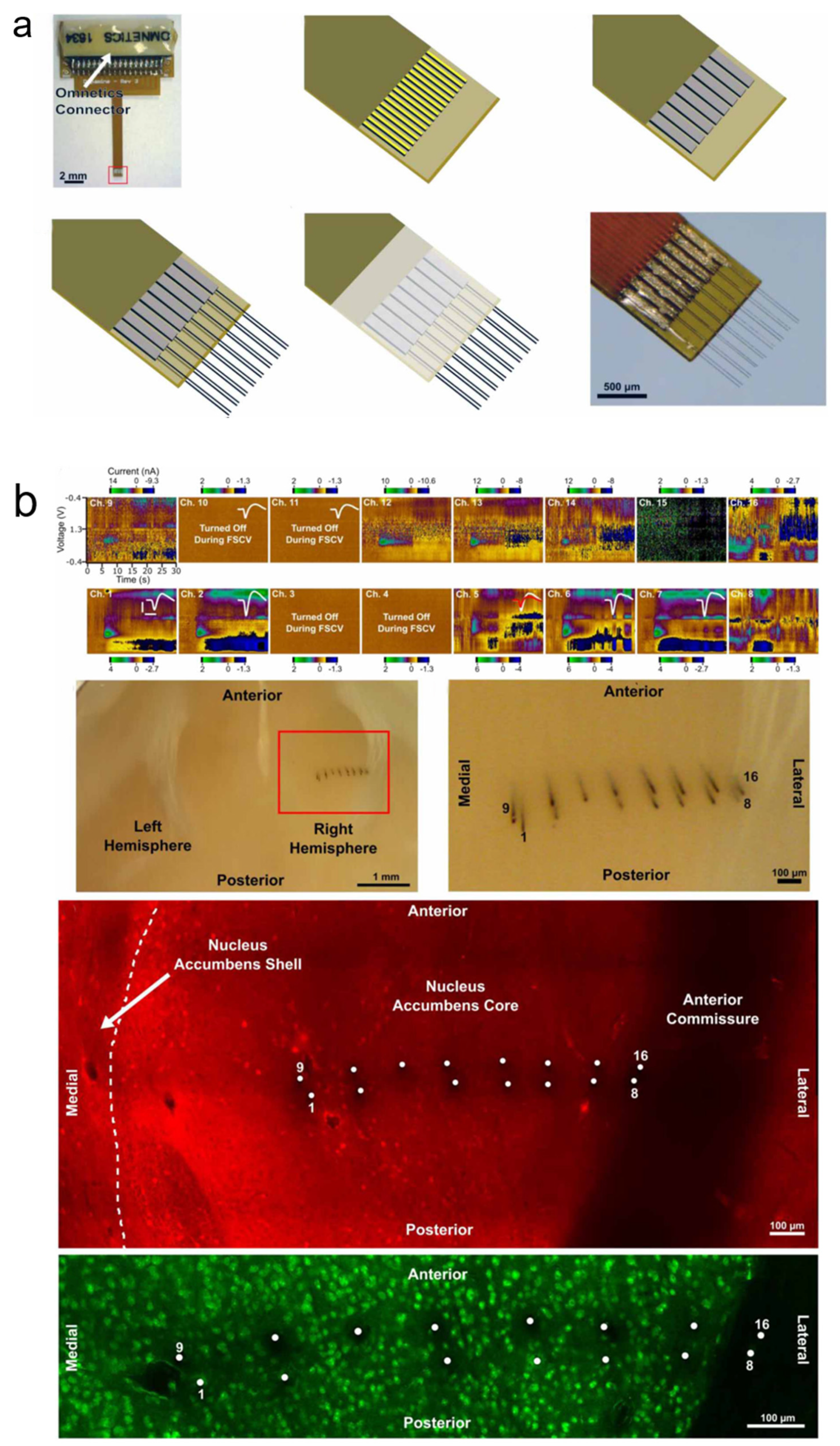
4.1.1. Carbon Fiber Microelectrodes (CFEs)
4.1.2. Graphene-Based Microelectrodes
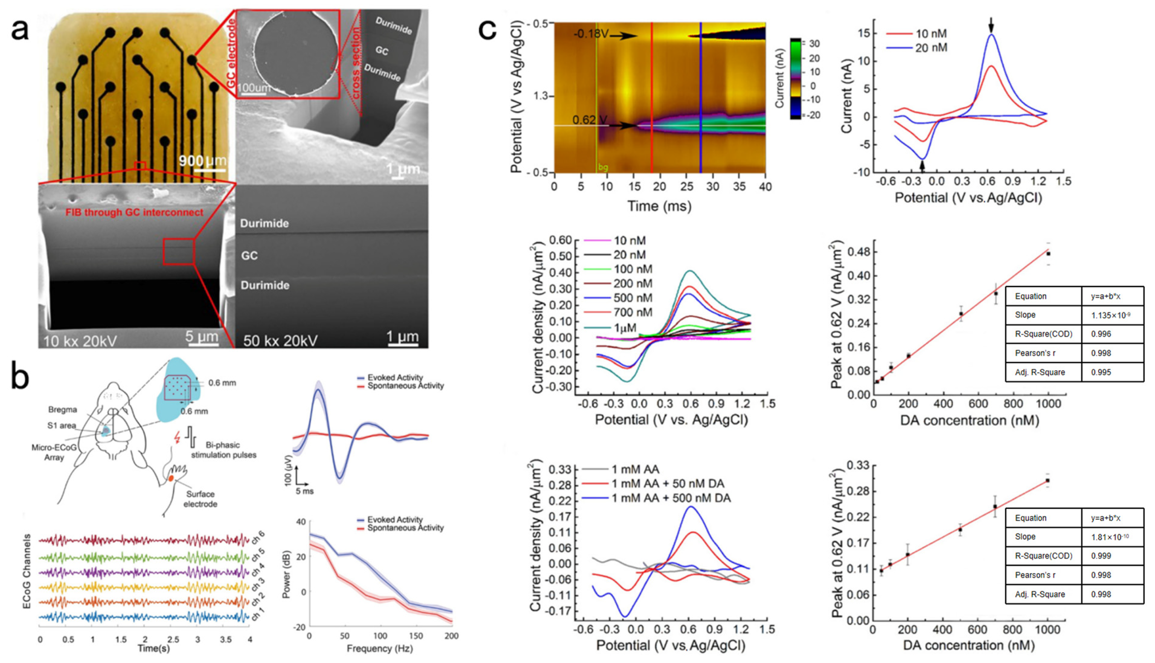
4.1.3. Glassy Carbon (GC) Microelectrodes
4.1.4. Diamond Microelectrodes
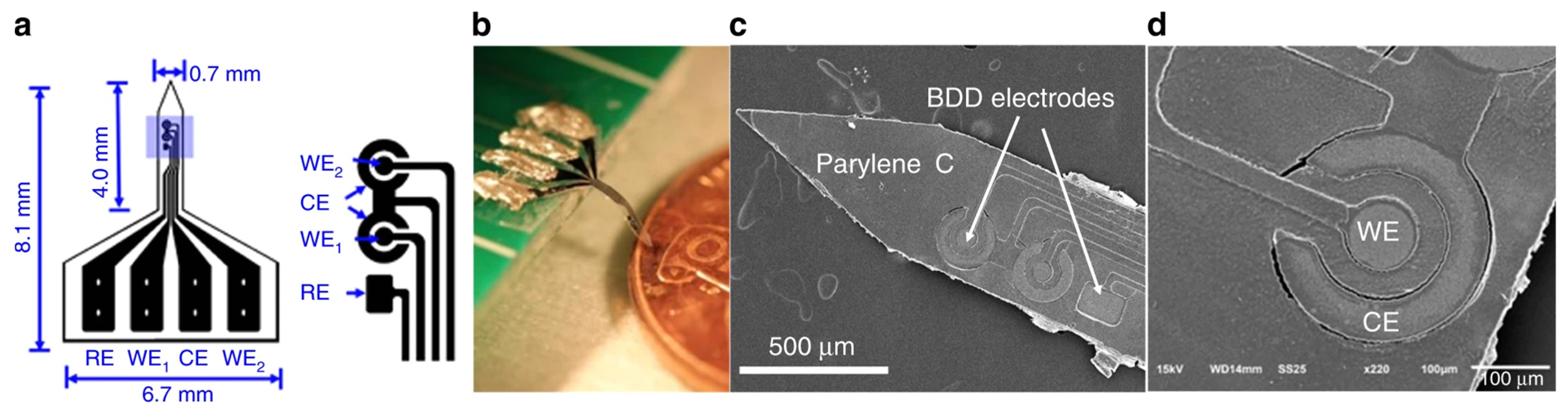
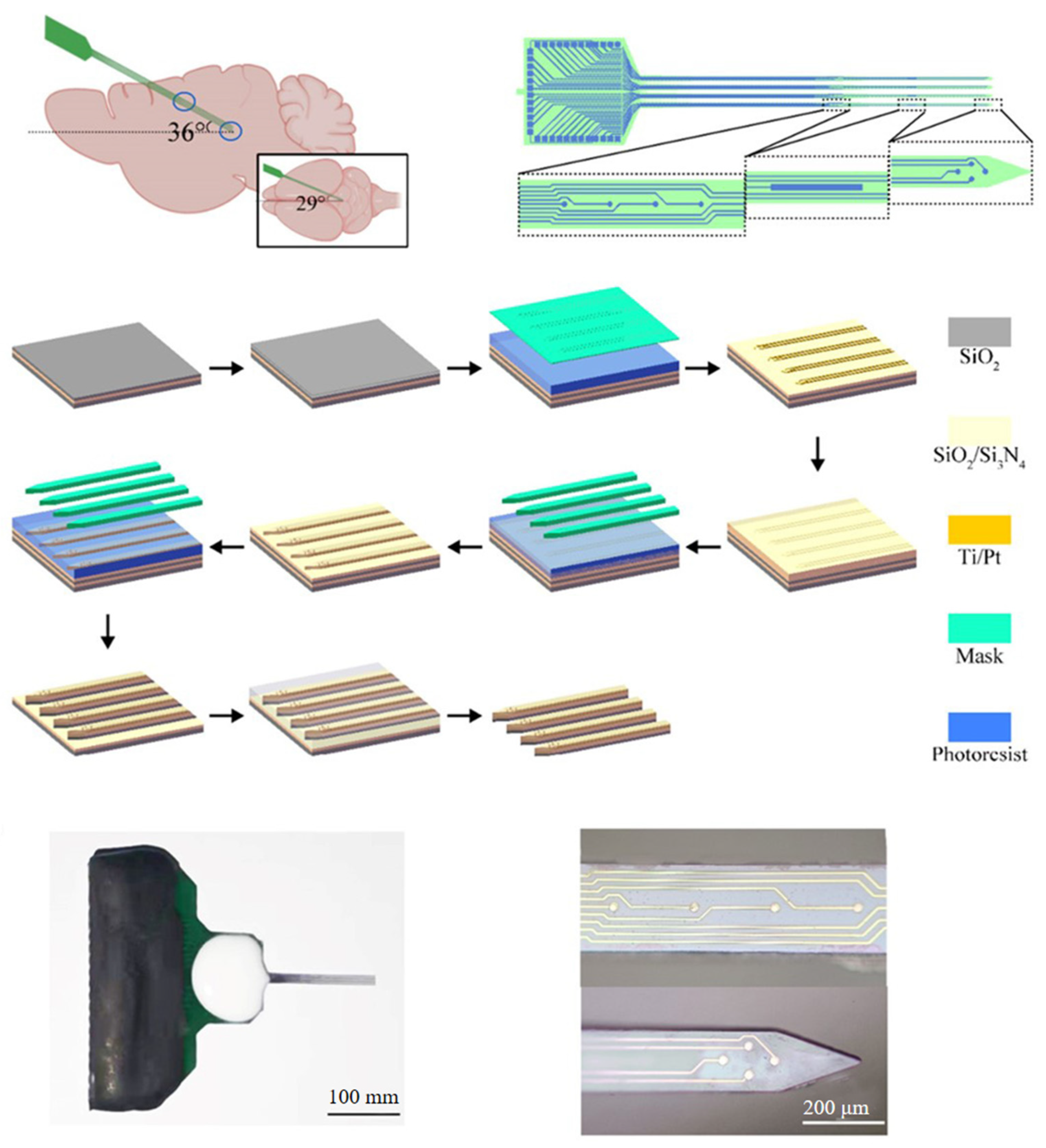
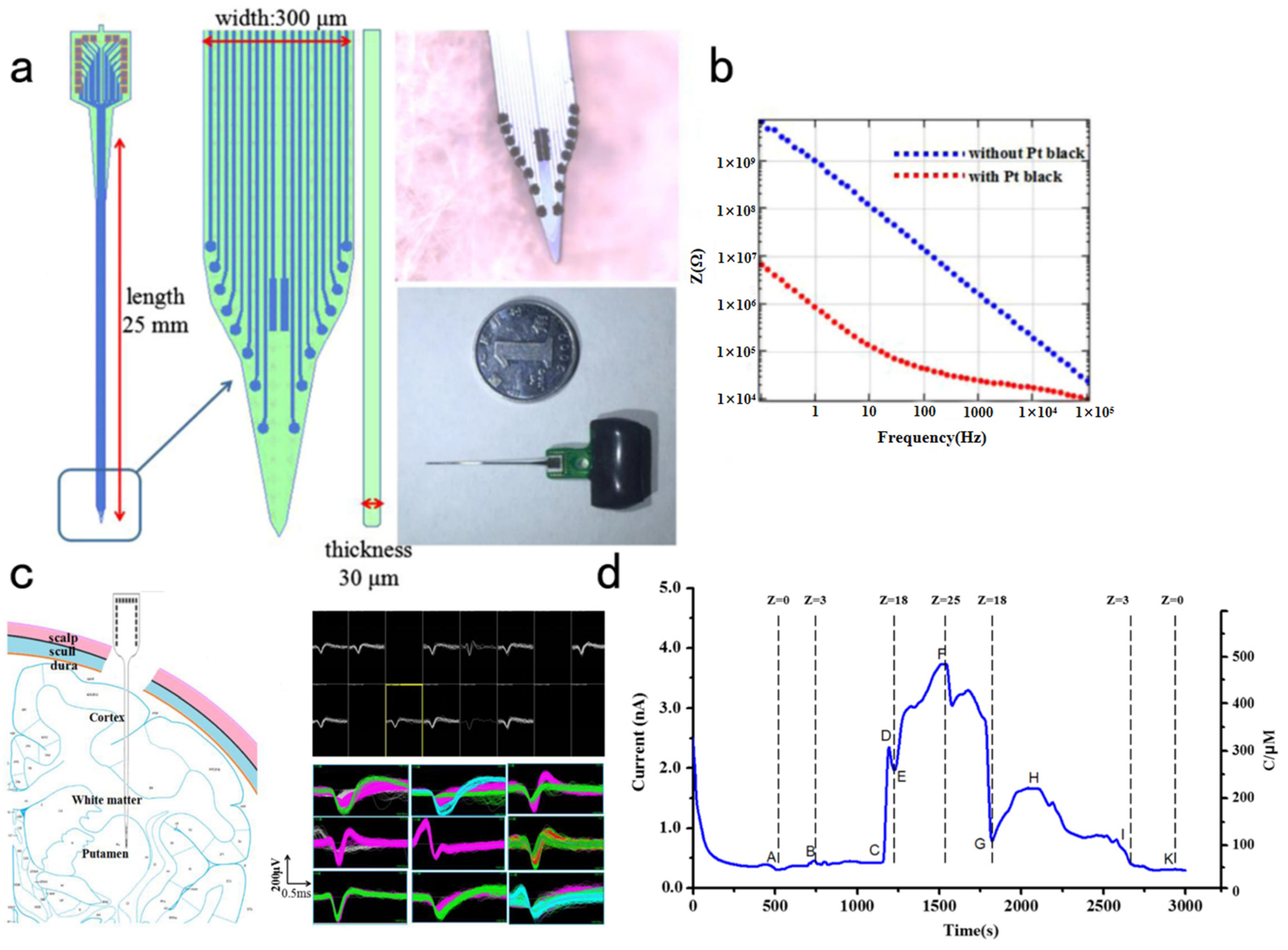
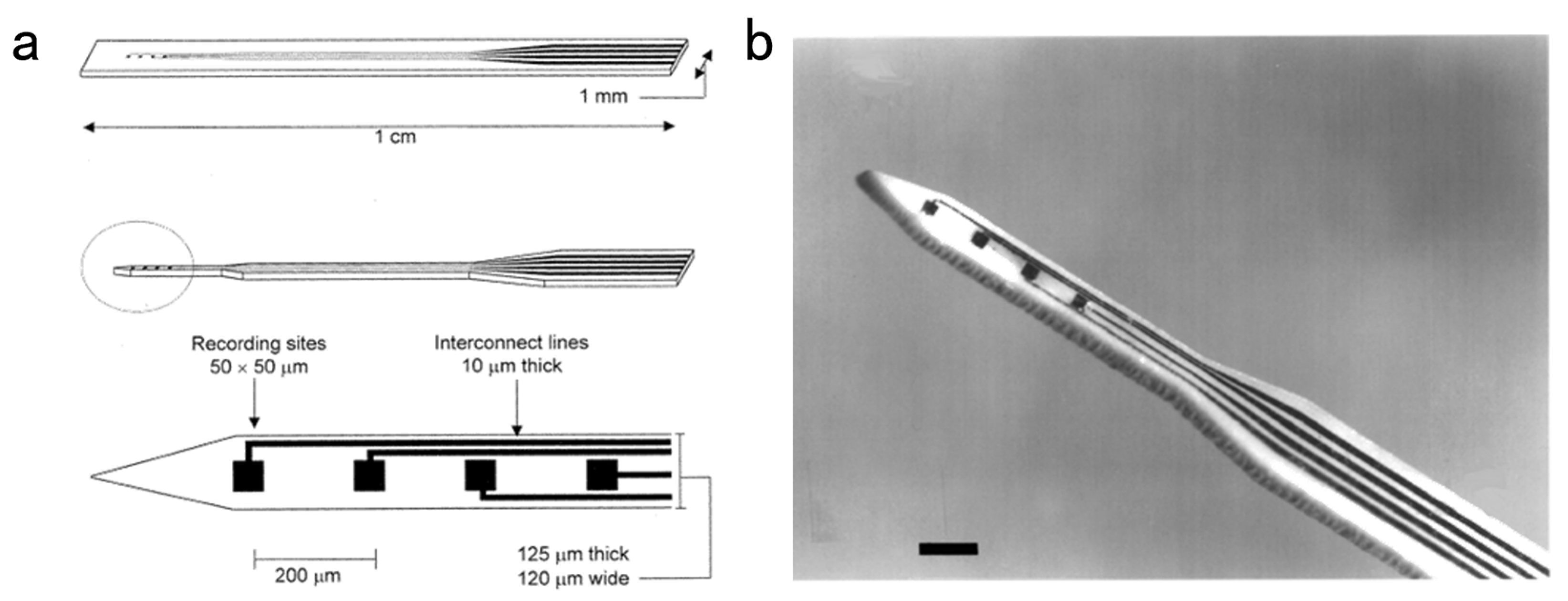
4.2. Silicon-Based Microelectrode Array
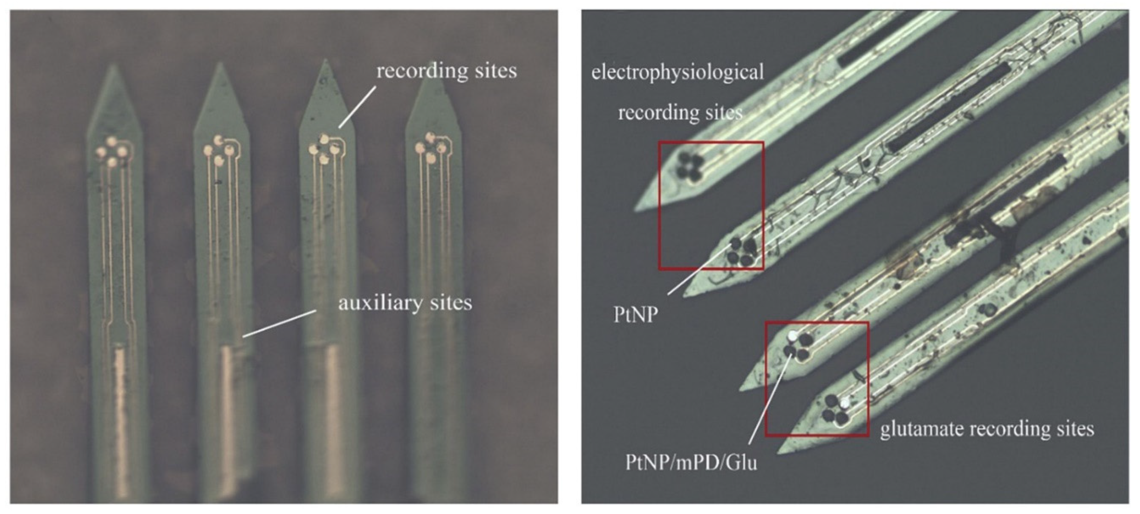
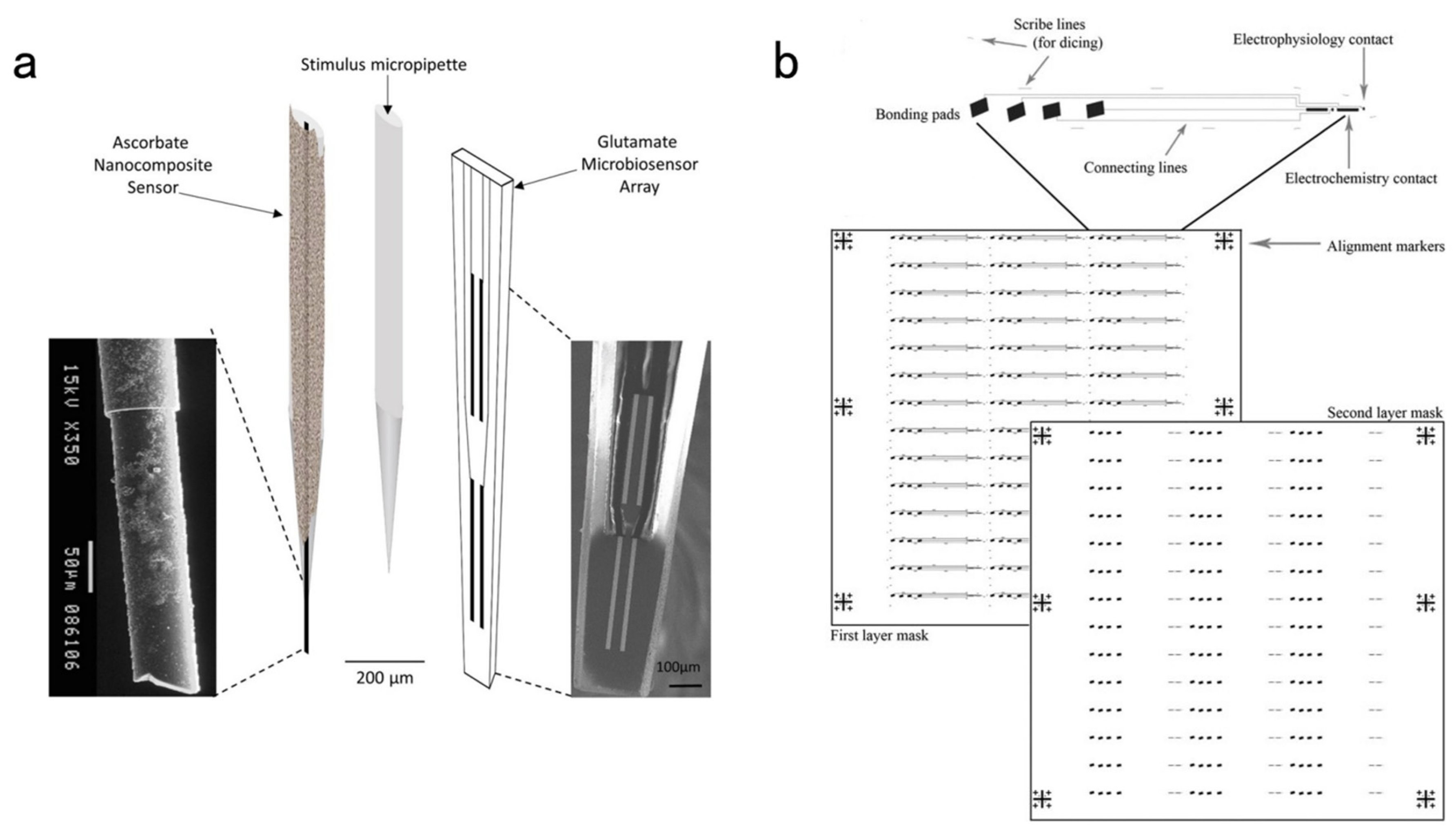
4.3. Ceramic-Based MEAs
5. Conclusions
Author Contributions
Funding
Institutional Review Board Statement
Informed Consent Statement
Data Availability Statement
Conflicts of Interest
References
- Xu, Y.; Jia, Y.; Ma, J.; Hayat, T.; Alsaedi, A. Collective responses in electrical activities of neurons under field coupling. Sci. Rep. 2018, 8, 1349. [Google Scholar] [CrossRef] [PubMed] [Green Version]
- Pereda, A.E. Electrical synapses and their functional interactions with chemical synapses. Nat. Rev. Neurosci. 2014, 15, 250–263. [Google Scholar] [CrossRef] [PubMed]
- Hormuzdi, S.G.; Filippov, M.A.; Mitropoulou, G.; Monyer, H.; Bruzzone, R. Electrical synapses: A dynamic signaling system that shapes the activity of neuronal networks. Biochim. Biophys. Acta 2004, 1662, 113–137. [Google Scholar] [CrossRef] [Green Version]
- Wu, Z.F.; Lin, D.Y.; Li, Y.L. Pushing the frontiers: Tools for monitoring neurotransmitters and neuromodulators. Nat. Rev. Neurosci. 2022, 23, 257–274. [Google Scholar] [CrossRef]
- Fisher, R.S.; Velasco, A.L. Electrical brain stimulation for epilepsy. Nat. Rev. Neurol. 2014, 10, 261–270. [Google Scholar] [CrossRef] [PubMed]
- Ju, Y.J.; Tam, K.Y. Pathological mechanisms and therapeutic strategies for Alzheimer’s disease. Neural Regen. Res. 2022, 17, 543. [Google Scholar] [PubMed]
- Li, Y.T.; Zhang, S.H.; Wang, L.; Xiao, R.R.; Liu, W.; Zhang, X.W.; Zhou, Z.; Amatore, C.; Huang, W.H. Nanoelectrode for Amperometric Monitoring of Individual Vesicular Exocytosis Inside Single Synapses. Angew. Chem. Int. Ed. 2014, 53, 12456–12460. [Google Scholar]
- Qiu, Q.F.; Zhang, F.L.; Tang, Y.; Zhang, X.W.; Jiang, H.; Liu, Y.L.; Huang, W.H. Real-time Monitoring of Exocytotic Glutamate Release from Single Neuron by Amperometry at an Enzymatic Biosensor. Electroanalysis 2018, 30, 1054–1059. [Google Scholar] [CrossRef]
- Shen, W.; Das, S.; Vitale, F.; Richardson, A.; Ananthakrishnan, A.; Struzyna, L.A.; Brown, D.P.; Song, N.; Ramkumar, M.; Lucas, T.; et al. Microfabricated intracortical extracellular matrix-microelectrodes for improving neural interfaces. Microsyst. Nanoeng. 2018, 4, 30. [Google Scholar] [CrossRef] [Green Version]
- Hong, G.; Lieber, C.M. Novel electrode technologies for neural recordings. Nat. Rev. Neurosci. 2019, 20, 330–345. [Google Scholar] [CrossRef]
- Wei, C.; Wang, Y.; Pei, W.; Han, X.; Lin, L.; Liu, Z.; Ming, G.; Chen, R.; Wu, P.; Yang, X.; et al. Distributed implantation of a flexible microelectrode array for neural recording. Microsyst. Nanoeng. 2022, 8, 50. [Google Scholar] [CrossRef] [PubMed]
- Zhang, X.W.; Hatamie, A.; Ewing, A.G. Nanoelectrochemical analysis inside a single living cell. Curr. Opin. Electrochem. 2020, 22, 94–101. [Google Scholar] [CrossRef]
- Phan, N.T.N.; Li, X.C.; Ewing, A.G. Measuring synaptic vesicles using cellular electrochemistry and nanoscale molecular imaging. Nat. Rev. Chem. 2017, 1, 0048. [Google Scholar] [CrossRef]
- Zhang, S.; Song, Y.; Wang, M.; Xiao, G.; Gao, F.; Li, Z.; Tao, G.; Zhuang, P.; Yue, F.; Chan, P.; et al. Real-time simultaneous recording of electrophysiological activities and dopamine overflow in the deep brain nuclei of a non-human primate with Parkinson’s disease using nano-based microelectrode arrays. Microsyst. Nanoeng. 2018, 4, 17070. [Google Scholar] [CrossRef] [Green Version]
- Hejazi, M.A.; Tong, W.; Stacey, A.; Soto-Breceda, A.; Ibbotson, M.R.; Yunzab, M.; Maturana, M.I.; Almasi, A.; Jung, Y.J.; Sun, S.; et al. Hybrid diamond/carbon fiber microelectrodes enable multimodal electrical/chemical neural interfacing. Biomaterials 2020, 230, 119648. [Google Scholar] [CrossRef] [PubMed]
- Venton, B.J.; Wightman, R.M. Psychoanalytical electrochemistry: Dopamine and behavior. Anal. Chem. 2003, 75, 414–421. [Google Scholar] [CrossRef] [Green Version]
- Hatamie, A.; He, X.; Zhang, X.-W.; Oomen, P.E.; Ewing, A.G. Advances in nano/microscale electrochemical sensors and biosensors for analysis of single vesicles, a key nanoscale organelle in cellular communication. Biosens. Bioelectron. 2023, 220, 114899. [Google Scholar] [CrossRef]
- Xu, C.; Wu, F.; Yu, P.; Mao, L.Q. In Vivo Electrochemical Sensors for Neurochemicals: Recent Update. ACS Sens. 2019, 4, 3102–3118. [Google Scholar] [CrossRef]
- Dale, N.; Hatz, S.; Tian, F.M.; Llaudet, E. Listening to the brain: Microelectrode biosensors for neurochemicals. Trends Biotechnol. 2005, 23, 420–428. [Google Scholar] [CrossRef]
- Deng, Z.X.; Zhao, L.J.; Mu, H.J.; Jiang, L.P.; Xi, W.Y.; Xu, X.X.; Zheng, W. High selective property of gelatin/MWCNTs functionalized carbon fiber microelectrode: Toward real-time monitoring of ascorbate. J. Electroanal. Chem. 2022, 914, 116315. [Google Scholar] [CrossRef]
- della Valle, E.; Beomseo, K.; Patel, P.R.; Whitsitt, Q.; Purcell, E.K.; Chestek, C.A.; Weiland, J.D. Electrodeposited Platinum Iridium Enables Microstimulation with Carbon Fiber Electrodes. Front. Nanotechnol. 2021, 3, 782883. [Google Scholar] [CrossRef]
- Jiman, A.A.; Ratze, D.C.; Welle, E.J.; Patel, P.R.; Richie, J.M.; Bottorff, E.C.; Seymour, J.P.; Chestek, C.A.; Bruns, T.M. Multi-channel intraneural vagus nerve recordings with a novel high-density carbon fiber microelectrode array. Sci. Rep. 2020, 10, 15501. [Google Scholar] [CrossRef] [PubMed]
- Ledo, A.; Lourenco, C.F.; Laranjinha, J.; Gerhardt, G.A.; Barbosa, R.M. Concurrent measurements of neurochemical and electrophysiological activity with microelectrode arrays: New perspectives for constant potential amperometry. Curr. Opin. Electrochem. 2018, 12, 129–140. [Google Scholar] [CrossRef]
- Wei, H.; Wu, F.; Yu, P.; Mao, L.Q. Advances in Electrochemical Biosensors for in Vivo Analysis. Chin. J. Anal. Chem. 2019, 47, 1466–1479. [Google Scholar]
- Zestos, A.G.; Rafi, H.; Ardabili, N.G. Carbon Fiber Multielectrode Arrays for Multiplexing Neurotransmitter Detection in Heterogeneous Brain Regions. FASEB J. 2022, 36, 1. [Google Scholar] [CrossRef]
- Desai, N.; Rutledge, K.M.; Caliguri, E.J. Electrochemical Measurement and Quantification of Biogenic Amine Neurotransmitters at Micro Electrode Surfaces Using In Vivo Voltammetry and Chronoamperometry; Abstracts of Papers of the American Chemical Society; American Chemical Society: Washington, DC, USA, 2012; Volume 243. [Google Scholar]
- Nasr, B.; Chatterton, R.; Yong, J.H.M.; Jamshidi, P.; D’Abaco, G.M.; Bjorksten, A.R.; Kavehei, O.; Chana, G.; Dottori, M.; Skafidas, E. Self-Organized Nanostructure Modified Microelectrode for Sensitive Electrochemical Glutamate Detection in Stem Cells-Derived Brain Organoids. Biosensors 2018, 8, 14. [Google Scholar] [CrossRef] [Green Version]
- Burmeister, J.J.; Gerhardt, G.A. Self-Referencing Ceramic-Based Multisite Microelectrodes for the Detection and Elimination of Interferences from the Measurement of l-Glutamate and Other Analytes. Anal. Chem. 2001, 73, 1037–1042. [Google Scholar] [CrossRef]
- Johnson, J.A.; Wightmanabout, R.M. Cyclic Voltammetric Measurements of Neurotransmitters. Electrochem. Soc. Interface 2017, 26, 53–57. [Google Scholar] [CrossRef] [Green Version]
- Castagnola, E.; Thongpang, S.; Hirabayashi, M.; Nava, G.; Nimbalkar, S.; Nguyen, T.; Lara, S.; Oyawale, A.; Bunnell, J.; Moritz, C.; et al. Glassy carbon microelectrode arrays enable voltage-peak separated simultaneous detection of dopamine and serotonin using fast scan cyclic voltammetry. Analyst 2021, 146, 3955–3970. [Google Scholar] [CrossRef]
- Xiao, T.F.; Wu, F.; Hao, J.; Zhang, M.N.; Yu, P.; Mao, L.Q. In Vivo Analysis with Electrochemical Sensors and Biosensors. Anal. Chem. 2017, 89, 300–313. [Google Scholar] [CrossRef]
- Monteiro, T.; Dias, C.; Lourenco, C.F.; Ledo, A.; Barbosa, R.M.; Almeida, M.G. Microelectrode Sensor for Real-Time Measurements of Nitrite in the Living Brain, in the Presence of Ascorbate. Biosensors 2021, 11, 277. [Google Scholar] [CrossRef] [PubMed]
- Thielen, B.; Meng, E.L. A comparison of insertion methods for surgical placement of penetrating neural interfaces. J. Neural Eng. 2021, 18, 041003. [Google Scholar] [CrossRef] [PubMed]
- Strumwasser, F. Long-Term Recording from Single Neurons in Brain of Unrestrained Mammals. Science 1958, 127, 469–470. [Google Scholar] [CrossRef] [PubMed]
- Campbell, P.K.; Jones, K.E.; Huber, R.J.; Horch, K.W.; Normann, R.A. A Silicon-Based, Three-Dimensional Neural Interface: Manufacturing Processes for an Intracortical Electrode Array. IEEE Trans. Biomed. Eng. 1991, 38, 758–768. [Google Scholar] [CrossRef]
- Wise, K.D.; Angell, J.B.; Starr, A. An Integrated-Circuit Approach to Extracellular Microelectrodes. IEEE Trans. Biomed. Eng. 1970, BME-17, 238–247. [Google Scholar] [CrossRef] [Green Version]
- Sui, Y.A.; Tian, Y.; Ko, W.K.D.; Wang, Z.Y.; Jia, F.M.; Horn, A.; De Ridder, D.; Choi, K.S.; Bari, A.A.; Wang, S.Y.; et al. Deep Brain Stimulation Initiative: Toward Innovative Technology, New Disease Indications, and Approaches to Current and Future Clinical Challenges in Neuromodulation Therapy. Front. Neurol. 2021, 11, 1706. [Google Scholar] [CrossRef]
- Delgado, J.M.R.; Mark, V.; Sweet, W.; Ervin, F.; Weiss, G.; Bachyrit, G.; Hagiwara, R. Intracerebral radio stimulation and recording in completely free patients. J. Nerv. Ment. Dis. 1968, 147, 329. [Google Scholar] [CrossRef]
- McNaughton, B.L.; Barnes, C.A.; O’Keefe, J. The contributions of position, direction, and velocity to single unit activity in the hippocampus of freely-moving rats. Exp. Brain Res. 1983, 52, 41–49. [Google Scholar] [CrossRef]
- Liu, J.; Fu, T.-M.; Cheng, Z.; Hong, G.; Zhou, T.; Jin, L.; Duvvuri, M.; Jiang, Z.; Kruskal, P.; Xie, C.; et al. Syringe-injectable electronics. Nat. Nanotechnol. 2015, 10, 629–636. [Google Scholar] [CrossRef] [Green Version]
- Khodagholy, D.; Gelinas, J.N.; Thesen, T.; Doyle, W.; Devinsky, O.; Malliaras, G.G.; Buzsáki, G. NeuroGrid: Recording action potentials from the surface of the brain. Nat. Neurosci. 2015, 18, 310–315. [Google Scholar] [CrossRef] [Green Version]
- Rios, G.; Lubenov, E.V.; Chi, D.; Roukes, M.L.; Siapas, A.G. Nanofabricated Neural Probes for Dense 3-D Recordings of Brain Activity. Nano Lett. 2016, 16, 6857–6862. [Google Scholar] [CrossRef]
- Jun, J.J.; Steinmetz, N.A.; Siegle, J.H.; Denman, D.J.; Bauza, M.; Barbarits, B.; Lee, A.K.; Anastassiou, C.A.; Andrei, A.; Aydın, Ç.; et al. Fully integrated silicon probes for high-density recording of neural activity. Nature 2017, 551, 232–236. [Google Scholar] [CrossRef]
- Guan, S.; Wang, J.; Gu, X.; Zhao, Y.; Hou, R.; Fan, H.; Zou, L.; Gao, L.; Du, M.; Li, C.; et al. Elastocapillary self-assembled neurotassels for stable neural activity recordings. Sci. Adv. 2019, 5, eaav2842. [Google Scholar] [CrossRef] [Green Version]
- Lane, R.F.; Hubbard, A.T.; Fukunaga, K.; Blanchard, R.J. Brain catecholamines-detection invivo by means of differential pulse voltammetry at surface-modified platinum-electrodes. Brain Res. 1976, 114, 346–352. [Google Scholar] [CrossRef] [PubMed]
- Gonon, F.; Buda, M.; Cespuglio, R.; Jouvet, M.; Pujol, J.F. In vivo electrochemical detection of catechols in the neostriatum of anesthetized rats-dopamine or dopac. Nature 1980, 286, 902–904. [Google Scholar] [CrossRef] [PubMed]
- Chung, T.; Wang, J.Q.; Wang, J.; Cao, B.; Li, Y.; Pang, S.W. Electrode modifications to lower electrode impedance and improve neural signal recording sensitivity. J. Neural Eng. 2015, 12, 056018. [Google Scholar] [CrossRef] [PubMed]
- Leber, M.; Bhandari, R.; Mize, J.; Warren, D.J.; Shandhi, M.M.H.; Solzbacher, F.; Negi, S. Long term performance of porous platinum coated neural electrodes. Biomed. Microdevices 2017, 19, 62. [Google Scholar] [CrossRef]
- Liu, B.; Lu, Y.; Wang, D.; Li, T.; Zhou, Q.; Cao, Y.; Duan, Y. Modification of implantable Pt-Ir alloy microelectrode and impedance monitoring in vivo. J. Wuhan Univ. 2009, 55, 253–257. [Google Scholar]
- Vafaiee, M.; Mohammadpour, R.; Vossoughi, M.; Asadian, E.; Janahmadi, M.; Sasanpour, P. Carbon Nanotube Modified Microelectrode Array for Neural Interface. Front. Bioeng. Biotechnol. 2021, 8, 582713. [Google Scholar] [CrossRef]
- Ryu, M.; Yang, J.H.; Ahn, Y.; Sim, M.; Lee, K.H.; Kim, K.; Lee, T.; Yoo, S.J.; Kim, S.Y.; Moon, C.; et al. Enhancement of Interface Characteristics of Neural Probe Based on Graphene, ZnO Nanowires, and Conducting Polymer PEDOT. ACS Appl. Mater. Interfaces 2017, 9, 10577–10586. [Google Scholar] [CrossRef]
- Wu, D.; Chen, X.Y.; Chen, T.C.; Ding, C.M.; Wu, W.; Li, J.S. Substrate-anchored and degradation-sensitive anti-inflammatory coatings for implant materials. Sci. Rep. 2015, 5, 11105. [Google Scholar] [CrossRef] [PubMed] [Green Version]
- Winter, J.O.; Han, N.; Jensen, R.; Cogan, S.F.; Rizzo, J.F. Adhesion Molecules Promote Chronic Neural Interfaces Following Neurotrophin Withdrawal. In Proceedings of the 2009 Annual International Conference of the IEEE Engineering in Medicine and Biology Society, Minneapolis, MN, USA, 3–6 September 2009; p. 7151. [Google Scholar]
- Yu, K.; Hou, J.; Jin, Z.; Wu, K.; Xu, S.; Yang, N.; Shen, Y.; Tang, T.; Guo, S. A cochlear implant loaded with dexamethasone and coated with hyaluronic acid to inhibit fibroblast adhesion and proliferation. J. Drug Deliv. Sci. Technol. 2018, 46, 173–181. [Google Scholar] [CrossRef]
- Patil, A.C.; Thakor, N.V. Implantable neurotechnologies: A review of micro- and nanoelectrodes for neural recording. Med. Biol. Eng. Comput. 2016, 54, 23–44. [Google Scholar] [CrossRef]
- Salatino, J.W.; Ludwig, K.A.; Kozai, T.D.Y.; Purcell, E.K. Glial responses to implanted electrodes in the brain. Nat. Biomed. Eng. 2017, 1, 862–877. [Google Scholar] [CrossRef] [PubMed]
- Tian, H.-C.; Liu, J.-Q.; Kang, X.-Y.; Tang, L.-J.; Wang, M.-H.; Ji, B.-W.; Yang, B.; Wang, X.-L.; Chen, X.; Yang, C.-S. Enhanced Flexible Tubular Microelectrode with Conducting Polymer for Multi-Functional Implantable Tissue-Machine Interface. Sci. Rep. 2016, 6, 26910. [Google Scholar] [CrossRef] [PubMed] [Green Version]
- Spencer, K.C.; Sy, J.C.; Ramadi, K.B.; Graybiel, A.M.; Langer, R.; Cima, M.J. Characterization of Mechanically Matched Hydrogel Coatings to Improve the Biocompatibility of Neural Implants. Sci. Rep. 2017, 7, 1952. [Google Scholar] [CrossRef] [PubMed] [Green Version]
- Filho, G.; Junior, C.; Spinelli, B.; Damasceno, I.; Fiuza, F.; Morya, E. All-Polymeric Electrode Based on PEDOT:PSS for In Vivo Neural Recording. Biosensors 2022, 12, 853. [Google Scholar] [CrossRef]
- Wei, W.; Hao, M.M.; Zhou, K.; Wang, Y.F.; Lu, Q.F.; Zhang, H.; Wu, Y.; Zhang, T.; Liu, Y.B. In situ multimodal transparent electrophysiological hydrogel for in vivo miniature two-photon neuroimaging and electrocorticogram analysis. Acta Biomater. 2022, 152, 86–99. [Google Scholar] [CrossRef]
- Heo, D.N.; Kim, H.J.; Lee, Y.J.; Heo, M.; Lee, S.J.; Lee, D.; Do, S.H.; Lee, S.H.; Kwon, I.K. Flexible and Highly Biocompatible Nanofiber-Based Electrodes for Neural Surface Interfacing. ACS Nano 2017, 11, 2961–2971. [Google Scholar] [CrossRef]
- Yan, D.X.; Jiman, A.A.; Bottorff, E.C.; Patel, P.R.; Meli, D.; Welle, E.J.; Ratze, D.C.; Havton, L.A.; Chestek, C.A.; Kemp, S.W.P.; et al. Ultraflexible and Stretchable Intrafascicular Peripheral Nerve Recording Device with Axon-Dimension, Cuff-Less Microneedle Electrode Array. Small 2022, 18, 2200311. [Google Scholar] [CrossRef]
- Abidian, M.R.; Ludwig, K.A.; Marzullo, T.C.; Martin, D.C.; Kipke, D.R. Interfacing Conducting Polymer Nanotubes with the Central Nervous System: Chronic Neural Recording using Poly (3,4-ethylenedioxythiophene) Nanotubes. Adv. Mater. 2009, 21, 3764–3770. [Google Scholar] [CrossRef] [PubMed] [Green Version]
- Gerwig, R.; Fuchsberger, K.; Schroeppel, B.; Link, G.S.; Heusel, G.; Kraushaar, U.; Schuhmann, W.; Stett, A.; Stelzle, M. PEDOT-CNT Composite Microelectrodes for Recording and Electrostimulation Applications: Fabrication, Morphology, and Electrical Properties. Front. Neuroeng. 2012, 5, 8. [Google Scholar] [CrossRef] [PubMed] [Green Version]
- Yang, J.Y.; Martin, D.C. Microporous conducting polymers on neural microelectrode arrays II. Physical characterization. Sens. Actuators A-Phys. 2004, 113, 204–211. [Google Scholar] [CrossRef]
- Abidian, M.R.; Martin, D.C. Experimental and theoretical characterization of implantable neural microelectrodes modified with conducting polymer nanotubes. Biomaterials 2008, 29, 1273–1283. [Google Scholar] [CrossRef] [Green Version]
- Cui, X.Y.; Wiler, J.; Dzaman, M.; Altschuler, R.A.; Martin, D.C. In vivo studies of polypyrrole/peptide coated neural probes. Biomaterials 2003, 24, 777–787. [Google Scholar] [CrossRef]
- Qi, D.; Liu, Z.; Liu, Y.; Jiang, Y.; Leow, W.R.; Pal, M.; Pan, S.; Yang, H.; Wang, Y.; Zhang, X.; et al. Highly Stretchable, Compliant, Polymeric Microelectrode Arrays for In Vivo Electrophysiological Interfacing. Adv. Mater. 2017, 29, 1702800. [Google Scholar] [CrossRef]
- Deng, M.; Yang, X.; Silke, M.; Qiu, W.; Xu, M.; Borghs, G.; Chen, H. Electrochemical deposition of polypyrrole/graphene oxide composite on microelectrodes towards tuning the electrochemical properties of neural probes. Sens. Actuators B-Chem. 2011, 158, 176–184. [Google Scholar] [CrossRef]
- Cui, X.Y.; Martin, D.C. Fuzzy gold electrodes for lowering impedance and improving adhesion with electrodeposited conducting polymer films. Sens. Actuators A-Phys. 2003, 103, 384–394. [Google Scholar] [CrossRef]
- Brahim, S.; Wilson, A.M.; Narinesingh, D.; Iwuoha, E.; Guiseppi-Elie, A. Chemical and Biological Sensors Based on Electrochemical Detection Using Conducting Electroactive Polymers. Microchim. Acta 2003, 143, 123–137. [Google Scholar] [CrossRef]
- Zhao, Z.; Gong, R.; Zheng, L.; Wang, J. In Vivo Neural Recording and Electrochemical Performance of Microelectrode Arrays Modified by Rough-Surfaced AuPt Alloy Nanoparticles with Nanoporosity. Sensors 2016, 16, 1851. [Google Scholar] [CrossRef] [Green Version]
- Kim, Y.H.; Kim, A.Y.; Kim, G.H.; Han, Y.H.; Chung, M.-A.; Jung, S.-D. Electrochemical and in vitro neuronal recording characteristics of multi-electrode arrays surface-modified with electro-co-deposited gold-platinum nanoparticles. Biomed. Microdevices 2016, 18, 14. [Google Scholar] [CrossRef] [PubMed]
- Rose, T.L.; Robblee, L.S. Electrical-stimulation with pt electrodes.8. electrochemically safe charge injection limits with 0.2 ms pulses. IEEE Trans. Biomed. Eng. 1990, 37, 1118–1120. [Google Scholar] [CrossRef] [PubMed]
- Keefer, E.W.; Botterman, B.R.; Romero, M.I.; Rossi, A.F.; Gross, G.W. Carbon nanotube coating improves neuronal recordings. Nat. Nanotechnol. 2008, 3, 434–439. [Google Scholar] [CrossRef] [PubMed]
- Patan, M.; Shah, T.; Sahin, M. Charge injection capacity of TiN electrodes for an extended voltage range. In Proceedings of the 2006 International Conference of the IEEE Engineering in Medicine and Biology Society, New York, NY, USA, 30 August–3 September 2006; p. 3. [Google Scholar]
- Rodrigues, F.; Ribeiro, J.F.; Anacleto, P.A.; Fouchard, A.; David, O.; Sarro, P.M.; Mendes, P.M. Fabrication and characterization of polyimide-based ‘smooth’ titanium nitride microelectrode arrays for neural stimulation and recording. J. Neural Eng. 2020, 17, 016010. [Google Scholar] [CrossRef] [PubMed]
- Apollo, N.V.; Maturana, M.I.; Tong, W.; Nayagam, D.A.X.; Shivdasani, M.N.; Foroughi, J.; Wallace, G.G.; Prawer, S.; Ibbotson, M.R.; Garrett, D.J. Soft, Flexible Freestanding Neural Stimulation and Recording Electrodes Fabricated from Reduced Graphene Oxide. Adv. Funct. Mater. 2015, 25, 3551–3559. [Google Scholar] [CrossRef] [Green Version]
- Nimbalkar, S.; Castagnola, E.; Balasubramani, A.; Scarpellini, A.; Samejima, S.; Khorasani, A.; Boissenin, A.; Thongpang, S.; Moritz, C.; Kassegne, S. Ultra-Capacitive Carbon Neural Probe Allows Simultaneous Long-Term Electrical Stimulations and High-Resolution Neurotransmitter Detection. Sci. Rep. 2018, 8, 6958. [Google Scholar] [CrossRef] [Green Version]
- Huffman, M.L.; Venton, B.J. Carbon-fiber microelectrodes for in vivo applications. Analyst 2009, 134, 18–24. [Google Scholar] [CrossRef] [Green Version]
- Dias, C.; Fernandes, E.; Barbosa, R.M.; Ledo, A. A Platinized Carbon Fiber Microelectrode-Based Oxidase Biosensor for Amperometric Monitoring of Lactate in Brain Slices. Sensors 2022, 22, 7011. [Google Scholar] [CrossRef]
- Xiang, L.; Yu, P.; Hao, J.; Zhang, M.N.; Zhu, L.; Dai, L.M.; Mao, L.Q. Vertically Aligned Carbon Nanotube-Sheathed Carbon Fibers as Pristine Microelectrodes for Selective Monitoring of Ascorbate in Vivo. Anal. Chem. 2014, 86, 3909–3914. [Google Scholar] [CrossRef]
- Xiang, L.; Yu, P.; Zhang, M.N.; Hao, J.; Wang, Y.X.; Zhu, L.; Dai, L.M.; Mao, L.Q. Platinized Aligned Carbon Nanotube-Sheathed Carbon Fiber Microelectrodes for In Vivo Amperometric Monitoring of Oxygen. Anal. Chem. 2014, 86, 5017–5023. [Google Scholar] [CrossRef]
- Wei, H.; Wu, F.; Li, L.J.; Yang, X.T.; Xu, C.; Yu, P.; Ma, F.R.; Mao, L.Q. Natural Leukocyte Membrane-Masked Microelectrodes with an Enhanced Antifouling Ability and Biocompatibility for In Vivo Electrochemical Sensing. Anal. Chem. 2020, 92, 11374–11379. [Google Scholar] [CrossRef] [PubMed]
- Hou, H.F.; Jin, Y.; Wei, H.; Ji, W.L.; Xue, Y.F.; Hu, J.B.; Zhang, M.N.; Jiang, Y.; Mao, L.Q. A Generalizable and Noncovalent Strategy for Interfacing Aptamers with a Microelectrode for the Selective Sensing of Neurotransmitters In Vivo. Angew. Chem. Int. Ed. 2020, 59, 18996–19000. [Google Scholar] [CrossRef] [PubMed]
- Regiart, M.; Ledo, A.; Fernandes, E.; Messina, G.A.; Brett, C.M.A.; Bertotti, M.M.; Barbosa, R. Highly sensitive and selective nanostructured microbiosensors for glucose and lactate simultaneous measurements in blood serum and in vivo in brain tissue. Biosens. Bioelectron. 2022, 199, 113874. [Google Scholar] [CrossRef] [PubMed]
- Guitchounts, G.; Cox, D. 64-Channel Carbon Fiber Electrode Arrays for Chronic Electrophysiology. Sci. Rep. 2020, 10, 3830. [Google Scholar] [CrossRef] [PubMed] [Green Version]
- Cheng, H.J.; Xiao, T.F.; Wang, D.L.; Hao, J.; Yu, P.; Mao, L.Q. Simultaneous in vivo ascorbate and electrophysiological recordings in rat brain following ischemia/reperfusion. J. Electroanal. Chem. 2016, 781, 90–96. [Google Scholar] [CrossRef]
- Patel, P.R.; Popov, P.; Caldwell, C.M.; Welle, E.J.; Egert, D.; Pettibone, J.R.; Roossien, D.H.; Becker, J.B.; Berke, J.D.; Chestek, C.A.; et al. High density carbon fiber arrays for chronic electrophysiology, fast scan cyclic voltammetry, and correlative anatomy. J. Neural Eng. 2020, 17, 056029. [Google Scholar] [CrossRef]
- Wei, W.C.; Wang, X.J. Graphene-Based Electrode Materials for Neural Activity Detection. Materials 2021, 14, 6170. [Google Scholar] [CrossRef]
- Gonzalez-Gonzalez, M.A.; Bendale, G.S.; Wang, K.; Wallace, G.G.; Romero-Ortega, M. Platinized graphene fiber electrodes uncover direct spleen-vagus communication. Commun. Biol. 2021, 4, 1097. [Google Scholar] [CrossRef]
- Lu, Y.; Lyu, H.; Richardson, A.G.; Lucas, T.H.; Kuzum, D. Flexible Neural Electrode Array Based-on Porous Graphene for Cortical Microstimulation and Sensing. Sci. Rep. 2016, 6, 33526. [Google Scholar] [CrossRef]
- Li, J.X.; Liu, Y.X.; Yuan, L.; Zhang, B.B.; Bishop, E.S.; Wang, K.C.; Tang, J.; Zheng, Y.Q.; Xu, W.H.; Niu, S.M.; et al. A tissue-like neurotransmitter sensor for the brain and gut. Nature 2022, 606, 94. [Google Scholar] [CrossRef]
- Castagnola, E.; Vahidi, N.W.; Nimbalkar, S.; Rudraraju, S.; Thielk, M.; Zucchini, E.; Cea, C.; Carli, S.; Gentner, T.Q.; Ricci, D.; et al. In Vivo Dopamine Detection and Single Unit Recordings Using Intracortical Glassy Carbon Microelectrode Arrays. MRS Adv. 2018, 3, 1629–1634. [Google Scholar] [CrossRef] [PubMed]
- Ogata, G.; Ishii, Y.; Asai, K.; Sano, Y.; Nin, F.; Yoshida, T.; Higuchi, T.; Sawamura, S.; Ota, T.; Hori, K.; et al. A microsensing system for the in vivo real-time detection of local drug kinetics. Nat. Biomed. Eng. 2017, 1, 654–666. [Google Scholar] [CrossRef] [PubMed]
- Varney, M.W.; Zongliang, C.; Aslam, D.M. All-diamond micro-electrode arrays for neural recordings and diamond electrochemistry. In Proceedings of the 2010 5th IEEE International Conference on Nano/Micro Engineered and Molecular Systems, Xiamen, China, 20–23 January 2010; pp. 1116–1119. [Google Scholar]
- Chan, H.Y.; Aslam, D.M.; Wiler, J.A.; Casey, B. A Novel Diamond Microprobe for Neuro-Chemical and -Electrical Recording in Neural Prosthesis. J. Microelectromechanical Syst. 2009, 18, 511–521. [Google Scholar] [CrossRef]
- Lu, B.T.; Fan, P.H.; Wang, Y.D.; Dai, Y.C.; Xie, J.Y.; Yang, G.C.; Mo, F.; Xu, Z.J.; Song, Y.L.; Liu, J.T.; et al. Neuronal Electrophysiological Activities Detection of Defense Behaviors Using an Implantable Microelectrode Array in the Dorsal Periaqueductal Gray. Biosensors 2022, 12, 193. [Google Scholar] [CrossRef]
- Wang, Y.; Wang, M.X.; Dai, Y.C.A.; Song, Y.L.; Wang, Y.D.; Lu, B.T.; Li, Y.H.; Cai, X.X. PtNPs/Short MWCNT-PEDOT: PSS-Modified Microelectrode Array to Detect Neuronal Firing Patterns in the Dorsal Raphe Nucleus and Hippocampus of Insomnia Rats. Micromachines 2022, 13, 488. [Google Scholar] [CrossRef]
- Xu, H.; Scholten, K.; Jiang, W.; Ortigoza-Diaz, J.-L.; Lu, Z.; Liu, X.; Meng, E.; Song, D. Acute in vivo Recording with a Generic Parylene Microelectrode Array Implanted with Dip-coating Method into the Rat Brain. Annu. Int. Conf. IEEE Eng. Med. Biol. Soc. 2022, 2022, 214–217. [Google Scholar]
- Wu, Y.-b.; Hou, A.-z.; Ni, H.-n.; Xu, A.-l.; Hui, C.; Ren, Q.-s. Study of microfabrication process of flexible microelectrodes arrays based on parylene. Semicond. Technol. 2007, 32, 1018–1020,1036. [Google Scholar]
- Mo, F.; Xu, Z.; Yang, G.; Fan, P.; Wang, Y.; Lu, B.; Liu, J.; Wang, M.; Jing, L.; Xu, W.; et al. Single-neuron detection of place cells remapping in short-term memory using motion microelectrode arrays. Biosens. Bioelectron. 2022, 217, 114726. [Google Scholar] [CrossRef]
- Tanwar, A.; Gandhi, H.A.; Kushwaha, D.; Bhattacharya, J. A review on microelectrode array fabrication techniques and their applications. Mater. Today Chem. 2022, 26, 101153. [Google Scholar] [CrossRef]
- Fekete, Z. Recent advances in silicon-based neural microelectrodes and microsystems: A review. Sens. Actuators B-Chem. 2015, 215, 300–315. [Google Scholar] [CrossRef]
- Johnson, M.D.; Franklin, R.K.; Scott, K.A.; Brown, R.B.; Kipke, D.R. Neural probes for concurrent detection of neurochemical and electrophysiological signals in vivo. In Proceedings of the 2005 27th Annual International Conference of the IEEE Engineering in Medicine and Biology Society, Shanghai, China, 31 August–3 September 2005; pp. 7325–7328. [Google Scholar]
- Zhang, S.; Song, Y.; Wang, M.; Zhang, Z.; Fan, X.; Song, X.; Zhuang, P.; Yue, F.; Chan, P.; Cai, X. A silicon based implantable microelectrode array for electrophysiological and dopamine recording from cortex to striatum in the non-human primate brain. Biosens. Bioelectron. 2016, 85, 53–61. [Google Scholar] [CrossRef] [PubMed]
- Xu, S.; Zhang, Y.; Zhang, S.; Xiao, G.; Wang, M.; Song, Y.; Gao, F.; Li, Z.; Zhuang, P.; Chan, P.; et al. An integrated system for synchronous detection of neuron spikes and dopamine activities in the striatum of Parkinson monkey brain. J. Neurosci. Methods 2018, 304, 83–91. [Google Scholar] [CrossRef] [PubMed]
- Xiao, G.; Song, Y.; Zhang, Y.; Xing, Y.; Zhao, H.; Xie, J.; Xu, S.; Gao, F.; Wang, M.; Xing, G.; et al. Microelectrode Arrays Modified with Nanocomposites for Monitoring Dopamine and Spike Firings under Deep Brain Stimulation in Rat Models of Parkinson’s Disease. ACS Sens. 2019, 4, 1992–2000. [Google Scholar] [CrossRef] [PubMed]
- Li, Z.; Song, Y.; Xiao, G.; Gao, F.; Xu, S.; Wang, M.; Zhang, Y.; Guo, F.; Liu, J.; Xia, Y.; et al. Bio-electrochemical microelectrode arrays for glutamate and electrophysiology detection in hippocampus of temporal lobe epileptic rats. Anal. Biochem. 2018, 550, 123–131. [Google Scholar] [CrossRef]
- Xiao, G.H.; Song, Y.L.; Zhang, Y.; Xing, Y.; Xu, S.W.; Wang, M.X.; Wang, J.B.; Chen, D.Y.; Chen, J.; Cai, X.X. Dopamine and Striatal Neuron Firing Respond to Frequency-Dependent DBS Detected by Microelectrode Arrays in the Rat Model of Parkinson’s Disease. Biosensors 2020, 10, 136. [Google Scholar] [CrossRef]
- Du, Z.J.; Bi, G.Q.; Cui, X.T. Electrically Controlled Neurochemical Release from Dual-Layer Conducting Polymer Films for Precise Modulation of Neural Network Activity in Rat Barrel Cortex. Adv. Funct. Mater. 2018, 28, 1703988. [Google Scholar] [CrossRef]
- He, E.H.; Xu, S.W.; Dai, Y.C.; Wang, Y.D.; Xiao, G.H.; Xie, J.Y.; Xu, S.H.; Fan, P.H.; Mo, F.; Wang, M.X.; et al. SWCNTs/PEDOT:PSS-Modified Microelectrode Arrays for Dual-Mode Detection of Electrophysiological Signals and Dopamine Concentration in the Striatum under Isoflurane Anesthesia. ACS Sens. 2021, 6, 3377–3386. [Google Scholar] [CrossRef]
- Pranti, A.S.; Schander, A.; Bodecker, A.; Lang, W. PEDOT: PSS coating on gold microelectrodes with excellent stability and high charge injection capacity for chronic neural interfaces. Sens. Actuators B-Chem. 2018, 275, 382–393. [Google Scholar] [CrossRef]
- Lee, S.; Eom, T.; Kim, M.K.; Yang, S.G.; Shim, B.S. Durable soft neural micro-electrode coating by an electrochemical synthesis of PEDOT:PSS/graphene oxide composites. Electrochim. Acta 2019, 313, 79–90. [Google Scholar] [CrossRef]
- Sui, L.; Song, X.J.; Ren, J.; Cai, W.J.; Ju, L.H.; Wang, Y.; Wang, L.Y.; Chen, M. In vitro and in vivo evaluation of poly(3,4-ethylenedioxythiophene)/poly(styrene sulfonate)/dopamine-coated electrodes for dopamine delivery. J. Biomed. Mater. Res. Part A 2014, 102, 1681–1696. [Google Scholar] [CrossRef]
- Shen, X.F.; Zhao, Y.; Li, Q.; Chen, T. Construction and electrochemical properties determination of carbon nanotubes/porous-network carbon micron tubes/silk fibroin composite film neural electrode. AIP Adv. 2020, 10, 045223. [Google Scholar] [CrossRef] [Green Version]
- Burmeister, J.J.; Gerhardt, G.A. Ceramic-based multisite microelectrode arrays for in vivo electrochemical recordings of glutamate and other neurochemicals. Trac-Trends Anal. Chem. 2003, 22, 498–502. [Google Scholar] [CrossRef]
- Burmeister, J.J.; Moxon, K.; Gerhardt, G.A. Ceramic-based multisite microelectrodes for electrochemical recordings. Anal. Chem. 2000, 72, 187–192. [Google Scholar] [CrossRef] [PubMed]
- Burmeister, J.J.; Price, D.A.; Pomerleau, F.; Huettl, P.; Quintero, J.E.; Gerhardt, G.A. Challenges of simultaneous measurements of brain extracellular GABA and glutamate in vivo using enzyme-coated microelectrode arrays. J. Neurosci. Methods 2020, 329, 108435. [Google Scholar] [CrossRef]
- Ferreira, N.R.; Ledo, A.; Laranjinha, J.; Gerhardt, G.A.; Barbosa, R.M. Simultaneous measurements of ascorbate and glutamate in vivo in the rat brain using carbon fiber nanocomposite sensors and microbiosensor arrays. Bioelectrochemistry 2018, 121, 142–150. [Google Scholar] [CrossRef] [PubMed]
- Disney, A.A.; McKinney, C.; Grissom, L.; Lu, X.; Reynolds, J.H. A multi-site array for combined local electrochemistry and electrophysiology in the non-human primate brain. J. Neurosci. Methods 2015, 255, 29–37. [Google Scholar] [CrossRef] [PubMed]


| Electrode Material | Electrical Property | Type of Electrode | Others | Reference |
|---|---|---|---|---|
| PEDOT nanotube | Impedance:17 kΩ (before implantation); 87 kΩ (after implantation); 2.21 ± 0.7 MΩ (6–8 days after implantation) | Si-based electrode; PEDOT nanotube is deposited on the record sites | Targeting: barrel cortex | [63] |
| PEDOT/PSS | Conductivity: 155 S/cm; Impedance:7.4 kΩ | 3D print for Pure Polymeric Electrode | Targeting: primary motor cortex (M1); HTU:12 dB | [59] |
| PEDOT film | Impedance ≈ 20 kΩ (Φ30 μm); Specific capacitance: 3.6 mF/cm2 | PEDOT/CNT is coated on the Au sites | - | [64] |
| PEDOT/LiClO4 | Impedance: 9 kΩ (1250 μm2) | Single and four-shank neural probes; Au sites | - | [65] |
| PPy nanotube | Impedance: 80 kΩ (1250 μm2) | Eight-channel Si substrate acute probe; Au sites | - | [66] |
| PPy/peptide | Impedance: 500–1700 kΩ | Michigan electrode; Au sites | Targeting: inferior colliculus or auditory Cortex; Coatings establish strong connection with the neural structure | [67] |
| PPy: PTSA 1:2 nanowire | Conductivity: 800 S/cm; CIC:67.1 mC/cm2 at 50 mV/s | PDMS substrate | Targeting: visual cortex for normal and epileptic rat; Yang’s modulus: 1.9 MPa | [68] |
| PPy/GO | Impedance: 26 kΩ (Φ50 μm); Charge Capacity Density: 278.83 mC/cm2 at 50 mV/s | Silicon substrate and Pt sites | - | [69] |
| PPy film | Impedance: 115 kΩ (Φ50 μm); Charge Capacity Density:190.98 mC/cm2 at 50 mV/s | Silicon substrate and Pt sites | - | [69] |
| PPy/PSS | Impedance: 30 kΩ (1250 μm2) | - | - | [70] |
| PPy/SLPE | Impedance: 390 kΩ (3900 μm2) | - | - | [71] |
| AuPt alloy nanoparticles | Impedance: 230 kΩ (2870 µm2) | Michigan electrode | Targeting: lateral globus pallidus | [72,73] |
| AuNPs | Impedance: 900 kΩ (2870 µm2) | Michigan electrode | SNR = 3.4 | [73] |
| Pt | Impedance: 5 kΩ (Φ33 μm); CSC: 50–100 μC/cm2 | Sixteen electrodes of 33 μm-diameter arranged in two rows of 8 electrodes | - | [74] |
| CNT/Au | Impedance: 38 kΩ (Φ20 μm) | 32 electrodes with CNT/Au composite | Targeting: motor cortex from rat; cortical area V4 from monkey | [75] |
| CNT/PPy | Impedance: 770 kΩ; Capacitance: 755 mF/cm2 | MEA | Targeting: motor cortex from rat; cortical area V4 from monkey | [75] |
| TiN | CIC: 4.45 mC/cm2 | MEA | - | [76,77] |
| RGO fiber | CIC: 14 mC/cm2 | MEA | Targeting: feline visual cortex | [78] |
| PI | Impedance: 59 kΩ (6400 µm2); CSC: 154 μC/cm2 | 2 mm long array with 14 TiN square-shaped microelectrodes | - | [77] |
Disclaimer/Publisher’s Note: The statements, opinions and data contained in all publications are solely those of the individual author(s) and contributor(s) and not of MDPI and/or the editor(s). MDPI and/or the editor(s) disclaim responsibility for any injury to people or property resulting from any ideas, methods, instructions or products referred to in the content. |
© 2022 by the authors. Licensee MDPI, Basel, Switzerland. This article is an open access article distributed under the terms and conditions of the Creative Commons Attribution (CC BY) license (https://creativecommons.org/licenses/by/4.0/).
Share and Cite
Xu, M.; Zhao, Y.; Xu, G.; Zhang, Y.; Sun, S.; Sun, Y.; Wang, J.; Pei, R. Recent Development of Neural Microelectrodes with Dual-Mode Detection. Biosensors 2023, 13, 59. https://doi.org/10.3390/bios13010059
Xu M, Zhao Y, Xu G, Zhang Y, Sun S, Sun Y, Wang J, Pei R. Recent Development of Neural Microelectrodes with Dual-Mode Detection. Biosensors. 2023; 13(1):59. https://doi.org/10.3390/bios13010059
Chicago/Turabian StyleXu, Meng, Yuewu Zhao, Guanghui Xu, Yuehu Zhang, Shengkai Sun, Yan Sun, Jine Wang, and Renjun Pei. 2023. "Recent Development of Neural Microelectrodes with Dual-Mode Detection" Biosensors 13, no. 1: 59. https://doi.org/10.3390/bios13010059