Strategies for Enhancing the Sensitivity of Electrochemiluminescence Biosensors

 ,
,
Abstract
:1. Introduction
2. DNA-Assisted Amplification Strategies

| Targets | Signal Amplification Strategy | Detection Range | Limit of Detection | Ref. |
|---|---|---|---|---|
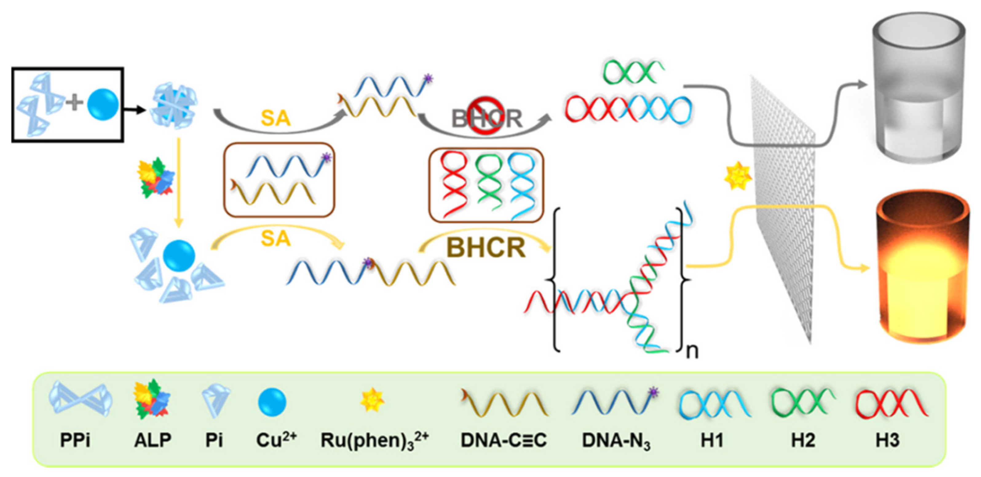
| miRNA | bHCR | 0.05–500 fM | 0.18 fM | [26] |
| pyrophosphatase | Cu+-catalyzed azide–alkyne cycloaddition (CuAAC) with high-efficiency hybridization chain reaction (HCR) | 0.025–50 mU | 8 μU | [27] |
| Bisphenol A | Ru(phen)32+ can integrate into the grooves of HCR products (dsDNA) | 2.0–50 × 103 pM | 1.5 pM | [28] |
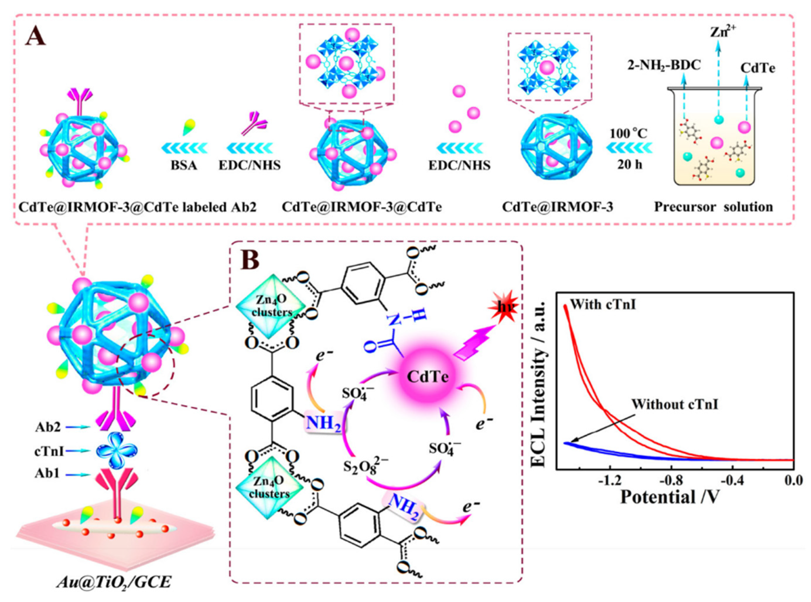
| cTnI | Au nanoclusters and HCR signal amplification | 5–5 × 10⁴ fg/mL | 1.01 fg/mL | [29] |
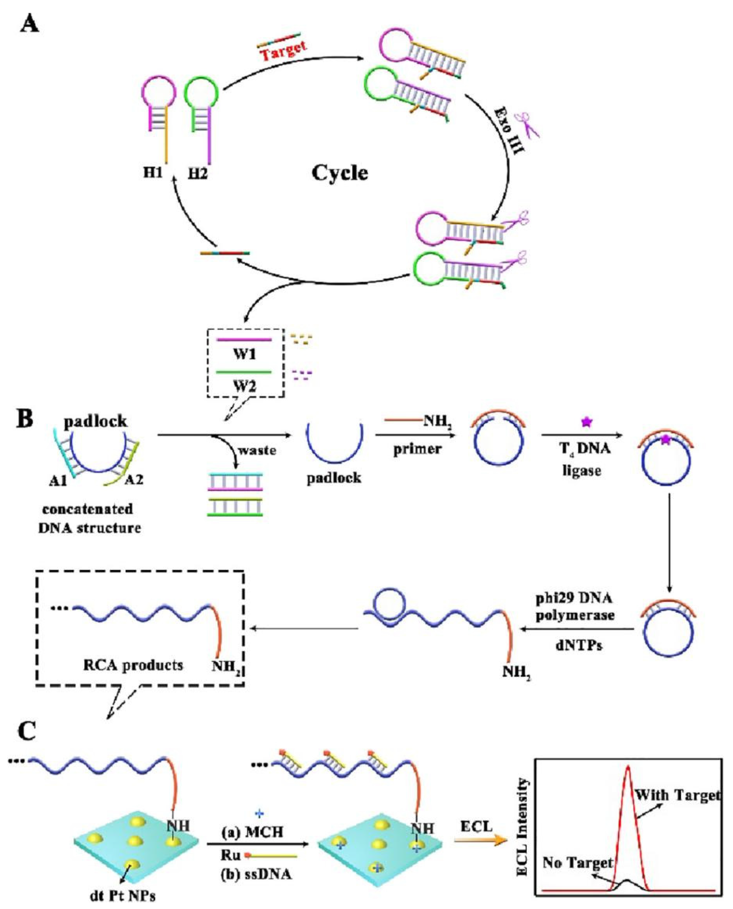
| Human immunodeficiency virus DNA | Target DNA triggered RCA signal amplification (RCA) | 100–1 × 10⁸ aM | 27.0 aM | [30] |
| HPV DNA | Bovine serum albumin carrier platforms and hyperbranched rolling circle amplification | 10–1.5 × 10⁴ fM | 7.6 fM | [31] |
| Hg2+ | Exonuclease III-assisted CRISPR/Cas12a | 0–1 × 10⁶ fM | 0.45 fM | [32] |
| BT63DNA | ExoIII enzyme-assisted hybridization chain reaction combined with nanoparticle-loaded multiple probes | 0.1–1 × 10⁴ fM | 0.036 fM | [33] |
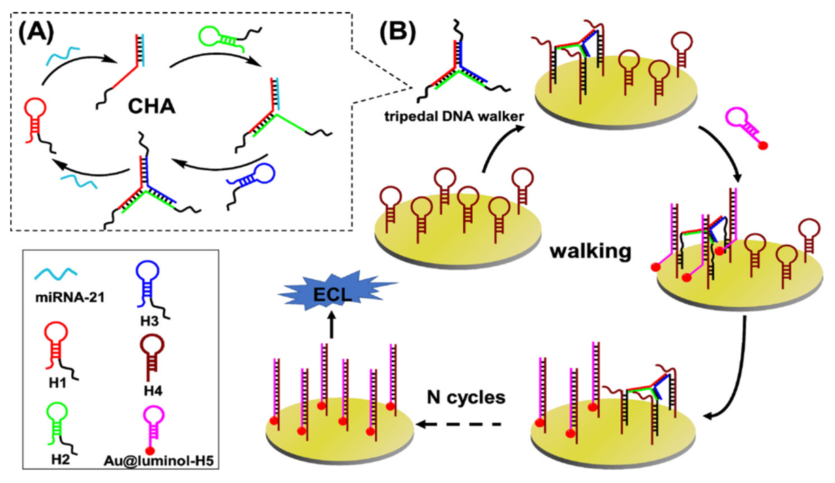
| MicroRNA | A synergistic promotion strategy for 3D DNA walker amplification | 10–1 × 10⁸ aM | 2.9 aM | [34] |
| miRNA-141 | 3D DNA walker-assisted CRISPR/Cas12a trans-cleavage | 1–1 × 10⁷ fM | 0.33 fM | [35] |
| SARS-CoV-2 | Target DNA-participated entropy-driven amplified reaction | 1–1 × 10⁵ fM | 2.67 fM | [36] |
| MicroRNA let-7a | Swing arm location-controllable DNA walker based on the DNA tetrahedral nanostructures (DTNs) | 10–1 × 10⁸ fM | 4.92 fM | [37] |
| 8-hydroxy-2′-deoxyguanosine | Target-induced multi-DNA release and nicking enzyme amplification strategy | 100–1 × 10⁷ fM | 25 fM | [38] |
| ochratoxin A | Nicking endonuclease-powered DNA walking machine | 0.05–5 nM | 0.012 nM | [39] |
| Myocardial miRNA | DNAzyme-regulated resonance energy transfer | 10–1 × 10⁷ fM | 2.44 fM | [40] |
| carcinoembryonic antigen | DNAzyme-driven DNA walker amplification | 1–1 × 10⁸ fg/mL | 0.21 fg/mL | [41] |
| 5-Hydroxymethylcytosine | DNAzyme motor triggered by strand displacement amplification | 1–1 × 10⁶ fM | 0.49 fM | [42] |
| MircoRNA-21 | Localized DNA cascade reaction (LDCR) in a DNA nanomachine | 100–1 × 10⁹ aM | 10.7 aM | [43] |
2.1. Enzyme-Assisted DNA Amplification Strategies
2.2. Enzyme-Free Amplification Strategies
3. Efficient ECL Luminophores
4. Ratiometric Strategies
4.1. Potential-Resolved Ratiometric Strategies
4.2. Spectrum-Resolved Ratiometric Strategies
5. Surface-Enhanced Strategies
6. Other Types of Signal-Amplification Strategies
7. Conclusions and Perspectives
Author Contributions
Funding
Institutional Review Board Statement
Informed Consent Statement
Data Availability Statement
Conflicts of Interest
References
- Alarfaj, N.A.; El-Tohamy, M.F.; Oraby, H.F. New immunosensing-fluorescence detection of tumor marker cytokeratin-19 fragment (CYFRA 21-1) via carbon quantum dots/zinc oxide nanocomposite. Nanoscale Res. Lett. 2020, 15, 1–14. [Google Scholar] [CrossRef] [PubMed]
- Su, M.; Liu, S. Solid-state electrochemiluminescence analysis with coreactant of the immobilized tris (2, 2′-bipyridyl) ruthenium. Anal. Biochem. 2010, 1, 1–12. [Google Scholar] [CrossRef] [PubMed]
- Zhu, L.; Ye, J.; Yan, M.; Zhu, Q.; Yang, X. A wavelength-resolved electrochemiluminescence resonance energy transfer ratiometric immunosensor for detection of cardiac troponin I. Analyst 2019, 144, 6554–6560. [Google Scholar] [CrossRef]
- Neves, M.M.; González-García, M.B.; Hernández-Santos, D.; Fanjul-Bolado, P. A miniaturized flow injection analysis system for electrogenerated chemiluminescence-based assays. ChemElectroChem 2017, 4, 1686–1689. [Google Scholar] [CrossRef]
- Forry, S.P.; Wightman, R.M. Electrogenerated chemiluminescence detection in reversed-phase liquid chromatography. Anal. Chem. 2002, 74, 528–532. [Google Scholar] [CrossRef]
- Sun, S.; Wei, Y.; Wang, H.; Cao, Y.; Deng, B. A novel electrochemiluminescence sensor coupled with capillary electrophoresis for simultaneous determination of quinapril hydrochloride and its metabolite quinaprilat hydrochloride in human plasma. Talanta 2018, 179, 213–220. [Google Scholar] [CrossRef]
- Huang, Y.; Lu, Y.; Huang, X.; Wang, J.; Qiu, B.; Luo, F.; Lin, Z. Design of an electrochemiluminescence detection system through the regulation of charge density in a microchannel. Chem. Sci. 2021, 12, 13151–13157. [Google Scholar] [CrossRef]
- Hercules, D.M. Chemiluminescence resulting from electrochemically generated species. Science 1964, 145, 808–809. [Google Scholar] [CrossRef]
- Santhanam, K.; Bard, A.J. Chemiluminescence of electrogenerated 9, 10-Diphenylanthracene anion radical1. J. Am. Chem. Soc. 1965, 87, 139–140. [Google Scholar] [CrossRef]
- Lee, W.-Y. Tris (2, 2′-bipyridyl) ruthenium (II) electrogenerated chemiluminescence in analytical science. Microchim. Acta 1997, 127, 19–39. [Google Scholar] [CrossRef]
- Fähnrich, K.A.; Pravda, M.; Guilbault, G.G. Recent applications of electrogenerated chemiluminescence in chemical analysis. Talanta 2001, 54, 531–559. [Google Scholar] [CrossRef]
- Liu, Z.; Qi, W.; Xu, G. Recent advances in electrochemiluminescence. Chem. Soc. Rev. 2015, 44, 3117–3142. [Google Scholar] [CrossRef] [PubMed]
- Hai-Juan, L.; Shuang, H.; Lian-Zhe, H.; Guo-Bao, X. Progress in Ru (bpy)32+ electrogenerated chemiluminescence. Chin. J. Anal. Chem. 2009, 37, 1557–1565. [Google Scholar]
- Wang, Q.; Ren, Z.-H.; Zhao, W.-M.; Wang, L.; Yan, X.; Zhu, A.-s.; Qiu, F.-m.; Zhang, K.-K. Research advances on surface plasmon resonance biosensors. Nanoscale 2022, 14, 564–591. [Google Scholar] [CrossRef] [PubMed]
- Zhao, L.D.; Yang, X.; Zhong, X.; Zhuo, Y. Advances in Electrochemiluminescence Biosensors Based on DNA Walkers. ChemPlusChem 2022, 87, e202200070. [Google Scholar] [CrossRef]
- Luo, W.; Chu, H.; Wu, X.; Ma, P.; Wu, Q.; Song, D. Disposable biosensor based on novel ternary Ru-PEI@ PCN-333 (Al) self-enhanced electrochemiluminescence system for on-site determination of caspase-3 activity. Talanta 2022, 239, 123083. [Google Scholar] [CrossRef]
- Kurup, C.P.; Lim, S.A.; Ahmed, M.U. Nanomaterials as signal amplification elements in aptamer-based electrochemiluminescent biosensors. Bioelectrochemistry 2022, 147, 108170. [Google Scholar] [CrossRef]
- Sun, T.; Du, J.; Li, Z.; Zhao, F. Recent Advancement in the development of hybridization chain reaction-based electrochemiluminescence biosensors. Int. J. Electrochem. Sci. 2022, 17, 2. [Google Scholar] [CrossRef]
- Miao, P.; Chai, H.; Tang, Y. DNA Hairpins and Dumbbell-Wheel Transitions Amplified Walking Nanomachine for Ultrasensitive Nucleic Acid Detection. ACS Nano 2022, 16, 4726–4733. [Google Scholar] [CrossRef]
- Yan, Y.; Li, J.; Li, W.; Wang, Y.; Song, W.; Bi, S. DNA flower-encapsulated horseradish peroxidase with enhanced biocatalytic activity synthesized by an isothermal one-pot method based on rolling circle amplification. Nanoscale 2018, 10, 22456–22465. [Google Scholar] [CrossRef]
- Feng, Q.-M.; Guo, Y.-H.; Xu, J.-J.; Chen, H.-Y. Self-assembled DNA tetrahedral scaffolds for the construction of electrochemiluminescence biosensor with programmable DNA cyclic amplification. ACS Appl. Mater. Interfaces 2017, 9, 17637–17644. [Google Scholar] [CrossRef] [PubMed]
- Wang, L.; Liu, P.; Liu, Z.; Zhao, K.; Ye, S.; Liang, G.; Zhu, J.-J. Simple tripedal DNA walker prepared by target-triggered catalytic hairpin assembly for ultrasensitive electrochemiluminescence detection of microRNA. ACS Sens. 2020, 5, 3584–3590. [Google Scholar] [CrossRef] [PubMed]
- Oishi, M.; Saito, K. Simple single-legged DNA walkers at diffusion-limited nanointerfaces of gold nanoparticles driven by a DNA circuit mechanism. ACS Nano 2020, 14, 3477–3489. [Google Scholar] [CrossRef] [PubMed]
- Shang, L.; Wang, X.; Zhang, W.; Jia, L.-P.; Ma, R.-N.; Jia, W.-L.; Wang, H.-S. A dual-potential electrochemiluminescence sensor for ratiometric detection of carcinoembryonic antigen based on single luminophor. Sens. Actuators B Chem. 2020, 325, 128776. [Google Scholar] [CrossRef]
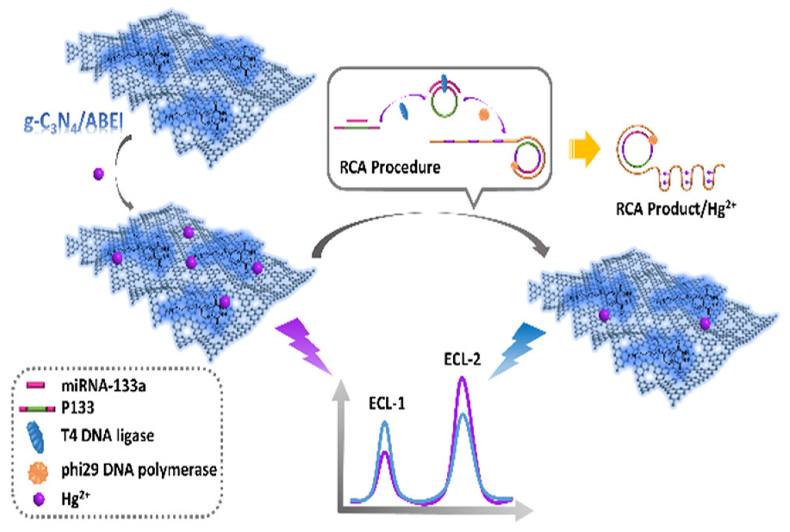
- Wang, J.; Haghighatbin, M.A.; Shen, W.; Mi, L.; Cui, H. Metal ion-mediated potential-resolved ratiometric electrochemiluminescence bioassay for efficient determination of miR-133a in early diagnosis of acute myocardial infarction. Anal. Chem. 2020, 92, 7062–7070. [Google Scholar] [CrossRef] [PubMed]
- Li, Y.; Huang, C.Z.; Li, Y.F. Ultrasensitive electrochemiluminescence detection of MicroRNA via one-step introduction of a target-triggered branched hybridization chain reaction circuit. Anal. Chem. 2019, 91, 9308–9314. [Google Scholar] [CrossRef] [PubMed]
- Huang, X.; Jia, J.; Lin, Y.; Qiu, B.; Lin, Z.; Chen, H. A highly sensitive electrochemiluminescence biosensor for pyrophosphatase detection based on click chemistry-triggered hybridization chain reaction in homogeneous solution. ACS Appl. Mater. Interfaces 2020, 12, 34716–34722. [Google Scholar] [CrossRef] [PubMed]
- Zhang, H.; Luo, F.; Wang, P.; Guo, L.; Qiu, B.; Lin, Z. Signal-on electrochemiluminescence aptasensor for bi s p h e n o l A based on hybridization chain reaction and electrically heated electrode. Biosens. Bioelectron. 2019, 129, 36–41. [Google Scholar] [CrossRef]
- Zhu, L.; Ye, J.; Yan, M.; Zhu, Q.; Wang, S.; Huang, J.; Yang, X. Electrochemiluminescence immunosensor based on Au nanocluster and hybridization chain reaction signal amplification for ultrasensitive detection of cardiac troponin I. ACS Sens. 2019, 4, 2778–2785. [Google Scholar] [CrossRef]
- Zhang, X.; Zhou, Y.; Chai, Y.; Yuan, R. Double hairpin DNAs recognition induced a novel cascade amplification for highly specific and ultrasensitive electrochemiluminescence detection of DNA. Anal. Chem. 2021, 93, 7987–7992. [Google Scholar] [CrossRef]
- He, Y.; Liu, Y.; Cheng, L.; Yang, Y.; Qiu, B.; Guo, L.; Wang, Y.; Lin, Z.; Hong, G. Highly reproducible and sensitive electrochemiluminescence biosensors for HPV detection based on bovine serum albumin carrier platforms and hyperbranched rolling circle amplification. ACS Appl. Mater. Interfaces 2020, 13, 298–305. [Google Scholar] [CrossRef] [PubMed]
- Hang, X.-M.; Zhao, K.-R.; Wang, H.-Y.; Liu, P.-F.; Wang, L. Exonuclease III-assisted CRISPR/Cas12a electrochemiluminescence biosensor for sub-femtomolar mercury ions determination. Sens. Actuators B Chem. 2022, 368, 132208. [Google Scholar] [CrossRef]
- Hai, H.; Chen, C.; Chen, D.; Li, P.; Shan, Y.; Li, J. A sensitive electrochemiluminescence DNA biosensor based on the signal amplification of ExoIII enzyme-assisted hybridization chain reaction combined with nanoparticle-loaded multiple probes. Microchim. Acta 2021, 188, 1–8. [Google Scholar] [CrossRef]
- Yang, F.; Yang, F.; Tu, T.-T.; Liao, N.; Chai, Y.-Q.; Yuan, R.; Zhuo, Y. A synergistic promotion strategy remarkably accelerated electrochemiluminescence of SnO2 QDs for MicroRNA detection using 3D DNA walker amplification. Biosens. Bioelectron. 2021, 173, 112820. [Google Scholar] [CrossRef] [PubMed]
- Wang, Q.; Liu, Y.; Yan, J.; Liu, Y.; Gao, C.; Ge, S.; Yu, J. 3D DNA walker-assisted CRISPR/Cas12a trans-cleavage for ultrasensitive electrochemiluminescence detection of miRNA-141. Anal. Chem. 2021, 93, 13373–13381. [Google Scholar] [CrossRef] [PubMed]
- Fan, Z.; Yao, B.; Ding, Y.; Zhao, J.; Xie, M.; Zhang, K. Entropy-driven amplified electrochemiluminescence biosensor for RdRp gene of SARS-CoV-2 detection with self-assembled DNA tetrahedron scaffolds. Biosens. Bioelectron. 2021, 178, 113015. [Google Scholar] [CrossRef]
- Liao, N.; Pan, M.-C.; Wang, L.; Yang, F.; Yuan, R.; Zhuo, Y. Swing arm location-controllable DNA walker for electrochemiluminescence biosensing. Anal. Chem. 2021, 93, 4051–4058. [Google Scholar] [CrossRef]
- Zhao, R.-N.; Jia, L.-P.; Feng, Z.; Ma, R.-N.; Zhang, W.; Shang, L.; Xue, Q.-W.; Wang, H.-S. Ultrasensitive electrochemiluminescence aptasensor for 8-hydroxy-2′-deoxyguanosine detection based on target-induced multi-DNA release and nicking enzyme amplification strategy. Biosens. Bioelectron. 2019, 144, 111669. [Google Scholar] [CrossRef]
- Wei, M.; Wang, C.; Xu, E.; Chen, J.; Xu, X.; Wei, W.; Liu, S. A simple and sensitive electrochemiluminescence aptasensor for determination of ochratoxin A based on a nicking endonuclease-powered DNA walking machine. Food Chem. 2019, 282, 141–146. [Google Scholar] [CrossRef]
- Sun, Y.; Fang, L.; Han, Y.; Feng, A.; Liu, S.; Zhang, K.; Xu, J.-J. Reversible Ratiometric Electrochemiluminescence Biosensor Based on DNAzyme Regulated Resonance Energy Transfer for Myocardial miRNA Detection. Anal. Chem. 2022, 94, 7035–7040. [Google Scholar] [CrossRef]
- Zhao, Y.; Tan, L.; Jie, G. Ultrasensitive electrochemiluminescence biosensor for the detection of carcinoembryonic antigen based on multiple amplification and a DNA walker. Sens. Actuators B Chem. 2021, 333, 129586. [Google Scholar] [CrossRef]
- Li, X.-R.; Wang, L.; Liang, W.-B.; Yuan, R.; Zhuo, Y. Epigenetic Quantification of 5-Hydroxymethylcytosine Signatures via Regulatable DNAzyme Motor Triggered by Strand Displacement Amplification. Anal. Chem. 2022, 94, 3313–3319. [Google Scholar] [CrossRef] [PubMed]
- Jiang, X.; Wang, H.; Chai, Y.; Li, H.; Shi, W.; Yuan, R. DNA cascade reaction with high-efficiency target conversion for ultrasensitive electrochemiluminescence microRNA detection. Anal. Chem. 2019, 91, 10258–10265. [Google Scholar] [CrossRef] [PubMed]
- Wang, L.; Liu, P.; Liu, Z.; Cao, H.; Ye, S.; Zhao, K.; Liang, G.; Zhu, J.-J. A dual-potential ratiometric electrochemiluminescence biosensor based on Au@CDs nanoflowers, Au@luminol nanoparticles and an enzyme-free DNA nanomachine for ultrasensitive p53 DNA detection. Sens. Actuators B Chem. 2021, 327, 128890. [Google Scholar] [CrossRef]
- Mason, J.T.; Xu, L.; Sheng, Z.-m.; O’Leary, T.J. A liposome-PCR assay for the ultrasensitive detection of biological toxins. Nat. Biotechnol. 2006, 24, 555–557. [Google Scholar] [CrossRef]
- Ye, J.; Yan, M.; Zhu, L.; Huang, J.; Yang, X. Novel electrochemiluminescence solid-state pH sensor based on an i-motif forming sequence and rolling circle amplification. Chem. Commun. 2020, 56, 8786–8789. [Google Scholar] [CrossRef]
- Li, D.; Li, Y.; Luo, F.; Qiu, B.; Lin, Z. Ultrasensitive homogeneous electrochemiluminescence biosensor for a transcription factor based on target-modulated proximity hybridization and exonuclease III-powered recycling amplification. Anal. Chem. 2020, 92, 12686–12692. [Google Scholar] [CrossRef]
- Zhang, Y.; Xu, J.; Zhou, S.; Zhu, L.; Lv, X.; Zhang, J.; Zhang, L.; Zhu, P.; Yu, J. DNAzyme-triggered visual and ratiometric electrochemiluminescence dual-readout assay for Pb (II) based on an assembled paper device. Anal. Chem. 2020, 92, 3874–3881. [Google Scholar] [CrossRef]
- Xia, M.; Zhou, F.; Feng, X.; Sun, J.; Wang, L.; Li, N.; Wang, X.; Wang, G. A DNAzyme-based dual-stimuli responsive electrochemiluminescence resonance energy transfer platform for ultrasensitive anatoxin-A detection. Anal. Chem. 2021, 93, 11284–11290. [Google Scholar] [CrossRef]
- Wang, L.; Zhao, K.-R.; Liu, Z.-J.; Zhang, Y.-B.; Liu, P.-F.; Ye, S.-Y.; Zhang, Y.-W.; Liang, G.-X. An “on-off” signal-switchable electrochemiluminescence biosensor for ultrasensitive detection of dual microRNAs based on DNAzyme-powered DNA walker. Sens. Actuators B Chem. 2021, 348, 130660. [Google Scholar] [CrossRef]
- Ni, J.; Zhang, H.; Chen, Y.; Luo, F.; Wang, J.; Guo, L.; Qiu, B.; Lin, Z. DNAzyme-based Y-shaped label-free electrochemiluminescent biosensor for lead using electrically heated indium-tin-oxide electrode for in situ temperature control. Sens. Actuators B Chem. 2019, 289, 78–84. [Google Scholar] [CrossRef]
- Jiang, J.; Du, X. DNA-targeted formation and catalytic reactions of DNAzymes for label-free ratiometric electrochemiluminescence biosensing. Talanta 2021, 225, 121964. [Google Scholar] [CrossRef]
- Gong, L.; Zhao, Z.; Lv, Y.-F.; Huan, S.-Y.; Fu, T.; Zhang, X.-B.; Shen, G.-L.; Yu, R.-Q. DNAzyme-based biosensors and nanodevices. Chem. Commun. 2015, 51, 979–995. [Google Scholar] [CrossRef] [PubMed]
- Sun, Y.; Fang, L.; Zhang, Z.; Yi, Y.; Liu, S.; Chen, Q.; Zhang, J.; Zhang, C.; He, L.; Zhang, K. A multitargeted electrochemiluminescent biosensor coupling DNAzyme with cascading amplification for analyzing myocardial miRNAs. Anal. Chem. 2021, 93, 7516–7522. [Google Scholar] [CrossRef] [PubMed]
- Zhang, Y.; Li, X.; Xu, Z.; Chai, Y.; Wang, H.; Yuan, R. An ultrasensitive electrochemiluminescence biosensor for multiple detection of microRNAs based on a novel dual circuit catalyzed hairpin assembly. Chem. Commun. 2018, 54, 10148–10151. [Google Scholar] [CrossRef]
- Zhang, C.; Chen, J.; Sun, R.; Huang, Z.; Luo, Z.; Zhou, C.; Wu, M.; Duan, Y.; Li, Y. The recent development of hybridization chain reaction strategies in biosensors. ACS Sens. 2020, 5, 2977–3000. [Google Scholar] [CrossRef]
- Chai, H.; Cheng, W.; Jin, D.; Miao, P. Recent progress in DNA hybridization chain reaction strategies for amplified biosensing. ACS Appl. Mater. Interfaces 2021, 13, 38931–38946. [Google Scholar] [CrossRef]
- Augspurger, E.E.; Rana, M.; Yigit, M.V. Chemical and biological sensing using hybridization chain reaction. ACS Sens. 2018, 3, 878–902. [Google Scholar] [CrossRef]
- Yang, D.; Tang, Y.; Miao, P. Hybridization chain reaction directed DNA superstructures assembly for biosensing applications. TrAC Trends Anal. Chem. 2017, 94, 1–13. [Google Scholar] [CrossRef]
- Huang, X.; Bian, X.; Chen, L.; Guo, L.; Qiu, B.; Lin, Z. Highly sensitive homogeneous electrochemiluminescence biosensor for alkaline phosphatase detection based on click chemistry-triggered branched hybridization chain reaction. Anal. Chem. 2021, 93, 10351–10357. [Google Scholar] [CrossRef]
- Zhang, C.; Ma, X.; Zheng, X.; Ke, Y.; Chen, K.; Liu, D.; Lu, Z.; Yang, J.; Yan, H. Programmable allosteric DNA regulations for molecular networks and nanomachines. Sci. Adv. 2022, 8, eabl4589. [Google Scholar] [CrossRef] [PubMed]
- Sobhanie, E.; Salehnia, F.; Xu, G.; Hamidi, Y.; Arshian, S.; Firoozbakhtian, A.; Hosseini, M.; Ganjali, M.R.; Hanif, S. Recent trends and advancements in electrochemiluminescence biosensors for human virus detection. TrAC Trends Anal. Chem. 2022, 157, 116727. [Google Scholar] [CrossRef] [PubMed]
- Chen, L.; Doeven, E.H.; Wilson, D.J.; Kerr, E.; Hayne, D.J.; Hogan, C.F.; Yang, W.; Pham, T.T.; Francis, P.S. Co-reactant electrogenerated chemiluminescence of iridium(III) complexes containing an acetylacetonate ligand. ChemElectroChem 2017, 4, 1797–1808. [Google Scholar] [CrossRef]
- Guo, W.; Ding, H.; Gu, C.; Liu, Y.; Jiang, X.; Su, B.; Shao, Y. Potential-resolved multicolor electrochemiluminescence for multiplex immunoassay in a single sample. J. Am. Chem. Soc. 2018, 140, 15904–15915. [Google Scholar] [CrossRef] [PubMed]
- Ding, Z.; Quinn, B.M.; Haram, S.K.; Pell, L.E.; Korgel, B.A.; Bard, A.J. Electrochemistry and electrogenerated chemiluminescence from silicon nanocrystal quantum dots. Science 2002, 296, 1293–1297. [Google Scholar] [CrossRef]
- Chen, X.; Liu, Y.; Ma, Q. Recent advances in quantum dot-based electrochemiluminescence sensors. J. Mater. Chem. C 2018, 6, 942–959. [Google Scholar] [CrossRef]
- Xu, Y.; Liu, J.; Gao, C.; Wang, E. Applications of carbon quantum dots in electrochemiluminescence: A mini review. Electrochem. Commun. 2014, 48, 151–154. [Google Scholar] [CrossRef]
- Yang, E.; Zhang, Y.; Shen, Y. Quantum dots for electrochemiluminescence bioanalysis-A review. Anal. Chim. Acta 2021, 339140. [Google Scholar] [CrossRef]
- Wang, X.-Y.; Che, Z.-Y.; Bao, N.; Qing, Z.; Ding, S.-N. Recent advances in II-VI quantum dots based-signal strategy of electrochemiluminescence sensor. Talanta Open 2022, 5, 100088. [Google Scholar] [CrossRef]
- Yang, X.; Yu, Y.-Q.; Peng, L.-Z.; Lei, Y.-M.; Chai, Y.-Q.; Yuan, R.; Zhuo, Y. Strong electrochemiluminescence from MOF accelerator enriched quantum dots for enhanced sensing of trace cTnI. Anal. Chem. 2018, 90, 3995–4002. [Google Scholar] [CrossRef]
- Peng, H.; Huang, Z.; Wu, W.; Liu, M.; Huang, K.; Yang, Y.; Deng, H.; Xia, X.; Chen, W. Versatile high-performance electrochemiluminescence ELISA platform based on a gold nanocluster probe. ACS Appl. Mater. Interfaces 2019, 11, 24812–24819. [Google Scholar] [CrossRef] [PubMed]
- Han, S.; Zhao, Y.; Zhang, Z.; Xu, G. Recent advances in electrochemiluminescence and chemiluminescence of metal nanoclusters. Molecules 2020, 25, 5208. [Google Scholar] [CrossRef] [PubMed]
- Nie, Y.; Tao, X.; Zhang, H.; Chai, Y.-q.; Yuan, R. Self-assembly of gold nanoclusters into a metal–organic framework with efficient electrochemiluminescence and their application for sensitive detection of rutin. Anal. Chem. 2021, 93, 3445–3451. [Google Scholar] [CrossRef] [PubMed]
- Jiao, Y.; Hu, R.; Wang, Q.; Fu, F.; Chen, L.; Dong, Y.; Lin, Z. Tune the Fluorescence and Electrochemiluminescence of Graphitic Carbon Nitride Nanosheets by Controlling the Defect States. Chem. Eur. J. 2021, 27, 10925–10931. [Google Scholar] [CrossRef] [PubMed]
- Zou, R.; Teng, X.; Lin, Y.; Lu, C. Graphitic carbon nitride-based nanocomposites electrochemiluminescence systems and their applications in biosensors. TrAC Trends Anal. Chem. 2020, 132, 116054. [Google Scholar] [CrossRef]
- Liu, F.; Du, F.; Yuan, F.; Quan, S.; Guan, Y.; Xu, G. Electrochemiluminescence bioassays based on carbon nitride nanomaterials and 2D transition metal carbides. Curr. Opin. Electrochem. 2022, 34, 100981. [Google Scholar] [CrossRef]
- Ahmad, R.; Tripathy, N.; Khosla, A.; Khan, M.; Mishra, P.; Ansari, W.A.; Syed, M.A.; Hahn, Y.-B. Recent advances in nanostructured graphitic carbon nitride as a sensing material for heavy metal ions. J. Electrochem. Soc. 2019, 167, 037519. [Google Scholar] [CrossRef]
- Wang, Z.; Wei, W.; Shen, Y.; Liu, S.; Zhang, Y. Carbon nitride–based biosensors. In Biochemical Sensors: Nanomaterial-Based Biosensing and Application in Honor of the 90th Birthday of Prof. Shaojun Dong; World Scientific: Singapore, 2021; pp. 175–225. [Google Scholar]
- Ji, J.; Wen, J.; Shen, Y.; Lv, Y.; Chen, Y.; Liu, S.; Ma, H.; Zhang, Y. Simultaneous noncovalent modification and exfoliation of 2D carbon nitride for enhanced electrochemiluminescent biosensing. J. Am. Chem. Soc. 2017, 139, 11698–11701. [Google Scholar] [CrossRef]
- Huo, X.-L.; Lu, H.-J.; Xu, J.-J.; Zhou, H.; Chen, H.-Y. Recent advances of ratiometric electrochemiluminescence biosensors. J. Mater. Chem. B 2019, 7, 6469–6475. [Google Scholar] [CrossRef]
- Han, Z.; Shu, J.; Liang, X.; Cui, H. Label-free ratiometric electrochemiluminescence aptasensor based on nanographene oxide wrapped titanium dioxide nanoparticles with potential-resolved electrochemiluminescence. Anal. Chem. 2019, 91, 12260–12267. [Google Scholar] [CrossRef]
- Liu, Y.; Sun, Y.; Yang, M. A double-potential ratiometric electrochemiluminescence platform based on gC3N4 nanosheets (gC3N4 NSs) and graphene quantum dots for Cu2+ detection. Anal. Methods 2021, 13, 903–909. [Google Scholar] [CrossRef] [PubMed]
- Fu, X.; Tan, X.; Yuan, R.; Chen, S. A dual-potential electrochemiluminescence ratiometric sensor for sensitive detection of dopamine based on graphene-CdTe quantum dots and self-enhanced Ru(II) complex. Biosens. Bioelectron. 2017, 90, 61–68. [Google Scholar] [CrossRef] [PubMed]
- Zhao, H.-F.; Liang, R.-P.; Wang, J.-W.; Qiu, J.-D. A dual-potential electrochemiluminescence ratiometric approach based on graphene quantum dots and luminol for highly sensitive detection of protein kinase activity. Chem. Commun. 2015, 51, 12669–12672. [Google Scholar] [CrossRef] [PubMed]
- Ye, J.; Zhu, L.; Yan, M.; Zhu, Q.; Lu, Q.; Huang, J.; Cui, H.; Yang, X. Dual-wavelength ratiometric electrochemiluminescence immunosensor for cardiac troponin I detection. Anal. Chem. 2018, 91, 1524–1531. [Google Scholar] [CrossRef] [PubMed]
- Huo, X.-L.; Zhang, N.; Yang, H.; Xu, J.-J.; Chen, H.-Y. Electrochemiluminescence resonance energy transfer system for dual-wavelength ratiometric miRNA detection. Anal. Chem. 2018, 90, 13723–13728. [Google Scholar] [CrossRef]
- Fan, Z.; Yao, B.; Ding, Y.; Xu, D.; Zhao, J.; Zhang, K. Rational engineering the DNA tetrahedrons of dual wavelength ratiometric electrochemiluminescence biosensor for high efficient detection of SARS-CoV-2 RdRp gene by using entropy-driven and bipedal DNA walker amplification strategy. Chem. Eng. J. 2022, 427, 131686. [Google Scholar] [CrossRef]
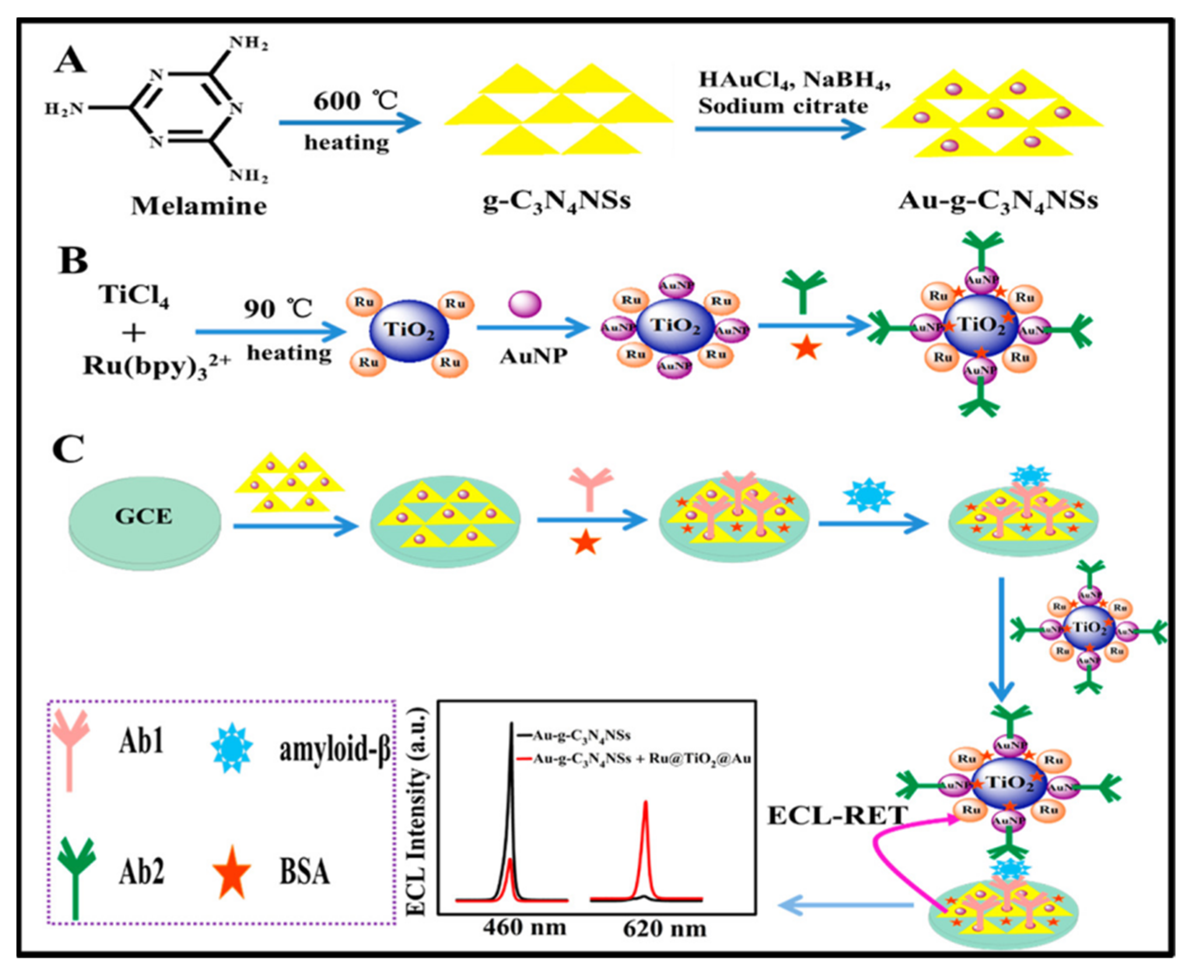
- Qin, D.; Meng, S.; Wu, Y.; Mo, G.; Jiang, X.; Deng, B. Design of a Dual-Wavelength Ratiometric Electrochemiluminescence Immunosensor for Sensitive Detection of Amyloid-β Protein in Human Serum. ACS Sustain. Chem. Eng. 2021, 9, 7541–7549. [Google Scholar] [CrossRef]
- Wang, P.; Nie, Y.; Tian, Y.; Liang, Z.; Xu, S.; Ma, Q. A whispering gallery mode-based surface enhanced electrochemiluminescence biosensor using biomimetic antireflective nanostructure. Chem. Eng. J. 2021, 426, 130732. [Google Scholar] [CrossRef]
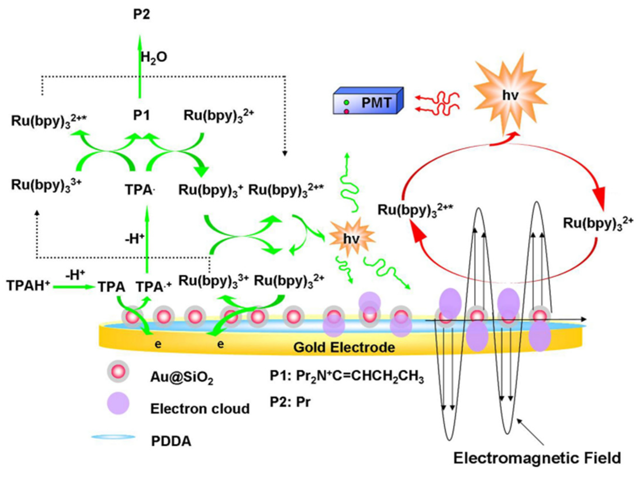
- Li, X.; Du, X. Surface enhanced electrochemiluminescence of the [Ru(bpy)3]2+/tripropylamine system by Au@SiO2 nanoparticles for highly sensitive and selective detection of dopamine. Microchem. J. 2022, 176, 107224. [Google Scholar] [CrossRef]
- Ding, L.; Xu, S.; Huang, Y.; Yao, Y.; Wang, Y.; Chen, L.; Zeng, Y.; Li, L.; Lin, Z.; Guo, L. Surface-Enhanced Electrochemiluminescence Imaging for Multiplexed Immunoassays of Cancer Markers in Exhaled Breath Condensates. Anal. Chem. 2022, 94, 7492–7499. [Google Scholar] [CrossRef]
- Li, M.-X.; Feng, Q.-M.; Zhou, Z.; Zhao, W.; Xu, J.-J.; Chen, H.-Y. Plasmon-enhanced electrochemiluminescence for nucleic acid detection based on gold nanodendrites. Anal. Chem. 2018, 90, 1340–1347. [Google Scholar] [CrossRef] [PubMed]
- Feng, X.; Han, T.; Xiong, Y.; Wang, S.; Dai, T.; Chen, J.; Zhang, X.; Wang, G. Plasmon-enhanced electrochemiluminescence of silver nanoclusters for microRNA detection. ACS Sens. 2019, 4, 1633–1640. [Google Scholar] [CrossRef] [PubMed]
- Wang, D.; Guo, L.; Huang, R.; Qiu, B.; Lin, Z.; Chen, G. Surface enhanced electrochemiluminescence of [Ru (bpy)3]2+. Sci. Rep. 2015, 5, 1–7. [Google Scholar]
- Wang, D.; Zhou, J.; Guo, L.; Qiu, B.; Lin, Z. A surface-enhanced electrochemiluminescence sensor based on Au-SiO2 core–shell nanocomposites doped with [Ru (bpy)3]2+ for the ultrasensitive detection of prostate-specific antigen in human serum. Analyst 2020, 145, 132–138. [Google Scholar] [CrossRef]
- Kitte, S.A.; Tafese, T.; Xu, C.; Saqib, M.; Li, H.; Jin, Y. Plasmon-enhanced quantum dots electrochemiluminescence aptasensor for selective and sensitive detection of cardiac troponin I. Talanta 2021, 221, 121674. [Google Scholar] [CrossRef]
- Chen, X.; Gui, W.; Ma, Q. Ultrasensitive detection of EGFR gene based on surface plasmon resonance enhanced electrochemiluminescence of CuZnInS quantum dots. Anal. Chim. Acta 2018, 1009, 73–80. [Google Scholar] [CrossRef]
- Zhang, Q.; Liu, Y.; Nie, Y.; Ma, Q.; Zhao, B. Surface plasmon coupling electrochemiluminescence assay based on the use of AuNP@C3N4QD@mSiO2 for the determination of the Shiga toxin-producing Escherichia coli (STEC) gene. Microchim. Acta 2019, 186, 1–9. [Google Scholar] [CrossRef]
- Wang, D.; Guo, L.; Huang, R.; Qiu, B.; Lin, Z.; Chen, G. Surface enhanced electrochemiluminescence for ultrasensitive detection of Hg2+. Electrochim. Acta 2014, 150, 123–128. [Google Scholar] [CrossRef]
- Zhang, Q.; Tian, Y.; Liang, Z.; Wang, Z.; Xu, S.; Ma, Q. DNA-mediated Au–Au dimer-based surface plasmon coupling electrochemiluminescence sensor for BRCA1 gene detection. Anal. Chem. 2021, 93, 3308–3314. [Google Scholar] [CrossRef]
- Zhang, Q.; Zhang, X.; Ma, Q. Recent advances in visual electrochemiluminescence analysis. J. Anal. Test. 2020, 4, 92–106. [Google Scholar] [CrossRef]
- Liu, Y.; Chen, X.; Wang, M.; Ma, Q. A visual electrochemiluminescence resonance energy transfer/surface plasmon coupled electrochemiluminescence nanosensor for Shiga toxin-producing Escherichia coli detection. Green Chem. 2018, 20, 5520–5527. [Google Scholar] [CrossRef]
- Xing, H.; Zhai, Q.; Zhang, X.; Li, J.; Wang, E. Boron nitride quantum dots as efficient coreactant for enhanced electrochemiluminescence of ruthenium (II) tris (2, 2′-bipyridyl). Anal. Chem. 2018, 90, 2141–2147. [Google Scholar] [CrossRef] [PubMed]
- Zhang, Z.; Du, P.; Pu, G.; Wei, L.; Wu, Y.; Guo, J.; Lu, X. Utilization and prospects of electrochemiluminescence for characterization, sensing, imaging and devices. Mater. Chem. Front. 2019, 3, 2246–2257. [Google Scholar] [CrossRef]


Publisher’s Note: MDPI stays neutral with regard to jurisdictional claims in published maps and institutional affiliations. |
© 2022 by the authors. Licensee MDPI, Basel, Switzerland. This article is an open access article distributed under the terms and conditions of the Creative Commons Attribution (CC BY) license (https://creativecommons.org/licenses/by/4.0/).
Share and Cite
Huang, Y.; Yao, Y.; Wang, Y.; Chen, L.; Zeng, Y.; Li, L.; Guo, L. Strategies for Enhancing the Sensitivity of Electrochemiluminescence Biosensors. Biosensors 2022, 12, 750. https://doi.org/10.3390/bios12090750
Huang Y, Yao Y, Wang Y, Chen L, Zeng Y, Li L, Guo L. Strategies for Enhancing the Sensitivity of Electrochemiluminescence Biosensors. Biosensors. 2022; 12(9):750. https://doi.org/10.3390/bios12090750
Chicago/Turabian StyleHuang, Yueyue, Yuanyuan Yao, Yueliang Wang, Lifen Chen, Yanbo Zeng, Lei Li, and Longhua Guo. 2022. "Strategies for Enhancing the Sensitivity of Electrochemiluminescence Biosensors" Biosensors 12, no. 9: 750. https://doi.org/10.3390/bios12090750