Progress of Enzymatic and Non-Enzymatic Electrochemical Glucose Biosensor Based on Nanomaterial-Modified Electrode


Abstract
:1. Introduction
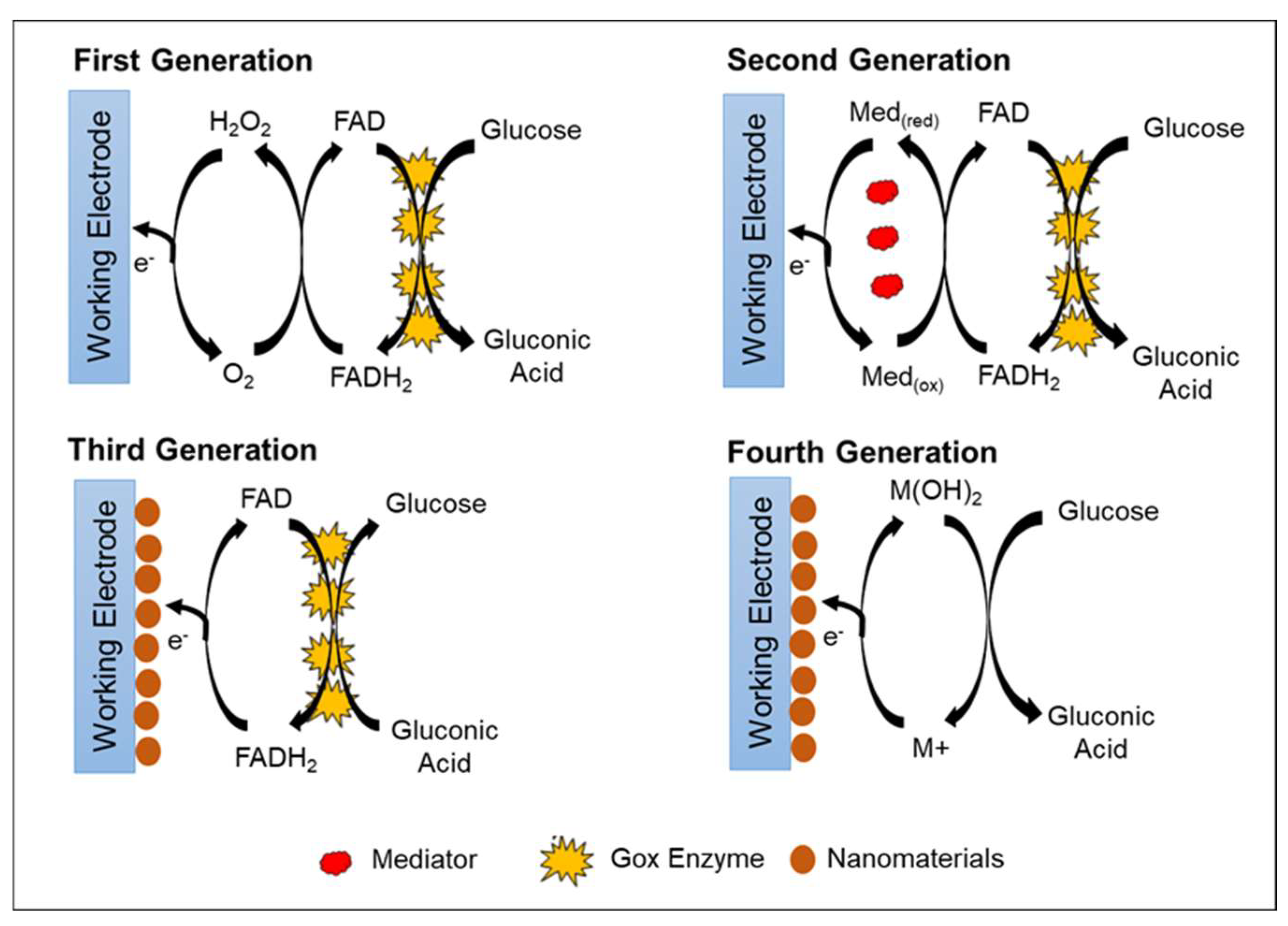
2. Generation of Glucose Biosensor

| Types of Glucose Sensor | Advantages | Disadvantages | Reference |
|---|---|---|---|
| First Generation (Enzymatic) |
|
| [82] |
| Second Generation (Enzymatic) |
|
| [66] |
| Third Generation (Enzymatic) |
|
| [79] |
| Fourth Generation (Non-Enzymatic) |
|
| [35] |
3. Parameters Controlling Enzymatic and Non-Enzymatic Glucose Biosensors
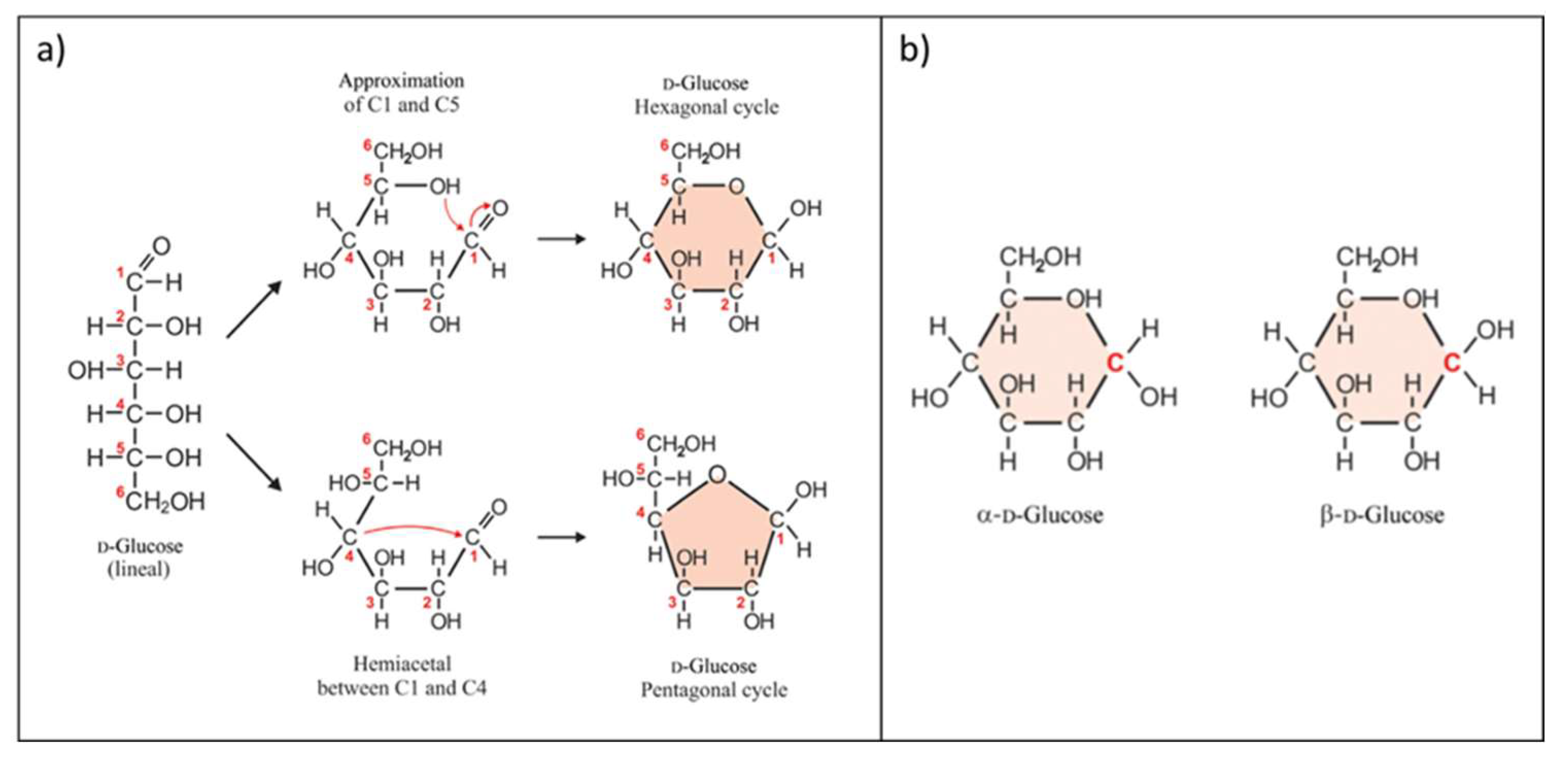
3.1. Glucose
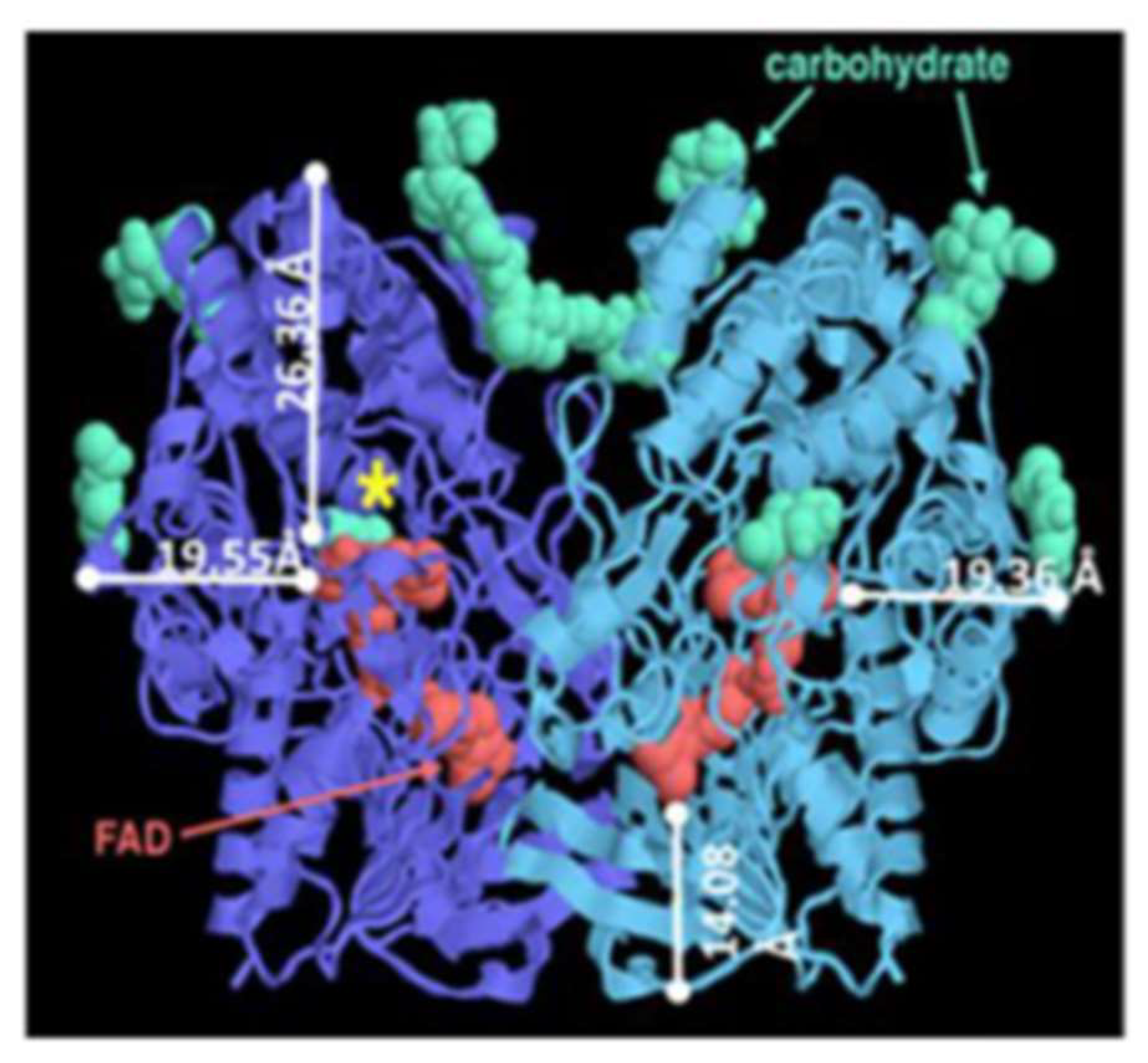
3.2. Enzymes

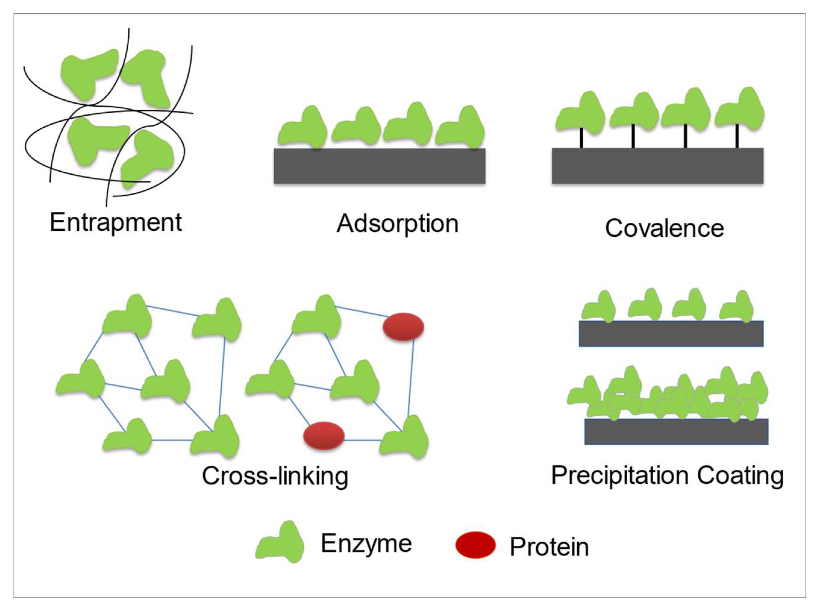
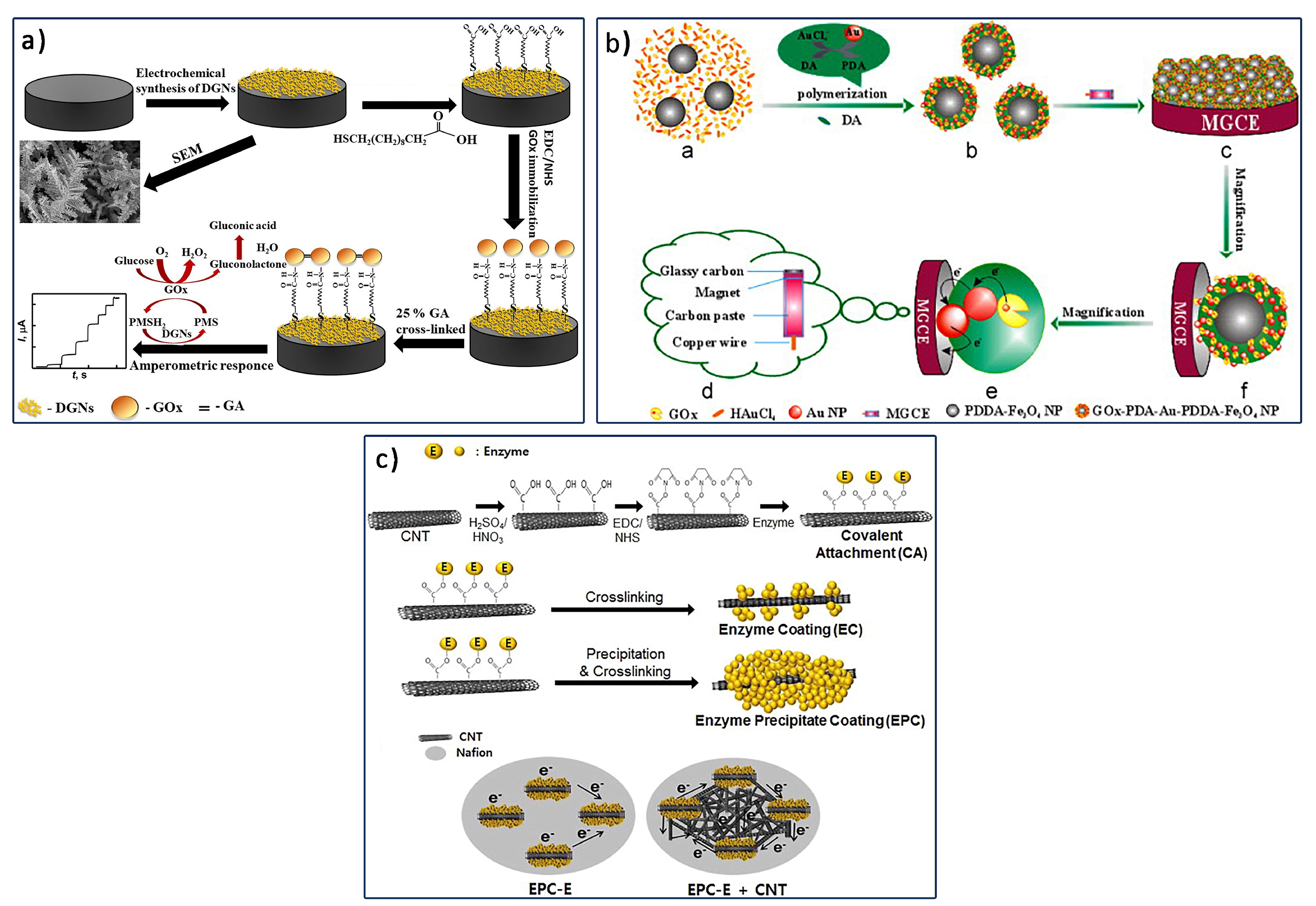
Enzyme Immobilization Technique

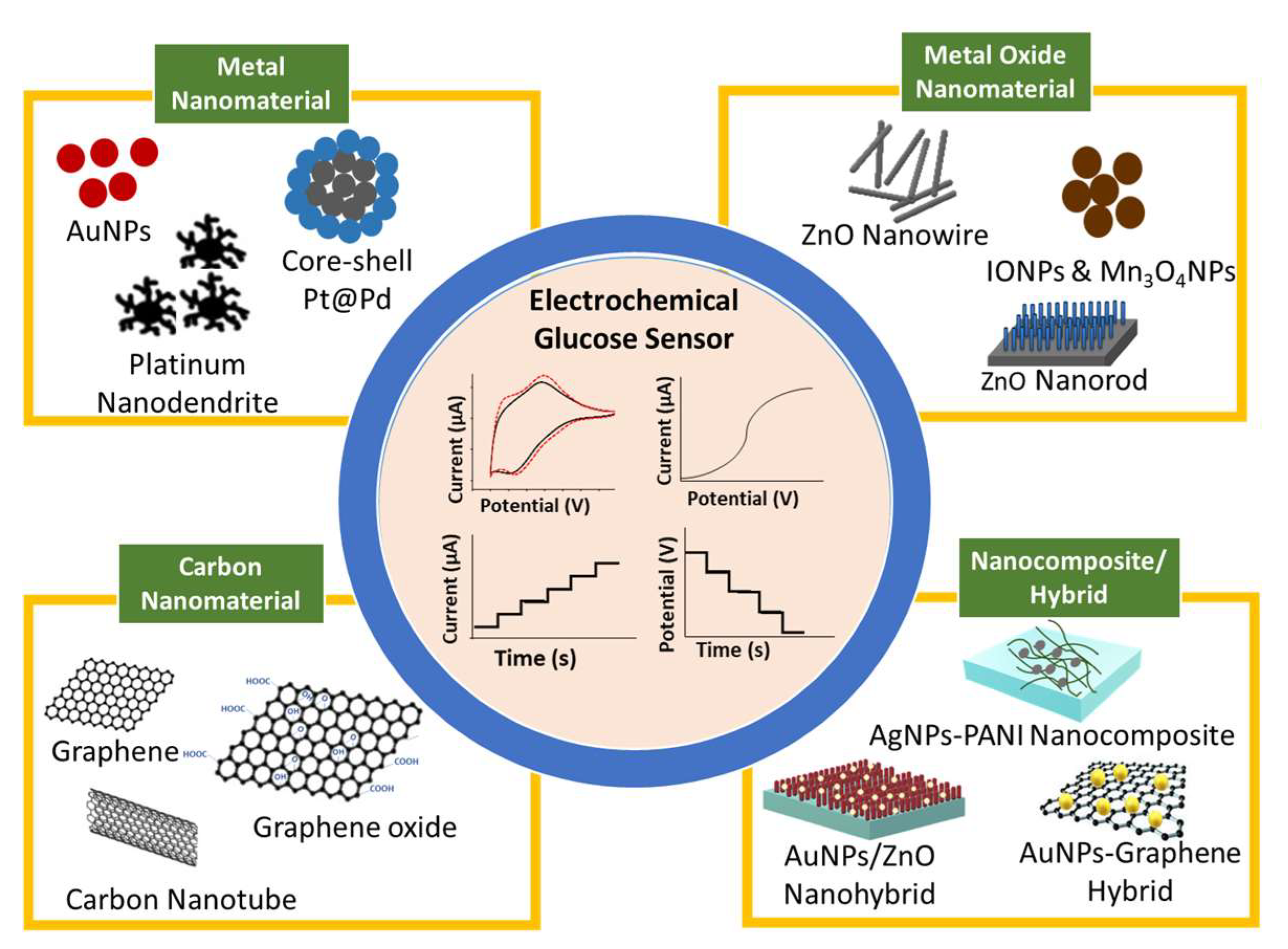
3.3. Electrode Materials
3.4. Type of Electrolyte and pH of Electrolyte
4. Recent Development of Nanomaterial-Modified Electrode for Enzymatic Glucose Biosensor
4.1. Metal-Based Enzymatic Glucose Biosensor
| Electrode Modification | Nanomaterials Modified Electrode | Enzyme/ Immobilization Technique | Applied Potential | Linearity (mM) | Sensitivity (µA mM−1 cm−2) | LOD (µM) | Stability/Lifetime | Sample | Reference |
|---|---|---|---|---|---|---|---|---|---|
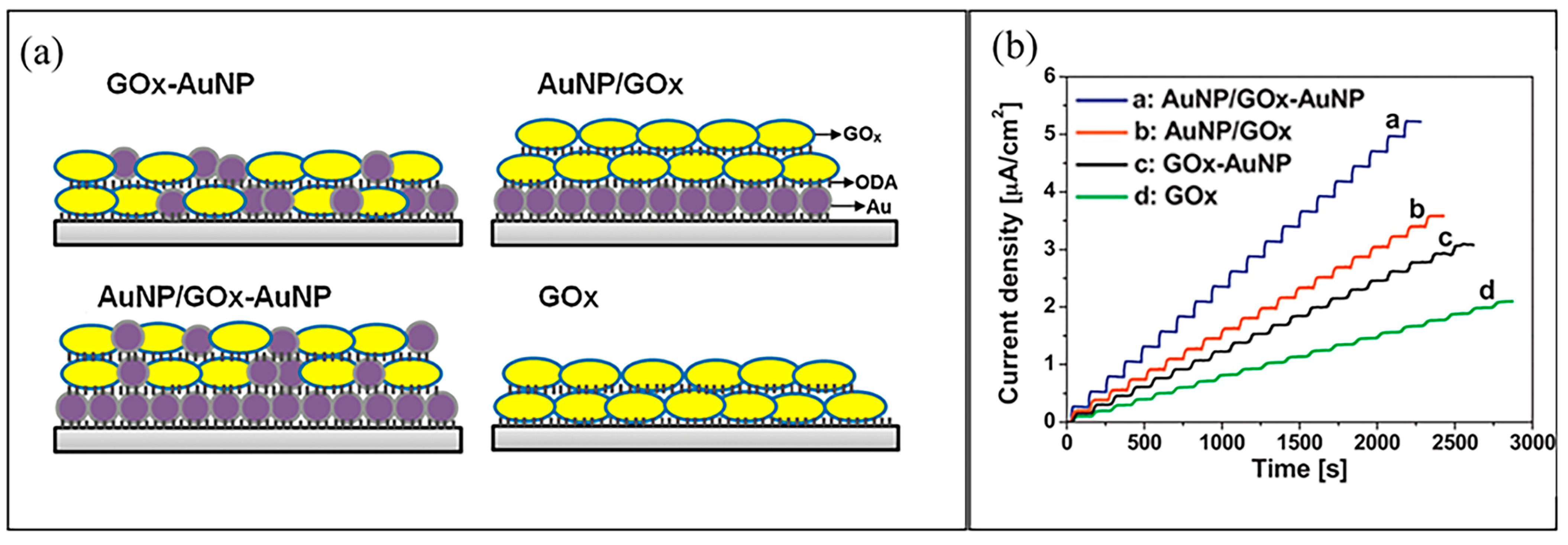
| AuNP-GOx-AuNPs/ODA-Pt | Langmuir–Blodgett deposition | GOx-Adsorption | 0.60 V | 0.1–5 | 0.52 | 63 | 95% 6 month | - | [127] |
| AuNP/GOx/ODA-Pt | Langmuir–Blodgett deposition | GOx-Adsorption | 0.60 V | 0.1–5 | 0.36 | 59 | 95% 6 month | - | |
| GOx/AuNPs/Pt/ODA-Pt | Langmuir–Blodgett deposition | GOx-Adsorption | 0.60 V | 0.1–5 | 0.31 | 59 | 95% 6 month | - | |
| GOx/ODA-Pt | Langmuir–Blodgett deposition | GOx-Adsorption | 0.60 V | 0.1–5 | 0.21 | 7 | 95% 6 month | - | |
| Nafion/GOx/AuNPs/OPPy/Au-PLA-MNs | Electrodeposition | GOx-Crosslink (GA) | 0.75 V | 0–2.6 | 8.09 | 40 | 14 days | - | [134] |
| GOx/AuNPs/PHCQE-Graphite | Electropolymerization | GOx-Crosslink (GA) | –0.70 V | 0.75–3.125 | 0.13 | 17 | 42 days | Beverage | [136] |
| Nafion/GOx-TCA/Au Microneedle | Electropolymerization | GOx-Covalent (EDC/NHS) | 0.45 V | 0–22.2 | 0.22 | 19.4 | 94% 30 days | Human Serum | [135] |
| PPy/GOx/AuNPs/ Graphite Rod | Electrodeposition | GOx-Crosslink (GA) | 0.30 V | 0–19.9 | 21.70 | 200 | 9.8 days | Human Serum | [139] |
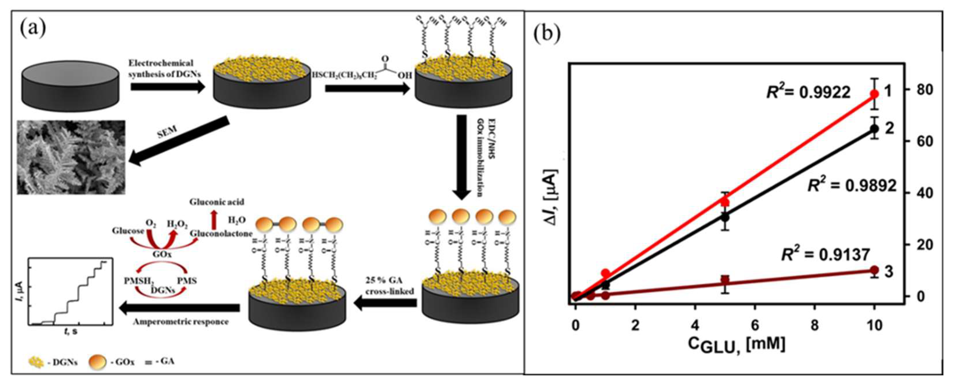
| GOx-SAM/Dendritic Au Nanostructure/ Graphite rod | Electrodeposition | GOx-Covalent (EDC/NHS) | 0.30 V | 0.1–10 | - | 19 | 73.25% 12 days | Human blood glucose | [105] |
| GA-GOx/Dendritic Au Nanostructure/Graphite rod | Electrodeposition | GOx-Crosslink (GA) | 0.30 V | 0.1–10 | - | 22 | 66.20% 12 days | Human blood glucose | |
| GOx/3D Au/carbon paper | Electrodeposition | GOx-Covalent (EDC/NHS) and Crosslink (GA) | 0.25 V | 0.002–21.97 | 96.27 | 0.6 | 80% 30 days | Human serum | [140] |
| GOx/PANI hydrogel/Pt | Chemical reduction | GOx-Crosslink (GA) | 0.56 V | 0.01–8 | 96.1 | 0.7 | - | - | [133] |
| GOx-PoPD/PtNPs/ PVF + ClO4−/Pt | Electrodeposition | GOx-Electropolymerization | 0.60 V | 0.06–9.64 | 17.4 | 18 | 95% 15 days | Blood serum sample | [137] |
| GOx-PtNPs-PAA-aSPCEs | PtNPs-Electrodeposition PAA-Elctropolymerization | GOx-Adsorption | 0.20 V | 0.02–2.3 | 42.7 | 7.6 | 50% 7 days | Commercial juices | [138] |
| GOx/Pt film/o-phenylenediamine-ß-cyclodextrin/Au | Electrodeposition | GOx-Electropolymerization | 0.25 V | 2.5–15 | 111.21 | 0.75 mM | 93.22% 4 days | Human serum sample | [146] |
| PU-PEG/GOx/Pt film/Au-PET | Electroplatting Pt film | GOx-Crosslink (GA) | 0.65 V | 0.5–25 | 3.418 | 0.25 | 90% 23 days | Beverage | [147] |
| GOx/Pt/rGO/poly(3-aminobenzoic acid/SPCE | Co-Electrodeposition | GOx-Covalent (EDC, NHS) | 0.50 V | 0.25–6.00 | 22.0 | 44.3 | 86% 7 days | Serum sample | [148] |
| Nafion/GOx/PtNP-CGr-f@MWCNTs /Au | Pt-Electrodeposition CGr-F@MWCNT-Drop Casted | GOx-Covalent (EDC, NHS) | 0.50 V | 0.005–13 | 26.5 | 5 | 21 days | - | [149] |
| Nafion/GOx/Graphene/ PtNPs | Drop Casted | GOx-Crosslink (GA) | 0.60 V | 0.005–0.5 | - | 0.01 | 75.45% 31 days | Human serum sample | [150] |
| GOx/Fc-bPEI-AuNPs/GCE | Drop Casted | GOx-Crosslink (GA) | 0.43 V | 0.5–10 | 800 | 0.04 | - | - | [151] |
| GOx/PPy/AuNPs/SP-Graphene Ink-PET | Electrodeposition | GOx-Electropolymerization | 0.40 V | 1–10 | 14.453 nA/mM | - | 90% 30 days | - | [122] |
| Nafion/GOx/Au–Ni coaxial nanorod array/Au electrode | Nano electroforming and immersion gold | GOx-Adsorption | 0.40 V | 0.028–27.5 | 778.2 | 5.5 | 87% 30 days | - | [152] |
| Nafion/GOx/Pd-MWCNT-SPCE Bulk | MWCNT-CVD Impregnate Pd | GOx-Adsorption | –0.20 V | 0.41–4.12 | –6.36 | 0.02 | 14 days | Human blood glucose | [95] |
| Nafion/GOx/Pd-MWCNT/SPCE | MWCNT-CVD Impregnate Pd | GOx-Absorption | –0.20 V | 0.41–4.12 | –5.05 | 0.14 | 14 days | Human blood glucose | |
| GOx-Graphene-PEI-AuNPs/Au Electrode | Microwave-irradiation | GOx-Crosslink (GA) | –0.35 V | 0.001–0.1 | 93 | 0.32 | 88% 10 days | Human Serum | [111] |
| GOx-Graphene-Thiol/Au Nanocube/ Au disk | Au Nanocube-Electrodeposition | GOx-Adsorption | –0.40 V | 0–0.8 | 221.0 | - | 79.3% 14 days | - | [143] |
| GOx-Chitosan/rGO-AuNPs/ | rGO-AuNPs- Drop Casted | GOx-Covalent (Chitosan) | –0.30 V | 0.1–1.3 | 34 | 76 | 70% 36 days | - | [153] |
| GOx/AuNPs/PENDI/PGE | AuNPs-Electrodeposition PENDI-Electropolymerization | GOx-Adsorption | - | 0.0009–0.33 | 0.172 | 0.0407 | - | - | [154] |
| Nafion/GOx/Carbon Fibre-Hemain AuNP/ Graphite Electrode | Carbon Fibre-AuNPs- Drop Casted | GOx-Nanoenzyme | –0.10 V | 0.1–0.9 | 909.5 A⋅M−1⋅m−2. | 0.05 | - | Beverage | [155] |
| Au@rGO/PIn/Ferritin/GOx/GCE | Electrodeposition | GOx | - | 50 | 7.2 mA cm−2 | - | - | - | [156] |
| Graphite NPs-Pyrene-GOx/GCE | Drop Casted | GOx-Crosslink (pyrenebutyric-NHS) | 0.60 V | 0–2.2 | 7.29 × 10⁻² nA | 50 | 30 days | Urine | [157] |
| GOD-CS/AgNWs/GCE | Drop Casted | GOx-Covalent (Chitosan) | –0.15 V | 0.01–0.8 | - | 2.83 | 83% 10 days | Human blood glucose | [158] |
4.2. Metal Oxide-Based Enzymatic Glucose Biosensor
| Electrode Modification | Nanomaterials Modified Electrode | Enzyme/ Immobilization Technique | Applied Potential | Linearity (mM) | Sensitivity (µA mM−1 cm−2) | Stability/ Lifetime | LOD (µM) | Sample | Reference |
|---|---|---|---|---|---|---|---|---|---|
| GOx-PVA-IONPs/Sn | Drop-casted | GOx-Adsorption | –0.19 V | 0.005–30 | 9.36 | 81% 30 days | 8 | - | [96] |
| Nafion/GOx/Pt/IONPs-MWCNTs-CS/MGCE | Electrodeposition | GOx-Adsorption | 0.30 V | 0.006–6.2 | - | 86.8% 14 days | 2 | - | [175] |
| Nafion/GOx/Nafion-IONPs@SiO2-MWCNT/GCE | Drop-casted | GOx-Adsorption | 0.10 V | 0.001–30 | - | - | 0.8 | - | [97] |
| GOx/IONPs-AuNPs-CS/Au | Electrochemical deposition | GOx-Adsorption | –0.40 V | 0.003–0.57 | - | 82.6% 14 days | 1.2 | - | [176] |
| Nf/CS-IONPs-GOx/Pt | Drop-casted | GOx-Crosslink (GA) | 0.40 V | 0.006–2.2 | 11.54 | 84% 30 days | 6 | Human Serum | [177] |
| Nf-GOx-HRP/AuNPs-IONPs@SiO2/ITO | Drop-casted | GOx-Crosslink (GA) | –0.20 V | 0.05–1.0 1.0–8.0 | 92.14 15.00 | 94.8% 30 days | 10 | Human Serum | [191] |
| GOx/AuNPs/BSA-IONPs/Pt | Immersed | Covalent GOx-BSA | 0.40 V | 0.25–7.0 | 115.13 | 81% 30 days | 3.54 | [112] | |
| GOx/IONPs/CS-Graphene/Pt | Drop-casted | Gox- Covalent (EDC/NHS) | 0.50 V | 0–26 | 5.658 | 75.7% 30 days | 16 | - | [192] |
| GOx-Au-PDA-IONPs/MGCE | Drop-casted | Co-polymerization GOx | –0.50 V | 0.02–1.875 | - | 95% 30 days | 6.5 | Human Serum | [31] |
| GOx/rGO-IONPs/MSPCE | Drop-casted | Electrostatic interaction GOx | –0.45 V | 0.05–1.0 | 5.90 | 95.1% 30 days | 0.1 | - | [142] |
| GOx-IONPs@AuNPs/MnO2-SPCE | Drop-casted | GOx-Adsorption | 0.38 V | 0.2–9.0 | 2.52 | 80% 30 days | 13.2 | Beverage | [193] |
| Nafion/GOx/IONPs-CA/SPCE | Drop-casted | GOx-Adsorption | –0.43 V. | 0.02–0.25 0.25–8.00 | 175 5.31 | 60% 30 days | 7 | - | [171] |
| Nafion/GOx/IONPs-CA/ITO | Drop-casted | GOx-Adsorption | 0.0001–0.005 0.005–20.0 | 995.57 5.81 | - | - | [163] | ||
| L-Cys/GOx/PVA/ ZnO/Au | Sputtered Deposition and Direct Growth | GOx-Adsorption | 0.06 V | 0.25–19 | 70.2 | 94% 45 days | 1 | Urine | [179] |
| GOx/ZnO Nanotube/AuCS | Hydrothermal Direct Growth | GOx-Adsorption | 0.80 V | 0–6.5 | 2.63 | 80.8% 20 days | 8 | Human Serum | [194] |
| GOx/ZnO Nanowire/ Carbon/Polymide | Flexographic printing | GOx-Adsorption | 0.80 V | 0–1.7 | - | - | 1200 | - | [181] |
| Nafion/GOx/ZnO Nanorod/Zn foil | Hydrothermal Direct Growth | GOx-Adsorption | 0.50 V | 0.006–0.38 | 35.1 | - | - | - | [180] |
| Nafion/GOx/ZnO/Au | Drop-casted | GOx-Adsorption | 0.80 V | 0.01–5.9 | 23.43 | - | 10 | - | [178] |
| Nafion/GOx/ZnO Nanorod/Au/ SiO2-Si | Hydrothermal Direct Growth | GOx-Adsorption | 0.40 V | 1–10 | 315 | 89% 11 days | 166.6 | - | [195] |
| GOx/ZnO Nanowire/Au-PET | Electrodeposition | GOx-Adsorption | 0.80 V | 0.2–2 | 19.5 | - | 50 | - | [196] |
| GA-GOx/rGO-Fc/GCE | Drop-casted | GOx-Crosslink (GA) Ferrocene | 0.35 V | 2–10 | - | 70% 14 days | 0.02 | Human Serum and Juice | [197] |
| GOx/rGO-Fc(COOH)2/GCE | Drop-casted | GOx-Adsorption -COOH F(x) | 0.35 V | 1–10 | - | 70% 14 days | 0.04 | Human Serum and Juice | |
| GOx-SiO2/Lig/Fc/CPE | Casted | Gox absorption with ferrocene mediator | 0.60 V | 0.5–9 | 0.78 | 73% 21 days | 145 | Liquid Glucose | [114] |
| Au@rGO/PIn/Ferritin/GOx/GCE | Electropolyme-rization | GOx-Elctropolymeri zation | LSV | 50 | 7.2 mA cm−2 | - | - | - | [156] |
| Graphite NPs-Pyrene-Gox/GCE | Drop-casted | GOx-Crosslink (pyrenebutyric acid/NHS) | 0.60 V | 0–2.2 | 7.29 × 10⁻² nA | 30 days | 50 | Urine | [157] |
| GOx-CS/AgNWs/GCE | Drop-casted | GOx-CS | -0.15 V | 0.01– 0.8 | - | 83% 10 days | 2.83 | Human Blood Glucose | [158] |
| Nafion/GOx/Fe3O4 /ZnONFs/Au/PET | ZnO-Hydrothermal Direct Growth Fe3O4-Drop-casting | GOx-Adsorption | 0.80 V | 0.089–12.5 | 4.52 | - | 0.089 | Human Blood Glucose | [161] |
| Nafion/GOx/ZnO Nanoflower/Au/PET | Hydrothermal Direct Growth | GOx-Adsorption | 0.80 V | 0.15–8.5 | 0.57 | - | 0.105 | Human Blood Glucose | [161] |
| Nafion/GOx/Pt Nanodendrite/ZnO NR/ITO | Hydrothermal Direct Growth and Spin-coated | GOx-Adsorption | - | 1–18 | 5.85 | - | 1.56 | - | [169] |
| Nafion/ZnONR/ITO | Hydrothermal Direct Growth | GOx-Adsorption | –0.5 V | 0.05–1 1–20 | 48.75 3.87 | 85% 14 days | - | Human Blood Glucose | [189] |
| Nafion/GOx/AuNP/ ZnONR/ITO | Hydrothermal Direct Growth and Drop-casted | GOx-Adsorption | –0.5 V | 0.05–1.0 1.0–20 | 14.53 2.54 | - | - | - | [198] |
| Nafion/GOx/PtNDs/ZnONRs/ITO | Hydrothermal Direct Growth and Drop-casted | GOx-Adsorption | –0.5 V | 0.05–1 1–18 | 98.34 9.76 | 76.1% 30 days | 0.03 | Human Blood Glucose | [190] |
| Nafion/GOx/Au/ZnONRs/ITO | Electrodeposition | GOx-Adsorption | 0.80 V | 0–20 | 20.19 | - | 0.5 | - | [118] |
5. Recent Development of Nanomaterial-Modified Electrode for Non-Enzymatic Glucose Biosensor
5.1. Metal-Based Non-Enzymatic Glucose Biosensor
5.2. Metal Oxide-Based Non-Enzymatic Glucose Biosensor

| Electrode Modification | Nanomaterials Modified Electrode | Applied Potential | Linearity (mM) | Sensitivity (µA mM−1 cm−2) | LOD (µM) | Sample | Reference |
|---|---|---|---|---|---|---|---|
| Nanoporous Pt/GCE | Alloying–dealloying via electrochemical deposition | 0.45 V | 0.0001–8.14 | - | 7.75 | Human blood serum | [208] |
| Nanoporous Pt | Alloying–dealloying via electrochemical deposition | 0.40 V | 1–13 | - | - | Human blood serum | [203] |
| Nanoporous Au/Au | Alloying–dealloying via electrochemical deposition | 0.10 V | 0.002–8.11 | 4374.6 | 0.36 | Human blood serum | [204] |
| AuNPs/PANI/Carbon cloth | Electropolymerization | - | 0.01–10 | 150.0 | 3.08 | - | [209] |
| AuNP/GCE | Seed-mediated growth | - | 0.1–25 | 87.5 | 50 | - | [205] |
| AuNPs/ITO | Electrodeposition | 0.10 V | 0.002–8.11 | 4374.6 | 0.36 | Human blood serum | [210] |
| Concave Pt-Pd/Au | Drop-casted Pt-Pd ink | −0.05 V | 2.4–10.6 | 11.06 | 0.15 | Serum R: 98.8–99.85% | [34] |
| Core-shell Pt-Pd/Au | Drop-casted Pt-Pd ink | −0.05 V | 2.4–9.4 | 9.939 | 0.14 | - | |
| Core-shell Pt-Pd-Pt Island/Au | Drop-casted Pt-Pd ink | −0.05 V | 1.8–9.4 | 9.715 | 0.14 | - | |
| Stellated Pt-Pd/Au | Drop-casting Pt-pd ink in isopropanol and Nafion | −0.05 V | 1.8−6 | 11.62 | 0.17 | - | |
| CuNP-Polyaniline/GCE | Electropolymerization | 0.20 V | 0.4−4 | 0.474 | - | - | [212] |
| Cu microspheroids/Carbon-Nomex | Electroplating | 0.50 V | 0.001−3.3 | 250 | 1.75 | - | [213] |
| CuO urchin/Carbon-Nomex | Electroplating | 0.50 V | 0.001−3.3 | 320 | 7.56 | - | |
| Nanoporous Cu/SPCE | Colloidal crystal templating technique | 0.60 V | 0.2–1 4–100 | 3411 | 0.1 | - | [214] |
| Ni nanostructure/ AuNPs/FTO | Physical vapour deposition; electrodeposition | 0.65 V | 0.005–3.5 3.5–7 | 893 | 0.7 | Human blood serum | [206] |
| Nafion/Pd@PtOctahedral/CE (76.2 nm) | Seed-mediated growth | −0.05 V | 0.25–6 6–20 0.25–5 | 74.71 28.1 53.14 | 20.4 | - | [219] |
| Nafion/Pd@PtNanocubic/CE (62.7 nm) | Seed-mediated growth | −0.05 V | 5–20 0.25–5 | 22.9 44.3 | 24.1 | - | |
| Nafion/Pd@PtRhobohe- dral/CE (79.3 nm) | Seed-mediated growth | −0.05 V | 5–20 | 20.1 | 33.5 | - | |
| 1D IONRs-Array/foil | Electrochemical anodization | +0.6 V | 0.005–0.77 0.76–3.67 | 406.9 134.1 | 0.1 | Human blood serum | [222] |
| IO-ZNRs/FET | Hydrothermal growth | - | 0.05–22 | 105.75 | 12 | Mouse Blood Serum | [221] |
| IONTs-Array/FTO | Hydrothermal growth | +0.6 V | 0.0001–0.125 0.125–1 1–5 | 673.3 71.2 9.58 | 0.1 | - | [220] |
| IONWs-MGCE | Drop-casting | +0.52 V | 0.015–8 | 726.9 | 6 | Human blood serum | [225] |
| PPy-Chitosan-IONPs/ITO | Electrochemical deposition | +0.18 V | 1–16 | 12 | 234 | [186] | |
| IONPs/Graphene-Chitosan/Pt | Immersion | +0.5 V | 0–26 | 5.658 | 16 | - | [192] |
| CO defected-CO3O4/GCE | Drop-casting | 0.55 V | 0.2 µM–0.5 mM | 2595.7 | 0.16 | Glucose drink | [207] |
| SnOx-CuO Nanorod/GCE | Drop-casting and Nafion mix | 0.60 V | 0.001–6 | 2303 | 3.08 | Saliva | [223] |
| Pt-gC3N4/ZnO/Au | Simple pyrolysis and chemical reduction | 0.20 V | 0.25–110 mM | 3.34 | 0.1 | Human blood serum and urine | [202] |
| N2-doped carbon aerogel (NCA) embedded with CoNx/GCE | Drop-casting and Nafion mix | 0.30 | 0.5 μM to 6 mM | - | 0.1 | Saliva and human serum | [226] |
| Ni-Cu LDH@Cu(OH)2 NWs/CuF electrode | Device | 0.5 | 0.006–1.6 | 7.08 | 1.3 | - | [227] |
| Pd nanowire- 3D-PANI/GCE | Electrodeposition of PdNW | 0.05 | 0.005–9.8 | 146.6 | 0.7 | Serum sample RE: 98.1 to 102.6% | [228] |
| Pt Nanoporous/ SPCE | Drop-cast | 0.4 | 0–29.97 | - | - | Human whole blood | [125] |
| CS-PPy/TiO2/FTO | Electrodeposition | 0.13 | 1–11 | 302.0 | 6.7 | - | [229] |
| PPy/GOx/DGNs/ Graphite | Electrodeposition/polymerization enzymatic | 0.30 V | 19.9 | 59.4 | 0.07 | Human serum, saliva, wine, milk, juice | [230] |
6. Conclusions and Future Perspectives
Author Contributions
Funding
Institutional Review Board Statement
Informed Consent Statement
Data Availability Statement
Conflicts of Interest
References
- Bruen, D.; Delaney, C.; Florea, L.; Diamond, D. Glucose Sensing for Diabetes Monitoring: Recent Developments. Sensors 2017, 17, 1866. [Google Scholar] [CrossRef] [PubMed] [Green Version]
- Turner, A.P.F. Biosensors: Sense and sensibility. Chem. Soc. Rev. 2013, 42, 3184–3196. [Google Scholar] [PubMed] [Green Version]
- Güemes, M.; Rahman, S.A.; Hussain, K. What is a normal blood glucose? Arch. Dis. Child. 2016, 101, 569. [Google Scholar] [CrossRef] [PubMed]
- National Institute for Health and Clinical Excellence (NICE). Type 2 Diabetes: Prevention in People at High Risk (NICE Public Health Guideline 38); National Institute for Health and Clinical Excellence, Ed.; National Institute for Health and Care Excellence (NICE): London, UK, 2012; pp. 1–42. [Google Scholar]
- American Diabetes, A. Diagnosis and Classification of Diabetes Mellitus. Diabetes Care 2010, 33, S62–S69. [Google Scholar]
- Clark, L.J.; Lyons, C. Electrode systems for continuous monitoring in cardiovascular surgery. Ann. N. Y. Acad. Sci. 1962, 102, 29. [Google Scholar] [CrossRef]
- Baghayeri, M.; Veisi, H.; Ghanei-Motlagh, M. Amperometric glucose biosensor based on immobilization of glucose oxidase on a magnetic glassy carbon electrode modified with a novel magnetic nanocomposite. Sens. Actuator B Chem. 2017, 249, 321–330. [Google Scholar] [CrossRef]
- Khun, K.; Ibupoto, Z.H.; Lu, J.; AlSalhi, M.S.; Atif, M.; Ansari, A.A.; Willander, M. Potentiometric glucose sensor based on the glucose oxidase immobilized iron ferrite magnetic particle/chitosan composite modified gold coated glass electrode. Sens. Actuator B Chem. 2012, 173, 698–703. [Google Scholar]
- Nouira, W.; Maaref, A.; Elaissari, H.; Vocanson, F.; Siadat, M.; Jaffrezic-Renault, N. Comparative study of conductometric glucose biosensor based on gold and on magnetic nanoparticles. Mater. Sci. Eng. C 2013, 33, 298–303. [Google Scholar] [CrossRef]
- Xu, S.; Qi, H.; Zhou, S.; Zhang, X.; Zhang, C. Mediatorless amperometric bienzyme glucose biosensor based on horseradish peroxidase and glucose oxidase cross-linked to multiwall carbon nanotubes. Microchim. Acta 2014, 181, 535–541. [Google Scholar]
- Cheng, Y.; Gong, X.; Yang, J.; Zheng, G.; Zheng, Y.; Li, Y.; Xu, Y.; Nie, G.; Xie, X.; Chen, M.; et al. A touch-actuated glucose sensor fully integrated with microneedle array and reverse iontophoresis for diabetes monitoring. Biosens. Bioelectron. 2022, 203, 114026. [Google Scholar]
- Chen, H.; Mei, Z.; Qi, K.; Wang, Y.; Chen, R. A wearable enzyme-free glucose sensor based on nickel nanoparticles decorated laser-induced graphene. J. Electroanal. Chem. 2022, 920, 116585. [Google Scholar] [CrossRef]
- Liu, N.; Xiang, X.; Sun, M.; Li, P.; Qin, H.; Liu, H.; Zhou, Y.; Wang, L.; Wu, L.; Zhu, J. Flexible hydrogel non-enzymatic QCM sensor for continuous glucose monitoring. Biosens. Bioelectron. X 2022, 10, 100110. [Google Scholar]
- Scholten, K.; Meng, E. A review of implantable biosensors for closed-loop glucose control and other drug delivery applications. Int. J. Pharm. 2018, 544, 319–334. [Google Scholar] [CrossRef] [PubMed]
- Fedalto, L.; de Oliveira, P.R.; Agustini, D.; Kalinke, C.; Banks, C.E.; Bergamini, M.F.; Marcolino-Junior, L.H. Novel and highly stable strategy for the development of microfluidic enzymatic assays based on the immobilization of horseradish peroxidase (HRP) into cotton threads. Talanta 2023, 252, 123889. [Google Scholar] [CrossRef] [PubMed]
- Lee, J.; Ji, J.; Hyun, K.; Lee, H.; Kwon, Y. Flexible, disposable, and portable self-powered glucose biosensors visible to the naked eye. Sens. Actuator B Chem. 2022, 372, 132647. [Google Scholar] [CrossRef]
- Yang, J.; Chen, H.; Zhu, C.; Huang, Z.; Ou, R.; Gao, S.; Yang, Z. A miniature CuO nanoarray sensor for noninvasive detection of trace salivary glucose. Anal. Biochem. 2022, 656, 114857. [Google Scholar] [CrossRef]
- Liu, Y.; Zhong, L.; Zhang, S.; Wang, J.; Liu, Z. An ultrasensitive and wearable photoelectrochemical sensor for unbiased and accurate monitoring of sweat glucose. Sens. Actuator B Chem. 2022, 354, 131204. [Google Scholar] [CrossRef]
- Eslami, R.; Azizi, N.; Ghaffarian, S.R.; Mehrvar, M.; Zarrin, H. Highly sensitive and selective non-enzymatic measurement of glucose using arraying of two separate sweat sensors at physiological pH. Electrochim. Acta 2022, 404, 139749. [Google Scholar] [CrossRef]
- Zhang, X.; Zhao, J.; Wang, C.; Zhu, L.; Pan, X.; Liu, Y.; Li, J.; Guo, X.; Chen, D. Measurement of sucrose in beverages using a blood glucose meter with cascade-catalysis enzyme particle. Food Chem. 2023, 398, 133951. [Google Scholar] [CrossRef]
- Wang, H.; Zhu, W.; Xu, T.; Zhang, Y.; Tian, Y.; Liu, X.; Wang, J.; Ma, M. An integrated nanoflower-like MoS2@CuCo2O4 heterostructure for boosting electrochemical glucose sensing in beverage. Food Chem. 2022, 396, 133630. [Google Scholar]
- Arduini, F.; Micheli, L.; Moscone, D.; Palleschi, G.; Piermarini, S.; Ricci, F.; Volpe, G. Electrochemical biosensors based on nanomodified screen-printed electrodes: Recent applications in clinical analysis. TrAC Trends Anal. Chem. 2016, 79, 114–126. [Google Scholar] [CrossRef] [Green Version]
- Goud, K.Y.; Reddy, K.K.; Khorshed, A.; Kumar, V.S.; Mishra, R.K.; Oraby, M.; Ibrahim, A.H.; Kim, H.; Gobi, K.V. Electrochemical diagnostics of infectious viral diseases: Trends and challenges. Biosens. Bioelectron. 2021, 180, 113112. [Google Scholar] [PubMed]
- Mohamad Nor, N.; Ramli, N.H.; Poobalan, H.; Qi Tan, K.; Abdul Razak, K. Recent Advancement in Disposable Electrode Modified with Nanomaterials for Electrochemical Heavy Metal Sensors. Crit. Rev. Anal. Chem. 2021, 1–36. [Google Scholar] [CrossRef] [PubMed]
- Umapathi, R.; Ghoreishian, S.M.; Rani, G.M.; Cho, Y.; Huh, Y.S. Review—Emerging Trends in the Development of Electrochemical Devices for the On-Site Detection of Food Contaminants. ECS Sens. Plus 2022, 1, 044601. [Google Scholar] [CrossRef]
- Umapathi, R.; Ghoreishian, S.M.; Sonwal, S.; Rani, G.M.; Huh, Y.S. Portable electrochemical sensing methodologies for on-site detection of pesticide residues in fruits and vegetables. Coord. Chem. Rev. 2022, 453, 214305. [Google Scholar] [CrossRef]
- Grieshaber, D.; MacKenzie, R.; Voros, J.; Reimhult, E. Electrochemical Biosensors—Sensor Principles and Architectures. Sensors 2008, 8, 1400. [Google Scholar]
- Shruthi, G.; Amitha, C.; Mathew, B.B. Biosensors: A Modern Day Achievement. J. Instrum. Tech. 2014, 2, 26–39. [Google Scholar]
- Witkowska Nery, E.; Kundys, M.; Jeleń, P.S.; Jönsson-Niedziółka, M. Electrochemical Glucose Sensing: Is There Still Room for Improvement? Anal. Chem. 2016, 88, 11271–11282. [Google Scholar] [CrossRef]
- Bankar, S.B.; Bule, M.V.; Singhal, R.S.; Ananthanarayan, L. Glucose oxidase—An overview. Biotechnol. Adv. 2009, 27, 489–501. [Google Scholar] [CrossRef]
- Peng, H.-P.; Liang, R.-P.; Zhang, L.; Qiu, J.-D. Facile preparation of novel core–shell enzyme–Au–polydopamine–Fe3O4 magnetic bionanoparticles for glucosesensor. Biosens. Bioelectron. 2013, 42, 293–299. [Google Scholar] [CrossRef]
- Batool, R.; Rhouati, A.; Nawaz, M.H.; Hayat, A.; Marty, J.L. A Review of the Construction of Nano-Hybrids for Electrochemical Biosensing of Glucose. Biosensors 2019, 9, 46. [Google Scholar] [PubMed] [Green Version]
- Sehit, E.; Altintas, Z. Significance of nanomaterials in electrochemical glucose sensors: An updated review (2016–2020). Biosens. Bioelectron. 2020, 159, 112165. [Google Scholar] [PubMed]
- Wang, S.-S.; Qiu, W.-J.; Wang, T.-P.; Lee, C.-L. Tuning structures of Pt shells on Pd nanocubes as neutral glucose oxidation catalysts and sensors. Appl. Surf. Sci. 2022, 605, 154670. [Google Scholar] [CrossRef]
- Naikoo, G.A.; Salim, H.; Hassan, I.U.; Awan, T.; Arshad, F.; Pedram, M.Z.; Ahmed, W.; Qurashi, A. Recent Advances in Non-Enzymatic Glucose Sensors Based on Metal and Metal Oxide Nanostructures for Diabetes Management—A Review. Front. Chem. 2021, 9, 748957. [Google Scholar] [CrossRef]
- Alexander, S.; Baraneedharan, P.; Balasubrahmanyan, S.; Ramaprabhu, S. Highly sensitive and selective non enzymatic electrochemical glucose sensors based on Graphene Oxide-Molecular Imprinted Polymer. Mater. Sci. Eng. C 2017, 78, 124–129. [Google Scholar] [CrossRef]
- Dhara, K.; Thiagarajan, R.; Nair, B.G.; Thekkedath, G.S.B. Highly sensitive and wide-range nonenzymatic disposable glucose sensor based on a screen printed carbon electrode modified with reduced graphene oxide and Pd-CuO nanoparticles. Microchim. Acta 2015, 182, 2183–2192. [Google Scholar] [CrossRef]
- He, C.; Wang, J.; Gao, N.; He, H.; Zou, K.; Ma, M.; Zhou, Y.; Cai, Z.; Chang, G.; He, Y. A gold electrode modified with a gold-graphene oxide nanocomposite for non-enzymatic sensing of glucose at near-neutral pH values. Microchim. Acta 2019, 186, 722. [Google Scholar] [CrossRef] [PubMed]
- De Lima, L.F.; de Freitas, A.d.S.M.; Ferreira, A.L.; Maciel, C.C.; Ferreira, M.; de Araujo, W.R. Enzymeless glucose sensor based on disposable Ecoflex®/graphite thermoplastic composite substrate modified with Au@GQDs. Sens. Actuators Rep. 2022, 4, 100102. [Google Scholar]
- Zhang, J.; Guan, P.; Li, Y.; Li, W.; Guo, Q. Polyaniline/Cerium Oxide Hybrid Modified Carbon Paste Electrode for Non-Enzymatic Glucose Detection. Bull. Korean Chem. Soc. 2016, 37, 985–986. [Google Scholar] [CrossRef]
- Sun, F.; Wang, X.; You, Z.; Xia, H.; Wang, S.; Jia, C.; Zhou, Y.; Zhang, J. Sandwich structure confined gold as highly sensitive and stable electrochemical non-enzymatic glucose sensor with low oxidation potential. J. Mater. Sci. Technol. 2022, 123, 113–122. [Google Scholar] [CrossRef]
- Hassan, M.H.; Vyas, C.; Grieve, B.; Bartolo, P. Recent Advances in Enzymatic and Non-Enzymatic Electrochemical Glucose Sensing. Sensors 2021, 21, 4672. [Google Scholar] [CrossRef] [PubMed]
- Thatikayala, D.; Ponnamma, D.; Sadasivuni, K.K.; Cabibihan, J.-J.; Al-Ali, A.K.; Malik, R.A.; Min, B. Progress of Advanced Nanomaterials in the Non-Enzymatic Electrochemical Sensing of Glucose and H2O2. Biosensors 2020, 10, 151. [Google Scholar] [CrossRef] [PubMed]
- Mohd Yazid, S.N.A.; Md Isa, I.; Abu Bakar, S.; Hashim, N.; Ab Ghani, S. A Review of Glucose Biosensors Based on Graphene/Metal Oxide Nanomaterials. Anal. Lett. 2014, 47, 1821–1834. [Google Scholar] [CrossRef]
- Yoo, E.-H.; Lee, S.-Y. Glucose Biosensors: An Overview of Use in Clinical Practice. Sensors 2010, 10, 4558–4576. [Google Scholar] [PubMed] [Green Version]
- Taguchi, M.; Ptitsyn, A.; McLamore, E.S.; Claussen, J.C. Nanomaterial-mediated Biosensors for Monitoring Glucose. J. Diabetes Sci. Technol. 2014, 8, 403–411. [Google Scholar] [CrossRef] [Green Version]
- Vaddiraju, S.; Burgess, D.J.; Tomazos, I.; Jain, F.C.; Papadimitrakopoulos, F. Technologies for Continuous Glucose Monitoring: Current Problems and Future Promises. J. Diabetes Sci. Technol. 2010, 4, 1540–1562. [Google Scholar] [CrossRef] [Green Version]
- Harper, A.; Anderson, M.R. Electrochemical Glucose Sensors—Developments Using Electrostatic Assembly and Carbon Nanotubes for Biosensor Construction. Sensors 2010, 10, 8248. [Google Scholar]
- Rahman, M.M.; Ahammad, A.J.S.; Jin, J.H.; Ahn, S.J.; Lee, J.J. A comprehensive review of glucose biosensors based on nanostructured metal-oxides. Sensors 2010, 10, 4855–4886. [Google Scholar] [CrossRef] [Green Version]
- Wang, J.; Lu, F. Oxygen-Rich Oxidase Enzyme Electrodes for Operation in Oxygen-Free Solutions. J. Am. Chem. Soc. 1998, 120, 1048–1050. [Google Scholar] [CrossRef]
- Kulkarni, T.; Slaughter, G. Application of Semipermeable Membranes in Glucose Biosensing. Membranes 2016, 6, 55. [Google Scholar] [CrossRef]
- Murphy, L.J. Reduction of Interference Response at a Hydrogen Peroxide Detecting Electrode Using Electropolymerized Films of Substituted Naphthalenes. Anal. Chem. 1998, 70, 2928–2935. [Google Scholar] [CrossRef]
- Yang, S.; Atanasov, P.; Wilkins, E. A glucose biosensor based on an oxygen electrode: In-vitro performances in model buffer solution and in blood plasma. Biomed. Instrum. Technol. 1996, 30, 55–61. [Google Scholar] [PubMed]
- Wang, J.; Liu, J.; Chen, L.; Lu, F. Highly Selective Membrane-Free, Mediator-Free Glucose Biosensor. Anal. Chem. 1994, 66, 3600–3603. [Google Scholar]
- Nagarale, R.K.; Lee, J.M.; Shin, W. Electrochemical properties of ferrocene modified polysiloxane/chitosan nanocomposite and its application to glucose sensor. Electrochim. Acta 2009, 54, 6508–6514. [Google Scholar]
- Dzyadevich, S.V.; Arkhipova, V.N.; Soldatkin, A.P.; El’skaya, A.V.; Shul’ga, A.A. Glucose conductometric biosensor with potassium hexacyanoferrate(III) as an oxidizing agent. Anal. Chim. Acta 1998, 374, 11–18. [Google Scholar] [CrossRef]
- Chaubey, A.; Malhotra, B.D. Mediated biosensors. Biosens. Bioelectron. 2002, 17, 441–456. [Google Scholar]
- Saleem, M.; Yu, H.; Wang, L.; Zain ul, A.; Khalid, H.; Akram, M.; Abbasi, N.M.; Huang, J. Review on synthesis of ferrocene-based redox polymers and derivatives and their application in glucose sensing. Anal. Chim. Acta 2015, 876, 9–25. [Google Scholar]
- Toghill, K.E.; Compton, R.G. Electrochemical non-enzymatic glucose sensors: A perspective and an evaluation. Int. J. Electrochem. Sci. 2010, 5, 1246–1301. [Google Scholar]
- Dominguez-Benetton, X.; Srikanth, S.; Satyawali, Y.; Vanbroekhoven, K.; Pant, D. Enzymatic Electrosynthesis: An Overview on the Progress in Enzyme-Electrodes for the Production of Electricity, Fuels and Chemicals. J. Microb. Biochem. Technol. 2013, S6, 1–20. [Google Scholar]
- Godet, C.; Boujtita, M.; El Murr, N. Direct electron transfer involving a large protein: Glucose oxidase. New J. Chem. 1999, 23, 795–797. [Google Scholar]
- Conghaile, P.; Kamireddy, S.; MacAodha, D.; Kavanagh, P.; Leech, D. Mediated glucose enzyme electrodes by cross-linking films of osmium redox complexes and glucose oxidase on electrodes. Anal. Bioanal. Chem. 2013, 405, 3807–3812. [Google Scholar] [CrossRef] [PubMed]
- Heller, A.; Feldman, B. Electrochemical Glucose Sensors and Their Applications in Diabetes Management. Chem. Rev. 2008, 108, 2482–2505. [Google Scholar] [CrossRef] [PubMed] [Green Version]
- Åženel, M.; Nergiz, C.; Çevik, E. Novel reagentless glucose biosensor based on ferrocene cored asymmetric PAMAM dendrimers. Sens. Actuator B. Chem. 2013, 176, 299–306. [Google Scholar]
- Åženel, M. Construction of reagentless glucose biosensor based on ferrocene conjugated polypyrrole. Synth. Met. 2011, 161, 1861–1868. [Google Scholar]
- Jiang, Z.; Shangguan, Y.; Zheng, Q. Ferrocene-Modified Polyelectrolyte Film-Coated Electrode and Its Application in Glucose Detection. Polymers 2019, 11, 551. [Google Scholar]
- Wang, Z.; Liu, S.; Wu, P.; Cai, C. Detection of Glucose Based on Direct Electron Transfer Reaction of Glucose Oxidase Immobilized on Highly Ordered Polyaniline Nanotubes. Anal. Chem. 2009, 81, 1638–1645. [Google Scholar]
- Chen, J.; Zheng, X.; Li, Y.; Zheng, H.; Liu, Y.; Suye, S.-i. A Glucose Biosensor Based on Direct Electron Transfer of Glucose Oxidase on PEDOT Modified Microelectrode. J. Electrochem. Soc. 2020, 167, 067502. [Google Scholar] [CrossRef]
- Sapountzi, E.; Braiek, M.; Vocanson, F.; Chateaux, J.-F.O.; Jaffrezic-Renault, N.; Lagarde, F. Gold nanoparticles assembly on electrospun poly(vinyl alcohol)/poly(ethyleneimine)/glucose oxidase nanofibers for ultrasensitive electrochemical glucose biosensing. Sens. Actuator B. Chem. 2017, 238, 392–401. [Google Scholar] [CrossRef]
- Parrilla, M.; Detamornrat, U.; Domínguez-Robles, J.; Donnelly, R.F.; De Wael, K. Wearable hollow microneedle sensing patches for the transdermal electrochemical monitoring of glucose. Talanta 2022, 249, 123695. [Google Scholar]
- Dudkaitė, V.; Bagdžiūnas, G. Functionalization of Glucose Oxidase in Organic Solvent: Towards Direct Electrical Communication across Enzyme-Electrode Interface. Biosensors 2022, 12, 335. [Google Scholar] [CrossRef]
- Shukla, S.K.; Deshpande, S.R.; Shukla, S.K.; Tiwari, A. Fabrication of a tunable glucose biosensor based on zinc oxide/chitosan-graft-poly(vinyl alcohol) core-shell nanocomposite. Talanta 2012, 99, 283–287. [Google Scholar] [CrossRef] [PubMed] [Green Version]
- Sadak, O. One-pot scalable synthesis of rGO/AuNPs nanocomposite and its application in enzymatic glucose biosensor. Nanocomposites 2021, 7, 44–52. [Google Scholar]
- Kafi, A.K.M.; Bin Kasri, A.; Jose, R. Glucose Biosensor Based on Glucose Oxidase-Horseradish Peroxidase/Multiporous Tin Oxide (SnO2) Modified Electrode. J. Nanosci. Nanotechnol. 2021, 21, 3059–3064. [Google Scholar] [CrossRef] [PubMed]
- Myndrul, V.; Coy, E.; Babayevska, N.; Zahorodna, V.; Balitskyi, V.; Baginskiy, I.; Gogotsi, O.; Bechelany, M.; Giardi, M.T.; Iatsunskyi, I. MXene nanoflakes decorating ZnO tetrapods for enhanced performance of skin-attachable stretchable enzymatic electrochemical glucose sensor. Biosens. Bioelectron. 2022, 207, 114141. [Google Scholar]
- Zhou, F.; Jing, W.; Liu, S.; Mao, Q.; Xu, Y.; Han, F.; Wei, Z.; Jiang, Z. Electrodeposition of gold nanoparticles on ZnO nanorods for improved performance of enzymatic glucose sensors. Mater. Sci. Semicond. 2020, 105, 104708. [Google Scholar] [CrossRef]
- Liébana, S.; Drago, G.A. Bioconjugation and stabilisation of biomolecules in biosensors. Essays Biochem. 2016, 60, 59–68. [Google Scholar]
- Gupta, A.K.; Gupta, M. Synthesis and surface engineering of iron oxide nanoparticles for biomedical applications. Biomaterials 2005, 26, 3995–4021. [Google Scholar] [CrossRef]
- Bollella, P.; Gorton, L.; Ludwig, R.; Antiochia, R. A Third Generation Glucose Biosensor Based on Cellobiose Dehydrogenase Immobilized on a Glassy Carbon Electrode Decorated with Electrodeposited Gold Nanoparticles: Characterization and Application in Human Saliva. Sensors 2017, 17, 1912. [Google Scholar] [CrossRef] [Green Version]
- Si, P.; Huang, Y.; Wang, T.; Ma, J. Nanomaterials for electrochemical non-enzymatic glucose biosensors. RSC Adv. 2013, 3, 3487–3502. [Google Scholar]
- Tian, K.; Prestgard, M.; Tiwari, A. A review of recent advances in nonenzymatic glucose sensors. Mater. Sci. Eng. C 2014, 41, 100–118. [Google Scholar]
- Putzbach, W.; Ronkainen, N. Immobilization Techniques in the Fabrication of Nanomaterial-Based Electrochemical Biosensors: A Review. Sensors 2013, 13, 4811. [Google Scholar] [CrossRef] [PubMed]
- Kost, J.; Goldbart, R. Natural and Modified Polysacharide. In Handbook of Biodegradable Polymers; Abraham, J.D., Joseph, K., David, W., Eds.; CRC Press: Boca Raton, FL, USA, 1998; p. 275. [Google Scholar]
- Garrett, R.H.; Grisham, C.M. Chapter 13 Enzymes—Kinetics and Specificity. In Biochemistry; Mary Finch: Hoboken, NJ, USA, 2010; p. 1008. [Google Scholar]
- Blanco, A.; Blanco, G. Chapter 4—Carbohydrates. In Medical Biochemistry; Blanco, A., Blanco, G., Eds.; Academic Press: Cambridge, MA, USA, 2017; pp. 73–97. [Google Scholar]
- Narla, S.N.; Jones, M.; Hermayer, K.L.; Zhu, Y. Chapter Four—Critical Care Glucose Point-of-Care Testing. In Advances in Clinical Chemistry; Makowski, G.S., Ed.; Elsevier: Amsterdam, The Netherlands, 2016; pp. 97–121. [Google Scholar]
- Ferri, S.; Kojima, K.; Sode, K. Review of Glucose Oxidases and Glucose Dehydrogenases: A Bird’s Eye View of Glucose Sensing Enzymes. J. Diabetes Sci. Technol. 2011, 5, 1068–1076. [Google Scholar] [CrossRef] [PubMed] [Green Version]
- Wilson, R.; Turner, A.P.F. Glucose oxidase: An ideal enzyme. Biosens. Bioelectron. 1992, 7, 165–185. [Google Scholar]
- Raba, J.; Mottola, H.A. Glucose Oxidase as an Analytical Reagent. Crit. Rev. Anal. Chem. 1995, 25, 1–42. [Google Scholar] [CrossRef]
- John, J.; Crennell, S.J.; Hough, D.W.; Danson, M.J.; Taylor, G.L. The crystal structure of glucose dehydrogenase from Thermoplasma acidophilum. Structure 1994, 2, 385–393. [Google Scholar] [CrossRef] [Green Version]
- Viswanathan, S.; Li, P.; Choi, W.; Filipek, S.; Balasubramaniam, T.A.; Renugopalakrishnan, V. Chapter Nine—Protein–Carbon Nanotube Sensors: Single Platform Integrated Micro Clinical Lab for Monitoring Blood Analytes. In Methods in Enzymology; Düzgüneş, N., Ed.; Academic Press: Cambridge, MA, USA, 2012; pp. 165–194. [Google Scholar]
- Brena, B.; González-Pombo, P.; Batista-Viera, F. Immobilization of enzymes: A literature survey. Methods Mol. Biol. 2013, 1051, 15–31. [Google Scholar]
- Ansari, A.A.; Alhoshan, M.; Alsalhi, M.S.; Aldwayyan, A.S.; Serra, P.A. Nanostructured Metal Oxides Based Enzymatic Electrochemical Biosensors. In Biosensors; InTech: Rijeka, Croatia, 2010; p. Ch.02. [Google Scholar]
- Mohamad, N.R.; Marzuki, N.H.C.; Buang, N.A.; Huyop, F.; Wahab, R.A. An overview of technologies for immobilization of enzymes and surface analysis techniques for immobilized enzymes. Biotechnol. Biotechnol. Equip. 2015, 29, 205–220. [Google Scholar] [CrossRef]
- Guzsvány, V.; Anojčić, J.; Radulović, E.; Vajdle, O.; Stanković, I.; Madarász, D.; Kónya, Z.; Kalcher, K. Screen-printed enzymatic glucose biosensor based on a composite made from multiwalled carbon nanotubes and palladium containing particles. Microchim. Acta 2017, 184, 1987–1996. [Google Scholar]
- Sanaeifar, N.; Rabiee, M.; Abdolrahim, M.; Tahriri, M.; Vashaee, D.; Tayebi, L. A novel electrochemical biosensor based on Fe3O4 nanoparticles-polyvinyl alcohol composite for sensitive detection of glucose. Anal. Biochem. 2017, 519, 19–26. [Google Scholar] [CrossRef] [Green Version]
- Baby, T.T.; Ramaprabhu, S. SiO2 coated Fe3O4 magnetic nanoparticle dispersed multiwalled carbon nanotubes based amperometric glucose biosensor. Talanta 2010, 80, 2016–2022. [Google Scholar] [CrossRef]
- Shukla, M.; Pramila; Dixit, T.; Prakash, R.; Palani, I.A.; Singh, V. Influence of aspect ratio and surface defect density on hydrothermally grown ZnO nanorods towards amperometric glucose biosensing applications. Appl. Surf. Sci. 2017, 422, 798–808. [Google Scholar]
- Sassolas, A.; Blum, L.J.; Leca-Bouvier, B.D. Immobilization strategies to develop enzymatic biosensors. Biotechnol. Adv. 2012, 30, 489–511. [Google Scholar] [PubMed]
- Ramon-Marquez, T.; Medina-Castillo, A.L.; Fernandez-Sanchez, J.F.; Fernández-Gutiérrez, A. Evaluation of different functional groups for covalent immobilization of enzymes in the development of biosensors with oxygen optical transduction. Anal. Methods 2015, 7, 2943–2949. [Google Scholar]
- Jung, J.; Lim, S. ZnO nanowire-based glucose biosensors with different coupling agents. Appl. Surf. Sci. 2013, 265, 24–29. [Google Scholar] [CrossRef]
- Shukla, M.; Pramila, I.A.P.; Singh, V. Effect of immobilization technique on performance ZnO nanorods based enzymatic electrochemical glucose biosensor. J. Phys. Conf. Ser. 2016, 924, 012013. [Google Scholar]
- Lipińska, W.; Siuzdak, K.; Ryl, J.; Barski, P.; Śliwiński, G.; Grochowska, K. The optimization of enzyme immobilization at Au-Ti nanotextured platform and its impact onto the response towards glucose in neutral media. Mater. Res. Express 2019, 6, 1150e3. [Google Scholar]
- Kowalewska, B.; Jakubow, K. The impact of immobilization process on the electrochemical performance, bioactivity and conformation of glucose oxidase enzyme. Sens. Actuator B. Chem. 2017, 238, 852–861. [Google Scholar] [CrossRef]
- Sakalauskiene, L.; Popov, A.; Kausaite-Minkstimiene, A.; Ramanavicius, A.; Ramanaviciene, A. The Impact of Glucose Oxidase Immobilization on Dendritic Gold Nanostructures on the Performance of Glucose Biosensors. Biosensors 2022, 12, 320. [Google Scholar]
- Sadik, O.A.; Brenda, S.; Joasil, P.; Lord, J. Electropolymerized Conducting Polymers as Glucose Sensors. J. Chem. Educ. 1999, 76, 967. [Google Scholar]
- Kim, J.H.; Jun, S.-A.; Kwon, Y.; Ha, S.; Sang, B.-I.; Kim, J. Enhanced electrochemical sensitivity of enzyme precipitate coating (EPC)-based glucose oxidase biosensors with increased free CNT loadings. Bioelectrochemistry 2015, 101, 114–119. [Google Scholar] [CrossRef]
- Kim, B.C.; Zhao, X.; Ahn, H.-K.; Kim, J.H.; Lee, H.-J.; Kim, K.W.; Nair, S.; Hsiao, E.; Jia, H.; Oh, M.-K.; et al. Highly stable enzyme precipitate coatings and their electrochemical applications. Biosens. Bioelectron. 2011, 26, 1980–1986. [Google Scholar]
- Bi, R.; Ma, X.; Miao, K.; Ma, P.; Wang, Q. Enzymatic biosensor based on dendritic gold nanostructure and enzyme precipitation coating for glucose sensing and detection. Enzym. Microb. Technol. 2023, 162, 110132. [Google Scholar]
- Bagheri, S.; Julkapli, N.M. Modified iron oxide nanomaterials: Functionalization and application. J. Magn. Magn. Mater. 2016, 416, 117–133. [Google Scholar] [CrossRef]
- Rafighi, P.; Tavahodi, M.; Haghighi, B. Fabrication of a third-generation glucose biosensor using graphene-polyethyleneimine-gold nanoparticles hybrid. Sens. Actuator B. Chem. 2016, 232, 454–461. [Google Scholar] [CrossRef]
- He, C.; Xie, M.; Hong, F.; Chai, X.; Mi, H.; Zhou, X.; Fan, L.; Zhang, Q.; Ngai, T.; Liu, J. A Highly Sensitive Glucose Biosensor Based on Gold Nanoparticles/Bovine Serum Albumin/Fe3O4 Biocomposite Nanoparticles. Electrochim. Acta 2016, 222, 1709–1715. [Google Scholar] [CrossRef]
- Cardosi, M.; Liu, Z. Amperometric Glucose Sensors for Whole Blood Measurement Based on Dehydrogenase Enzymes. In Dehydrogenases; Canuto, R.A., Ed.; InTech: Rijeka, Croatia, 2012; p. Ch.13. [Google Scholar]
- Jędrzak, A.; Rębiś, T.; Klapiszewski, Ł.; Zdarta, J.; Milczarek, G.; Jesionowski, T. Carbon paste electrode based on functional GOx/silica-lignin system to prepare an amperometric glucose biosensor. Sens. Actuator B. Chem. 2018, 256, 176–185. [Google Scholar]
- Comba, F.N.; Rubianes, M.D.; Herrasti, P.; Rivas, G.A. Glucose biosensing at carbon paste electrodes containing iron nanoparticles. Sens. Actuator B. Chem. 2010, 149, 306–309. [Google Scholar]
- Hernández-Ramírez, D.; Mendoza-Huizar, L.H.; Galán-Vidal, C.A.; Aguilar-Lira, G.Y.; Álvarez-Romero, G.A. Development of a Non-Enzymatic Glucose Sensor Based on Fe2O3 Nanoparticles-Carbon Paste Electrodes. J. Electrochem. Soc. 2022, 169, 067507. [Google Scholar]
- Silah, H.; Erkmen, C.; Demir, E.; Uslu, B. Modified indium tin oxide electrodes: Electrochemical applications in pharmaceutical, biological, environmental and food analysis. TrAC—Trends Anal. Chem. 2021, 141, 116289. [Google Scholar] [CrossRef]
- Bhattacharya, A.; Rao, V.P.; Jain, C.; Ghose, A.; Banerjee, S. Bio-sensing property of gold coated ZnO nanorods. Mater. Lett. 2014, 117, 128–130. [Google Scholar]
- Fu, Y.; Liang, F.; Tian, H.; Hu, J. Nonenzymatic glucose sensor based on ITO electrode modified with gold nanoparticles by ion implantation. Electrochim. Acta 2014, 120, 314–318. [Google Scholar] [CrossRef]
- Annu; Sharma, S.; Jain, R.; Raja, A.N. Review—Pencil Graphite Electrode: An Emerging Sensing Material. J. Electrochem. Soc. 2019, 167, 037501. [Google Scholar]
- Parrilla, M.; Cánovas, R.; Andrade, F.J. Paper-based enzymatic electrode with enhanced potentiometric response for monitoring glucose in biological fluids. Biosens. Bioelectron. 2017, 90, 110–116. [Google Scholar]
- Zhang, B.L.; Jin, X.; Sun, L.H.; Guo, X.D. Needle-shaped glucose sensor based on polypyrrole doped with glucose oxidase. Microchem. J. 2020, 158, 105217. [Google Scholar] [CrossRef]
- Robinson, P.K. Enzymes: Principles and biotechnological applications. Essays Biochem. 2015, 59, 1–41. [Google Scholar] [CrossRef]
- Goodnight, L.; Butler, D.; Xia, T.; Ebrahimi, A. Non-Enzymatic Detection of Glucose in Neutral Solution Using PBS-Treated Electrodeposited Copper-Nickel Electrodes. Biosensors 2021, 11, 409. [Google Scholar]
- Lee, S.; Lee, J.; Park, S.; Boo, H.; Kim, H.C.; Chung, T.D. Disposable non-enzymatic blood glucose sensing strip based on nanoporous platinum particles. Appl. Mater. Today 2018, 10, 24–29. [Google Scholar]
- Strakosas, X.; Selberg, J.; Pansodtee, P.; Yonas, N.; Manapongpun, P.; Teodorescu, M.; Rolandi, M. A non-enzymatic glucose sensor enabled by bioelectronic pH control. Sci. Rep. 2019, 9, 10844. [Google Scholar] [PubMed] [Green Version]
- Wang, K.-H.; Wu, J.-Y.; Chen, L.-H.; Lee, Y.-L. Architecture effects of glucose oxidase/Au nanoparticle composite Langmuir-Blodgett films on glucose sensing performance. Appl. Surf. Sci. 2016, 366, 202–209. [Google Scholar]
- Teixeira, M.d.C.; Santini, A.; Souto, E.B. Chapter 8—Delivery of Antimicrobials by Chitosan-Composed Therapeutic Nanostructures. In Nanostructures for Antimicrobial Therapy; Ficai, A., Grumezescu, A.M., Eds.; Elsevier: Amsterdam, The Netherlands, 2017; pp. 203–222. [Google Scholar]
- Gu, H.; Liu, C.; Zhu, J.; Gu, J.; Wujcik, E.K.; Shao, L.; Wang, N.; Wei, H.; Scaffaro, R.; Zhang, J.; et al. Introducing advanced composites and hybrid materials. Adv. Compos. Mater. 2018, 1, 1–5. [Google Scholar] [CrossRef] [Green Version]
- Shrestha, B.K.; Ahmad, R.; Mousa, H.M.; Kim, I.-G.; Kim, J.I.; Neupane, M.P.; Park, C.H.; Kim, C.S. High-performance glucose biosensor based on chitosan-glucose oxidase immobilized polypyrrole/Nafion/functionalized multi-walled carbon nanotubes bio-nanohybrid film. J. Colloid Interface Sci. 2016, 482, 39–47. [Google Scholar] [CrossRef]
- Arslan, F.; UstabaÅŸ, S.; Arslan, H. An Amperometric Biosensor for Glucose Determination Prepared from Glucose Oxidase Immobilized in Polyaniline-Polyvinylsulfonate Film. Sensors 2011, 11, 8152. [Google Scholar] [CrossRef] [PubMed]
- Hansen, B.; Hocevar, M.A.; Ferreira, C.A. A facile and simple polyaniline-poly(ethylene oxide) based glucose biosensor. Synth. Met. 2016, 222, 224–231. [Google Scholar] [CrossRef]
- Zhai, D.; Liu, B.; Shi, Y.; Pan, L.; Wang, Y.; Li, W.; Zhang, R.; Yu, G. Highly Sensitive Glucose Sensor Based on Pt Nanoparticle/Polyaniline Hydrogel Heterostructures. ACS Nano 2013, 7, 3540–3546. [Google Scholar] [CrossRef] [PubMed]
- Zhang, B.L.; Yang, Y.; Zhao, Z.Q.; Guo, X.D. A gold nanoparticles deposited polymer microneedle enzymatic biosensor for glucose sensing. Electrochim. Acta 2020, 358, 136917. [Google Scholar]
- Kim, K.B.; Lee, W.-C.; Cho, C.-H.; Park, D.-S.; Cho, S.J.; Shim, Y.-B. Continuous glucose monitoring using a microneedle array sensor coupled with a wireless signal transmitter. Sens. Actuator B Chem. 2019, 281, 14–21. [Google Scholar]
- Tan, B.; Baycan, F. A new donor-acceptor conjugated polymer-gold nanoparticles biocomposite materials for enzymatic determination of glucose. Polymer 2020, 210, 123066. [Google Scholar] [CrossRef]
- Turkmen, E.; Bas, S.Z.; Gulce, H.; Yildiz, S. Glucose biosensor based on immobilization of glucose oxidase in electropolymerized poly(o-phenylenediamine) film on platinum nanoparticles-polyvinylferrocenium modified electrode. Electrochim. Acta 2014, 123, 93–102. [Google Scholar]
- Jiménez-Fiérrez, F.; González-Sánchez, M.I.; Jiménez-Pérez, R.; Iniesta, J.; Valero, E. Glucose Biosensor Based on Disposable Activated Carbon Electrodes Modified with Platinum Nanoparticles Electrodeposited on Poly(Azure A). Sensors 2020, 20, 4489. [Google Scholar] [CrossRef]
- German, N.; Ramanavicius, A.; Ramanaviciene, A. Amperometric Glucose Biosensor Based on Electrochemically Deposited Gold Nanoparticles Covered by Polypyrrole. Electroanalysis 2017, 29, 1267–1277. [Google Scholar] [CrossRef]
- Yan, L.; Ma, P.; Liu, Y.; Ma, X.; Chen, F.; Li, M. 3D coral-like gold/carbon paper electrode modified with covalent and cross-linked enzyme aggregates for electrochemical sensing of glucose. Microchem. J. 2020, 159, 105347. [Google Scholar] [CrossRef]
- Cazorla-Amorós, D. Grand Challenges in Carbon-Based Materials Research. Front. Mater. Sci. 2014, 1, 6. [Google Scholar]
- Pakapongpan, S.; Poo-arporn, R.P. Self-assembly of glucose oxidase on reduced graphene oxide-magnetic nanoparticles nanocomposite-based direct electrochemistry for reagentless glucose biosensor. Mater. Sci. Eng. C. 2017, 76, 398–405. [Google Scholar]
- Chu, Z.; Liu, Y.; Xu, Y.; Shi, L.; Peng, J.; Jin, W. In-situ fabrication of well-distributed gold nanocubes on thiol graphene as a third-generation biosensor for ultrasensitive glucose detection. Electrochim. Acta 2015, 176, 162–171. [Google Scholar] [CrossRef]
- Han, Z.; Zhang, X.; Yuan, H.; Li, Z.; Li, G.; Zhang, H.; Tan, Y. Graphene oxide/gold nanoparticle/graphite fiber microelectrodes for directing electron transfer of glucose oxidase and glucose detection. J. Power Sources 2022, 521, 230956. [Google Scholar] [CrossRef]
- Cai, Y.; Tu, T.; Li, T.; Zhang, S.; Zhang, B.; Fang, L.; Ye, X.; Liang, B. Research on direct electron transfer of native glucose oxidase at PEDOT:PSS hydrogels modified electrode. J. Electroanal. Chem. 2022, 922, 116738. [Google Scholar] [CrossRef]
- Madden, J.; Barrett, C.; Laffir, F.R.; Thompson, M.; Galvin, P.; O’Riordan, A. On-Chip Glucose Detection Based on Glucose Oxidase Immobilized on a Platinum-Modified, Gold Microband Electrode. Biosensors 2021, 11, 249. [Google Scholar] [CrossRef]
- Feng, R.; Chu, Y.; Wang, X.; Wu, Q.; Tang, F. A long-term stable and flexible glucose sensor coated with poly(ethylene glycol)-modified polyurethane. J. Electroanal. Chem. 2021, 895, 115518. [Google Scholar] [CrossRef]
- Phetsang, S.; Jakmunee, J.; Mungkornasawakul, P.; Laocharoensuk, R.; Ounnunkad, K. Sensitive amperometric biosensors for detection of glucose and cholesterol using a platinum/reduced graphene oxide/poly(3-aminobenzoic acid) film-modified screen-printed carbon electrode. Bioelectrochemistry 2019, 127, 125–135. [Google Scholar]
- Hossain, M.F.; Kim, I.; Slaughter, G. Platinum Nanoparticles Decorated Multiwalled Carbon Nanotubes and Chemically Modified Graphene based Electrochemical Biosensor for Highly Sensitive and Selective Glucose Detection. In Proceedings of the 2020 IEEE 15th International Conference on Nano/Micro Engineered and Molecular System (NEMS), San Diego, CA, USA, 27–30 September 2020. [Google Scholar]
- Özbek, M.A.; Yaşar, A.; Çete, S.; Er, E.; Erk, N. A novel biosensor based on graphene/platinum nanoparticles/Nafion composites for determination of glucose. J. Solid State Electrochem. 2021, 25, 1601–1610. [Google Scholar] [CrossRef]
- Dilusha Cooray, M.C.; Sandanayake, S.; Li, F.; Langford, S.J.; Bond, A.M.; Zhang, J. Efficient Enzymatic Oxidation of Glucose Mediated by Ferrocene Covalently Attached to Polyethylenimine Stabilized Gold Nanoparticles. Electroanalysis 2016, 28, 2728–2736. [Google Scholar]
- Hsu, C.-W.; Wang, G.-J. Highly sensitive glucose biosensor based on Au–Ni coaxial nanorod array having high aspect ratio. Biosens. Bioelectron. 2014, 56, 204–209. [Google Scholar] [CrossRef]
- Bai, X.; Shiu, K.-K. Investigation of the optimal weight contents of reduced graphene oxide–gold nanoparticles composites and theirs application in electrochemical biosensors. J. Electroanal. Chem. 2014, 720–721, 84–91. [Google Scholar] [CrossRef]
- Tan, B.; Baycan, F. An Enzymatic Glucose Biosensor Based on a Pencil Graphite Electrode Modified with Naphthalenedimide/3,4-Ethylenedioxythiophene Conjugated Polymer and Enriched with Au Nanoparticles. Chem. Sel. 2022, 7, e202103437. [Google Scholar]
- Smutok, O.; Kavetskyy, T.; Prokopiv, T.; Serkiz, R.; Wojnarowska-Nowak, R.; Šauša, O.; Novák, I.; Berek, D.; Melman, A.; Gonchar, M. New micro/nanocomposite with peroxidase-like activity in construction of oxidases-based amperometric biosensors for ethanol and glucose analysis. Anal. Chim. Acta 2021, 1143, 201–209. [Google Scholar] [CrossRef] [PubMed]
- Alharthi, M.A.; Luqman, M.; Shakeel, N.; Ahamed, M.I.; Inamuddin. Gold nanoparticles decorated on reduced graphene oxide as a supporting material for enzymatic bioanode. J. Nanostruct. Chem. 2022. [Google Scholar] [CrossRef]
- Piao, Y.; Han, D.J.; Seo, T.S. Highly conductive graphite nanoparticle based enzyme biosensor for electrochemical glucose detection. Sens. Actuator B Chem. 2014, 194, 454–459. [Google Scholar]
- Wang, L.; Gao, X.; Jin, L.; Wu, Q.; Chen, Z.; Lin, X. Amperometric glucose biosensor based on silver nanowires and glucose oxidase. Sens. Actuator B Chem. 2013, 176, 9–14. [Google Scholar]
- Karuppiah, C.; Palanisamy, S.; Chen, S.-M.; Veeramani, V.; Periakaruppan, P. Direct electrochemistry of glucose oxidase and sensing glucose using a screen-printed carbon electrode modified with graphite nanosheets and zinc oxide nanoparticles. Microchim. Acta 2014, 181, 1843–1850. [Google Scholar] [CrossRef]
- Tian, K.; Alex, S.; Siegel, G.; Tiwari, A. Enzymatic glucose sensor based on Au nanoparticle and plant-like ZnO film modified electrode. Mater. Sci. Eng. C 2015, 46, 548–552. [Google Scholar]
- Mao, Q.; Jing, W.; Gao, W.; Wei, Z.; Tian, B.; Liu, M.; Ren, W.; Jiang, Z. High-Sensitivity Enzymatic Glucose Sensor Based on ZnO Urchin-like Nanostructure Modified with Fe3O4 Magnetic Particles. Micromachines 2021, 12, 977. [Google Scholar] [PubMed]
- Abbasi, M.; Amiri, R.; Bordbar, A.-K.; Ranjbakhsh, E.; Khosropour, A.-R. Improvement of the stability and activity of immobilized glucose oxidase on modified iron oxide magnetic nanoparticles. Appl. Surf. Sci. 2016, 364, 752–757. [Google Scholar] [CrossRef]
- Nor, N.M.; Razak, K.A.; Lockman, Z. Glucose-sensing properties of citrate-functionalized maghemite nanoparticle–modified indium tin oxide electrodes. J. Mater. Res. 2020, 35, 1279–1289. [Google Scholar]
- Cheng, C.-E.; Tangsuwanjinda, S.; Cheng, H.-M.; Lee, P.-H. Copper Oxide Decorated Zinc Oxide Nanostructures for the Production of a Non-Enzymatic Glucose Sensor. Coatings 2021, 11, 936. [Google Scholar] [CrossRef]
- Phetsang, S.; Kidkhunthod, P.; Chanlek, N.; Jakmunee, J.; Mungkornasawakul, P.; Ounnunkad, K. Copper/reduced graphene oxide film modified electrode for non-enzymatic glucose sensing application. Sci. Rep. 2021, 11, 9302. [Google Scholar] [PubMed]
- De, S.; Mohanty, S.; Nayak, S.K. Nano-CeO2 decorated graphene based chitosan nanocomposites as enzymatic biosensing platform: Fabrication and cellular biocompatibility assessment. Bioprocess Biosyst. Eng. 2015, 38, 1671–1683. [Google Scholar] [CrossRef]
- Vilian, A.T.E.; Mani, V.; Chen, S.-M.; Dinesh, B.; Huang, S.-T. The Immobilization of Glucose Oxidase at Manganese Dioxide Particles-Decorated Reduced Graphene Oxide Sheets for the Fabrication of a Glucose Biosensor. Ind. Eng. Chem. Res. 2014, 53, 15582–15589. [Google Scholar] [CrossRef]
- Vukojević, V.; Djurdjić, S.; Ognjanović, M.; Fabián, M.; Samphao, A.; Kalcher, K.; Stanković, D.M. Enzymatic glucose biosensor based on manganese dioxide nanoparticles decorated on graphene nanoribbons. J. Electroanal. Chem. 2018, 823, 610–616. [Google Scholar] [CrossRef]
- Abdul Razak, K.; Neoh, S.H.; Ridhuan, N.S.; Mohamad Nor, N. Effect of platinum-nanodendrite modification on the glucose-sensing properties of a zinc-oxide-nanorod electrode. Appl. Surf. Sci. 2016, 380, 32–39. [Google Scholar]
- Anusha, J.R.; Kim, H.J.; Fleming, A.T.; Das, S.J.; Yu, K.H.; Kim, B.C.; Raj, C.J. Simple fabrication of ZnO/Pt/chitosan electrode for enzymatic glucose biosensor. Sens. Actuator B Chem. 2014, 202, 827–833. [Google Scholar]
- Mohamad Nor, N.; Abdul Razak, K.; Lockman, Z. Physical and Electrochemical Properties of Iron Oxide Nanoparticles-modified Electrode for Amperometric Glucose Detection. Electrochim. Acta 2017, 248, 160–168. [Google Scholar] [CrossRef]
- Weina, X.; Guanlin, L.; Chuanshen, W.; Hu, C.; Wang, X. A novel β-MnO2 micro/nanorod arrays directly grown on flexible carbon fiber fabric for high-performance enzymeless glucose sensing. Electrochim. Acta 2017, 225, 121–128. [Google Scholar] [CrossRef]
- Howes, P.D.; Chandrawati, R.; Stevens, M.M. Colloidal nanoparticles as advanced biological sensors. Science 2014, 346, 1247390. [Google Scholar] [CrossRef] [PubMed] [Green Version]
- Abdul Amir Al-Mokaram, A.M.A.; Yahya, R.; Abdi, M.M.; Muhammad Ekramul Mahmud, H.N. One-step electrochemical deposition of Polypyrrole Chitosan Iron oxide nanocomposite films for non-enzymatic glucose biosensor. Mater. Lett. 2016, 183, 90–93. [Google Scholar]
- Li, J.; Yuan, R.; Chai, Y.; Che, X. Fabrication of a novel glucose biosensor based on Pt nanoparticles-decorated iron oxide-multiwall carbon nanotubes magnetic composite. J. Mol. Catal. B Enzym. 2010, 66, 8–14. [Google Scholar] [CrossRef]
- Li, J.; Yuan, R.; Chai, Y. Simple construction of an enzymatic glucose biosensor based on a nanocomposite film prepared in one step from iron oxide, gold nanoparticles, and chitosan. Microchim. Acta 2011, 173, 369–374. [Google Scholar] [CrossRef]
- Yang, L.; Ren, X.; Tang, F.; Zhang, L. A practical glucose biosensor based on Fe3O4 nanoparticles and chitosan/nafion composite film. Biosens. Bioelectron. 2009, 25, 889–895. [Google Scholar] [CrossRef]
- Lei, Y.; Yan, X.; Zhao, J.; Liu, X.; Song, Y.; Luo, N.; Zhang, Y. Improved glucose electrochemical biosensor by appropriate immobilization of nano-ZnO. Colloids Surf. B Biointerfaces 2011, 82, 168–172. [Google Scholar]
- Ahmad, R.; Tripathy, N.; Kim, J.H.; Hahn, Y.-B. Highly selective wide linear-range detecting glucose biosensors based on aspect-ratio controlled ZnO nanorods directly grown on electrodes. Sens. Actuator B Chem. 2012, 174, 195–201. [Google Scholar] [CrossRef]
- Sarkar, N.K.; Bhattacharyya, S.K. High electro-catalytic activities of glucose oxidase embedded one-dimensional ZnO nanostructures. Nanotechnology 2013, 24, 225502. [Google Scholar]
- Fung, C.M.; Lloyd, J.S.; Samavat, S.; Deganello, D.; Teng, K.S. Facile fabrication of electrochemical ZnO nanowire glucose biosensor using roll to roll printing technique. Sens. Actuator B Chem. 2017, 247, 807–813. [Google Scholar]
- Hwa, K.-Y.; Subramani, B. Synthesis of zinc oxide nanoparticles on graphene–carbon nanotube hybrid for glucose biosensor applications. Biosens. Bioelectron. 2014, 62, 127–133. [Google Scholar] [PubMed]
- Palanisamy, S.; Cheemalapati, S.; Chen, S.-M. Enzymatic glucose biosensor based on multiwalled carbon nanotubes-zinc oxide composite. Int. J. Electrochem. Sci. 2012, 7, 8394. [Google Scholar]
- Chou, J.-C.; Chen, J.-S.; Liao, Y.-H.; Lai, C.-H.; Huang, M.-S.; Wu, T.-Y.; Zhuang, B.-Y.; Yan, S.-J.; Chou, H.-T.; Hsu, C.-C. Effect of different contents of magnetic beads on enzymatic IGZO glucose biosensor. Mater. Lett. 2016, 175, 241–243. [Google Scholar] [CrossRef]
- Gallay, P.; Tosi, E.; Madrid, R.; Tirado, M.; Comedi, D. Glucose biosensor based on functionalized ZnO nanowire/graphite films dispersed on a Pt electrode. Nanotechnology 2016, 27, 425501. [Google Scholar] [CrossRef] [PubMed]
- Saei, A.A.; Dolatabadi, J.E.N.; Najafi-Marandi, P.; Abhari, A.; de la Guardia, M. Electrochemical biosensors for glucose based on metal nanoparticles. TrAC—Trends Anal. Chem. 2013, 42, 216–227. [Google Scholar] [CrossRef]
- Zhao, Y.; Li, W.; Pan, L.; Zhai, D.; Wang, Y.; Li, L.; Cheng, W.; Yin, W.; Wang, X.; Xu, J.-B.; et al. ZnO-nanorods/graphene heterostructure: A direct electron transfer glucose biosensor. Sci. Rep. 2016, 6, 32327. [Google Scholar]
- Li, J.; Lu, M.; Tan, Z.; Xu, Y.; Zhang, Y.; Hu, X.; Yang, Z. One-step solvothermal preparation of silver-ZnO hybrid nanorods for use in enzymatic and direct electron-transfer based biosensing of glucose. Microchim. Acta 2016, 183, 1705–1712. [Google Scholar]
- Ridhuan, N.S.; Abdul Razak, K.; Lockman, Z. Fabrication and Characterization of Glucose Biosensors by Using Hydrothermally Grown ZnO Nanorods. Sci. Rep. 2018, 8, 13722. [Google Scholar]
- Ridhuan, N.S.; Mohamad Nor, N.; Abdul Razak, K.; Lockman, Z.; Zakaria, N.D. ITO electrode modified with Pt nanodendrites-decorated ZnO nanorods for enzymatic glucose sensor. J. Solid State Electrochem. 2021, 25, 1065–1072. [Google Scholar]
- Chen, X.; Zhu, J.; Chen, Z.; Xu, C.; Wang, Y.; Yao, C. A novel bienzyme glucose biosensor based on three-layer Au–Fe3O4@SiO2 magnetic nanocomposite. Sens. Actuator B. Chem. 2011, 159, 220–228. [Google Scholar]
- Zhang, W.; Li, X.; Zou, R.; Wu, H.; Shi, H.; Yu, S.; Liu, Y. Multifunctional glucose biosensors from Fe3O4 nanoparticles modified chitosan/graphene nanocomposites. Sci. Rep. 2015, 5, 11129. [Google Scholar] [CrossRef] [Green Version]
- Samphao, A.; Butmee, P.; Jitcharoen, J.; Å vorc, Ä.u.; Raber, G.; Kalcher, K. Flow-injection amperometric determination of glucose using a biosensor based on immobilization of glucose oxidase onto Au seeds decorated on core Fe3O4 nanoparticles. Talanta 2015, 142, 35–42. [Google Scholar] [CrossRef]
- Zhou, F.; Jing, W.; Wu, Q.; Gao, W.; Jiang, Z.; Shi, J.; Cui, Q. Effects of the surface morphologies of ZnO nanotube arrays on the performance of amperometric glucose sensors. Mater. Sci. Semicond. 2016, 56, 137–144. [Google Scholar]
- Marie, M.; Manasreh, O. Investigation of the Influence of the As-Grown ZnO Nanorods and Applied Potentials on an Electrochemical Sensor for In-Vitro Glucose Monitoring. Chemosensors 2017, 5, 4. [Google Scholar] [CrossRef] [Green Version]
- Pradhan, D.; Niroui, F.; Leung, K.T. High-Performance, Flexible Enzymatic Glucose Biosensor Based on ZnO Nanowires Supported on a Gold-Coated Polyester Substrate. ACS Appl. Mater. Interfaces 2010, 2, 2409–2412. [Google Scholar] [CrossRef]
- Matysiak-Brynda, E.; Sęk, J.P.; Kasprzak, A.; Królikowska, A.; Donten, M.; Patrzalek, M.; Poplawska, M.; Nowicka, A.M. Reduced graphene oxide doping with nanometer-sized ferrocene moieties—New active material for glucose redox sensors. Biosens. Bioelectron. 2019, 128, 23–31. [Google Scholar]
- Hoo, X.F.; Abdul Razak, K.; Ridhuan, N.S.; Mohamad Nor, N.; Zakaria, N.D. Electrochemical glucose biosensor based on ZnO nanorods modified with gold nanoparticles. J. Mater. Sci. Mater. Electron. 2019, 30, 7460–7470. [Google Scholar] [CrossRef]
- Pletcher, D. Electrocatalysis: Present and future. J. Appl. Electrochem. 1984, 14, 403–415. [Google Scholar] [CrossRef]
- Hwang, D.-W.; Lee, S.; Seo, M.; Chung, T.D. Recent advances in electrochemical non-enzymatic glucose sensors—A review. Anal. Chim. Acta 2018, 1033, 1–34. [Google Scholar] [CrossRef]
- Tian, H.; Jia, M.; Zhang, M.; Hu, J. Nonenzymatic glucose sensor based on nickel ion implanted-modified indium tin oxide electrode. Electrochim. Acta 2013, 96, 285–290. [Google Scholar]
- Imran, H.; Vaishali, K.; Antony Francy, S.; Manikandan, P.N.; Dharuman, V. Platinum and zinc oxide modified carbon nitride electrode as non-enzymatic highly selective and reusable electrochemical diabetic sensor in human blood. Bioelectrochemistry 2021, 137, 107645. [Google Scholar]
- McCormick, W.; McCrudden, D. Development of a highly nanoporous platinum screen-printed electrode and its application in glucose sensing. J. Electroanal. Chem. 2020, 860, 113912. [Google Scholar] [CrossRef]
- Pei, Y.; Hu, M.; Tu, F.; Tang, X.; Huang, W.; Chen, S.; Li, Z.; Xia, Y. Ultra-rapid fabrication of highly surface-roughened nanoporous gold film from AuSn alloy with improved performance for nonenzymatic glucose sensing. Biosens. Bioelectron. 2018, 117, 758–765. [Google Scholar] [CrossRef]
- Chang, G.; Shu, H.; Ji, K.; Oyama, M.; Liu, X.; He, Y. Gold nanoparticles directly modified glassy carbon electrode for non-enzymatic detection of glucose. Appl. Surf. Sci. 2014, 288, 524–529. [Google Scholar] [CrossRef]
- Pak, M.; Moshaii, A.; Nikkhah, M.; Abbasian, S.; Siampour, H. Nickel-gold bimetallic nanostructures with the improved electrochemical performance for non-enzymatic glucose determination. J. Electroanal. Chem. 2021, 900, 115729. [Google Scholar]
- Qi, C.; Zhang, C.; Yang, Z. Engineering Co3O4 with Co defects for highly sensitive nonenzymatic detection of glucose. Colloids Surf. A Physicochem. Eng. Asp. 2022, 654, 130096. [Google Scholar] [CrossRef]
- Xu, Q.; Yin, L.; Hou, C.; Liu, X.; Hu, X. Facile fabrication of nanoporous platinum by alloying–dealloying process and its application in glucose sensing. Sens. Actuator B Chem. 2012, 173, 716–723. [Google Scholar] [CrossRef]
- Xu, M.; Song, Y.; Ye, Y.; Gong, C.; Shen, Y.; Wang, L.; Wang, L. A novel flexible electrochemical glucose sensor based on gold nanoparticles/polyaniline arrays/carbon cloth electrode. Sens. Actuator B Chem. 2017, 252, 1187–1193. [Google Scholar]
- Wang, J.; Cao, X.; Wang, X.; Yang, S.; Wang, R. Electrochemical Oxidation and Determination of Glucose in Alkaline Media Based on Au (111)-Like Nanoparticle Array on Indium Tin Oxide Electrode. Electrochim. Acta 2014, 138, 174–186. [Google Scholar]
- Naikoo, G.A.; Awan, T.; Salim, H.; Arshad, F.; Hassan, I.U.; Pedram, M.Z.; Ahmed, W.; Faruck, H.L.; Aljabali, A.A.A.; Mishra, V.; et al. Fourth-generation glucose sensors composed of copper nanostructures for diabetes management: A critical review. Bioeng. Transl. Med. 2022, 7, e10248. [Google Scholar] [CrossRef]
- Choudhary, M.; Shukla, S.K.; Taher, A.; Siwal, S.; Mallick, K. Organic–Inorganic Hybrid Supramolecular Assembly: An Efficient Platform for Nonenzymatic Glucose Sensor. ACS Sustain. Chem. Eng. 2014, 2, 2852–2858. [Google Scholar]
- Mamleyev, E.R.; Weidler, P.G.; Nefedov, A.; Szabó, D.V.; Islam, M.; Mager, D.; Korvink, J.G. Nano- and Microstructured Copper/Copper Oxide Composites on Laser-Induced Carbon for Enzyme-Free Glucose Sensors. ACS Appl. Nano Mater. 2021, 4, 13747–13760. [Google Scholar]
- Metelka, R.; Vlasáková, P.; Smarzewska, S.; Guziejewski, D.; Vlček, M.; Sýs, M. Screen-Printed Carbon Electrodes with Macroporous Copper Film for Enhanced Amperometric Sensing of Saccharides. Sensors 2022, 22, 3466. [Google Scholar]
- Chen, K.-J.; Lee, C.-F.; Rick, J.; Wang, S.-H.; Liu, C.-C.; Hwang, B.-J. Fabrication and application of amperometric glucose biosensor based on a novel PtPd bimetallic nanoparticle decorated multi-walled carbon nanotube catalyst. Biosens. Bioelectron. 2012, 33, 75–81. [Google Scholar] [CrossRef]
- Batvani, N.; Tehrani, M.A.; Alimohammadi, S.; Kiani, M.A. Non-enzymatic amperometric glucose sensor based on bimetal-oxide modified carbon fiber ultra-microelectrode. Sens. Bio-Sens. Res. 2022, 38, 100532. [Google Scholar] [CrossRef]
- Cao, F.; Guo, S.; Ma, H.; Yang, G.; Yang, S.; Gong, J. Highly sensitive nonenzymatic glucose sensor based on electrospun copper oxide-doped nickel oxide composite microfibers. Talanta 2011, 86, 214–220. [Google Scholar] [CrossRef]
- Chu, D.; Wang, Y.; Li, D.; Chu, X.-Q.; Ge, D.; Chen, X. Prism-like bimetallic (Ni–Co) alkaline carboxylate-based non-enzymatic sensor capable of exceptionally high catalytic activity towards glucose. Dalton Trans. 2022, 51, 15354–15360. [Google Scholar]
- Wang, T.-P.; Hong, B.-D.; Lin, Y.-M.; Lee, C.-L. Catalysis of the D-glucose Oxidation Reaction Using Octahedral, Rhombic Dodecahedral, and Cubic Pd@Pt Core-Shell Nanoparticles. Appl. Catal. B Environ. 2020, 260, 118140. [Google Scholar] [CrossRef]
- Chen, Y.-C.; Hsu, J.-H.; Chen, Z.-B.; Lin, Y.-G.; Hsu, Y.-K. Fabrication of Fe3O4 nanotube arrays for high-performance non-enzymatic detection of glucose. J. Electroanal. Chem. 2017, 788, 144–149. [Google Scholar]
- Ahmad, R.; Ahn, M.-S.; Hahn, Y.-B. Fabrication of a non-enzymatic glucose sensor field-effect transistor based on vertically-oriented ZnO nanorods modified with Fe2O3. Electrochem. Commun. 2017, 77, 107–111. [Google Scholar]
- Zhang, C.; Ni, H.; Chen, R.; Zhan, W.; Zhang, B.; Lei, R.; Xiao, T.; Zha, Y. Enzyme-free glucose sensing based on Fe3O4 nanorod arrays. Microchim. Acta 2015, 182, 1811–1818. [Google Scholar] [CrossRef]
- Wang, D.; Zheng, C.; Li, Y.; Han, C.; Fang, H.; Fang, X.; Zhao, H. Sensitive non-invasive electrochemical sensing of glucose in saliva using amorphous SnOx decorated one-dimensional CuO nanorods rich in oxygen vacancy defects. Appl. Surf. Sci. 2022, 592, 153349. [Google Scholar]
- Zhong, X.; He, Z.; Chen, H.; Chen, Y. Enhancing the non-enzymatic glucose detection performance of Ni(OH)2 nanosheets via defect engineering. Surf. Interfaces 2021, 25, 101234. [Google Scholar]
- Cao, X.; Wang, N. A novel non-enzymatic glucose sensor modified with Fe2O3 nanowire arrays. Analyst 2011, 136, 4241–4246. [Google Scholar] [CrossRef]
- Song, Y.; He, T.; Zhang, Y.; Yin, C.; Chen, Y.; Liu, Q.; Zhang, Y.; Chen, S. Cobalt single atom sites in carbon aerogels for ultrasensitive enzyme-free electrochemical detection of glucose. J. Electroanal. Chem. 2022, 906, 116024. [Google Scholar]
- Zhao, Z.; Huang, Y.; Huang, Z.; Mei, H.; Xie, Y.; Long, D.; Zhu, F.; Gong, W. Nonenzymetic glucose sensitive device based on morchella shaped nickel-copper layered double hydroxide. Appl. Surf. Sci. 2022, 597, 153658. [Google Scholar]
- Li, Z.; Qian, W.; Guo, H.; Song, X.; Yan, H.; Jin, R.; Zheng, J. Facile preparation of novel Pd nanowire networks on a polyaniline hydrogel for sensitive determination of glucose. Anal. Bioanal. Chem. 2020, 412, 6849–6858. [Google Scholar] [CrossRef]
- Jeong, H.; Yoo, J.; Park, S.; Lu, J.; Park, S.; Lee, J. Non-Enzymatic Glucose Biosensor Based on Highly Pure TiO2 Nanoparticles. Biosensors 2021, 11, 149. [Google Scholar] [CrossRef]
- German, N.; Popov, A.; Ramanavicius, A.; Ramanaviciene, A. Development and Practical Application of Glucose Biosensor Based on Dendritic Gold Nanostructures Modified by Conducting Polymers. Biosensors 2022, 12, 641. [Google Scholar] [CrossRef]


| Types | Advantages | Disadvantages | Example | References |
|---|---|---|---|---|
| Carbon-based |
|
|
| [114,115,116] |
| Glass-based |
|
|
| [117,118,119] |
| Flexible-based (Paper, polymer and Textile) |
|
|
| [120,121,122] |
| Metal Oxide | Advantages | Disadvantages | References |
|---|---|---|---|
| ZnO |
|
| [169,170] |
| CuO/Cu2O |
|
| [164,165] |
| Fe3O4 |
|
| [110,171] |
| MnO2 |
|
| [168,172] |
| Metal Oxide | Advantages | Disadvantages | References |
|---|---|---|---|
| Pt Nanomaterials |
|
| [202,203] |
| Au Nanomaterials |
|
| [204,205] |
| Ni Nanomaterials |
|
| [124,206] |
| Co Nanomaterials |
|
| [35,207] |
| Cu Nanomaterials |
|
| [42] |
Publisher’s Note: MDPI stays neutral with regard to jurisdictional claims in published maps and institutional affiliations. |
© 2022 by the authors. Licensee MDPI, Basel, Switzerland. This article is an open access article distributed under the terms and conditions of the Creative Commons Attribution (CC BY) license (https://creativecommons.org/licenses/by/4.0/).
Share and Cite
Mohamad Nor, N.; Ridhuan, N.S.; Abdul Razak, K. Progress of Enzymatic and Non-Enzymatic Electrochemical Glucose Biosensor Based on Nanomaterial-Modified Electrode. Biosensors 2022, 12, 1136. https://doi.org/10.3390/bios12121136
Mohamad Nor N, Ridhuan NS, Abdul Razak K. Progress of Enzymatic and Non-Enzymatic Electrochemical Glucose Biosensor Based on Nanomaterial-Modified Electrode. Biosensors. 2022; 12(12):1136. https://doi.org/10.3390/bios12121136
Chicago/Turabian StyleMohamad Nor, Noorhashimah, Nur Syafinaz Ridhuan, and Khairunisak Abdul Razak. 2022. "Progress of Enzymatic and Non-Enzymatic Electrochemical Glucose Biosensor Based on Nanomaterial-Modified Electrode" Biosensors 12, no. 12: 1136. https://doi.org/10.3390/bios12121136