Picomolar or beyond Limit of Detection Using Molecularly Imprinted Polymer-Based Electrochemical Sensors: A Review

 ,
, 
Abstract
:1. Introduction
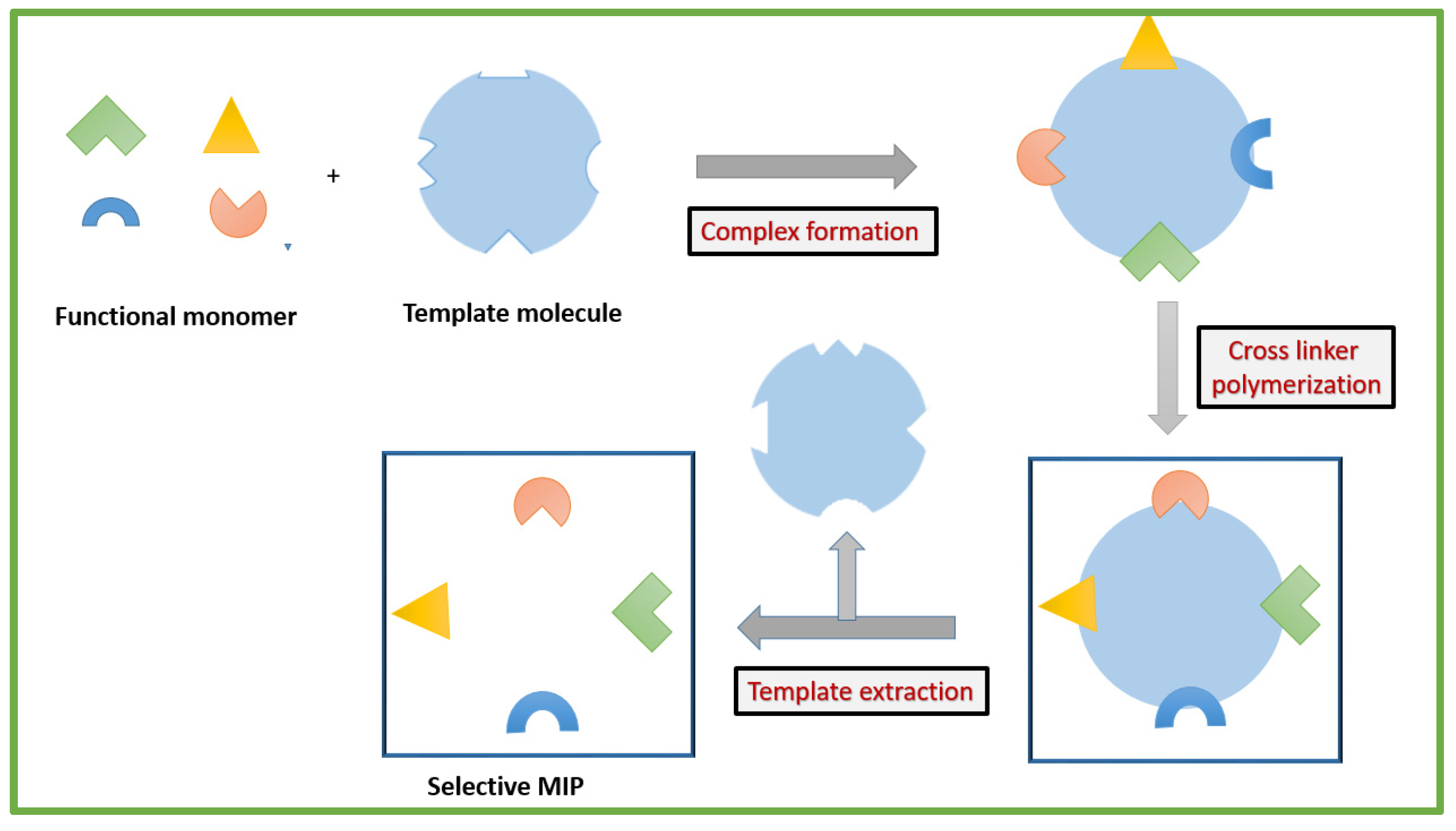
2. General Synthesis Procedure of an MIP
Polymerization Method of Molecular Imprinting
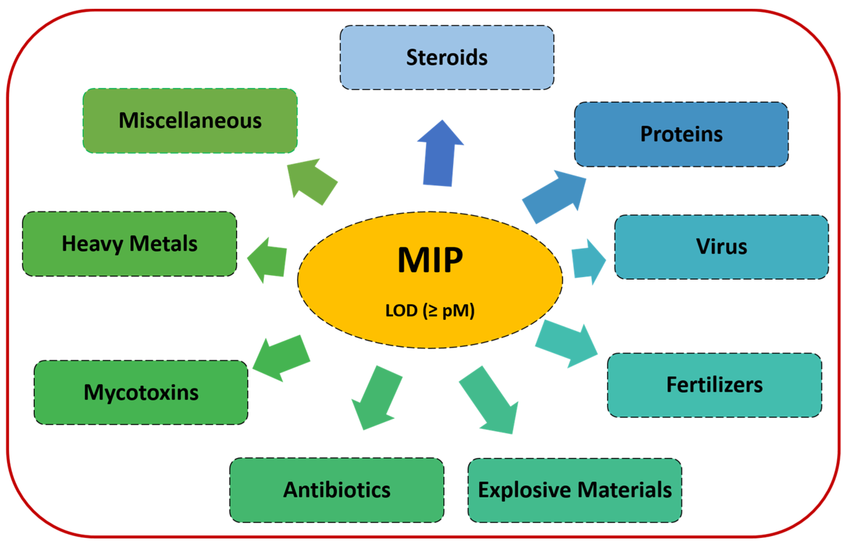
3. Determination of Various Analytes via MIPs
3.1. Steroids, Hormones, and Metabolites
3.2. Proteins
3.3. Virus
3.4. Fertilizers
3.5. Explosive Materials
3.6. Antibiotics
3.7. Mycotoxins
3.8. Heavy Metals
3.9. Miscellaneous
4. Critical Aspects of Binding Models and Imprinting in MIPs
5. Conclusions and Future Perspectives of MIPs Achieving Ultrasensitive LODs
Funding
Institutional Review Board Statement
Informed Consent Statement
Data Availability Statement
Conflicts of Interest
References
- Ramanavicius, S.; Jagminas, A.; Ramanavicius, A. Advances in molecularly imprinted polymers based affinity sensors. Polymers 2021, 13, 974. [Google Scholar] [CrossRef] [PubMed]
- Zaidi, S.A. An overview of Bio-inspired intelligent imprinted polymers for virus determination. Biosensors 2021, 11, 89. [Google Scholar] [CrossRef] [PubMed]
- Saylan, Y.; Akgönüllü, S.; Yavuz, H.; Ünal, S.; Denizli, A. Molecularly imprinted polymer based sensors for medical applications. Sensors 2019, 19, 1279. [Google Scholar] [CrossRef] [Green Version]
- Hussain, S.; Zaidi, S.A.; Vikraman, D.; Kim, H.-S.; Jung, J. Fabrication of molecularly imprinted polymer on tungsten carbide nanoparticles loaded glassy carbon as potential electrochemical sensors for oxalic acid. Microchem. J. 2020, 159, 105404. [Google Scholar] [CrossRef]
- Xu, S.; Wang, L.; Liu, Z. Molecularly imprinted polymer nanoparticles: An emerging versatile platform for cancer therapy. Angew. Chem. Int. 2021, 60, 3858–3869. [Google Scholar] [CrossRef] [PubMed]
- Zaidi, S.A. Bacterial Imprinting Methods and Their Applications: An overview. Crit. Rev. Anal. Chem. 2021, 51, 609–618. [Google Scholar] [CrossRef]
- Crapnell, R.D.; Dempsey-Hibbert, N.C.; Peeters, M.; Tridente, A.; Banks, C.E. Molecularly imprinted polymer based electrochemical biosensors: Overcoming the challenges of detecting vital biomarkers and speeding up diagnosis. Talanta Open 2020, 2, 100018. [Google Scholar] [CrossRef]
- Ozcelikay, G.; Kaya, S.; Ozkan, E.; Cetinkaya, A.; Nemutlu, E.; Kır, S.; Ozkan, S. Sensor-based MIP technologies for targeted metabolomics analysis. Trends Anal. Chem. 2022, 146, 116487. [Google Scholar] [CrossRef]
- Zidarič, T.; Finšgar, M.; Maver, U.; Maver, T. Artificial Biomimetic Electrochemical Assemblies. Biosensors 2022, 12, 44. [Google Scholar] [CrossRef]
- Leibl, N.; Haupt, K.; Gonzato, C.; Duma, L. Molecularly Imprinted Polymers for Chemical Sensing: A tutorial review. Chemosensors 2021, 9, 123. [Google Scholar] [CrossRef]
- Hussain, S.; Zaidi, S.A.; Vikraman, D.; Kim, H.S. Facile preparation of molybdenum carbide (Mo2C) nanoparticles and its effective utilization in electrochemical sensing of folic acid via imprinting. Biosens. Bioelectron. 2019, 140, 111330. [Google Scholar] [CrossRef] [PubMed]
- Zaidi, S.A. An account on the versatility of Dopamine as a functional monomer in molecular imprinting. ChemistrySelect 2019, 4, 5081–5090. [Google Scholar] [CrossRef]
- Mujahid, A.; Mustafa, G.; Dickert, F.L. Label-Free Bioanalyte Detection from Nanometer to Micrometer Dimensions—Molecular Imprinting and QCMs. Biosensors 2018, 8, 52. [Google Scholar] [CrossRef] [PubMed] [Green Version]
- Belbruno, J. Molecularly imprinted polymers. Chem. Rev. 2019, 119, 94–119. [Google Scholar] [CrossRef]
- Zaidi, S. Molecular Effective imprinting of an anticancer drug, 6-thioguanine, via mussel inspired self-polymerization of dopamine over reduced graphene oxide. Analyst 2019, 144, 2345–2352. [Google Scholar] [CrossRef]
- Zaidi, S.A. Molecular imprinting prevents environmental contamination and body toxicity from anticancer drugs: An update. Crit. Rev. Anal. Chem. 2019, 49, 324–335. [Google Scholar] [CrossRef]
- Leal, P.M.; Frías, I.A.M.; Alonso, E.V.; Errachid, A.; Renault, N.J. A Molecularly Imprinted Polypyrrole/GO@Fe3O4 Nanocomposite Modified Impedimetric Sensor for the Routine Monitoring of Lysozyme. Biosensors 2022, 12, 727. [Google Scholar] [CrossRef]
- Culver, H.R.; Peppas, N.A. Protein-imprinted polymers: The shape of things to come? Chem. Mater 2017, 29, 5753–5761. [Google Scholar] [CrossRef]
- Gama, M.R.; Bottoli, C.B.G. Molecularly imprinted polymers for bioanalytical sample preparation. J. Chromatogr. 2017, 1043, 107–121. [Google Scholar] [CrossRef]
- Zaidi, S.A. Utilization of an Environmentally-Friendly Monomer for an Efficient and Sustainable Adrenaline Imprinted Electrochemical Sensor using graphene. Electrochim. Acta 2018, 274, 370–377. [Google Scholar]
- Haupt, K.; Linares, A.V.; Bompart, M.; Bui, B.T. Molecularly imprinted polymers. Top. Curr. Chem. 2012, 325, 1–28. [Google Scholar] [CrossRef] [PubMed]
- Bossi, A.; Bonnini, F.; Turner, A.P.F.; Piletksy, S.A. Moleculary imprinted polymers for the recognition of proteins: The state of the art. Biosens. Bioelectron. 2007, 22, 1131–1137. [Google Scholar] [CrossRef] [PubMed]
- Xu, S.; Xu, Z.; Liu, Z. Paper-Based Molecular-Imprinting Technology and Its Application. Biosensors 2022, 12, 595. [Google Scholar] [CrossRef] [PubMed]
- Jang, R.; Kim, K.H.; Zaidi, S.A.; Cheong, W.J.; Mun, M.H. Analysis of phospholipids using an open-tubular capillary column with a monolithic layer of molecularly imprinted polymer in capillary electrochromatography-electrospray ionization-tandem mass spectrometry. Electrophoresis 2011, 32, 2167–2173. [Google Scholar] [CrossRef]
- Vasapollo, G.; Sole, R.D.; Mergola, L.; Lazzoi, M.R.; Scardino, A.; Scorrano, S.; Mele, G. Molecularly imprinted polymers: Present and future prospective. Int. J. Mol. Sci. 2011, 12, 5908–5945. [Google Scholar] [CrossRef] [Green Version]
- Kryscio, D.R.; Peppas, N.A. Critical review and perspective of macromolecularly imprinted polymers. Acta Biomater. 2012, 8, 461–473. [Google Scholar] [CrossRef] [PubMed]
- Zaidi, S.A. Molecular imprinted polymer as drug delivery vehicles. Drug Deliv. 2014, 161, 1–10. [Google Scholar] [CrossRef] [PubMed]
- Haupt, K.; Rangel, X.P.; Bui, B.T.S. Molecularly imprinted polymers: Antibody mimics for bioimaging and therapy. Chem. Rev. 2020, 120, 9554–9582. [Google Scholar] [CrossRef]
- Poma, A.; Guerreiro, A.; Whitcombe, M.J. Solid-phase synthesis of molecularly imprinted nanoparticles with a reusable template—plastic antibodies. Adv. Funct. Mater. 2013, 23, 2821–2827. [Google Scholar] [CrossRef] [Green Version]
- Uzun, L.; Turner, A.P.F. Molecularly-imprinted polymer sensors: Realizing their potential. Biosens. Bioelectron. 2016, 76, 131–144. [Google Scholar] [CrossRef]
- Goncalves, L.M. Electroploymerized molecularly imprinted polymers: Perceptions based on recent literature for soon-to-be world-class scientists. Curr. Opin. Electrochem. 2021, 25, 100640. [Google Scholar] [CrossRef]
- Erdossy, J.; Horvath, V.; Yarman, A.; Scheller, F.W.; Gyurcsanyi, R.E. Electro synthesized molecularly imprinted polymers for protein recognition. Trends Anal. Chem. 2016, 79, 179–190. [Google Scholar] [CrossRef] [Green Version]
- Spivak, D.A. Optimization, evaluation, and characterization of molecularly imprinted polymers. Adv. Drug Deliv. Rev. 2005, 57, 1779–1794. [Google Scholar] [CrossRef] [PubMed]
- Wloch, M.; Datta, J. Synthesis and polymerization techniques of molecularly imprinted polymers. Compr. Anal. Chem. 2019, 86, 17–40. [Google Scholar] [CrossRef]
- Mayes, A.G.; Mosbach, K. Molecularly imprinted polymers: Useful materials for analytical chemistry? Trends Anal. Chem. 1997, 16, 321–332. [Google Scholar] [CrossRef]
- Zhang, H.; Ye, L.; Mosbach, K. Non-covalent molecular imprinting with emphasis on its application in separation and drug development. J. Mol. Recognit. 2006, 19, 248–259. [Google Scholar] [CrossRef]
- Mayes, A.G.; Whitcombe, M.J. Synthetic strategies for the generation of molecularly imprinted organic polymers. Adv. Drug Deliv. Rev. 2005, 57, 1742–1778. [Google Scholar] [CrossRef]
- Sellergren, B. Noncovalent molecular imprinting: Antibody-like molecular recognition in polymeric network materials. Trends Anal. Chem. 1997, 16, 310–320. [Google Scholar] [CrossRef]
- Schirhagl, R. Bioapplications for molecularly imprinted polymers. Anal. Chem. 2014, 86, 250–261. [Google Scholar] [CrossRef]
- Canfarotta, F.; Cecchini, A.; Piletsky, S. Chapter 1: Nano sized Moleculary Imprinted Polymers as Artificial Antibodies. Mol. Impr. Polym. Anal. Chem. Appl. 2018, 1–27. [Google Scholar] [CrossRef]
- Archana, S.N. Molecularly Imprinted Polymer Composites: Synthesis, Characterization and Application, 1st ed.; Sooraj, M.P., Archana, S.N., Beena, M., Sabu, T., Eds.; Elsevier Science & Technology: Amsterdam, The Netherlands, 2021. [Google Scholar]
- Yan, H.; Row, K.H. Characteristic and Synthetic Approach of Molecularly Imprinted Polymer. Int. J. Mol. Sci. 2006, 7, 155–178. [Google Scholar] [CrossRef]
- Jian, J.; Zhihui, Z.; Zhao, X.; Sun, J. Electrochemical sensor based on molecularly imprinted film at Au nanoparticles-carbon nanotubes modified electrode for determination of cholesterol. Biosens. Bioelectron. 2015, 66, 590–595. [Google Scholar] [CrossRef]
- Florea, A.; Cristea, C.; Vocanson, F.; Səndulescu, R.; Renault, N.J. Electrochemical Sensor for the Detection of Estradiol Based on Electropolymerized Molecularly Imprinted Polythioaniline Film with Signal Amplification Using Gold Nanoparticles. Electrochem. Commun. 2015, 59, 36–39. [Google Scholar] [CrossRef]
- Liu, W.; Li, H.; Yu, S.; Zhang, J.; Zheng, W.; Niu, L.; Li, G. Poly(3,6-Diamino-9-Ethylcarbazole) Based Molecularly Imprinted Polymer Sensor for Ultra-Sensitive and Selective Detection of 17-β-Estradiol in Biological Fluids. Biosens. Bioelectron. 2018, 104, 79–86. [Google Scholar] [CrossRef] [PubMed]
- Zhang, Q.; Jing, L.; Wang, Y.; Zhang, J.; Ren, Y.; Wang, Y.; Wei, T.; Liedberg, B. Surface Plasmon Resonance Sensor for Femtomolar Detection of Testosterone with Water-Compatible Macroporous Molecularly Imprinted Film. Anal. Biochem. 2014, 463, 7–14. [Google Scholar] [CrossRef] [PubMed]
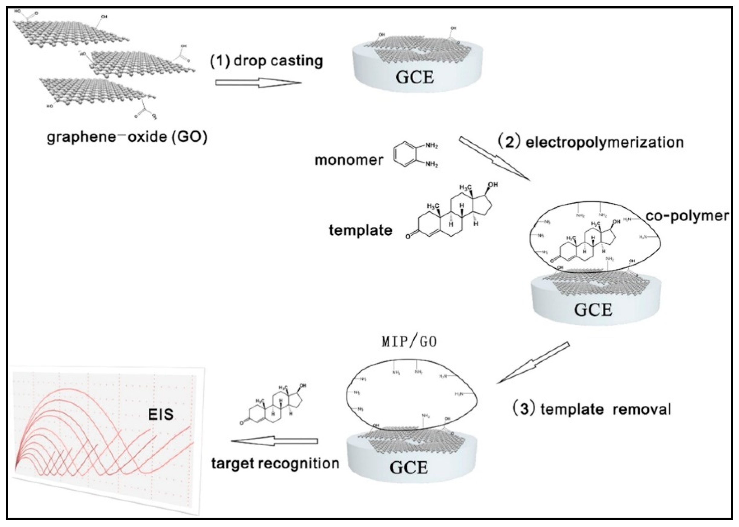
- Liu, W.; Ma, Y.; Sun, G.; Wang, S.; Deng, J.; Wei, H. Molecularly Imprinted Polymers on Graphene Oxide Surface for EIS Sensing of Testosterone. Biosens. Bioelectron. 2017, 92, 305–312. [Google Scholar] [CrossRef]
- Pareek, S.; Jain, U.; Balayan, S.; Chauhan, N. Ultra-Sensitive Nano- Molecular Imprinting Polymer-Based Electrochemical Sensor for Follicle-Stimulating Hormone (FSH) Detection. Biochem. Eng. J. 2022, 180, 108329. [Google Scholar] [CrossRef]
- Ozkan, E.; Çorman, M.E.; Nemutlu, E.; Ozkan, S.A.; Kır, S. Development of an electrochemical sensor based on porous molecularly imprinted polymer via photopolymerization for detection of somatostatin in pharmaceuticals and human serum. J. Electroanal. Chem. 2022, 919, 116554. [Google Scholar] [CrossRef]
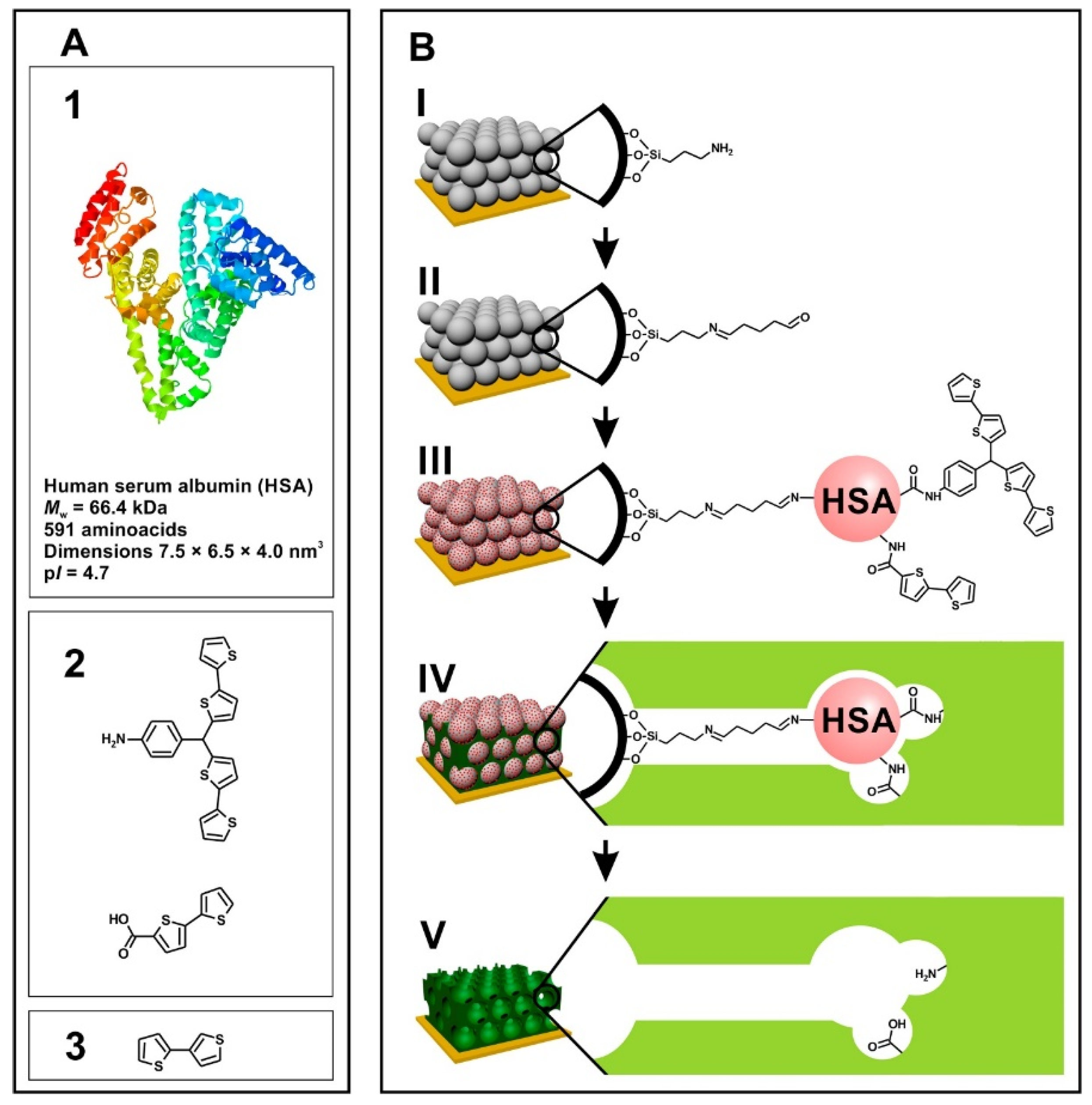
- Dabrowski, M.; Cieplak, M.; Sharma, P.S.; Borowicz, P.; Noworyta, K.; Lisowski, W.; D’Souza, F.; Kuhn, A.; Kutner, W. Hierarchical templating in deposition of semi-covalently imprinted inverse opal polythiophene film for femtomolar determination of human serum albumin. Biosens. Bioelectron. 2017, 94, 155–161. [Google Scholar] [CrossRef]
- Piloto, A.M.; Ribeiro, D.S.M.; Rodrigues, S.S.M.; Santos, C.; Santos, J.L.; Sales, M.G.F. Plastic Antibodies Tailored on Quantum Dots for an Optical Detection of Myoglobin down to the Femtomolar Range. Sci. Rep. 2018, 8, 4944. [Google Scholar] [CrossRef] [Green Version]
- You, M.; Yang, S.; An, Y.; Zhang, F.; He, P. A novel electrochemical biosensor with molecularly imprinted polymers and aptamer-based sandwich assay for determining amyloid-β oligomer. J. Electroanal. Chem. 2020, 862, 114017. [Google Scholar] [CrossRef]
- Babamiri, B.; Salimi, A.; Hallaj, R. A Molecularly Imprinted Electrochemiluminescence Sensor for Ultrasensitive HIV-1 Gene Detection Using EuS Nanocrystals as Luminophore. Biosens. Bioelectron. 2018, 117, 332–339. [Google Scholar] [CrossRef] [PubMed]
- Raziq, A.; Kidakova, A.; Boroznjak, R.; Reut, J.; Öpik, A.; Syritski, V. Development of a portable MIP-based electrochemical sensor for detection of SARS-CoV-2 antigen. Biosens. Bioelectron. 2021, 178, 113099. [Google Scholar] [CrossRef]
- Ayankojo, A.G.; Boroznjak, R.; Reut, J.; Öpik, A.; Syritski, V. Molecularly Imprinted Polymer Based Electrochemical Sensor for Quantitative Detection of SARS-CoV-2 Spike Protein. Sens. Actuators B Chem. 2022, 353, 131160. [Google Scholar] [CrossRef]
- Amouzadeh Tabrizi, M.; Fernández-Blázquez, J.P.; Medina, D.M.; Acedo, P. An Ultrasensitive Molecularly Imprinted Polymer-Based Electrochemical Sensor for the Determination of SARS-CoV-2-RBD by Using Macroporous Gold Screen-Printed Electrode. Biosens. Bioelectron. 2022, 196, 113729. [Google Scholar] [CrossRef]
- Das, K.; Penelle, J.; Rotello, V.M. Selective Picomolar Detection of Hexachlorobenzene in Water Using a Quartz Crystal Microbalance Coated with a Molecularly Imprinted Polymer Thin Film. Langmuir 2003, 19, 3921–3925. [Google Scholar] [CrossRef]
- Mazouz, Z.; Rahali, S.; Fourati, N.; Zerrouki, C.; Aloui, N.; Seydou, M.; Yaakoubi, N.; Chehimi, M.M.; Othmane, A.; Kalfat, R. Highly Selective Polypyrrole MIP-Based Gravimetric and Electrochemical Sensors for Picomolar Detection of Glyphosate. Sensors 2017, 17, 2586. [Google Scholar] [CrossRef] [Green Version]
- Li, S.; Li, J.; Luo, J.; Xu, Z.; Ma, X. A microfluidic chip containing a molecularly imprinted polymer and a DNA aptamer for voltammetric determination of carbofuran. Microchim. Acta 2018, 185, 295. [Google Scholar] [CrossRef]
- Roushani, M.; Nezhadali, A.; Jalilian, Z. An electrochemical chlorpyrifos aptasensor based on the use of a glassy carbon electrode modified with an electropolymerized aptamer-imprinted polymer and gold nanorods. Mikrochim. Acta 2018, 185, 551. [Google Scholar] [CrossRef]
- Guo, Z.; Florea, A.; Cristea, C.; Bessueille, F.; Vocanson, F.; Goutaland, F.; Zhang, A.; Sandulescu, R.; Lagrade, F.; Renault, N.J. 1,3,5-Trinitrotoluene detection by a molecularly imprinted polymer sensor based on electropolymerization of a microporous-metal-organic framework. Sens. Actuators B Chem. 2015, 207, 960–966. [Google Scholar] [CrossRef]
- Shahdost-fard, F.; Roushani, M. Impedimetric detection of trinitrotoluene by using a glassy carbon electrode modified with a gold nanoparticle@fullerene composite and an aptamer-imprinted polydopamine. Microchim. Acta 2017, 184, 3997–4006. [Google Scholar] [CrossRef]
- Alizadeh, T.; Atashi, F.; Ganjali, M.R. Molecularly imprinted polymer nano-sphere/multi-walled carbon nanotube coated glassy carbon electrode as an ultra-sensitive voltammetric sensor for picomolar level determination of RDX. Talanta 2019, 194, 415–421. [Google Scholar] [CrossRef] [PubMed]
- Rad, A.O.; Azadbakht, A. An Aptamer Embedded in a Molecularly Imprinted Polymer for Impedimetric Determination of Tetracycline. Microchim. Acta 2019, 186, 56. [Google Scholar] [CrossRef] [PubMed]
- Bougrini, M.; Florea, A.; Cristea, C.; Sandulescu, R.; Vocanson, F.; Errachid, A.; Bouchikhi, B.; El Bari, N.; Jaffrezic-Renault, N. Development of a Novel Sensitive Molecularly Imprinted Polymer Sensor Based on Electropolymerization of a Microporous-Metal-Organic Framework for Tetracycline Detection in Honey. Food Control 2016, 59, 424–429. [Google Scholar] [CrossRef]
- Li, S.; Liu, C.; Yin, G.; Zhang, Q.; Luo, J.; Wu, N. Aptamer-Molecularly Imprinted Sensor Base on Electrogenerated Chemiluminescence Energy Transfer for Detection of Lincomycin. Biosens. Bioelectron. 2017, 91, 687–691. [Google Scholar] [CrossRef]
- Roushani, M.; Rahmati, Z.; Hoseini, S.J.; Hashemi Fath, R. Impedimetric Ultrasensitive Detection of Chloramphenicol Based on Aptamer MIP Using a Glassy Carbon Electrode Modified by 3-Ampy-RGO and Silver Nanoparticle. Colloids Surf. B Biointerfaces 2019, 183, 110451. [Google Scholar] [CrossRef]
- Lu, H.; Liu, M.; Cui, H.; Huang, Y.; Li, L.; Ding, Y. An advanced molecularly imprinted electrochemical sensor based bifunctional monomers for highly sensitive detection of nitrofurazone. Electrochim. Acta 2022, 427, 140858. [Google Scholar] [CrossRef]
- Guo, W.; Pi, F.; Zhang, H.; Sun, J.; Zhang, Y.; Sun, X. A novel molecularly imprinted electrochemical sensor modified with carbon dots, chitosan, gold nanoparticles for the determination of patulin. Biosens. Bioelectron. 2017, 98, 299–304. [Google Scholar] [CrossRef] [PubMed]
- Munawar, H.; Garcia-Cruz, A.; Majewska, M.; Karim, K.; Kutner, W.; Piletsky, S.A. Electrochemical Determination of Fumonisin B1 Using a Chemosensor with a Recognition Unit Comprising Molecularly Imprinted Polymer Nanoparticles. Sens. Actuators B Chem. 2020, 321, 128552. [Google Scholar] [CrossRef]
- Alizadeh, T.; Hamidi, N.; Ganjali, M.R.; Rafiei, F. An extraordinarily sensitive voltammetric sensor with picomolar detection limit for Pb2+ determination based on carbon paste electrode impregnated with nano-sized imprinted polymer and multi-walled carbon nanotubes. J. Environ. Chem. Eng. 2017, 5, 4327–4336. [Google Scholar] [CrossRef]
- Li, S.; Ma, X.; Pang, C.; Tian, H.; Xu, Z.; Yang, Y.; Lv, D.; Ge, H. Fluorometric aptasensor for cadmium(II) by using an aptamer-imprinted polymer as the recognition element. Microchim. Acta 2019, 186, 823. [Google Scholar] [CrossRef] [PubMed]
- Gupta, G.; Singh, P.K.; Boopathi, M.; Kamboj, D.V.; Singh, B.; Vijayaraghavan, R. Molecularly imprinted polymer for the recognition of biological warfare agent staphylococcal enterotoxin B based on Surface Plasmon Resonance. Thin Solid Film. 2010, 519, 1115–1121. [Google Scholar] [CrossRef]
- Cenci, L.; Andreetto, E.; Vestri, A.; Bovi, M.; Barozzi, M.; Lacob, E.; Busato, M.; Castagna, A.; Girelli, D.; Bossi, A.M. Surface plasmon resonance based on molecularly imprinted nanoparticles for the picomolar detection of the iron regulating hormone Hepcidin-25. J. Nanobiotechnol. 2015, 13, 51. [Google Scholar] [CrossRef] [Green Version]
- Yarahmadi, S.; Azadbakht, A.; Derikvand, R.M. Hybrid synthetic receptor composed of molecularly imprinted polydopamine and aptamers for impedimetric biosensing of urea. Microchim. Acta 2019, 186, 71. [Google Scholar] [CrossRef]
- Parnianchi, F.; Kashanian, S.; Nazari, M.; Santoro, C.; Bollella, P.; Varmira, K. Highly selective and sensitive molecularly imprinting electrochemical sensing platform for bilirubin detection in saliva. Microchem. J. 2021, 168, 106367. [Google Scholar] [CrossRef]
- Roy, S.; Nagabooshanam, S.; Wadhwa, S.; Chauhan, N.; Mathur, A.; Khan, S.A.; Davis, J. Ultra-sensitive detection of l-tyrosine using molecularly imprinted electrochemical sensor towards diabetic foot ulcer detection. Electrochem. Commun. 2020, 117, 106782. [Google Scholar] [CrossRef]
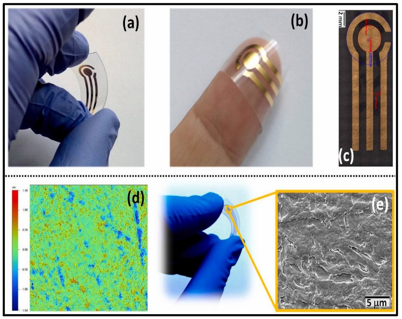
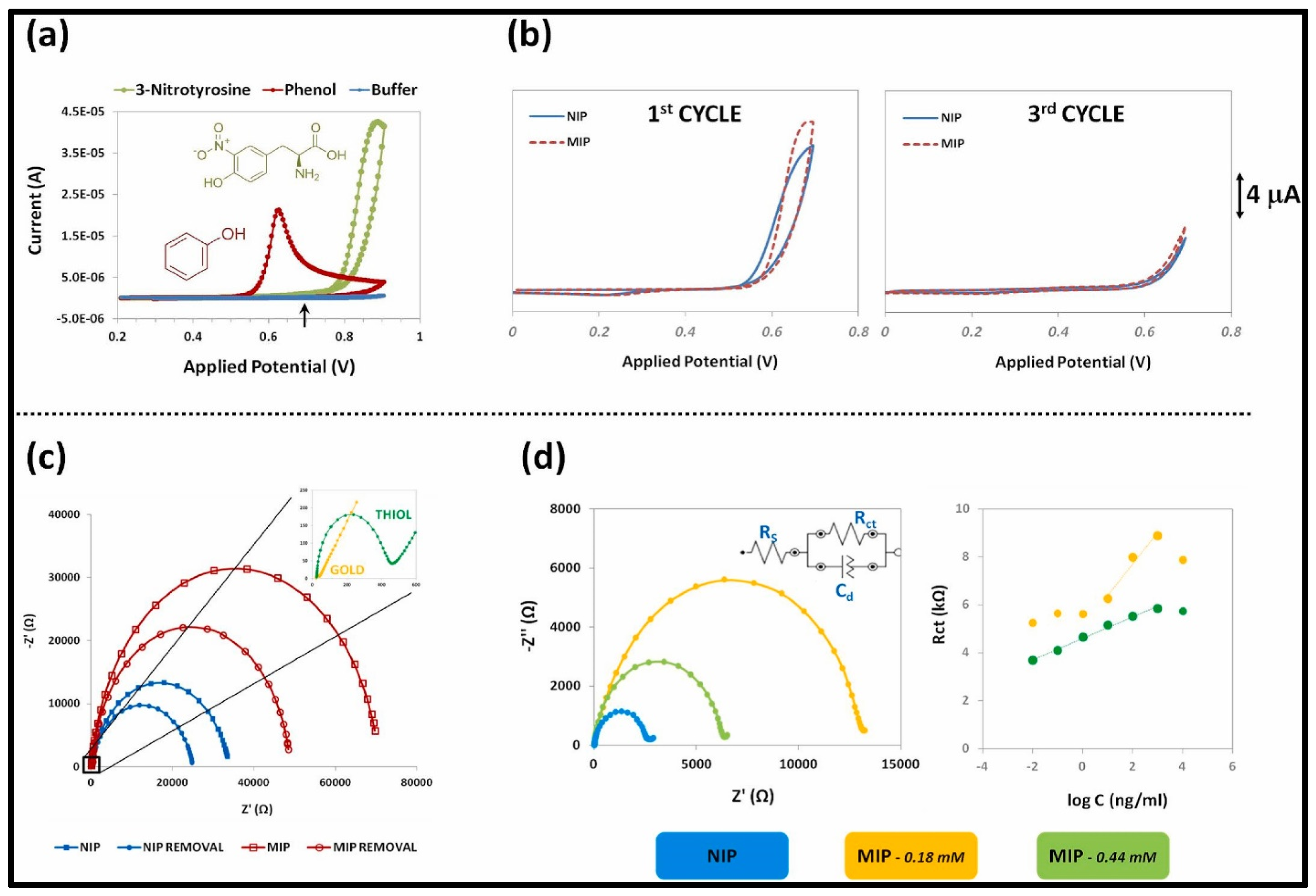
- Martins, G.V.; Riveiro, A.; Chiussi, S.; Sales, M.G.F. Flexible sensing devices integrating molecularly-imprinted polymers for the detection of 3-nitrotyrosine biomarker. Biosens. Bioelectron. 2022, 10, 100107. [Google Scholar] [CrossRef]
- Moncer, F.; Adhoum, N.; Catak, D.; Monser, L. Electrochemical sensor based on MIP for highly sensitive detection of 5-hydroxyindole-3-acetic acid carcinoid cancer biomarker in human biological fluids. Anal. Chim. Acta 2021, 1181, 338925. [Google Scholar] [CrossRef] [PubMed]
- Alizadeh, T.; Akhoundian, M. An ultra-sensitive and highly selective impedimetric sensor for vitamin D measurement based on a novel imprinted polymer synthesized utilizing template-derived functional monomer. Anal. Chim. Acta 2022, 1223, 340206. [Google Scholar] [CrossRef]
- Ensafi, A.A.; Amini, M.; Rezaei, B. Molecularly imprinted electrochemical aptasensor for the attomolar detection of bisphenol A. Microchim. Acta 2018, 185, 265. [Google Scholar] [CrossRef]
- Kaya, S.I.; Corman, M.E.; Uzun, L.; Ozkan, S.A. A porous molecularly imprinted electrochemical sensor for specific determination of bisphenol S from human serum and bottled water samples in femtomolar level. Anal. Bioanal. Chem. 2022, 414, 2775–2785. [Google Scholar] [CrossRef]
- Bakas, I.; Salmi, Z.; Jouini, M.; Geneste, F.; Mazerie, I.; Floner, D.; Carbonnier, B.; Yagci, Y.; Chehimi, M.M. Picomolar Detection of Melamine Using Molecularly Imprinted Polymer-Based Electrochemical Sensors Prepared by UV-Graft Photopolymerization. Electroanalysis 2015, 27, 429–439. [Google Scholar] [CrossRef]
- Elizabeth, N.N. Molecularly imprinted polymers—A closer look at the control polymer used in determining the imprinting effect: A mini review. J. Mol. Recognit. 2020, 33, 11. [Google Scholar]
- Molecularly Imprinted Polymers in Biotechnology; Mattiasson, B.; Ye, L. (Eds.) Springer: Berlin/Heidelberg, Germany, 2015; Volume 150, pp. 51–93. [Google Scholar] [CrossRef]
- Toth, B.; Pap, T.; Horvath, V.; Horvai, G. Nonlinear adsorption isotherm as a tool for understanding and characterizing molecularly imprinted polymers. J. Chromatogr. A 2006, 1119, 29–33. [Google Scholar] [CrossRef]
- Toth, B.; Pap, T.; Horvath, V.; Horvai, G. Which molecularly imprinted polymer is better? Anal. Chim. Acta 2007, 591, 17–21. [Google Scholar] [CrossRef] [PubMed]
- Castell, O.K.; Barrow, D.A.; Kamarudin, A.R.; Allender, C.J. Current practices for describing the performance of molecularly imprinted polymers can be misleading and may be hamper in the development of the field. J. Mol. Recognit. 2011, 24, 1115–1122. [Google Scholar] [CrossRef]
- Baggiani, C.; Giovannoli, C.; Anfossi, L.; Passini, C.; Baravalle, P.; Giraudi, G. A Connection between the Binding Properties of Imprinted and Nonimprinted Polymers: A Change of Perspective in Molecular Imprinting. J. Am. Chem. Soc. 2012, 134, 1513–1518. [Google Scholar] [CrossRef] [Green Version]
- Kastritis, P.L.; Bonvin, A.M.J.J. On the binding affinity of macromolecular interactions: Daring to ask why proteins interact. J. R. Soc. Interface 2013, 10, 2012835. [Google Scholar] [CrossRef] [PubMed]


| Matrix | Analyte | Linear Range | LOD/LOQ * | Detection or /Electrochemcial Readout Method | % Recovery in Real Samples | Ref. |
|---|---|---|---|---|---|---|
| MIP/AuNPs–MWNTs/GCE | Cholesterol | 0.1 pM–1 nM | 0.33 pM | DPV | NR | [43] |
| MIP-AuNP | 17-β-estradiol | 3.6 fM–3.6 nM | 1.09 fM/3.6 fM * | LSV | river water (95.6–103.6) | [44] |
| 3,6-diamino-9-ethylcarbazole/MIP | 1 aM–10 μM | 0.36 aM | EIS | human serum (96.6–104.6) | [45] | |
| MIP/ PSNPs | Testosterone | NR | 3.5 fM | SPR | NR | [46] |
| MIP/GO | 1 fM–1µM | 0.4 fM | EIS | human serum (98.6–104.2) | [47] | |
| NiCo2O4/rGO/MIP/ITO | Follicle stimulating hormone (FSH) | 0.1 pM–1µM | 0.1 pM | EIS | blood samples (90–98.79) | [48] |
| MAAsp/MIP/GCE | Somatostatin (SOM) | 10–100 fM | 0.175 fM/ 0.584 fM * | DPV | serum samples (99.32–100.25) | [49] |
| MIP- EG-FET | Human Serum Albumin | 15 fM–150 fM | 13 fM | EG-FET-Potentiostat | NR | [50] |
| Cd-Te-MPA QDs/MIP | Myoglobin (Myo) | 50.6 fM–95 pM | 7.6 fM | Fluorescence | NR | [51] |
| Aptamer/MIP/SiO2@Ag NPs | Amyloid-β oligomers (AβOs) | 5–10 pg mL−1 | 1.22 pg/ml | DPV | human serum (93–107.7) | [52] |
| EuSNCs/ECL-MIP/ITO | Human immunodeficiency virus (HIV) | 3.0 fM–0.3 nM | 0.3 fM | ECL | human serum (95–102.1) | [53] |
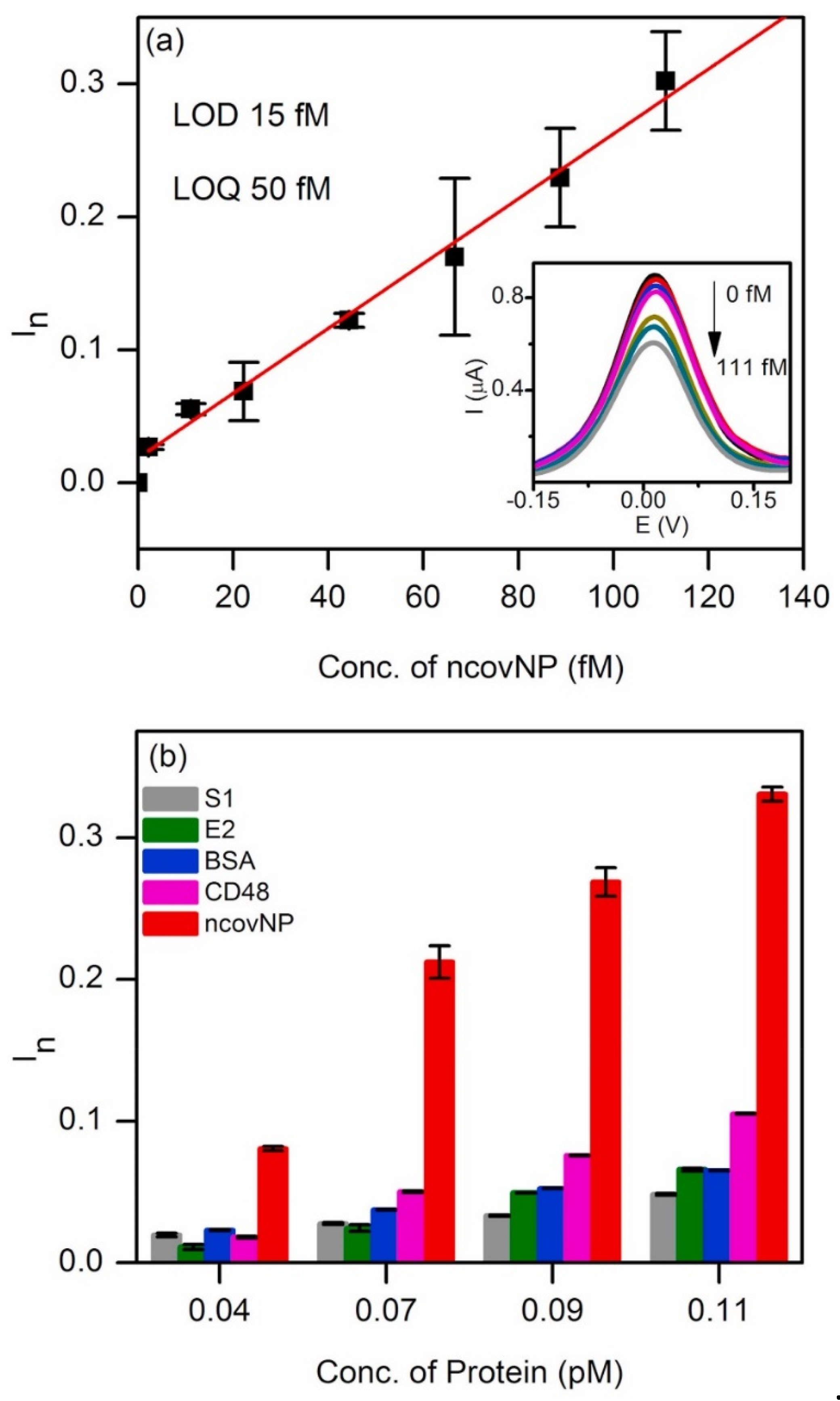
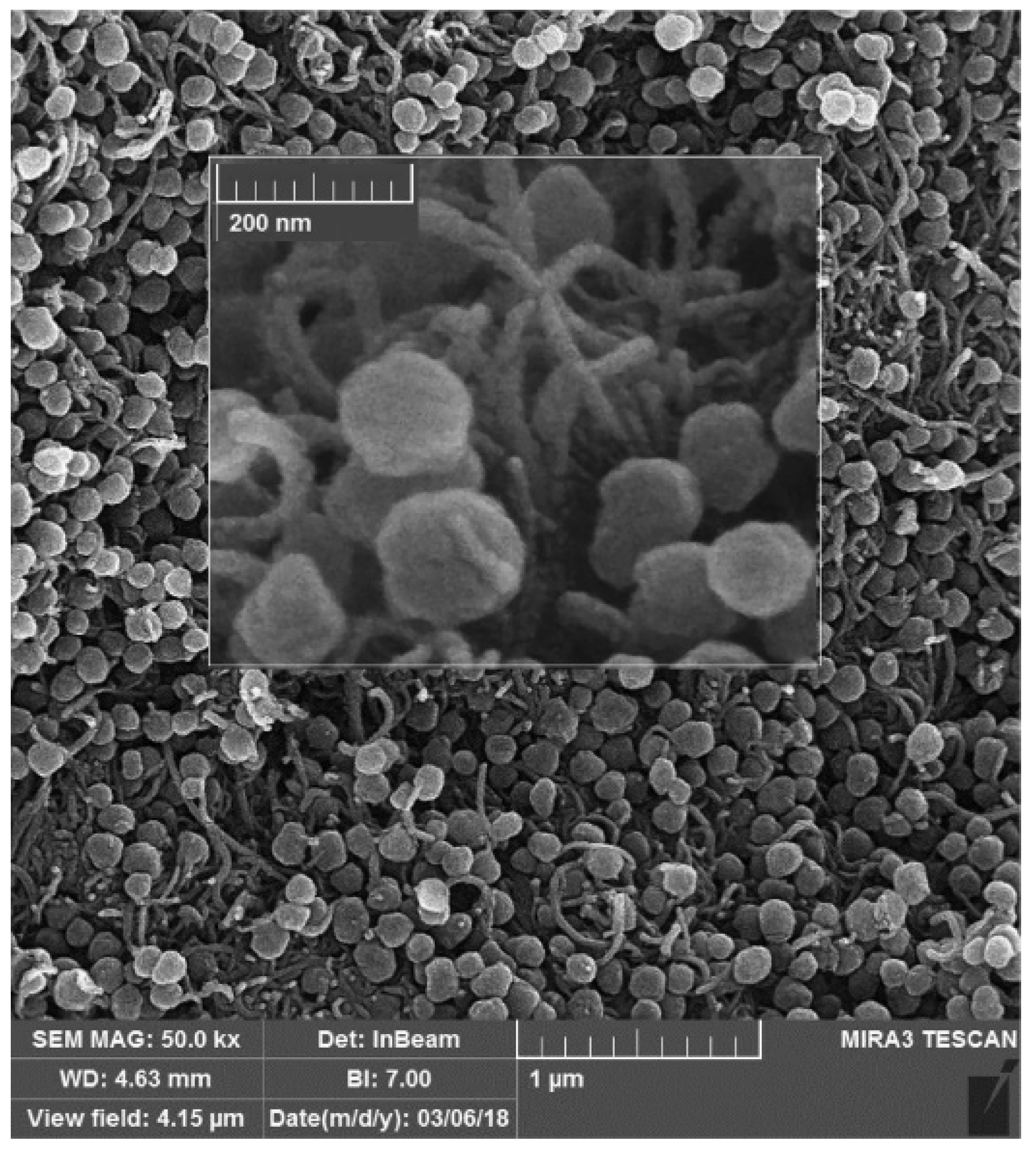
| ncovNP/MIP/AuTFME | SARS-CoV-2 | 2.22–111 fM | 15 fM/50 fM * | DPV | NR | [54] |
| ncovS1/MIP/AuTFME | 2.0–40.0 pg mL−1 | 15 fM/ 51 fM * (PBS) 64 fM/213 fM * (NPS) | SWV | NR | [55] | |
| MPAu/MIP/SPE | 2.0–40.0 pg mL−1 | 20 fM | EIS | NR | [56] | |
| MIP-QCM | Hexachlorobenzene | NR | 1 pM | QCM | NR | [57] |
| PPy/MIP | Glyphosate | 1 pM–1 nM | 1 pM | SWV& Gravimetry (SAW) | NR | [58] |
| Aptamer-AuNP/MIP/GCE | Carbofuran | 0.2 nM–50 nM | 67 pM | DPV | Chinese cabbage (95.6), chili (110.4) lettuce (101.5) tomato (94.5), apple (92.6) banana (91.2) tangerine (89.6) watermelon (102.6) | [59] |
| aptamer-MIP/AuNP/GCE | Chlorpyrifos | 1.0 fM–0.4 pM | 0.35 fM | DPV | Apple(97.65–102.5) lettuce (98.2–103) | [60] |
| PATP/AuNP/MIP | 1,3,5--TNT | 44 nM–4.4 fM | 0.044 fM | LSV | tap water (96.70–102.20), river water (100.2) | [61] |
| aptamer-MIP nanohybrid/AuNP@C60/GCE | 2,4,6-TNT | 0.01 fM–1.5 µM | 3.5 aM | EIS | soil (98.50–100.50) river water (97.0–100.80) | [62] |
| MIP/MWCNTs-GCE | RDX | 0.01–1.00 µM | 20 pM/ 0.2 nM * | DPV | tap water (97.00–106.0) sea water (94.00–108.0) river water (90.00–97.50) | [63] |
| MMOF/MIP/AuNps | Tetracycline (TC) | 224 fM- 22.4 nM | 0.22 fM | LSV | spiked honey (101.84–106.1) | [64] |
| Aptamer/MIP/Au-GCE | Tetracycline (TET) | 0.5–100 pM and 1–1000 nM | 144 fM | EIS | milk (94.90–106.2) | [65] |
| Aptamer/ECL-MIP/Au-GO/GCE | Lincomycin | 5.0 pM–1.0 nM | 0.16 pM | ECL | Chicken (97.2),duck (90.2) crucian (100.1), pork (89.9), crab (103.1), beef (94.5), mutton (104.5) | [66] |
| AgNPs/3-ampy-RGO/MIP/GCE | Chloramphenicol (CAP) | 1.0 pM–1.0 nM | 0.3 pM | EIS | milk (90–103) | [67] |
| c-MWCNTs/MIP/ZIF | Nitrofurazone (NFZ) | 0.1 pM–1.0 µM | 0.067 pM | CV, DPV | Urine (99.6), water (98.7) | [68] |
| MIP-Au/CS-CDs/GCE | Patulin | 1 pM–1 nM | 75 pM | CV, DPV | Apple juice (96%–98.7%) | [69] |
| Nano-MIP (MIP/PPy-ZnP/Pt) | Fumonisins | 1 fM–10 pM | 0.03 fM 0.7 fM | EIS, DPV | Maize (96–102) | [70] |
| IIP/MWCNTs | Lead, Pb2+ | 0.1 pM–0.8 nM | 3.8 pM | EIS, SWV | sea water (95.40–101.96), river water (97.64–102.40) | [71] |
| SN-CQD/Au/MIP/ITO | Cadmium, Cd2+ | 20 pM–12 nM | 1.2 pM | CV,EIS | Water, soil, vegetable (82.1–113.9) | [72] |
| MIP | Staphylococcal enterotoxin B (SEB) | 3.2–25.6 pM | 0.05 fM | SPR | NR | [73] |
| MIP-NPs | Hepcidin-25 | 7.2–720 pM | 5.0 PM | SPR | NR | [74] |
| HSA | Urea | 0.005–0.1 nM, 1–500 nM | 900 fM | EIS | soil (98.30–104.1), water (99.5–102.0) | [75] |
| OPD/MWCNT/GCE | Bilirubin (BR) | 12.08 fM–91.81 fM | 7.80 fM | DPV | human serum (95.23–103.80), saliva (92.85–102.21) | [76] |
| MIPPy/ITO | L-Tyrosine (TYR) | 100 fM–1 mM | 1.73 pM, 6.63 pM | CV, EIS | NR | [77] |
| Au/MIP | 3-nitro tyrosine (3-NT) | 10 pg mL−1–1 μg mL−1 | 24.9 pM | CV | NR | [78] |
| MIP/GCE | 5-hydroxyindole-3-acetic acid (5-HIAA) | 50 pM–50 µM | 15 pM | DPV | serum (99.40–100.21), plasma (99.84–100.46), urine (98.97–101.52) | [79] |
| MIPNPs-CPE | Vitamin D3 (VD3) | 1.0–100.0 pM | 0.22 pM/ 0.73 pM * | EIS | plasma samples (94.7–104.6) | [80] |
| BPA@p-63aptamer/AuNP/GCE | Bisphenol A | 0.5 fM–5 pM | 80 aM | EIS | Fresh Milk (96.0), Milk Powder (102.0), Tap Water (94.0), Pretreated water in baby glass (96) | [81] |
| MA-TyrMA-Tyr@MIP/GCE | Bisphenol S | 1 fM–10 fM | 0.17 fM/ 0.569 fM * | CV, DPV | Serum (102.9) water (98.30–101.56) | [82] |
| Au-DMA/MIP/GCE | Melamine | NR | 1.75 pM | SWV | Milk Sample (~95%) | [83] |
Publisher’s Note: MDPI stays neutral with regard to jurisdictional claims in published maps and institutional affiliations. |
© 2022 by the authors. Licensee MDPI, Basel, Switzerland. This article is an open access article distributed under the terms and conditions of the Creative Commons Attribution (CC BY) license (https://creativecommons.org/licenses/by/4.0/).
Share and Cite
Shah, N.S.; Thotathil, V.; Zaidi, S.A.; Sheikh, H.; Mohamed, M.; Qureshi, A.; Sadasivuni, K.K. Picomolar or beyond Limit of Detection Using Molecularly Imprinted Polymer-Based Electrochemical Sensors: A Review. Biosensors 2022, 12, 1107. https://doi.org/10.3390/bios12121107
Shah NS, Thotathil V, Zaidi SA, Sheikh H, Mohamed M, Qureshi A, Sadasivuni KK. Picomolar or beyond Limit of Detection Using Molecularly Imprinted Polymer-Based Electrochemical Sensors: A Review. Biosensors. 2022; 12(12):1107. https://doi.org/10.3390/bios12121107
Chicago/Turabian StyleShah, Naheed Sidiq, Vandana Thotathil, Shabi Abbas Zaidi, Hanan Sheikh, Maimoona Mohamed, Ahmadyar Qureshi, and Kishor Kumar Sadasivuni. 2022. "Picomolar or beyond Limit of Detection Using Molecularly Imprinted Polymer-Based Electrochemical Sensors: A Review" Biosensors 12, no. 12: 1107. https://doi.org/10.3390/bios12121107