Review

Review of Thin-Layer Chromatography Tandem with Surface-Enhanced Raman Spectroscopy for Detection of Analytes in Mixture Samples

1
School of Petrochemical Engineering, Liaoning Petrochemical University, Fushun 113001, China
2
Jiangsu Co-Innovation Center for Efficient Processing and Utilization of Forest Resources and Joint International Research Lab of Lignocellulosic Functional Materials, Nanjing Forestry University, Nanjing 210037, China
3
School of Electrical Engineering and Computer Science, Oregon State University, Corvallis, OR 97331, USA
*
Author to whom correspondence should be addressed.
Biosensors 2022, 12(11), 937; https://doi.org/10.3390/bios12110937
Submission received: 28 September 2022 / Revised: 20 October 2022 / Accepted: 25 October 2022 / Published: 28 October 2022

Abstract

:
In the real world, analytes usually exist in complex systems, and this makes direct detection by surface-enhanced Raman spectroscopy (SERS) difficult. Thin layer chromatography tandem with SERS (TLC-SERS) has many advantages in analysis such as separation effect, instant speed, simple process, and low cost. Therefore, the TLC-SERS has great potential for detecting analytes in mixtures without sample pretreatment. The review demonstrates TLC-SERS applications in diverse analytical relevant topics such as environmental pollutants, illegal additives, pesticide residues, toxic ingredients, biological molecules, and chemical substances. Important properties such as stationary phase, separation efficiency, and sensitivity are discussed. In addition, future perspectives for improving the efficiency of TLC-SERS in real sample detecting are outlined.

1. Introduction

Surface-enhanced Raman spectroscopy (SERS) is a type of molecular vibrational spectroscopy with high selectivity and sensitivity. SERS could provide fingerprints information of analytes in a label-free, non-destructive, instant, and simple way. Since the first discovery of enhanced Raman signals of pyridine from roughened silver surfaces in the 1970s, SERS has been widely applied in analytical areas such as food safety [1], environmental detection [2], disease diagnosis [3], pesticide residues [4], and biochemistry [5]. The common application for SERS is direct detection of the target analyte near or adsorbed on the plasmonic-enhanced substrate. The development of nanotechnology has led to an expansion of SERS applications by using metallic nanomaterials as active substrates. Metallic nanomaterials, especially those that are part of the noble metals, can provide excellent SERS sensitivity even down to the single molecule level. In the real world, the target analytes usually exist in complex systems, and it is difficult to obtain information on analytes from complex systems by the SERS method.
It is necessary to develop an advanced analytical method, combined with a separation technique such as extraction or chromatography [6]. Chromatography is a commonly used separation technique for detecting analytes in mixture samples. Examples are high performance liquid chromatography-mass spectrometry (HPLC-MS) [7], high performance liquid chromatography-ultraviolet (HPLC-UV) [8], and gas chromatography-mass spectrometry (GC-MS) [9]. These combined analytical methods are reliable. However, the GC and HPLC are expensive, time consuming, and complex, which is not convenient for on-site detection [10]. Thin-layer chromatography (TLC) is a simple and instant separation method, which has been commonly used as a cost-effective technique for separating and identifying products in organic synthesis. The tandem of TLC and SERS (TLC-SERS) is an efficient technology for separating and identifying analytes from complex systems. The TLC-SERS has been employed in food safety [11], healthcare [12], chemical analysis [13], and environment protection [14].
In this review, we will focus on the research of the application of TLC-SERS. First, we will briefly overview the phenomenon of SERS and the basic concept of the TLC method. Next, we will discuss the construction and application of TLC-SERS, which show excellent performance in detecting analytes from complex samples. Finally, we will conclude with the challenges and prospects of TLC-SERS, as well, such as how to improve the sensitivity, uniformity, and separation efficiency of TLC-SERS. Meanwhile, it can be predicted that TLC-SERS technology will have broad development space in terms of analysis and instrumentation. Therefore, it is particularly important to review the progress of TLC-SERS.

2. SERS

SERS is a powerful analytical technique that has received considerable attention [15]. In the 1970s, the intense Raman spectra of pyridine was observed from the rough surface of a silver electrode [16]. In 1977, Van Duyne et al. found a new discovery through electrochemical experiments [17,18]. The Raman signal of pyridine adsorbed on the rough silver surface showed higher intensity compared with ordinary Raman spectroscopy, and the increase was nearly 105–106 times. The significant enhancement of Raman signal was highly related to the feature of noble metal substrate [19,20,21,22,23,24,25]. Additionally, the SERS phenomenon was observed from transition metals and semiconductor materials. The detection performance of SERS is highly dependent on the enhancement substrate [26]. Thus, numerous nanomaterials were developed and used as SERS substrates. There are many strategies for preparing SERS substrates [27,28,29,30,31,32] such as chemical synthesis, photolithography, and self-assembly. The colloidal nanomaterials is one of the most commonly used substrates in SERS [33]. The nanoparticles with different geometry such as nanorods, nanocubes, and nanostars have shown high performance in SERS sensing. However, in the real world, the analytes mostly exist in complex systems, and the interference from other components hinder the detection performance of SERS. Therefore, the sample pretreatment or separation process was necessary in tandem with SERS [34,35].

3. TLC

TLC is an effective separation technology and has shown excellent performance in isolating analytes from mixture samples. The mixture sample was firstly spotted onto the TLC plate, then the TLC plate was placed in a chromatographic chamber with a suitable mobile phase. Next, the analytes migrated with the development of the mobile phase. After that, the TLC plate was illuminated under the UV light or sprayed with a color reagent. Different components undergo adsorption–desorption equilibrium during the development process. The adsorption capacity of the mixture is proportional to the polarity. The components with strong adsorption move slowly with the developing agent, while the components with weak adsorption move faster. The position of the analytes on the TLC plate is represented by the ratio shift value (Rf), and the components in the mixture are separated due to their different inherent polarities [36]. TLC has the advantages of low cost, fast separation speed, simple process, and low requirements for sample pretreatment in detection.
The stationary phase of the TLC plate is a key factor in the separation process. In the conventional TLC process, the silica gel, alumina, diatomaceous earth, and cellulose were used as the stationary phase. The mobile phase is divided into single- and multi-solvent according to the target mixture. Ewa Bębenek et al. studied the lipophilicity parameters of birch resin and betulin ester derivatives on silica TLC plates [37], and the mobile phase comprised the mixture of acetone and tris buffer. High-performance thin-layer chromatography (HPTLC) was proposed to improve the separation efficiency, in which the particles with small diameter and narrow size distribution were used as the stationary phase. The smaller size of particles in the stationary phase could provide a high theoretical plate height (H) and, hence, higher separation efficiency. HPTLC has been widely used in quantitative analysis in various fields. Oellig et.al. first separated ricinoleic acid from rye by using HPTLC on a silica-gel plate [38], in which the mixture solution of cyclohexane/diisopropyl ether/formic acid (86:14:1) was chosen as the mobile phase. After separation, the ricinoleic acid spot on the HPTLC plate was visualized by UV light (254 nm) irradiation, the limit of detection could achieve 0.1 ppm. In order to obtain the quantitative and qualitative information in a more reliable and sensitive way, the analytical instrument was tandem with TLC, such as the TLC-Fourier transform–infrared microscopy [39], TLC-mass spectroscopy [40,41,42] and TLC-Raman spectroscopy [43].

4. TLC-SERS

SERS spectroscopy is a facile and powerful analytical method, which could provide the inherent molecular information of analytes. The combination of chromatography and SERS has presented obvious advantages in separating and identifying analytes from mixture samples [44,45,46,47]. The simple, instant, and cost-effective merits of TLC-SERS make it widely applicable in analytical and organic chemistry [48]. Zhang et al. used a simple TLC-SERS analysis technique to effectively in situ monitor the chemical reaction process as shown in Figure 1 [49]. Since the pioneering research of TLC-SERS was developed by Zeiss and his coworkers [50], this technology has been successfully applied for separating and detecting analytes in complex samples. The interference from the molecules of the mobile phase is negligible in TLC-SERS. First, the mobile phase used in TLC is commonly an organic solvent, and after separation, the TLC plate is dried in air or heat conditions; thus, the molecules of the mobile phase are evaporated as their volatile property [51,52]. Second, the control experiment is usually developed in the TLC-SERS method, and no SERS signal of molecules of the mobile phase is measured from the TLC plate [53,54]. Following the understanding of the SERS and TLC, we will discuss the application of TLC-SERS in real cases, which include the detection of environmental pollutants [55], pesticides, food additives [12], food spoilage, biological sample [56], and chemical substances. Thereby, we hope TLC-SERS could be effectively applied to detect analytes from practical samples.

4.1. Environmental Pollutants

The environment is one of the most important issues for human beings. With the development of industry and economy, environmental pollution has occurred and presents a serious trend. There are several kinds of pollutants commonly presented in the environment such as organic pollutants, heavy metal pollutants, inorganic pollutants, biological, and radioactive pollutants. Inorganic, radioactive, and biological pollutants have relative single sources and are easily identified. Organic pollutants have strong toxicity and difficult degradation features. With a wide range of characteristics, once the pollutants are enriched for a long time, this will seriously threaten the ecological environment and human health. Therefore, it is urgent to develop analytical technologies that can detect organic pollutants in a simple and rapid way. There are several methods commonly used for detecting organic pollutants including gas chromatography [57], FTIR spectroscopy [58], and electrochemical analysis [59]. These methods are reliable but the complicated sample pretreatment hinders the wide application. Li et al. proposed TLC-SERS technology for on-site detection of benzene pollutants in water as shown in Figure 2 [14], in which the aggregating agent, concentration of Ag colloids, integration time, and laser power were optimized. The qualitative and quantitative detection of p-toluidine, p-nitroaniline, and m-phenylenediamine from mixture samples was achieved, and the detection accuracy was comparable with the GC-MS method. No sample pretreatment was needed before the TLC-SERS method, and the detection process took a short time. Thereby, errors caused by transportation and storage were reduced. It provides a fast and convenient way for on-site monitoring of environmental pollutants. Takei’s group prepared a TLC plate with a built-in SERS layer [11], in which the Au layer (40 nm) was firstly deposited onto the surface of the quasi-monodisperse silica nanoparticles by vacuum evaporation. The mixture of 1,2-bis(4-pyridyl)ethylene (BPE), crystal violet (CV), and rhodamine 6G (R6G) was successfully separated and detected. The built-in SERS layer TLC is different with normal TLC-SERS whereby plasmonic colloids are added after separation of the sample. Incidentally, having a built-in enhanced substrate significantly facilitates detection and provides better uniformity of SERS signals. This method provides a fast and convenient route for on-site monitoring of environmental pollutants. Zhang et. al. demonstrated an advanced strategy to prepare the TLC plate with metal-organic frameworks (MOFs), and the gold nanoparticles were composed in the MOF layer to form an eTLC-SERS device [60], which was used for detecting R6G with outstanding sensitivity. Compared with the normal TLC-SERS method, the eTLC-SERS device showed several merits. First, it eliminates interference from the additional nanoparticles and has a simpler process. Second, the eTLC platform could provide sensitive instant SERS sensing with excellent uniformity. The eTLC-SERS method based on the MOF layer can shorten analytical time and be used in a wider range of applications.
The TLC-SERS technology has been continuously improved in the detection of environmental pollutants, from the optimization of experimental conditions to new materials used to fabricate the TLC plate. Guaranteeing the feasibility of TLC-SERS for detecting more types of pollutants in the environment is one direction of this technology. Moreover, the miniaturization and simplification of the instrument would also be beneficial in detecting pollutants in the environment by TLC-SERS.

4.2. Illegal Additives

Recently, food safety issues have created considerable concerns as foodborne diseases are highly related with the morbidity in the world. Nearly one third of people in industrialized countries suffer from foodborne illness every year. Food additives were commonly used in the modern food industry as they could provide brilliant color, rich flavor, or long shelf life. Meanwhile, chemicals are illegally added in food stuffs for imparting special standards or effect, which brings serious problems to food safety. Thus, instant, accurate, and simple methods for detecting food additives is of high significance to ensure food safety. Many technologies have been used to detect illegal additives in food; most of them involve the application of chromatography coupled with different detectors [61,62,63]. TLC-SERS technology has been widely applied for separating and detecting harmful ingredients in food.
Botanical dietary supplements (BDS) have shown positive effects on chronic or systematic diseases from long-term ingesting. Lu’s group applied the TLC-SERS method for rapid on-site detecting adulteration of antidiabetes drugs in botanical dietary supplements [12], in which the accuracy was determined by liquid chromatography–triple quadrupole mass spectrometry. As adding ephedrine analogues into BDS, which brought difficulty in detecting adulteration of BDS. Lv et al. applied the TLC-SERS method for detecting ephedrine and its analogues in BDS [64], in which the characteristic peaks of additives were firstly measured. The supervised PLS-DA method was used to clarify the differences of the four ephedrine analogues. The detection method was effective for detection of BDS adulterated with ephedrine analogues, and the novel TLC-SERS mode could ensure the quality of BDS in a simple and instant way. The enhanced substrate used in most TLC-SERS was hydrophilic, which hinders the sensitive detection of hydrophobic analytes as their incompatibility with water. Zhu et al. synthesized a universal silver colloid, which is applicable for both hydrophilic and hydrophobic analyte sensing by TLC-SERS [65]. After optimizing the preparation conditions of the silver colloid, they successfully detected hydrophilic and hydrophobic adulterants in real BDS samples by TLC-SERS.
The performance of TLC-SERS is highly related with the chromatographic materials of the TLC plate. Currently, most TLC-SERS were based on a silica gel or cellulose TLC plate. Several strategies were proposed for fabricating the TLC plate with a novel stationary phase. Gao et al. [66] developed molecularly imprinted polymer TLC-SERS (MIPs-TLC-SERS) to detect Sudan red in pepper powder, and the limit of detection reached 1 ppm. The pretreatment of the pepper sample was nearly eliminated and the separation process was extremely instant due to the “lock and key” principle between MIPs and Sudan I. Kong et al. have fabricated a diatomite TLC plate; the periodic pores on diatomite have photonic crystal features that could bring additional SERS enhancement [67]. The TLC-SERS method based on the diatomite plate successfully separates and detects Sudan I from real chili product as shown in Figure 3. Zhao and coworkers used the silver nanorod arrays as the stationary phase for constructing the ultra-thin-layer chromatography-SERS (UTLC-SERS) method, in which the nanorod arrays were prepared by oblique angle deposition (OAD) [68,69]. The novel plate was used for the detection of PAHs from cooking oil by TLC-SERS, the ultra-thin-layer stationary of silver nanorod arrays provided uniform SERS signals and with little amounts of sample consuming. The TLC-SERS method can also be applied for detecting poppy peels in hot pot condiments, melamine from milk, and hydrophilic vitamins from food [45,70,71,72]. The TLC-SERS method is preferable for on-site detection of additives in food to enhance food safety.

4.3. Pesticide Residues

In modern agriculture, pesticides play an important role as the chemical control method, but their residues on the surfaces of vegetables and fruits are difficult to be avoided totally. TLC-SERS has been applied for detecting pesticide residues in food samples. Our group used TLC-SERS for separating and detecting carbendazim in orange juice and kale, in which the diatomite chip exhibited excellent separation and detection performance [73]. The detection could be finished in 5 min, and the pyrimethanil, pymetrozine, and carbendazim could be detected simultaneously by the TLC-SERS method as shown in Figure 4. TLC-SERS was also used to separate and detect organophosphate pesticides from tea leaves [74]. In that research, the different TLC plate, Au/Ag colloid, and concentration of enhanced substrate were investigated; five different organophosphorus pesticides were identified with a limit of detection down to 0.1 ppm. The enhanced substrate is a critical issue that is associated with the performance of TLC-SERS. Metallic colloids are commonly used as enhanced substrates in TLC-SERS, and that enhancement effect usually varied from one substrate to another, and from one spot to another of the same TLC plate. Kang and coworkers fabricated dendritic-like gold nanomaterials and wrapped them with carbon fiber [54]; this novel substrate could provide a huge number of hotspots for SERS as the dendritic-like nanostructure. That material was successfully used as an enhanced substrate in TLC-SERS for detecting acetamiprid pesticides in cabbage. The needle tip of the SERS substrate was inserted into the stationary phase of the TLC plate to collect the molecule information of analytes. Recently, the TLC-SERS method with high sensitivity was achieved by using a state translation process of metallic colloid from wet state to dry state, namely thin-layer chromatography-dynamic metastable state nanoparticle-enhanced Raman spectroscopy (TLC-MSNERS) [75]. The metallic nanoparticles tended to move closer to produce hot spots during the solvent volatilization process. Additionally, it is a challenge to seize the state of metallic colloid before the solvent is completely dry to collect the best SERS signal. Du’s group prepared Ag nanoparticles by using amphiphilic polymer polyurethane as the stabilizing agent, in which the polyurethane could form micelle to adsorb metallic nanoparticles and analytes. The polymer-Ag nanocomposite was used as an enhanced substrate in the TLC-MSNERS method to successfully separate and detect the mixture pesticides of triazophos, phosmet, and thiabendazole from fruit [76]. The time-dependent SERS signal showed that the polymer could significantly improve the stability of metallic colloid during their volatilization process.

4.4. Toxic Ingredients

Corrosion and deterioration problems commonly exist in food stuff because the improper storage of food leads to infection with bacteria. In seafood, the bacteria could convert histidine into histamine, which is associated with pathological processes, such as acute allergies and the inflammation of the immune system. Our group applied TLC-SERS for separating and identifying histamine from real spoiled tuna [53]; the diatomite photonic crystal was used to construct the TLC plate. The spoiled tuna sample was directly applied on the diatomite TLC plate without pretreatment; the separation process was within 3 min. After separation, the molecule information of histamine was measured by SERS, and the concentration of histamine in the decomposed tuna was almost at 150 ppm. Histamine has a low Raman cross-section and colorless features, which hinder the sensitive detection by normal TLC-SERS. Derivatized TLC-SERS methods were developed, in which the fluorescamine was employed for derivatizing histamine [77,78]. As shown in Figure 5, after derivatization, the new compound presented a fluorescence feature and intense Raman signal, and the limit of detection of histamine from mixture sample by TLC-SERS could achieve 9 ppb. Another challenge in the TLC-SERS method for food spoilage detection is in the nonlinear relationship between the SERS signal and the concentration of analytes, which brings difficulty in quantitative detection. The machine learning analysis based on support vector regression and principal component analysis was introduced in TLC-SERS; the quantitative model achieved excellent predictive performance for monitoring the spoiled process of the tuna sample in 48 h [79]. This study indicates that the TLC-SERS combined with machine-learning analysis is a simple, reliable, and accurate method for on-site detection and quantification of toxic components in food.
Improper storage of nuts or soybeans can produce aflatoxins (AFs), which bring irreversible damage to the human body. Qu et al. established a portable, fast, and simple method for on-site detecting of AFs in peanuts by TLC-SERS [52], in which the concentrated gold colloid was used as the SERS substrate. This method showed high selectivity and sensitivity for identifying four kinds of AFs in complex samples from moldy peanuts. The result proves that TLC-SERS could be effectively applied for distinguishing four AFs, which shows good prospects for on-site qualitative monitoring toxic materials in food. The citrus flavonoids, benzidine, and 4-aminobiphenyl were also successfully detected from food stuff by TLC-SERS.

4.5. Biological Molecules

TLC-SERS has been used to detect biological molecules. Apomorphine is a type of short-acting dopamine agonist for treating Parkinson disease. The instant detection of apomorphine plays an important role in human health. Lucotti et al. developed a method for detecting apomorphine in blood plasma by TLC-SERS. The detection process could be finished in a few minutes, in which the information of drugs was observed from the SERS spectra after TLC separation. Furthermore, they proved the interaction between the analyte and metallic SERS substrate by density functional theory calculation [80].
Kong et. al. have fabricated plasmonic nanoparticles-decorated diatomite biosilica for separating and detecting analytes in blood plasma [48]. That device demonstrated significant potential in biomedical diagnosis; the phenethylamine and miR21cDNA were separated and identified in human plasma. The experimental results showed high sensitivity, which is more than 10 times compared to the commercial silica-gel TLC plate. In order to further improve the sensitivity in TLC-SERS, a microfluidic diatomite analytical device (μDADs) was developed [81]. The μDADs device was prepared by spin-coating and tape-stripping the diatomite channel with width and height at 400 and 30 μm, respectively. The μDADs device showed high confinement of the analyte due to the ultra-small dimension of the diatomite channels, which demonstrated high sensitivity for sensing cocaine (10 ppb) in human plasma. Sivashanmugan et al. applied TLC-SERS for monitoring cannabis-based drug abuse; a trace number of cannabis biomarkers was successfully detected in biofluids, and the multivariate statistical method of PCA was used to quantitatively evaluate the concentration of tetrahydrocannabinol and its metabolites [82]. Such a portable sensing platform can play a pivotal role in future forensic and biological applications. TLC-SERS has been used for detecting nicotine metabolites and paracetamol in urine samples [83,84]. The simple and instant features of TLC-SERS enable the potential application in bioanalysis.

4.6. Chemical Substances

TLC has advance features such as simple operation and instant process, which plays an important role in differentiating chemicals and monitoring synthetic organic reactions. The limited specificity and low sensitivity hinder the application of TLC in identifying chemicals. SERS could provide specific information of target molecules with high sensitivity. Ian White’s group has performed advance work on TLC-SERS based on plasmonic paper substrates [85,86,87], in which the plasmonic Ag NPs were firstly deposited onto the surface of the filter paper by inkjet printing. The plasmonic paper substrates provide a high SERS enhancement effect as the dense Ag NPs. In addition, the inherent porous structure of cellulose fiber in the paper provides unique capabilities, which function as the TLC plate. The paper-based TLC-SERS methods have successfully separated and detected different pigments from mixture. Van Duyne’s group has applied TLC-SERS for detecting artist dyestuffs [88]. The low sensitivity of TLC was overcome by combing it with SERS, and the colorants such as alizarin, purpurin, carminic acid, lac dye, crocin, and Cape jasmine were successfully distinguished. Zhang et al. applied TLC-SERS for continuous and automatic on-site monitoring the chemical reaction processes [49]. After separated by TLC, the Raman signals provide all the information of the different components. The Raman mapping could cover a large area on the TLC plate, and the byproducts were identified by SERS spectra. This facile TLC-SERS method can be exploited in monitoring the progresses of organic reactions. The separation efficiency is a key factor in TLC-SERS for identifying chemicals. The normal silica-gel TLC plate is hard to separate chemicals with similar structure or polarity. Several types of materials such as silver/polymer nanocomposite, plasmonic diatomite, monolithic silica gel, silicon nanowires array, and silver nanorod array were used to fabricate the TLC plates, which could significantly improve the separation capability and signal uniformity of TLC-SERS [13,89,90,91,92]. These TLC plates were used for distinguishing polycyclic aromatic hydrocarbons, diterpenoic acids, malachite green isothiocyanate, 4-aminothiophenol, and dyes by TLC-SERS.

5. Conclusions and Future Perspectives

As a facile analytical method, TLC-SERS is simply combined separation technology (TLC) and Raman spectroscopy (SERS). There has been considerable work on applying the TLC-SERS method for either multiplex analyte detection or identifying harmful ingredients from food and environments. The TLC-SERS method is instant, simple, and cost-effective for on-site detecting of analytes in complex systems. Nevertheless, opportunities and challenges remain in TLC-SERS, for example, to improve the separation efficiency, detection sensitivity and reproducibility in application. Researchers should fabricate high-performance TLC plates, optimize SERS substrates, and precisely control the distribution of enhanced substrates on the TLC plate.
Most research works have attempted to show proof-of concept about the TLC-SERS method as a facile analytical method. Despite possible impediments such as sensitivity, reproducibility, and separation efficiency issues, TLC-SERS has the potential to promote the field of analytical science. It also has some valuable research directions. In the future, research about TLC-SERS should also focus on moving the application from lab to on-site. Therefore, first, the portable Raman spectrometer is a future direction that would be beneficial for on-site sensing by TLC-SERS. Second, the engineering plasmonic-based stationary phase in the TLC plate construction could improve the detection sensitivity and reproducibility. Third, the separation efficiency could be improved by using porous materials or polymers. For example, the smaller size of particles in the stationary phase could provide a lower theoretical plate height and provide higher separation efficiency. Additionally, by taking advantage of the chemometric analysis or machine learning, more accurate and quantitative information could be obtained from the signal. All in all, we believe that this review may help many researchers’ work on analytical chemistry, environment protection, food safety, and biomedicine.

Author Contributions

Conceptualization, X.K.; Investigation, M.Z. and Q.Y.; Writing—review & editing, X.K., J.G. and B.W. All authors have read and agreed to the published version of the manuscript.

Funding

This research was funded by Science Research Project of Education Department of Liaoning Province of China (No. L2019011) and the talent scientific research fund of LSHU (No. 2017XJJ-037).

Institutional Review Board Statement

Not applicable.

Informed Consent Statement

Not applicable.

Data Availability Statement

The data presented in this study are available on request from the corresponding author.

Conflicts of Interest

The authors declare no conflict of interest.

References

  1. Gillibert, R.; Huang, J.Q.; Zhang, Y.; Fu, W.L.; Lamy de la Chapelle, M. Food quality control by Surface Enhanced Raman Scattering. TrAC Trends Anal. Chem. 2018, 105, 185–190. [Google Scholar] [CrossRef]
  2. Guo, H.; He, L.; Xing, B. Applications of surface-enhanced Raman spectroscopy in the analysis of nanoparticles in the environment. Environ. Sci. Nano 2017, 4, 2093–2107. [Google Scholar] [CrossRef]
  3. Tadesse, L.F.; Safir, F.; Ho, C.S.; Hasbach, X.; Khuri-Yakub, B.P.; Jeffrey, S.S.; Saleh, A.A.E.; Dionne, J. Toward rapid infectious disease diagnosis with advances in surface-enhanced Raman spectroscopy. J. Chem. Phys. 2020, 152, 240902. [Google Scholar] [CrossRef]
  4. Wei, W.; Du, Y.; Zhang, L.; Yang, Y.; Gao, Y. Improving SERS hot spots for on-site pesticide detection by combining silver nanoparticles with nanowires. J. Mater. Chem. C 2018, 6, 8793–8803. [Google Scholar] [CrossRef]
  5. Li, X.; Li, B.; Hong, J.; Zhou, X. Highly selective determination of acid phosphatase in biological samples using a biomimetic recognition-based SERS sensor. Sens. Actuators B Chem. 2018, 276, 421–428. [Google Scholar] [CrossRef]
  6. Ding, S.Y.; Zhang, X.M.; You, E.M.; Ren, B.; Tian, Z.Q. Surface-Enhanced Raman Spectroscopy: General Introduction. In Encyclopedia of Analytical Chemistry; Wiley Online Library: Hoboken, NJ, USA, 2020; pp. 1–42. [Google Scholar]
  7. Cindric, M.; Cepo, T.; Marinc, S.; Paskvan, I.; Mijic, I.; Bindila, L.; Peter-Katalinic, J. Determination of dithiothreitol in complex protein mixtures by HPLC-MS. J. Sep. Sci. 2008, 31, 3489–3496. [Google Scholar] [CrossRef]
  8. Barba, A.I.O.; Hurtado, M.C.; Mata, M.C.S.; Ruiz, V.F.; Tejada, M.L.S.d. Application of a UV–vis detection-HPLC method for a rapid determination of lycopene and β-carotene in vegetables. Food Chem. 2006, 95, 328–336. [Google Scholar] [CrossRef]
  9. Wei, X.; Koo, I.; Kim, S.; Zhang, X. Compound identification in GC-MS by simultaneously evaluating the mass spectrum and retention index. Analyst 2014, 139, 2507. [Google Scholar] [CrossRef] [Green Version]
  10. Khajuria, H.; Nayak, B.P. Detection and accumulation of morphine in hair using GC–MS. Egypt J. Forensic Sci. 2016, 6, 337–341. [Google Scholar] [CrossRef] [Green Version]
  11. Takei, H.; Saito, J.; Kato, K.; Vieker, H.; Beyer, A.; Gölzhäuser, A. TLC-SERS Plates with a Built-In SERS Layer Consisting of Cap-Shaped Noble Metal Nanoparticles Intended for Environmental Monitoring and Food Safety Assurance. J. Nanomater. 2015, 2015, 9. [Google Scholar] [CrossRef]
  12. Zhu, Q.; Cao, Y.; Cao, Y.; Chai, Y.; Lu, F. Rapid on-site TLC-SERS detection of four antidiabetes drugs used as adulterants in botanical dietary supplements. Anal. Bioanal. Chem. 2014, 406, 1877–1884. [Google Scholar] [CrossRef]
  13. Oriňák, A.; Talian, I.; Efremov, E.V.; Ariese, F.; Oriáaková, R. Diterpenoic Acids Analysis Using a Coupled TLC-Surface-Enhanced Raman Spectroscopy System. Chromatographia 2007, 67, 315–319. [Google Scholar] [CrossRef]
  14. Li, D.; Qu, L.; Zhai, W.; Xue, J.; Fossey, J.S.; Long, Y. Facile on-site detection of substituted aromatic pollutants in water using thin layer chromatography combined with surface-enhanced Raman spectroscopy. Env. Sci. Technol. 2011, 45, 4046–5402. [Google Scholar] [CrossRef]
  15. Sharma, B.; Frontiera, R.R.; Henry, A.-I.; Ringe, E.; Van Duyne, R.P. SERS: Materials, applications, and the future. Mater. Today 2012, 15, 16–25. [Google Scholar] [CrossRef]
  16. Fleischmann, M.; Hendra, P.J.; McQuillan, A.J. Raman spectra of pyridine adsorbed at a silver electrode. Chem. Phys. Lett. 1974, 26, 163–166. [Google Scholar] [CrossRef]
  17. Grant, M.A.; Creighton, J.A. Anomalously Intense Raman Spectra of Pyridine at a Silver Electrode. J. Am. Chem. Soc. 1977, 3, 5215–5217. [Google Scholar]
  18. Jeanmaire, D.L.; Van Duyne, R.P. Heterrocyclic, Aromatic, And Aliphatic Amines Adsorbed on The Anodized Silver Electrode. J. Electroanal. Chem. 1977, 84, 20. [Google Scholar]
  19. Yamamoto, Y.S.; Ishikawa, M.; Ozaki, Y.; Itoh, T. Fundamental studies on enhancement and blinking mechanism of surface-enhanced Raman scattering (SERS) and basic applications of SERS biological sensing. Front. Phys. 2013, 9, 31–46. [Google Scholar] [CrossRef]
  20. Yilmaz, M.; Babur, E.; Ozdemir, M.; Gieseking, R.L.; Dede, Y.; Tamer, U.; Schatz, G.C.; Facchetti, A.; Usta, H.; Demirel, G. Nanostructured organic semiconductor films for molecular detection with surface-enhanced Raman spectroscopy. Nat. Mater. 2017, 16, 918–924. [Google Scholar] [CrossRef]
  21. Bilmes, S.A. SERS of pyridine adsorbed on rhodium electrodes. Chem. Phys. Lett. 1990, 171, 6. [Google Scholar] [CrossRef]
  22. Lin, S.; Lin, X.; Shang, Y.; Han, S.; Hasi, W.; Wang, L. Self-Assembly of Faceted Gold Nanocrystals for Surface-Enhanced Raman Scattering Application. J. Phys. Chem. C 2019, 123, 24714–24722. [Google Scholar] [CrossRef]
  23. Lin, X.; Fang, G.; Liu, Y.; He, Y.; Wang, L.; Dong, B. Marangoni Effect-Driven Transfer and Compression at Three-Phase Interfaces for Highly Reproducible Nanoparticle Monolayers. J. Phys. Chem. Lett. 2020, 11, 3573–3581. [Google Scholar] [CrossRef]
  24. Jiang, X.; Zhang, J.; Xu, L.; Wang, W.; Du, J.; Qu, M.; Han, X.; Yang, L.; Zhao, B. Ultrasensitive SERS detection of antitumor drug methotrexate based on modified Ag substrate. Spectrochim. Acta Part A Mol. Biomol. Spectrosc. 2020, 240, 118589. [Google Scholar] [CrossRef]
  25. Hou, X.; Sivashanmugan, K.; Zhao, Y.; Zhang, B.; Wang, A.X. Multiplex sensing of complex mixtures by machine vision analysis of TLC-SERS images. Sens. Actuators B Chem. 2022, 357, 131355. [Google Scholar] [CrossRef]
  26. Ji, W.; Li, L.; Song, W.; Wang, X.; Zhao, B.; Ozaki, Y. Enhanced Raman scattering by ZnO superstructures: Synergistic effect of charge transfer and Mie resonances. Angew. Chem. Int. Ed. 2019, 58, 14452–14456. [Google Scholar] [CrossRef]
  27. Cho, W.J.; Kim, Y.; Kim, J.K. Ultrahigh-Density Array of Silver Nanoclusters for SERS Substrate with High Sensitivity and Excellent Reproducibility. ACS Nano 2012, 6, 7. [Google Scholar]
  28. Betz, J.F.; Yu, W.W.; Cheng, Y.; White, I.M.; Rubloff, G.W. Simple SERS substrates: Powerful, portable, and full of potential. Phys. Chem. Chem. Phys. 2014, 16, 2224–2239. [Google Scholar] [CrossRef]
  29. Ouyang, L.; Ren, W.; Zhu, L.; Irudayaraj, J. Prosperity to challenges: Recent approaches in SERS substrate fabrication. Rev. Anal. Chem. 2017, 36, 20160027. [Google Scholar] [CrossRef]
  30. Wang, A.X.; Kong, X. Review of Recent Progress of Plasmonic Materials and Nano-Structures for Surface-Enhanced Raman Scattering. Materials 2015, 8, 3024–3052. [Google Scholar] [CrossRef]
  31. Fu, Z.; Shen, Z.; Fan, Q.; Hao, S.; Wang, Y.; Liu, X.; Tong, X.; Kong, X.; Yang, Z. Preparation of multi-functional magnetic–plasmonic nanocomposite for adsorption and detection of thiram using SERS. J. Hazard. Mater. 2020, 392, 122356. [Google Scholar] [CrossRef]
  32. Zhang, C.; You, T.; Yang, N.; Gao, Y.; Jiang, L.; Yin, P. Hydrophobic paper-based SERS platform for direct-droplet quantitative determination of melamine. Food Chem. 2019, 287, 363–368. [Google Scholar] [CrossRef] [PubMed]
  33. Li, W.Q.; Wang, G.; Zhang, X.N.; Geng, H.P.; Shen, J.L.; Wang, L.S.; Zhao, J.; Xu, L.F.; Zhang, L.J.; Wu, Y.Q.; et al. Geometrical and morphological optimizations of plasmonic nanoarrays for high-performance SERS detection. Nanoscale 2015, 7, 15487–15494. [Google Scholar] [CrossRef] [PubMed] [Green Version]
  34. Lai, H.; Yu, Z.; Li, G.; Zhang, Z. Advanced sample preparation techniques for rapid surface-enhanced Raman spectroscopy analysis of complex samples. J. Chromatogr. A 2022, 1675, 463181. [Google Scholar] [CrossRef]
  35. Lai, H.; Li, G.; Zhang, Z. Advanced materials on sample preparation for safety analysis of aquatic products. J. Sep. Sci. 2021, 44, 1174–1194. [Google Scholar] [CrossRef] [PubMed]
  36. Starek, M.; Plenis, A.; Zagrobelna, M.; Dabrowska, M. Assessment of Lipophilicity Descriptors of Selected NSAIDs Obtained at Different TLC Stationary Phases. Pharmaceutics 2021, 13, 440. [Google Scholar] [CrossRef] [PubMed]
  37. Bebenek, E.; Bober-Majnusz, K.; Siudak, S.; Chrobak, E.; Kadela-Tomanek, M.; Wietrzyk, J.; Boryczka, S. Application of TLC to Evaluate the Lipophilicity of Newly Synthesized Betulin Derivatives. J. Chromatogr. Sci. 2020, 58, 323–333. [Google Scholar] [CrossRef]
  38. Oellig, C. Screening for Ricinoleic Acid as a Chemical Marker for Secale cornutum in Rye by High-Performance Thin-Layer Chromatography with Fluorescence Detection. J. Agric. Food Chem. 2016, 64, 8246–8253. [Google Scholar] [CrossRef] [PubMed]
  39. He, W.; Cheng, G.; Zao, F.; Lin, Y.; Huang, J.; Shanks, R. Separation and identification of multicomponent mixture by thin-layer chromatography coupled with Fourier transform–infrared microscopy. Spectrochim. Acta Part A Mol. Biomol. Spectrosc. 2005, 61, 1965–1970. [Google Scholar] [CrossRef]
  40. Sun, J.M.; Guo, Y.; Zhang, J.; Zhang, H.; Lin, Z. Screening and Isolation of Natural Antioxidants from Acer Ginnala Max by TLC-MS/MS Guided Bioautographic Method. Iran J. Pharm. Res. 2019, 18, 914–921. [Google Scholar]
  41. Zhang, N.; Wang, M.; Li, Y.; Zhou, M.; Wu, T.; Cheng, Z. TLC-MS identification of alkaloids in Leonuri Herba and Leonuri Fructus aided by a newly developed universal derivatisation reagent optimised by the response surface method. Phytochem. Anal. 2021, 32, 242–251. [Google Scholar] [CrossRef]
  42. Hu, B.; Xin, G.-z.; So, P.-K.; Yao, Z.-P. Thin layer chromatography coupled with electrospray ionization mass spectrometry for direct analysis of raw samples. J. Chromatogr. A 2015, 1415, 155–160. [Google Scholar] [CrossRef]
  43. István, K.; Keresztury, G.; Szép, A. Normal Raman and surface enhanced Raman spectroscopic experiments with thin layer chromatography spots of essential amino acids using different laser excitation sources. Spectrochim. Acta Part A Mol. Biomol. Spectrosc. 2003, 59, 1709–1723. [Google Scholar] [CrossRef]
  44. Minh, D.T.C.; Thi, L.A.; Huyen, N.T.T.; Van Vu, L.; Anh, N.T.K.; Ha, P.T.T. Detection of sildenafil adulterated in herbal products using thin layer chromatography combined with surface enhanced Raman spectroscopy: “Double coffee-ring effect” based enhancement. J. Pharm. Biomed. Anal. 2019, 174, 340–347. [Google Scholar] [CrossRef] [PubMed]
  45. Zhao, Y.; Tan, A.; Squire, K.; Sivashanmugan, K.; Wang, A.X. Quaternion-based parallel feature extraction: Extending the horizon of quantitative analysis using TLC-SERS sensing. Sens. Actuators B Chem. 2019, 299, 126902. [Google Scholar] [CrossRef] [PubMed]
  46. Yang, F.; Wang, C.; Yu, H.; Guo, Y.; Cheng, Y.; Yao, W.; Xie, Y. Establishment of the thin-layer chromatography-surface-enhanced Raman spectroscopy and chemometrics method for simultaneous identification of eleven illegal drugs in anti-rheumatic health food. Food Biosci. 2022, 49, 101842. [Google Scholar] [CrossRef]
  47. Yao, H.; Dong, X.; Xiong, H.; Liu, J.; Zhou, J.; Ye, Y. Functional cotton fabric-based TLC-SERS matrix for rapid and sensitive detection of mixed dyes. Spectrochim. Acta A Mol. Biomol. Spectrosc. 2022, 280, 121464. [Google Scholar] [CrossRef] [PubMed]
  48. Kong, X.; Li, E.; Squire, K.; Liu, Y.; Wu, B.; Cheng, L.J.; Wang, A.X. Plasmonic nanoparticles-decorated diatomite biosilica: Extending the horizon of on-chip chromatography and label-free biosensing. J. Biophotonics 2017, 10, 1473–1484. [Google Scholar] [CrossRef] [PubMed]
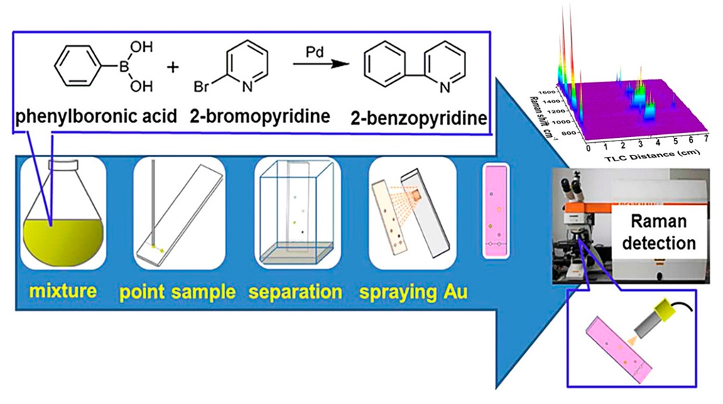
  49. Zhang, Z.M.; Liu, J.F.; Liu, R.; Sun, J.F.; Wei, G.H. Thin layer chromatography coupled with surface-enhanced Raman scattering as a facile method for on-site quantitative monitoring of chemical reactions. Anal. Chem. 2014, 86, 7286–7292. [Google Scholar] [CrossRef]
  50. Hezel, U.B.; Zeiss, C. Potential and experience in quantitative “high performance thin-layer chromatography” HPTLC. J. Chromatogr. Libr. 1977, 9, 147–188. [Google Scholar]
  51. Poole, C.F. Thin-layer chromatography: Challenges and opportunities. J. Chromatogr. A 2003, 1000, 963–984. [Google Scholar] [CrossRef]
  52. Qu, L.L.; Jia, Q.; Liu, C.; Wang, W.; Duan, L.; Yang, G.; Han, C.Q.; Li, H. Thin layer chromatography combined with surface-enhanced raman spectroscopy for rapid sensing aflatoxins. J. Chromatogr. A 2018, 1579, 115–120. [Google Scholar] [CrossRef] [PubMed]
  53. Kong, X.; Yu, Q.; Li, E.; Wang, R.; Liu, Q.; Wang, A.X. Diatomite Photonic Crystals for Facile On-Chip Chromatography and Sensing of Harmful Ingredients from Food. Materials 2018, 11, 539. [Google Scholar] [CrossRef] [PubMed] [Green Version]
  54. Kang, Y.; Wu, T.; Han, X.; Gu, H.; Zhang, X. A needle-like reusable surface-enhanced Raman scattering substrate, and its application to the determination of acetamiprid by combining SERS and thin-layer chromatography. Mikrochim. Acta 2018, 185, 504. [Google Scholar] [CrossRef] [PubMed]
  55. Cheng, H.; Lu, Y.; Zhu, D.; Rosa, L.; Han, F.; Ma, M.; Su, W.; Francis, P.S.; Zheng, Y. Plasmonic nanopapers: Flexible, stable and sensitive multiplex PUF tags for unclonable anti-counterfeiting applications. Nanoscale 2020, 12, 9471–9480. [Google Scholar] [CrossRef]
  56. Cai, G.; Ge, K.; Ouyang, X.; Hu, Y.; Li, G. Thin-layer chromatography combined with surface-enhanced Raman scattering for rapid detection of benzidine and 4-aminobiphenyl in migration from food contact materials based on gold nanoparticle doped metal-organic framework. J. Sep. Sci. 2020, 43, 2834–2841. [Google Scholar] [CrossRef]
  57. Muscalu, A.M.; Górecki, T. Comprehensive two-dimensional gas chromatography in environmental analysis. TrAC Trends Anal. Chem. 2018, 106, 225–245. [Google Scholar] [CrossRef]
  58. Li, D.-W.; Zhai, W.-L.; Li, Y.-T.; Long, Y.-T. Recent progress in surface enhanced Raman spectroscopy for the detection of environmental pollutants. Microchim. Acta 2013, 181, 23–43. [Google Scholar] [CrossRef]
  59. Al-Hamaiedh, H.D.; Maaitah, O.N. Treatment of oil polluted soil using electrochemical method. Alex. Eng. J. 2011, 50, 105–110. [Google Scholar] [CrossRef] [Green Version]
  60. Zhang, B.; Shi, Y.; Chen, H.; Zhu, Q.; Lu, F.; Li, Y. A separable surface-enhanced Raman scattering substrate modified with MIL-101 for detection of overlapping and invisible compounds after thin-layer chromatography development. Anal. Chim. Acta 2018, 997, 35–43. [Google Scholar] [CrossRef]
  61. Rebane, R.; Leito, I.; Yurchenko, S.; Herodes, K. A review of analytical techniques for determination of Sudan I–IV dyes in food matrixes. J. Chromatogr. A 2010, 1217, 2747–2757. [Google Scholar] [CrossRef]
  62. Calbiani, F.; Careri, M.; Elviri, L.; Mangia, A.; Pistara, L.; Zagnoni, I. Development and in-house validation of a liquid chromatography–electrospray–tandem mass spectrometry method for the simultaneous determination of Sudan I, Sudan II, Sudan III and Sudan IV in hot chilli products. J. Chromatogr. A 2004, 1042, 123–130. [Google Scholar] [CrossRef] [PubMed]
  63. He, L.; Su, Y.; Shen, X.; Zeng, Z.; Liu, Y. Determination of Sudan dye residues in eggs by liquid chromatography and gas chromatography—Mass spectrometry. Anal. Chim. Acta 2007, 594, 139–146. [Google Scholar] [CrossRef] [PubMed]
  64. Lv, D.; Cao, Y.; Lou, Z.; Li, S.; Chen, X.; Chai, Y.; Lu, F. Rapid on-site detection of ephedrine and its analogues used as adulterants in slimming dietary supplements by TLC-SERS. Anal. Bioanal. Chem. 2015, 407, 1313–1325. [Google Scholar] [CrossRef] [PubMed]
  65. Zhu, Q.; Cao, Y.; Li, D.; Fang, F.; Lu, F.; Yuan, Y. A fast response TLC-SERS substrate for on-site detection of hydrophilic and hydrophobic adulterants in botanical dietary supplements. New J. Chem. 2019, 43, 13873–13880. [Google Scholar] [CrossRef]
  66. Gao, F.; Hu, Y.; Chen, D.; Li-Chan, E.C.Y.; Grant, E.; Lu, X. Determination of Sudan I in paprika powder by molecularly imprinted polymers-thin layer chromatography-surface enhanced Raman spectroscopic biosensor. Talanta 2015, 143, 344–352. [Google Scholar] [CrossRef] [PubMed]
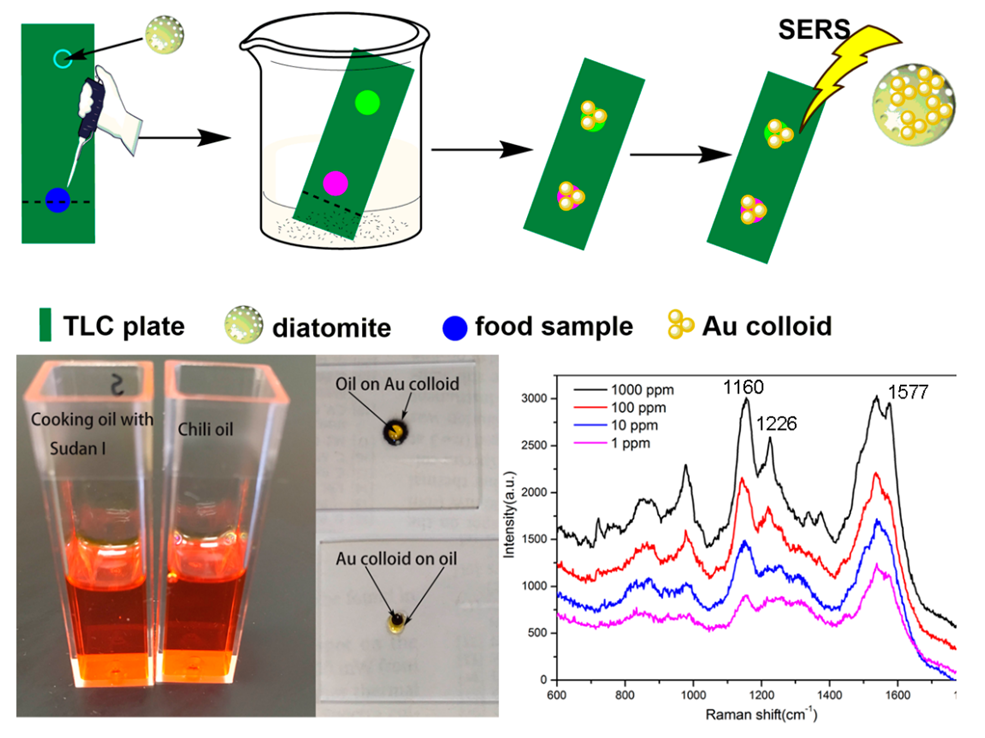
  67. Kong, X.; Squire, K.; Chong, X.; Wang, A.X. Ultra-sensitive lab-on-a-chip detection of Sudan I in food using plasmonics-enhanced diatomaceous thin film. Food Control 2017, 79, 258–265. [Google Scholar] [CrossRef]
  68. Wu, X.; Chen, J.; Park, B.; Huang, Y.-W.; Zhao, Y. The Use of Silver Nanorod Array-Based Surface-Enhanced Raman Scattering Sensor for Food Safety Applications. In Advances in Applied Nanotechnology for Agriculture; American Chemical Society: Washington, DC, USA, 2013; pp. 85–108. [Google Scholar]
  69. Chen, J.; Abell, J.; Huang, Y.-w.; Zhao, Y. On-Chip Ultra-Thin Layer Chromatography and Surface Enhanced Raman Spectroscopy. Lab Chip 2012, 12, 3096–3102. [Google Scholar] [CrossRef]
  70. Hu, X.; Fang, G.; Han, A.; Liu, J.; Wang, S. Rapid detection of Pericarpium papaveris in hot pot condiments using thin-layer chromatography and surface enhanced Raman spectroscopy combined with a support vector machine. Anal. Methods 2017, 9, 2177–2182. [Google Scholar] [CrossRef]
  71. Zhu, Q.; Chen, M.; Han, L.; Yuan, Y.; Lu, F. High efficiency screening of nine lipid-lowering adulterants in herbal dietary supplements using thin layer chromatography coupled with surface enhanced Raman spectroscopy. Anal. Methods 2017, 9, 1595–1602. [Google Scholar] [CrossRef]
  72. Cimpoiu, C.; Casoni, D.; Hosu, A.; Miclaus, V.; Hodisan, T.; Damian, G. Separation and Identification of Eight Hydrophilic Vitamins Using a New TLC Method and Raman Spectroscopy. J. Liq. Chromatogr. Relat. Technol. 2007, 28, 2551–2559. [Google Scholar] [CrossRef]
  73. Shen, Z.; Fan, Q.; Yu, Q.; Wang, R.; Wang, H.; Kong, X. Facile detection of carbendazim in food using TLC-SERS on diatomite thin layer chromatography. Spectrochim. Acta A Mol. Biomol. Spectrosc. 2021, 247, 119037. [Google Scholar] [CrossRef] [PubMed]
  74. Yao, C.; Cheng, F.; Wang, C.; Wang, Y.; Guo, X.; Gong, Z.; Fan, M.; Zhang, Z. Separation, identification and fast determination of organophosphate pesticide methidathion in tea leaves by thin layer chromatography–surface-enhanced Raman scattering. Anal. Methods 2013, 5, 5560–5564. [Google Scholar] [CrossRef]
  75. Fang, F.; Qi, Y.; Lu, F.; Yang, L. Highly sensitive on-site detection of drugs adulterated in botanical dietary supplements using thin layer chromatography combined with dynamic surface enhanced Raman spectroscopy. Talanta 2016, 146, 351–357. [Google Scholar] [CrossRef]
  76. Kang, Y.; Wu, T.; Chen, W.; Li, L.; Du, Y. A novel metastable state nanoparticle-enhanced Raman spectroscopy coupled with thin layer chromatography for determination of multiple pesticides. Food Chem. 2019, 270, 494–501. [Google Scholar] [CrossRef] [PubMed]
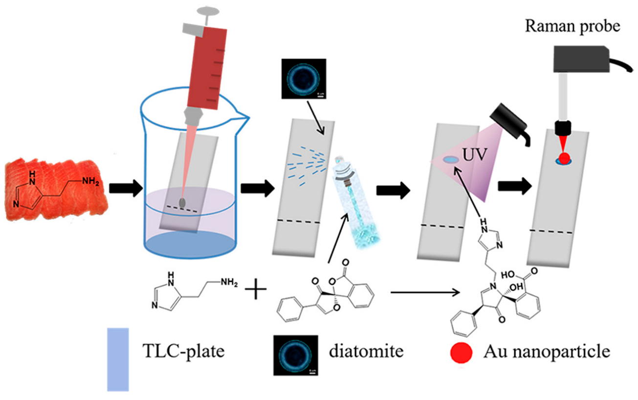
  77. Zhang, S.; Fan, Q.; Guo, J.; Jiao, X.; Kong, X.; Yu, Q. Surface-enhanced Raman spectroscopy tandem with derivatized thin-layer chromatography for ultra-sensitive on-site detection of histamine from fish. Food Control 2022, 138, 108987. [Google Scholar] [CrossRef]
  78. Xie, Z.; Wang, Y.; Chen, Y.; Xu, X.; Jin, Z.; Ding, Y.; Yang, N.; Wu, F. Tuneable surface enhanced Raman spectroscopy hyphenated to chemically derivatized thin-layer chromatography plates for screening histamine in fish. Food Chem. 2017, 230, 547–552. [Google Scholar] [CrossRef]
  79. Tan, A.; Zhao, Y.; Sivashanmugan, K.; Squire, K.; Wang, A.X. Quantitative TLC-SERS detection of histamine in seafood with support vector machine analysis. Food Control 2019, 103, 111–118. [Google Scholar] [CrossRef] [PubMed] [Green Version]
  80. Lucotti, A.; Tommasini, M.; Casella, M.; Morganti, A.; Gramatica, F.; Zerbi, G. TLC–surface enhanced Raman scattering of apomorphine in human plasma. Vib. Spectrosc. 2012, 62, 286–291. [Google Scholar] [CrossRef]
  81. Kong, X.; Chong, X.; Squire, K.; Wang, A.X. Microfluidic diatomite analytical devices for illicit drug sensing with ppb-Level sensitivity. Sens. Actuators B Chem. 2018, 259, 587–595. [Google Scholar] [CrossRef]
  82. Sivashanmugan, K.; Zhao, Y.; Wang, A.X. Tetrahydrocannabinol Sensing in Complex Biofluid with Portable Raman Spectrometer Using Diatomaceous SERS Substrates. Biosensors 2019, 9, 125. [Google Scholar] [CrossRef] [Green Version]
  83. Durucan, O.; Wu, K.; Viehrig, M.; Rindzevicius, T.; Boisen, A. Nanopillar-Assisted SERS Chromatography. ACS Sens. 2018, 3, 2492–2498. [Google Scholar] [CrossRef] [PubMed]
  84. Huang, R.; Han, S.; Li, X. Detection of tobacco-related biomarkers in urine samples by surface-enhanced Raman spectroscopy coupled with thin-layer chromatography. Anal. Bioanal. Chem. 2013, 405, 6815–6822. [Google Scholar] [CrossRef] [PubMed]
  85. Yu, W.W.; White, I.M. Inkjet Printed Surface Enhanced Raman Spectroscopy Array on Cellulose Paper. Anal. Chem. 2010, 82, 9626–9630. [Google Scholar] [CrossRef] [Green Version]
  86. Yu, W.W.; White, I.M. Inkjet-printed paper-based SERS dipsticks and swabs for trace chemical detection. Analyst 2013, 138, 1020–1025. [Google Scholar] [CrossRef] [PubMed]
  87. Yu, W.W.; White, I.M. Chromatographic separation and detection of target analytes from complex samples using inkjet printed SERS substrates. Analyst 2013, 138, 3679–3686. [Google Scholar] [CrossRef] [PubMed]
  88. Brosseau, C.L.; Gambardella, A.; Casadio, F.; Grzywacz, C.M.; Wouters, J.; Duyne, R.P.V. Ad-hoc Surface-Enhanced Raman Spectroscopy Methodologies for the Detection of Artist Dyestuffs Thin Layer Chromatography-Surface Enhanced Raman Spectroscopy and in Situ On the Fiber Analysis. Am. Chem. Soc. 2009, 81, 8. [Google Scholar] [CrossRef] [PubMed]
  89. Lee, B.S.; Lin, D.Z.; Huang, C.H.; Yen, T.J. A high-performance multifunctional substrate of ultrathin-layer chromatography (UTLC) and surface-enhancedRaman scattering (SERS) for rapid biochemical mixture screening. J. Raman Spectrosc. 2018, 49, 1920–1927. [Google Scholar] [CrossRef]
  90. Freye, C.E.; Crane, N.A.; Kirchner, T.B.; Sepaniak, M.J. Surface Enhanced Raman Scattering Imaging of Developed Thin-Layer Chromatography Plates. Anal. Chem. 2013, 85, 3991–3998. [Google Scholar] [CrossRef]
  91. Wang, S.; Yu, Q.; Guo, J.; Yuan, C.; Kong, X. On-site detection of pyrene from mixture with ppb level sensitivity by plasmonic TLC-DSERS. Spectrochim. Acta Part A Mol. Biomol. Spectrosc. 2022, 280, 121547. [Google Scholar] [CrossRef]
  92. Chen, J.; Huang, Y.-W.; Zhao, Y. Detection of polycyclic aromatic hydrocarbons from cooking oil using ultra-thin layer chromatography and surface enhanced Raman spectroscopy. J. Mater. Chem. B 2015, 3, 1898–1906. [Google Scholar] [CrossRef]
Figure 1. On-site quantitative monitoring of organic reaction by TLC-SERS. Reprinted with permission from ref. [49]; Copyright 2014 American Chemical Society.
Biosensors 12 00937 g001
Figure 2. On-site detecting of organic pollutants from wastewater by TLC-SERS. Reprinted with permission from ref. [14]; Copyright 2011 American Chemical Society.
Biosensors 12 00937 g002
Figure 3. Schematic representation of the TLC-SERS method by diatomite plate and the application for detecting Sudan dye in chili oil. Reprinted with permission from ref. [67]; Copyright 2017 Elsevier.
Biosensors 12 00937 g003
Figure 4. TLC-SERS detection of pesticide residue from food sample. Reprinted with permission from ref. [73]; Copyright 2021 Elsevier.
Biosensors 12 00937 g004
Figure 5. On-site derivatized-TLC-SERS for detecting histamine in tuna. Reprinted with permission from ref. [77]; Copyright 2022 Elsevier.
Biosensors 12 00937 g005
Publisher’s Note: MDPI stays neutral with regard to jurisdictional claims in published maps and institutional affiliations.

