Next Issue
Volume 12, December
Previous Issue
Volume 12, October


Biosensors, Volume 12, Issue 11 (November 2022) – 148 articles

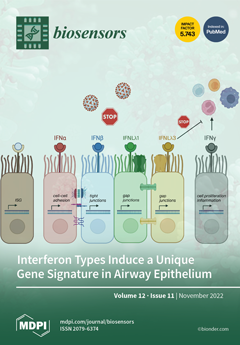
Cover Story (view full-size image): Type-I, -II and -III interferons (IFNs) are crucial for the first line of cell-intrinsic host defense. While type-I (IFNα, IFNβ) and type-III (IFNλs) induce transcription of interferon-stimulated genes (ISGs) via the JAK/STAT pathway, type-II IFN (IFNγ) is secreted by T helper type-1 cells and fuels the adaptive immune response to the pathogen. In addition, the airway epithelial barrier is important for the host cell intrinsic defense and orchestrates antiviral immune response by producing cytokines such as IL-33. Using a genome-wide gene expression biosensor chip sensing the gene expression in organotypic 3D air-liquid interface cultures, we were able to show that all types of IFNs induced similar ISGs. Type-I and type-III IFN stimulation positively correlated with the expression of cell–cell adhesion and intercellular signaling, and IFNγ promoted cell proliferation. View this paper
  • Issues are regarded as officially published after their release is announced to the table of contents alert mailing list.
  • You may sign up for e-mail alerts to receive table of contents of newly released issues.
  • PDF is the official format for papers published in both, html and pdf forms. To view the papers in pdf format, click on the "PDF Full-text" link, and use the free Adobe Reader to open them.
Order results
Result details
Section
Select all
Export citation of selected articles as:
12 pages, 4126 KiB  
Article
Electrochemical Immunosensor for Early Detection of β-Amyloid Alzheimer’s Disease Biomarker Based on Aligned Carbon Nanotubes Gold Nanocomposites
by Pushpesh Ranjan and Raju Khan
Biosensors 2022, 12(11), 1059; https://doi.org/10.3390/bios12111059 - 21 Nov 2022
Cited by 6 | Viewed by 2177
Abstract
Beta-amyloid (βA) peptides accompanying the physiological change in brain induce Alzheimer’s disease. In this work, a highly sensitive electrochemical (EC) immunosensor platform has been developed for the quantitative detection of βA peptides, using the gold nanoparticle functionalized chitosan-aligned carbon nanotube (CS-aCNT-Au) nanocomposites on [...] Read more.
Beta-amyloid (βA) peptides accompanying the physiological change in brain induce Alzheimer’s disease. In this work, a highly sensitive electrochemical (EC) immunosensor platform has been developed for the quantitative detection of βA peptides, using the gold nanoparticle functionalized chitosan-aligned carbon nanotube (CS-aCNT-Au) nanocomposites on glassy carbon electrodes (GCE). The immunosensor has been fabricated by immobilization of the anti-βA antibody upon CS-aCNT-Au/GCE. In the CS-aCNT nanocomposite, CS has high biocompatibility. Hydroxy and amine functionalities favor the antibody immobilization and prevent the leaching of nanocomposites of the modified electrode due to the adhesive environment. Moreover, aCNT offers high conductivity, stability, and a large surface area (the calculated effective surface area of the CS-aCNT/GCE is 8.594 × 10⁻² cm2). However, the incorporation of AuNPs further enhances the conductivity of the CS-aCNT-Au nanocomposite based on differential pulse voltammetry (DPV) results, and also improves the effective surface area (9.735 × 10⁻² cm2). The surface morphology and electrochemical studies of the nanocomposite, as well as its modifications by the anti-βA antibody and BSA, were carried out through field emission scanning electron microscope (FESEM), cyclic voltammetry (CV), and DPV. The quantitative immunosensing of the βA in phosphate-buffered saline (PBS) solution is accomplished via DPV, which reveals that the immunosensor has a high sensitivity of 157.60 µA pg−1 mL cm−2 and a broad detection range of 10.0 pg mL−1–100.0 µg mL−1, with a limit of detection (LOD) of 0.87 pg mL−1. Subsequently, we detected the spiked βA in diluted serum with a linear detection range of 10.0 pg mL−1–1.0 ng mL−1 and LOD of 0.95 pg mL−1. Moreover, a selectivity study exhibited a high affinity of immunosensors towards βA. Thus, we propose that this highly efficient immunosensor can potentially be applied for the point-of-care (POC) sensing of βA in clinical samples. Full article
(This article belongs to the Special Issue Electrochemical (Bio-) Sensors in Biological Applications)
Show Figures

Figure 1

12 pages, 2727 KiB  
Article
Development of Ic-ELISA and Colloidal Gold Lateral Flow Immunoassay for the Determination of Cypermethrin in Agricultural Samples
by Lianrun Huang, Fuxuan Zhang, Fangxuan Li, Yin Jia, Minghua Wang, Xiude Hua and Limin Wang
Biosensors 2022, 12(11), 1058; https://doi.org/10.3390/bios12111058 - 21 Nov 2022
Cited by 5 | Viewed by 1671
Abstract
Cypermethrin (CYP) is an insecticide in the pyrethroid family and is used widely in agriculture and for public health purposes. However, CYP has been shown to have negative impacts on reproduction, immunity and nerves in mammals. In this study, a monoclonal antibody (mAb) [...] Read more.
Cypermethrin (CYP) is an insecticide in the pyrethroid family and is used widely in agriculture and for public health purposes. However, CYP has been shown to have negative impacts on reproduction, immunity and nerves in mammals. In this study, a monoclonal antibody (mAb) against CYP was prepared and used to establish an indirect competitive immunosorbent assay (ic-ELISA) and colloidal gold lateral flow immunoassay (LFIA) for the quantitative and qualitative determination of CYP residues in agricultural products. The half inhibition concentration of the ic-ELISA was 2.49 ng/mL, and the cut-off value and visual limit of detection of the LFIA were 0.6 and 0.3 μg/mL, respectively. The recovery rates of the ic-ELISA ranged from 78.8% to 87.6% in tomato, cabbage and romaine lettuce. The qualitative results of LFIA and quantitative results of ic-ELISA and HPLC were in good agreement in blind samples. Overall, the established ic-ELISA and LFIA proved to be accurate and rapid methods for the determination of CYP in agricultural products. Full article
Show Figures

Figure 1

18 pages, 2707 KiB  
Review
Recent Advances in Multifunctional Wearable Sensors and Systems: Design, Fabrication, and Applications
by Shigang Jia, Hongwei Gao, Zhaoguo Xue and Xianhong Meng
Biosensors 2022, 12(11), 1057; https://doi.org/10.3390/bios12111057 - 21 Nov 2022
Cited by 10 | Viewed by 2920
Abstract
Multifunctional wearable sensors and systems are of growing interest over the past decades because of real-time health monitoring and disease diagnosis capability. Owing to the tremendous efforts of scientists, wearable sensors and systems with attractive advantages such as flexibility, comfort, and long-term stability [...] Read more.
Multifunctional wearable sensors and systems are of growing interest over the past decades because of real-time health monitoring and disease diagnosis capability. Owing to the tremendous efforts of scientists, wearable sensors and systems with attractive advantages such as flexibility, comfort, and long-term stability have been developed, which are widely used in temperature monitoring, pulse wave detection, gait pattern analysis, etc. Due to the complexity of human physiological signals, it is necessary to measure multiple physiological information simultaneously to evaluate human health comprehensively. This review summarizes the recent advances in multifunctional wearable sensors, including single sensors with various functions, planar integrated sensors, three-dimensional assembled sensors, and stacked integrated sensors. The design strategy, manufacturing method, and potential application of each type of sensor are discussed. Finally, we offer an outlook on future developments and provide perspectives on the remaining challenges and opportunities of wearable multifunctional sensing technology. Full article
(This article belongs to the Special Issue Wearable Sensing for Health Monitoring)
Show Figures

Figure 1

12 pages, 3173 KiB  
Article
Dual Response Site Fluorescent Probe for Highly Sensitive Detection of Cys/Hcy and GSH In Vivo through Two Different Emission Channels
by Huiling Hou, Qi Liu, Xiangbao Liu, Shuang Fu, Hongguang Zhang, Shuang Li, Song Chen and Peng Hou
Biosensors 2022, 12(11), 1056; https://doi.org/10.3390/bios12111056 - 21 Nov 2022
Cited by 6 | Viewed by 1674
Abstract
Much research has demonstrated that metabolic imbalances of biothiols are closely associated with the emergence of different types of disease. In view of the significant effect of biothiols, quantitative evaluation and discrimination of intracellular Cys/Hcy and GSH in complex biological environments is very [...] Read more.
Much research has demonstrated that metabolic imbalances of biothiols are closely associated with the emergence of different types of disease. In view of the significant effect of biothiols, quantitative evaluation and discrimination of intracellular Cys/Hcy and GSH in complex biological environments is very important. In this study, probe CDS-NBD, synthesized by attaching 2,4-dinitrobenzenesulfonate (DNBS, site 1) and nitrobenzoxadiazole (NBD, site 2) as the highly sensitive and selective dual response site for thiols onto the coumarin derivative 7-hydroxycoumarin-4-acetic acid, exhibited large separation of the emission wavelengths, fast response, notable fluorescence enhancement, excellent sensitivity and selectivity to Cys/Hcy and GSH over other biological species. Additionally, CDS-NBD could make a distinction between two different fluorescent signals, GSH (an obvious blue fluorescence) and Cys/Hcy (a mixed blue-green fluorescence). Further study on imaging of Cys/Hcy and GSH in vivo by employing probe CDS-NBD could also be successfully achieved. Full article
Show Figures

Figure 1

10 pages, 2788 KiB  
Article
Mechanical Force-Induced Blue-Shifted and Enhanced Emission for AIEgens
by Chang-Sheng Guo, Xiao-Long Su, Yu-Ting Yin, Bo-Xuan Zhang, Xin-Yi Liu, Rui-Peng Wang, Pu Chen, Hai-Tao Feng and Ben-Zhong Tang
Biosensors 2022, 12(11), 1055; https://doi.org/10.3390/bios12111055 - 21 Nov 2022
Cited by 1 | Viewed by 1863
Abstract
Mechanochromic (MC) luminescence of organic molecules has been emerging as a promising smart material for optical recording and memory devices. At the same time, pressure-induced blue-shifted and enhanced luminescence are rarely reported now. Herein, a series of cyanostilbene-based AIEgens with different substituents were [...] Read more.
Mechanochromic (MC) luminescence of organic molecules has been emerging as a promising smart material for optical recording and memory devices. At the same time, pressure-induced blue-shifted and enhanced luminescence are rarely reported now. Herein, a series of cyanostilbene-based AIEgens with different substituents were synthesized to evaluate the influence of morphology transformation and push-pull electronic effect on the MC luminescence. Among these luminophores, compound 1 with one cyano group and diethylamino group was more susceptible to mechanical stimuli and obtained blue-shifted and enhanced fluorescence in response to anisotropic grinding. Powder X-ray diffraction patterns indicated that the MC behaviors were ascribed to the solid-state morphology transition from crystal-to-crystal. Analysis of crystal structures revealed that loose molecular packing is a key factor for high high-contrast MC luminescence. The smart molecular design, together with the excellent performance, verified that luminophores with twisted structures are ideal candidates for MC luminogens. Full article
Show Figures

Graphical abstract

26 pages, 7575 KiB  
Review
Recent Progress of Biomimetic Tactile Sensing Technology Based on Magnetic Sensors
by Jiandong Man, Guangyuan Chen and Jiamin Chen
Biosensors 2022, 12(11), 1054; https://doi.org/10.3390/bios12111054 - 21 Nov 2022
Cited by 12 | Viewed by 2676
Abstract
In the past two decades, biomimetic tactile sensing technology has been a hot spot in academia. It has prospective applications in many fields such as medical treatment, health monitoring, robot tactile feedback, and human–machine interaction. With the rapid development of magnetic sensors, biomimetic [...] Read more.
In the past two decades, biomimetic tactile sensing technology has been a hot spot in academia. It has prospective applications in many fields such as medical treatment, health monitoring, robot tactile feedback, and human–machine interaction. With the rapid development of magnetic sensors, biomimetic tactile sensing technology based on magnetic sensors (which are called magnetic tactile sensors below) has been widely studied in recent years. In order to clarify the development status and application characteristics of magnetic tactile sensors, this paper firstly reviews the magnetic tactile sensors from three aspects: the types of magnetic sensors, the sources of magnetic field, and the structures of sensitive bodies used in magnetic tactile sensors. Secondly, the development of magnetic tactile sensors in four applications of robot precision grasping, texture characterization, flow velocity measurement, and medical treatment is introduced in detail. Finally, this paper analyzes technical difficulties and proposes prospective research directions for magnetic tactile sensors. Full article
(This article belongs to the Section Wearable Biosensors)
Show Figures

Figure 1

14 pages, 2141 KiB  
Article
Dual-Wavelength Fluorescence Polarization Immunoassay for Simultaneous Detection of Sulfonamides and Antibacterial Synergists in Milk
by Changfei Duan, Yingjie Zhang, Peipei Li, Qiang Li, Wenbo Yu, Kai Wen, Sergei A. Eremin, Jianzhong Shen, Xuezhi Yu and Zhanhui Wang
Biosensors 2022, 12(11), 1053; https://doi.org/10.3390/bios12111053 - 21 Nov 2022
Cited by 4 | Viewed by 1614
Abstract
Combinations of sulfonamides (SAs) and antibacterial synergists (ASGs) are frequently used for treating infectious diseases and promoting growth for animals, which cause potential hazards to food safety and human health. To realize the simultaneous detection of SAs and ASGs in food, a homogeneous [...] Read more.
Combinations of sulfonamides (SAs) and antibacterial synergists (ASGs) are frequently used for treating infectious diseases and promoting growth for animals, which cause potential hazards to food safety and human health. To realize the simultaneous detection of SAs and ASGs in food, a homogeneous and high-throughput screening dual-wavelength fluorescence polarization immunoassay (DWFPIA) was developed. In this study, three SAs tracers and three ASGs tracers were synthesized by fluoresceins with different linkers and paired with their corresponding monoclonal antibodies (mAbs), respectively. To achieve a high sensitivity and broad specificity, the combination of tracers SADMPM-HDF with the longest linker paring mAb 10E6 for SAs and tracer HaptenA-DSCA paring mAb 9C9 for ASGs were chosen for the development of DWFPIA, achieving surprising IC50 values for 23 SAs below 100 μg L−1 and 5 ASGs below 50 μg L−1. The accuracy of DWFPIA was applied in real milk samples by typical sulfamethazine (SMZ) and trimethoprim (TMP), with recoveries of 81.7–97.2% and 78.6–103.6%, and coefficient of variations (CVs) below 18.9%, which could be completed within 15 min, including sample pretreatment. We firstly developed a simultaneous screening DWFPIA, covering all of the SAs and ASGs used in clinic and providing a great application potential in food safety analysis. Full article
(This article belongs to the Special Issue Immunosensors and Immunoassays for the Control of Hazardous Compounds)
Show Figures

Graphical abstract

12 pages, 1724 KiB  
Perspective
Future Prospects of Luminescent Silicon Nanowires Biosensors
by Maria Josè Lo Faro, Antonio Alessio Leonardi, Francesco Priolo, Barbara Fazio and Alessia Irrera
Biosensors 2022, 12(11), 1052; https://doi.org/10.3390/bios12111052 - 21 Nov 2022
Cited by 2 | Viewed by 1691
Abstract
In this paper, we exploit the perspective of luminescent Si nanowires (NWs) in the growing field of commercial biosensing nanodevices for the selective recognition of proteins and pathogen genomes. We fabricated quantum confined fractal arrays of Si NWs with room temperature emission at [...] Read more.
In this paper, we exploit the perspective of luminescent Si nanowires (NWs) in the growing field of commercial biosensing nanodevices for the selective recognition of proteins and pathogen genomes. We fabricated quantum confined fractal arrays of Si NWs with room temperature emission at 700 nm obtained by thin-film, metal-assisted, chemical etching with high production output at low cost. The fascinating optical features arising from multiple scattering and weak localization of light promote the use of Si NWs as optical biosensing platforms with high sensitivity and selectivity. In this work, label-free Si NW optical sensors are surface modified for the selective detection of C-reactive protein through antigen–gene interaction. In this case, we report the lowest limit of detection (LOD) of 1.6 fM, fostering the flexibility of different dynamic ranges for detection either in saliva or for serum analyses. By varying the NW surface functionalization with the specific antigen, the luminescence quenching of NW biosensors is used to measure the hepatitis B-virus pathogen genome without PCR-amplification, with an LOD of about 20 copies in real samples or blood matrix. The promising results show that NW optical biosensors can detect and isolate extracellular vesicles (EV) marked with CD81 protein with unprecedented sensitivity (LOD 2 × 10⁵ sEV/mL), thus enabling their measurement even in a small amount of blastocoel fluid. Full article
(This article belongs to the Special Issue Biosensors State-of-the-Art in Italy)
Show Figures

Figure 1

11 pages, 1902 KiB  
Article
Micro-Raman Analysis of Sperm Cells on Glass Slide: Potential Label-Free Assessment of Sperm DNA toward Clinical Applications
by Shengrong Du, Qun Zhang, Haohao Guan, Guannan Chen, Sisi Wang, Yan Sun, Yuling Li, Rong Chen, Youwu He and Zufang Huang
Biosensors 2022, 12(11), 1051; https://doi.org/10.3390/bios12111051 - 21 Nov 2022
Cited by 1 | Viewed by 1695
Abstract
Routine assessment of sperm DNA integrity involves the time-consuming and complex process of staining sperm chromatin. Here, we report a Raman spectroscopy method combined with extended multiplicative signal correction (EMSC) for the extraction of characteristic fingerprints of DNA-intact and DNA-damaged sperm cells directly [...] Read more.
Routine assessment of sperm DNA integrity involves the time-consuming and complex process of staining sperm chromatin. Here, we report a Raman spectroscopy method combined with extended multiplicative signal correction (EMSC) for the extraction of characteristic fingerprints of DNA-intact and DNA-damaged sperm cells directly on glass slides. Raman results of sperm cell DNA integrity on glass substrates were validated one-to-one with clinical sperm cell staining. Although the overall Raman spectral pattern showed considerable similarity between DNA-damaged and DNA-intact sperm cells, differences in specific Raman spectral responses were observed. We then employed and compared multivariate statistical analysis based on principal component analysis-linear discriminant analysis (PCA-LDA) and partial least-squares-discriminant analysis (PLS-DA), and the classifications were validated by leave-one-out-cross-validation (LOOCV) and k-fold cross-validation methods. In comparison, the PLS-DA model showed relatively better results in terms of diagnostic sensitivity, specificity, and the classification rate between the sperm DNA damaged group and the DNA intact group. Our results demonstrate the potential of Raman based label-free DNA assessment of sperm cell on glass substrates as a simple method toward clinical applications. Full article
(This article belongs to the Special Issue Emerging Applications of Label-Free Optical Biosensors)
Show Figures

Graphical abstract

10 pages, 1974 KiB  
Article
Terahertz Biosensor Based on Mode Coupling between Defect Mode and Optical Tamm State with Dirac Semimetal
by Yuwen Bao, Mengjiao Ren, Chengpeng Ji, Jun Dong, Leyong Jiang and Xiaoyu Dai
Biosensors 2022, 12(11), 1050; https://doi.org/10.3390/bios12111050 - 21 Nov 2022
Cited by 2 | Viewed by 1314
Abstract
Bulk Dirac semimetal (BDS) has emerged as a “3D graphene” material for the development of optical devices in the past few years. In this study, a BDS-based tunable highly sensitive terahertz (THz) biosensor is proposed by using a Dirac semimetal/Bragg reflector multilayer structure. [...] Read more.
Bulk Dirac semimetal (BDS) has emerged as a “3D graphene” material for the development of optical devices in the past few years. In this study, a BDS-based tunable highly sensitive terahertz (THz) biosensor is proposed by using a Dirac semimetal/Bragg reflector multilayer structure. The high sensitivity of the biosensor originates from the sharp Fano resonance peak caused by coupling the Optical Tamm State (OTS) mode and defect mode. Besides, the sensitivity of the proposed structure is sensitive to the Fermi energy of Dirac semimetal and the refractive index of the sensing medium. The maximum sensitivity of 1022°/RIU is obtained by selecting structural and material parameter appropriately, which has certain competitiveness compared to conventional surface plasmon resonance (SPR) sensors. From the standpoint of the fabrication facility and integration, we judged that the BDS-based layered structure has the potential application in biosensor field. Full article
(This article belongs to the Special Issue SPR-Based Biochip)
Show Figures

Graphical abstract

15 pages, 2165 KiB  
Article
Nanoporous ZIF-8 Microparticles as Acetylcholinesterase and Alkaline Phosphatase Mimics for the Selective and Sensitive Detection of Ascorbic Acid Oxidase and Copper Ions
by Guo-Ying Chen, Shi-Jun Yin, Li Chen, Xi Zhou and Feng-Qing Yang
Biosensors 2022, 12(11), 1049; https://doi.org/10.3390/bios12111049 - 21 Nov 2022
Cited by 4 | Viewed by 1720
Abstract
In this study, the alkaline phosphatase (ALP)-like activity of zeolitic-imidazolate framework-8 (ZIF-8) is reported for the first time. Then, colorimetric sensors for the ascorbic acid oxidase (AAO) and copper ion (Cu2+) detection were developed based on the acetylcholinesterase (AChE)- and ALP-like [...] Read more.
In this study, the alkaline phosphatase (ALP)-like activity of zeolitic-imidazolate framework-8 (ZIF-8) is reported for the first time. Then, colorimetric sensors for the ascorbic acid oxidase (AAO) and copper ion (Cu2+) detection were developed based on the acetylcholinesterase (AChE)- and ALP-like activities of ZIF-8. The ZIF-8 has good mimetic enzyme activity and exhibits high affinity to the substrates. Its AChE- and ALP-like activities also have good reusability and storage stability. Good linear dependences are obtained in the range of 1.3−250.0 μM (AChE-like activity-based) and 4.5−454.5 μM (ALP-like activity based) for Cu2+ detection. Furthermore, good linear dependence is also obtained based on the ALP-like activity of ZIF-8 for AAO detection in the range of 2.3−454.5 U/L. Their limits of detection (LODs) are calculated to be 0.7 µM, 2.8 µM, and 1.8 U/L, respectively. Finally, the sample spiked recoveries of Cu2+ in tap water, Cu2+, and AAO in human serum and rabbit plasma were measured, and the results are in the range of 80.0−119.3%. In short, the preparation of ZIF-8 is simple, environmentally friendly, and harmless, and can realize highly selective detection of AAO and Cu2+ in an efficient and fast process. Full article
(This article belongs to the Special Issue Nanomaterial-Based Biosensors for Biomedical Applications)
Show Figures

Figure 1

9 pages, 2670 KiB  
Article
Label-Free Sensing of Biomolecular Adsorption and Desorption Dynamics by Interfacial Second Harmonic Generation
by Chuansheng Xia, Jianli Sun, Qiong Wang, Jinping Chen, Tianjie Wang, Wenxiong Xu, He Zhang, Yuanyuan Li, Jianhua Chang, Zengliang Shi, Chunxiang Xu and Qiannan Cui
Biosensors 2022, 12(11), 1048; https://doi.org/10.3390/bios12111048 - 20 Nov 2022
Cited by 2 | Viewed by 1483
Abstract
Observing interfacial molecular adsorption and desorption dynamics in a label-free manner is fundamentally important for understanding spatiotemporal transports of matter and energy across interfaces. Here, we report a label-free real-time sensing technique utilizing strong optical second harmonic generation of monolayer 2D semiconductors. BSA [...] Read more.
Observing interfacial molecular adsorption and desorption dynamics in a label-free manner is fundamentally important for understanding spatiotemporal transports of matter and energy across interfaces. Here, we report a label-free real-time sensing technique utilizing strong optical second harmonic generation of monolayer 2D semiconductors. BSA molecule adsorption and desorption dynamics on the surface of monolayer MoS2 in liquid environments have been all-optically observed through time-resolved second harmonic generation (SHG) measurements. The proposed SHG detection scheme is not only interface specific but also expected to be widely applicable, which, in principle, undertakes a nanometer-scale spatial resolution across interfaces. Full article
(This article belongs to the Special Issue Label-Free Biosensor)
Show Figures

Figure 1

11 pages, 2198 KiB  
Article
Polydopamine-Coated Co3O4 Nanoparticles as an Efficient Catalase Mimic for Fluorescent Detection of Sulfide Ion
by Trung Hieu Vu, Phuong Thy Nguyen and Moon Il Kim
Biosensors 2022, 12(11), 1047; https://doi.org/10.3390/bios12111047 - 19 Nov 2022
Cited by 9 | Viewed by 1927
Abstract
Surface engineering of nanozymes has been recognized as a potent strategy to improve their catalytic activity and specificity. We synthesized polydopamine-coated Co3O4 nanoparticles (PDA@Co3O4 NPs) through simple dopamine-induced self-assembly and demonstrated that these NPs exhibit catalase-like activity [...] Read more.
Surface engineering of nanozymes has been recognized as a potent strategy to improve their catalytic activity and specificity. We synthesized polydopamine-coated Co3O4 nanoparticles (PDA@Co3O4 NPs) through simple dopamine-induced self-assembly and demonstrated that these NPs exhibit catalase-like activity by decomposing H2O2 into oxygen and water. The activity of PDA@Co3O4 NPs was approximately fourfold higher than that of Co3O4 NPs without PDA, possibly due to the additional radical scavenging activity of the PDA shell. In addition, PDA@Co3O4 NPs were more stable than natural catalase under a wide range of pH, temperature, and storage time conditions. Upon the addition of a sample containing sulfide ion, the activity of PDA@Co3O4 NPs was significantly inhibited, possibly because of increased mass transfer limitations via the absorption of the sulfide ion on the PDA@Co3O4 NP surface, along with NP aggregation which reduced their surface area. The reduced catalase-like activity was used to determine the levels of sulfide ion by measuring the increased fluorescence of the oxidized terephthalic acid, generated from the added H2O2. Using this strategy, the target sulfide ion was sensitively determined to a lower limit of 4.3 µM and dynamic linear range of up to 200 µM. The fluorescence-based sulfide ion assay based on PDA@Co3O4 NPs was highly precise when applied to real tap water samples, validating its potential for conveniently monitoring toxic elements in the environment. Full article
(This article belongs to the Special Issue Feature Issue of Biosensor Materials Section)
Show Figures

Figure 1

18 pages, 2204 KiB  
Review
Nanomaterials-Based Electrochemiluminescence Biosensors for Food Analysis: Recent Developments and Future Directions
by Jiaojiao Zhou, Xuqin Lv, Jilai Jia, Zia-ud Din, Shiqi Cai, Jiangling He, Fang Xie and Jie Cai
Biosensors 2022, 12(11), 1046; https://doi.org/10.3390/bios12111046 - 18 Nov 2022
Cited by 10 | Viewed by 2677
Abstract
Developing robust and sensitive food safety detection methods is important for human health. Electrochemiluminescence (ECL) is a powerful analytical technique for complete separation of input source (electricity) and output signal (light), thereby significantly reducing background ECL signal. ECL biosensors have attracted considerable attention [...] Read more.
Developing robust and sensitive food safety detection methods is important for human health. Electrochemiluminescence (ECL) is a powerful analytical technique for complete separation of input source (electricity) and output signal (light), thereby significantly reducing background ECL signal. ECL biosensors have attracted considerable attention owing to their high sensitivity and wide dynamic range in food safety detection. In this review, we introduce the principles of ECL biosensors and common ECL luminophores, as well as the latest applications of ECL biosensors in food analysis. Further, novel nanomaterial assembly strategies have been progressively incorporated into the design of ECL biosensors, and by demonstrating some representative works, we summarize the development status of ECL biosensors in detection of mycotoxins, heavy metal ions, antibiotics, pesticide residues, foodborne pathogens, and other illegal additives. Finally, the current challenges faced by ECL biosensors are outlined and the future directions for advancing ECL research are presented. Full article
Show Figures

Figure 1

31 pages, 3098 KiB  
Review
Recent Advances of Organ-on-a-Chip in Cancer Modeling Research
by Xingxing Liu, Qiuping Su, Xiaoyu Zhang, Wenjian Yang, Junhua Ning, Kangle Jia, Jinlan Xin, Huanling Li, Longfei Yu, Yuheng Liao and Diming Zhang
Biosensors 2022, 12(11), 1045; https://doi.org/10.3390/bios12111045 - 18 Nov 2022
Cited by 13 | Viewed by 6431
Abstract
Although many studies have focused on oncology and therapeutics in cancer, cancer remains one of the leading causes of death worldwide. Due to the unclear molecular mechanism and complex in vivo microenvironment of tumors, it is challenging to reveal the nature of cancer [...] Read more.
Although many studies have focused on oncology and therapeutics in cancer, cancer remains one of the leading causes of death worldwide. Due to the unclear molecular mechanism and complex in vivo microenvironment of tumors, it is challenging to reveal the nature of cancer and develop effective therapeutics. Therefore, the development of new methods to explore the role of heterogeneous TME in individual patients’ cancer drug response is urgently needed and critical for the effective therapeutic management of cancer. The organ-on-chip (OoC) platform, which integrates the technology of 3D cell culture, tissue engineering, and microfluidics, is emerging as a new method to simulate the critical structures of the in vivo tumor microenvironment and functional characteristics. It overcomes the failure of traditional 2D/3D cell culture models and preclinical animal models to completely replicate the complex TME of human tumors. As a brand-new technology, OoC is of great significance for the realization of personalized treatment and the development of new drugs. This review discusses the recent advances of OoC in cancer biology studies. It focuses on the design principles of OoC devices and associated applications in cancer modeling. The challenges for the future development of this field are also summarized in this review. This review displays the broad applications of OoC technique and has reference value for oncology development. Full article
(This article belongs to the Special Issue Immunoassays and Biosensing)
Show Figures

Figure 1

16 pages, 4032 KiB  
Article
Slippery Epidural ECoG Electrode for High-Performance Neural Recording and Interface
by Md Eshrat E. Alahi, Yonghong Liu, Sara Khademi, Anindya Nag, Hao Wang, Tianzhun Wu and Subhas Chandra Mukhopadhyay
Biosensors 2022, 12(11), 1044; https://doi.org/10.3390/bios12111044 - 18 Nov 2022
Viewed by 2031
Abstract
Chronic implantation of an epidural Electrocorticography (ECoG) electrode produces thickening of the dura mater and proliferation of the fibrosis around the interface sites, which is a significant concern for chronic neural ECoG recording applications used to monitor various neurodegenerative diseases. This study describes [...] Read more.
Chronic implantation of an epidural Electrocorticography (ECoG) electrode produces thickening of the dura mater and proliferation of the fibrosis around the interface sites, which is a significant concern for chronic neural ECoG recording applications used to monitor various neurodegenerative diseases. This study describes a new approach to developing a slippery liquid-infused porous surface (SLIPS) on the flexible ECoG electrode for a chronic neural interface with the advantage of increased cell adhesion. In the demonstration, the electrode was fabricated on the polyimide (PI) substrate, and platinum (Pt)-gray was used for creating the porous nanocone structure for infusing the silicone oil. The combination of nanocone and the infused slippery oil layer created the SLIPS coating, which has a low impedance (4.68 kΩ) level favourable for neural recording applications. The electrochemical impedance spectroscopy and equivalent circuit modelling also showed the effect of the coating on the recording site. The cytotoxicity study demonstrated that the coating does not have any cytotoxic potentiality; hence, it is biocompatible for human implantation. The in vivo (acute recording) neural recording on the rat model also confirmed that the noise level could be reduced significantly (nearly 50%) and is helpful for chronic ECoG recording for more extended neural signal recording applications. Full article
(This article belongs to the Special Issue Novel Materials in Biosensing Devices)
Show Figures

Figure 1

17 pages, 4665 KiB  
Article
A Combination of Near-Infrared Hyperspectral Imaging with Two-Dimensional Correlation Analysis for Monitoring the Content of Alanine in Beef
by Fujia Dong, Yongzhao Bi, Jie Hao, Sijia Liu, Yu Lv, Jiarui Cui, Songlei Wang, Yafang Han and Argenis Rodas-González
Biosensors 2022, 12(11), 1043; https://doi.org/10.3390/bios12111043 - 18 Nov 2022
Cited by 9 | Viewed by 1694
Abstract
Alanine (Ala), as the most important free amino acid, plays a significant role in food taste characteristics and human health regulation. The feasibility of using near–infrared hyperspectral imaging (NIR–HSI) combined with two–dimensional correlation spectroscopy (2D–COS) analysis to predict beef Ala content quickly and [...] Read more.
Alanine (Ala), as the most important free amino acid, plays a significant role in food taste characteristics and human health regulation. The feasibility of using near–infrared hyperspectral imaging (NIR–HSI) combined with two–dimensional correlation spectroscopy (2D–COS) analysis to predict beef Ala content quickly and nondestructively is first proposed in this study. With Ala content as the external disturbance condition, the sequence of chemical bond changes caused by synchronous and asynchronous correlation spectrum changes in 2D–COS was analyzed, and local sensitive variables closely related to Ala content were obtained. On this basis, the simplified linear, nonlinear, and artificial neural network models developed by the weighted coefficient based on the feature wavelength extraction method were compared. The results show that with the change in Ala content in beef, the double-frequency absorption of the C-H bond of CH2 in the chemical bond sequence occurred prior to the third vibration of the C=O bond and the first stretching of O-H in COOH. Furthermore, the wavelength within the 1136–1478 nm spectrum range was obtained as the local study area of Ala content. The linear partial least squares regression (PLSR) model based on effective wavelengths was selected by competitive adaptive reweighted sampling (CARS) from 2D–COS analysis, and provided excellent results (R2C of 0.8141, R2P of 0.8458, and RPDp of 2.54). Finally, the visual distribution of Ala content in beef was produced by the optimal simplified combination model. The results show that 2D–COS combined with NIR–HSI could be used as an effective method to monitor Ala content in beef. Full article
Show Figures

Figure 1

11 pages, 2824 KiB  
Article
Biosensor Based on Peroxidase-Mimetic Nanozyme and Lactate Oxidase for Accurate L-Lactate Analysis in Beverages
by Oleh Smutok, Taras Kavetskyy, Tetiana Prokopiv, Roman Serkiz, Ondrej Šauša, Ivan Novák, Helena Švajdlenková, Igor Maťko, Mykhailo Gonchar and Evgeny Katz
Biosensors 2022, 12(11), 1042; https://doi.org/10.3390/bios12111042 - 18 Nov 2022
Cited by 8 | Viewed by 2004
Abstract
Precision analysis of the key biological metabolites such as L-lactate has great practical importance for many technological processes in food technology, including beverage production. Here we describe a new, highly selective, and sensitive biosensor for accurate L-lactate assay based on a combination of [...] Read more.
Precision analysis of the key biological metabolites such as L-lactate has great practical importance for many technological processes in food technology, including beverage production. Here we describe a new, highly selective, and sensitive biosensor for accurate L-lactate assay based on a combination of peroxidase-mimetic nanozymes with microbial lactate oxidase (LOx) immobilized onto the surface of a graphite-rod electrode (GE). The peroxidase-like nanozymes were synthesized using the debris of carbon microfibers (CFs) functionalized with hemin (H) and modified with gold nanoparticles (AuNPs) or platinum microparticles (PtMPs). The nanozyme formed with PtMPs as well as corresponding bioelectrodes based on it (LOx-CF-H-PtMPs/GE) is characterized by preferable catalytic and operational characteristics, so it was selected for the analysis of L-lactate content in real samples of grape must and red wine. The results of the L-lactate analysis obtained by the developed biosensors are highly correlated with a very selective spectrophotometric approach used as a reference. The developed biosensor, due to its high selectivity and sensitivity, is very prospective not only for the beverage industry and food technology, but also for clinical diagnostics and medicine, as well as in other applications where the accurate analysis of L-lactate is highly important. Full article
(This article belongs to the Special Issue Electrochemical (Bio-) Sensors in Biological Applications)
Show Figures

Figure 1

10 pages, 1598 KiB  
Article
Heterogeneous Optical Fiber Sensor System for Temperature and Turbidity Assessment in Wide Range
by Arnaldo Leal-Junior, Guilherme Lopes, Leandro C. Macedo, Welton Duque, Anselmo Frizera and Carlos Marques
Biosensors 2022, 12(11), 1041; https://doi.org/10.3390/bios12111041 - 18 Nov 2022
Cited by 3 | Viewed by 1678
Abstract
This paper presents the development of an optical fiber sensor system for multiparametric assessment of temperature and turbidity in liquid samples. The sensors are based on the combination between fiber Bragg gratings (FBGs), intensity variation and surface plasmon resonance (SPR) sensors. In this [...] Read more.
This paper presents the development of an optical fiber sensor system for multiparametric assessment of temperature and turbidity in liquid samples. The sensors are based on the combination between fiber Bragg gratings (FBGs), intensity variation and surface plasmon resonance (SPR) sensors. In this case, the intensity variation sensors are capable of detecting turbidity with a resolution of about 0.5 NTU in a limited range between 0.02 NTU and 100 NTU. As the turbidity increases, a saturation trend in the sensor is observed. In contrast, the SPR-based sensor is capable of detecting refractive index (RI) variation. However, RI measurements in the turbidity calibrated samples indicate a significant variation on the RI only when the turbidity is higher than 100 NTU. Thus, the SPR-based sensor is used as a complementary approach for the dynamic range increase of the turbidity assessment, where a linearity and sensitivity of 98.6% and 313.5 nm/RIU, respectively, are obtained. Finally, the FBG sensor is used in the temperature assessment, an assessment which is not only used for water quality assessment, but also in temperature cross-sensitivity mitigation of the SPR sensor. Furthermore, this approach also leads to the possibility of indirect assessment of turbidity through the differences in the heat transfer rates due to the turbidity increase. Full article
(This article belongs to the Special Issue New Progress in Optical Fiber-Based Biosensors)
Show Figures

Figure 1

16 pages, 1175 KiB  
Article
Advanced Lab-on-Fiber Optrodes Assisted by Oriented Antibody Immobilization Strategy
by Sarassunta Ucci, Sara Spaziani, Giuseppe Quero, Patrizio Vaiano, Maria Principe, Alberto Micco, Annamaria Sandomenico, Menotti Ruvo, Marco Consales and Andrea Cusano
Biosensors 2022, 12(11), 1040; https://doi.org/10.3390/bios12111040 - 17 Nov 2022
Cited by 3 | Viewed by 2033
Abstract
Lab-on-fiber (LoF) optrodes offer several advantages over conventional techniques for point-of-care platforms aimed at real-time and label-free detection of clinically relevant biomarkers. Moreover, the easy integration of LoF platforms in medical needles, catheters, and nano endoscopes offer unique potentials for in vivo biopsies [...] Read more.
Lab-on-fiber (LoF) optrodes offer several advantages over conventional techniques for point-of-care platforms aimed at real-time and label-free detection of clinically relevant biomarkers. Moreover, the easy integration of LoF platforms in medical needles, catheters, and nano endoscopes offer unique potentials for in vivo biopsies and tumor microenvironment assessment. The main barrier to translating the vision close to reality is the need to further lower the final limit of detection of developed optrodes. For immune-biosensing purposes, the assay sensitivity significantly relies on the capability to correctly immobilize the capture antibody in terms of uniform coverage and correct orientation of the bioreceptor, especially when very low detection limits are requested as in the case of cancer diagnostics. Here, we investigated the possibility to improve the immobilization strategies through the use of hinge carbohydrates by involving homemade antibodies that demonstrated a significantly improved recognition of the antigen with ultra-low detection limits. In order to create an effective pipeline for the improvement of biofunctionalization protocols to be used in connection with LoF platforms, we first optimized the protocol using a microfluidic surface plasmon resonance (mSPR) device and then transferred the optimized strategy onto LoF platforms selected for the final validation. Here, we selected two different LoF platforms: a biolayer interferometry (BLI)-based device (commercially available) and a homemade advanced LoF biosensor based on optical fiber meta-tips (OFMTs). As a clinically relevant scenario, here we focused our attention on a promising serological biomarker, Cripto-1, for its ability to promote tumorigenesis in breast and liver cancer. Currently, Cripto-1 detection relies on laborious and time-consuming immunoassays. The reported results demonstrated that the proposed approach based on oriented antibody immobilization was able to significantly improve Cripto-1 detection with a 10-fold enhancement versus the random approach. More interestingly, by using the oriented antibody immobilization strategy, the OFMTs-based platform was able to reveal Cripto-1 at a concentration of 0.05 nM, exhibiting detection capabilities much higher (by a factor of 250) than those provided by the commercial LoF platform based on BLI and similar to the ones shown by the commercial and well-established bench-top mSPR Biacore 8K system. Therefore, our work opened new avenues into the development of high-sensitivity LoF biosensors for the detection of clinically relevant biomarkers in the sub-ng/mL range. Full article
Show Figures

Figure 1

45 pages, 6926 KiB  
Review
Advances in Materials, Sensors, and Integrated Systems for Monitoring Eye Movements
by Seunghyeb Ban, Yoon Jae Lee, Ka Ram Kim, Jong-Hoon Kim and Woon-Hong Yeo
Biosensors 2022, 12(11), 1039; https://doi.org/10.3390/bios12111039 - 17 Nov 2022
Cited by 6 | Viewed by 5485
Abstract
Eye movements show primary responses that reflect humans’ voluntary intention and conscious selection. Because visual perception is one of the fundamental sensory interactions in the brain, eye movements contain critical information regarding physical/psychological health, perception, intention, and preference. With the advancement of wearable [...] Read more.
Eye movements show primary responses that reflect humans’ voluntary intention and conscious selection. Because visual perception is one of the fundamental sensory interactions in the brain, eye movements contain critical information regarding physical/psychological health, perception, intention, and preference. With the advancement of wearable device technologies, the performance of monitoring eye tracking has been significantly improved. It also has led to myriad applications for assisting and augmenting human activities. Among them, electrooculograms, measured by skin-mounted electrodes, have been widely used to track eye motions accurately. In addition, eye trackers that detect reflected optical signals offer alternative ways without using wearable sensors. This paper outlines a systematic summary of the latest research on various materials, sensors, and integrated systems for monitoring eye movements and enabling human-machine interfaces. Specifically, we summarize recent developments in soft materials, biocompatible materials, manufacturing methods, sensor functions, systems’ performances, and their applications in eye tracking. Finally, we discuss the remaining challenges and suggest research directions for future studies. Full article
(This article belongs to the Special Issue Biophysical Sensors for Biomedical/Health Monitoring Applications)
Show Figures

Graphical abstract

31 pages, 4974 KiB  
Review
Environmental Monitoring: A Comprehensive Review on Optical Waveguide and Fiber-Based Sensors
by Muhammad A. Butt, Grigory S. Voronkov, Elizaveta P. Grakhova, Ruslan V. Kutluyarov, Nikolay L. Kazanskiy and Svetlana N. Khonina
Biosensors 2022, 12(11), 1038; https://doi.org/10.3390/bios12111038 - 17 Nov 2022
Cited by 25 | Viewed by 4312
Abstract
Globally, there is active development of photonic sensors incorporating multidisciplinary research. The ultimate objective is to develop small, low-cost, sensitive, selective, quick, durable, remote-controllable sensors that are resistant to electromagnetic interference. Different photonic sensor designs and advances in photonic frameworks have shown the [...] Read more.
Globally, there is active development of photonic sensors incorporating multidisciplinary research. The ultimate objective is to develop small, low-cost, sensitive, selective, quick, durable, remote-controllable sensors that are resistant to electromagnetic interference. Different photonic sensor designs and advances in photonic frameworks have shown the possibility to realize these capabilities. In this review paper, the latest developments in the field of optical waveguide and fiber-based sensors which can serve for environmental monitoring are discussed. Several important topics such as toxic gas, water quality, indoor environment, and natural disaster monitoring are reviewed. Full article
Show Figures

Figure 1

12 pages, 15924 KiB  
Article
On-Chip Single-Cell Bioelectrical Analysis for Identification of Cell Electrical Phenotyping in Response to Sequential Electric Signal Modulation
by Seungyeop Choi, Insu Park, Sang Hyun Lee, Kang In Yeo, Gyeongjun Min, Sung-Hun Woo, Yoon Suk Kim, Sei Young Lee and Sang Woo Lee
Biosensors 2022, 12(11), 1037; https://doi.org/10.3390/bios12111037 - 17 Nov 2022
Cited by 1 | Viewed by 1453
Abstract
In recent years, an interesting biomarker called membrane breakdown voltage has been examined using artificial planar lipid bilayers. Even though they have great potential to identify cell electrical phenotyping for distinguishing similar cell lines or cells under different physiological conditions, the biomarker has [...] Read more.
In recent years, an interesting biomarker called membrane breakdown voltage has been examined using artificial planar lipid bilayers. Even though they have great potential to identify cell electrical phenotyping for distinguishing similar cell lines or cells under different physiological conditions, the biomarker has not been evaluated in the context of living cell electrical phenotyping. Herein, we present a single-cell analysis platform to continuously measure the electric response in a large number of cells in parallel using electric frequency and voltage variables. Using this platform, we measured the direction of cell displacement and transparent cell image alteration as electric polarization of the cell responds to signal modulation, extracting the dielectrophoretic crossover frequency and membrane breakdown voltage for each cell, and utilizing the measurement results in the same spatiotemporal environment. We developed paired parameters using the dielectrophoretic crossover frequency and membrane breakdown voltage for each cell and evaluated the paired parameter efficiency concerning the identification of two different breast cancer cells and cell drug response. Moreover, we showed that the platform was able to identify cell electrical phenotyping, which was generated by subtle changes in cholesterol depletion-induced cell membrane integrity disruption when the paired parameter was used. Our platform introduced in this paper is extremely useful for facilitating more accurate and efficient evaluation of cell electrical phenotyping in a variety of applications, such as cell biology and drug discovery. Full article
(This article belongs to the Section Biosensor and Bioelectronic Devices)
Show Figures

Graphical abstract

37 pages, 5956 KiB  
Review
A Comprehensive Review on Upconversion Nanomaterials-Based Fluorescent Sensor for Environment, Biology, Food and Medicine Applications
by Wei Jiang, Jiaqi Yi, Xiaoshuang Li, Fei He, Na Niu and Ligang Chen
Biosensors 2022, 12(11), 1036; https://doi.org/10.3390/bios12111036 - 17 Nov 2022
Cited by 10 | Viewed by 2652
Abstract
Near-infrared-excited upconversion nanoparticles (UCNPs) have multicolor emissions, a low auto-fluorescence background, a high chemical stability, and a long fluorescence lifetime. The fluorescent probes based on UCNPs have achieved great success in the analysis of different samples. Here, we presented the research results of [...] Read more.
Near-infrared-excited upconversion nanoparticles (UCNPs) have multicolor emissions, a low auto-fluorescence background, a high chemical stability, and a long fluorescence lifetime. The fluorescent probes based on UCNPs have achieved great success in the analysis of different samples. Here, we presented the research results of UCNPs probes utilized in analytical applications including environment, biology, food and medicine in the last five years; we also introduced the design and construction of upconversion optical sensing platforms. Future trends and challenges of the UCNPs used in the analytical field have also been discussed with particular emphasis. Full article
(This article belongs to the Special Issue Advances in Fluorescent Probe Biosensing)
Show Figures

Figure 1

15 pages, 3162 KiB  
Review
CRISPR-Cas-Integrated LAMP
by Nazente Atçeken, Defne Yigci, Berin Ozdalgic and Savas Tasoglu
Biosensors 2022, 12(11), 1035; https://doi.org/10.3390/bios12111035 - 17 Nov 2022
Cited by 6 | Viewed by 3222
Abstract
Pathogen-specific point-of-care (PoC) diagnostic tests have become an important need in the fight against infectious diseases and epidemics in recent years. PoC diagnostic tests are designed with the following parameters in mind: rapidity, accuracy, sensitivity, specificity, and ease of use. Molecular techniques are [...] Read more.
Pathogen-specific point-of-care (PoC) diagnostic tests have become an important need in the fight against infectious diseases and epidemics in recent years. PoC diagnostic tests are designed with the following parameters in mind: rapidity, accuracy, sensitivity, specificity, and ease of use. Molecular techniques are the gold standard for pathogen detection due to their accuracy and specificity. There are various limitations in adapting molecular diagnostic methods to PoC diagnostic tests. Efforts to overcome limitations are focused on the development of integrated molecular diagnostics by utilizing the latest technologies available to create the most successful PoC diagnostic platforms. With this point of view, a new generation technology was developed by combining loop-mediated isothermal amplification (LAMP) technology with clustered regularly interspaced short palindromic repeat (CRISPR)-associated (CRISPR-Cas) technology. This integrated approach benefits from the properties of LAMP technology, namely its high efficiency, short turnaround time, and the lack of need for a complex device. It also makes use of the programmable function of CRISPR-Cas technology and the collateral cleavage activity of certain Cas proteins that allow for convenient reporter detection. Thus, this combined technology enables the development of PoC diagnostic tests with high sensitivity, specificity, and ease of use without the need for complicated devices. In this review, we discuss the advantages and limitations of the CRISPR/Cas combined LAMP technology. We review current limitations to convert CRISPR combined LAMP into pathogen-specific PoC platforms. Furthermore, we point out the need to design more useful PoC platforms using microfabrication technologies by developing strategies that overcome the limitations of this new technology, reduce its complexity, and reduce the risk of contamination. Full article
(This article belongs to the Collection Recent Developments in Microfluidics)
Show Figures

Figure 1

11 pages, 2976 KiB  
Article
A Rapid, Fluorescence Switch-On Biosensor for Early Diagnosis of Sorghum Mosaic Virus
by Zhenlong Han, Congyuan Yang, Dan Xiao, Yinfu Lin, Ronghui Wen, Baoshan Chen and Xipu He
Biosensors 2022, 12(11), 1034; https://doi.org/10.3390/bios12111034 - 17 Nov 2022
Cited by 2 | Viewed by 1365
Abstract
For the first time, a nanobiosensor was established for Sorghum mosaic virus (SrMV) detection. The biosensor consists of cadmium telluride quantum dots (CdTe QDs) conjugated to the specific antibody (Ab) against SrMV coat protein (CP) and carbon quantum dots (C QDs) labeled with [...] Read more.
For the first time, a nanobiosensor was established for Sorghum mosaic virus (SrMV) detection. The biosensor consists of cadmium telluride quantum dots (CdTe QDs) conjugated to the specific antibody (Ab) against SrMV coat protein (CP) and carbon quantum dots (C QDs) labeled with SrMV coat protein. The formation of the fluorophore-quencher immunocomplex CdTe QDs-Ab+C QDs-CP led to a distinct decrease in the fluorescence intensity of CdTe QDs. Conversely, the emission intensity of CdTe QDs recovered upon the introduction of unlabeled CP. The developed biosensor showed a limit of detection of 44 nM in a linear range of 0.10–0.54 μM and exhibited the strongest fluorescence intensity (about 47,000 a.u.) at 552 nm. This strategy was applied to detect purified CP in plant sap successfully with a recovery rate between 93–103%. Moreover, the feasibility of the proposed method was further verified by the detection of field samples, and the results were consistent with an enzyme-linked immunosorbent assay (ELISA). Contrarily to ELISA, the proposed biosensor did not require excessive washing and incubation steps, thus the detection could be rapidly accomplished in a few minutes. The high sensitivity and short assay time of this designed biosensor demonstrated its potential application in situ and rapid detection. In addition, the fluorescence quenching of CdTe QDs was attributed to dynamic quenching according to the Stern-Volmer equation. Full article
(This article belongs to the Section Optical and Photonic Biosensors)
Show Figures

Figure 1

15 pages, 5867 KiB  
Article
Demonstrating a Filter-Free Wavelength Sensor with Double-Well Structure and Its Application
by Yong-Joon Choi, Kakeru Nakano, Tomoya Ide, Tsugumi Sakae, Ryosuke Ichikawa, Takeshi Hizawa, Daisuke Akai, Kazuhiro Takahashi, Toshihiko Noda and Kazuaki Sawada
Biosensors 2022, 12(11), 1033; https://doi.org/10.3390/bios12111033 - 17 Nov 2022
Cited by 7 | Viewed by 1918
Abstract
This study proposed a filter-free wavelength sensor with a double-well structure for detecting fluorescence without an optical filter. The impurity concentration was optimized and simulated to form a double-well-structured sensor, of which the result was consistent with the fabricated sensor. Furthermore, we proposed [...] Read more.
This study proposed a filter-free wavelength sensor with a double-well structure for detecting fluorescence without an optical filter. The impurity concentration was optimized and simulated to form a double-well-structured sensor, of which the result was consistent with the fabricated sensor. Furthermore, we proposed a novel wavelength detection method using the current ratio based on the silicon absorption coefficient. The results showed that the proposed method successfully detected single wavelengths in the 460–800 nm range. Additionally, we confirmed that quantification was possible using the current ratio of the sensor for a relatively wide band wavelength, such as fluorescence. Finally, the fluorescence that was emitted from the reagents ALEXA488, 594, and 680 was successfully identified and quantified. The proposed sensor can detect wavelengths without optical filters, which can be used in various applications in the biofield, such as POCT as a miniaturized wavelength detection sensor. Full article
(This article belongs to the Special Issue Recent Advances in Optical Biosensors)
Show Figures

Figure 1

10 pages, 2367 KiB  
Article
Spectroelectrochemical Enzyme Sensor System for Acetaldehyde Detection in Wine
by David Ibáñez, María Begoña González-García, David Hernández-Santos and Pablo Fanjul-Bolado
Biosensors 2022, 12(11), 1032; https://doi.org/10.3390/bios12111032 - 17 Nov 2022
Cited by 4 | Viewed by 2243
Abstract
A new spectroelectrochemical two-enzyme sensor system has been developed for the detection of acetaldehyde in wine. A combination of spectroscopy and electrochemistry improves the analytical features of the electrochemical sensor because the optical information collected with this system is only associated with acetaldehyde [...] Read more.
A new spectroelectrochemical two-enzyme sensor system has been developed for the detection of acetaldehyde in wine. A combination of spectroscopy and electrochemistry improves the analytical features of the electrochemical sensor because the optical information collected with this system is only associated with acetaldehyde and avoids the interferents also present in wines as polyphenols. Spectroelectrochemical detection is achieved by the analysis of the optical properties of the K3[Fe(CN)6]/K4[Fe(CN)6] redox couple involved in the enzymatic process: aldehyde dehydrogenase catalyzes the aldehyde oxidation using β-nicotinamide adenine dinucleotide hydrate (NAD+) as a cofactor and, simultaneously, diaphorase reoxidizes the NADH formed in the first enzymatic process due to the presence of K3[Fe(CN)6]. An analysis of the characteristic UV-vis bands of K3[Fe(CN)6] at 310 and 420 nm allows the detection of acetaldehyde, since absorption bands are only related to the oxidation of this substrate, and avoids the contribution of other interferents. Full article
Show Figures

Figure 1

15 pages, 2699 KiB  
Article
Study Hypoxic Response under Cyclic Oxygen Gradients Generated in Microfluidic Devices Using Real-Time Fluorescence Imaging
by Dao-Ming Chang and Yi-Chung Tung
Biosensors 2022, 12(11), 1031; https://doi.org/10.3390/bios12111031 - 17 Nov 2022
Viewed by 1337
Abstract
Oxygen plays important roles in regulating various biological activities under physiological and pathological conditions. However, the response of cells facing temporal variation in oxygen microenvironments has seldom been studied due to technical limitations. In this paper, an integrated approach to studying hypoxic response [...] Read more.
Oxygen plays important roles in regulating various biological activities under physiological and pathological conditions. However, the response of cells facing temporal variation in oxygen microenvironments has seldom been studied due to technical limitations. In this paper, an integrated approach to studying hypoxic response under cyclic oxygen gradients is developed. In the experiments, a cell culture system based on a microfluidic device is constructed to generate cyclic oxygen gradients with desired periods by alternately introducing gases with specific compositions into the microfluidic channels next to the cell culture channel separated by thin channel walls. Observation of the hypoxic responses is performed using real-time fluorescence imaging of dyes sensitive to extra- and intracellular oxygen tensions as well as intracellular calcium concentrations. Cellular hypoxic responses of human aortic smooth muscle cells (AoSMCs) and lung carcinoma epithelium (A549) cells, including intracellular oxygen and calcium levels, are measured. The results show that the two types of cells have different hypoxic responses to the applied cyclic oxygen gradients. With the capability of real-time cellular response monitoring under cyclic oxygen gradients, the developed approach provides a useful scheme to investigate hypoxic responses in vitro under microenvironments mimicking various in vivo physiological and pathological conditions. Full article
(This article belongs to the Special Issue Biosensors and Biochips for Cell Analysis)
Show Figures

Figure 1

12 pages, 2196 KiB  
Article
Sample-to-Answer Immuno-Magnetic Assay Using Thermally Responsive Alkane Partitions
by Micaela L. Everitt, David J. Boegner and Ian M. White
Biosensors 2022, 12(11), 1030; https://doi.org/10.3390/bios12111030 - 17 Nov 2022
Viewed by 1821
Abstract
To combat pandemics, there is a need for rapid point-of-care diagnostics to identify infected patients and to track the spread of the disease. While recent progress has been made in response to COVID-19, there continues to be a need for point-of-care diagnostics capable [...] Read more.
To combat pandemics, there is a need for rapid point-of-care diagnostics to identify infected patients and to track the spread of the disease. While recent progress has been made in response to COVID-19, there continues to be a need for point-of-care diagnostics capable of detecting biomarkers—such as antibodies—in whole blood. We have recently reported the development of thermally responsive alkane partitions (TRAPs) for the automation of point-of-care immuno-magnetic assays. Here, we demonstrate the use of TRAPs to enable sample-to-answer detection of antibodies against the SARS-CoV-2 virus in whole blood samples. We report a limit of detection of 84 pg/mL, well below the clinically relevant threshold. We anticipate that the TRAP-enabled sample-to-answer immunoassay can be used to track the progression of future pandemics, leading to a more informed and robust clinical and societal response. Full article
(This article belongs to the Section Biosensors and Healthcare)
Show Figures

Figure 1

Previous Issue
Next Issue
Back to TopTop